Page 1
REVIEW ARTICLE
Current concepts for the diagnosis and managementof eosinophilic granuloma of bone
Andrea Angelini1 • Andreas F. Mavrogenis2• Eugenio Rimondi3 • Giuseppe Rossi3 •
Pietro Ruggieri4
Received: 14 December 2015 / Accepted: 11 October 2016 / Published online: 21 October 2016
� The Author(s) 2016. This article is published with open access at Springerlink.com
Abstract This review summarizes current concepts in the
diagnosis and management of the patients with eosino-
philic granuloma. Given the benign biology, the clinical
course, and the pediatric group of patients that this con-
dition more commonly affects, a treatment approach that
carries a lower risk of complications while ensuring a
successful cure is desirable. Variable treatment options
have been reported with satisfactory results and a recur-
rence rate of less than 20 %. In this setting, symptomatic
lesions that are accessible in the spine or the extremities
may be treated with intralesional methylprednisolone
injection after tissue biopsy for histological diagnosis.
Keywords Eosinophilic granuloma � Methylprednisolone
injection � Spine � Extremities � Bone tumors
Introduction
Langerhans-cell histiocytosis is a rare disease involving
clonal proliferation of Langerhans cells [1, 2]. It is part of a
group of clinical syndromes called histiocytoses, which are
characterized by an abnormal proliferation of histiocytes
(an archaic term for activated dendritic cells and macro-
phages). These diseases are related to other forms of
abnormal proliferation of white blood cells, such as leu-
kemias and lymphomas. The disease, previously known as
histiocytosis X, was renamed in 1985 by the Histiocyte
Society as Langerhans-cell histiocytosis because of the
proliferation of Langerhans-cells. The spectrum includes
localized-to-bone eosinophilic granuloma, and the rare
multisystem syndromes Hand–Schuller–Christian disease
and Abt–Letterer–Siwe disease; the manifestations range
from isolated bone lesions to multisystem disease [1, 2].
Eosinophilic granuloma of bone
Eosinophilic granuloma is a rare, benign tumor-like dis-
order characterized by clonal proliferation of antigen-pre-
senting mononuclear cells of dendritic origin known as
Langerhans cells [1, 2]. It is the most common manifes-
tation of Langerhans-cell histiocytosis (60–80 % cases),
accounting for less than 1 % of all bone tumors [3]. In
80 % of cases it affects children and adolescents [4, 5]. It
can affect any bone in the skeleton; however, bone lesions
are more common in the skull, mandible, spine, ribs, and
long bones; the femur, humerus and clavicle are the most
frequent sites [6]. The pathogenesis is unclear; viruses such
as Epstein-Barr and human herpes virus-6, bacteria, and
genetic factors have been implicated [3, 7, 8]. An
immunological dysfunction including an increase of certain
cytokines such as interleukin-1 and interleukin-10 in
affected patients has also been reported; familial occur-
rence is very rare [1, 9]. In the spine, eosinophilic granu-
loma accounts for 6.5–25 % of all spinal bone tumors
[5, 10–16]. The most common location is the thoracic spine
followed by the lumbar and the cervical spine
& Andrea Angelini
[email protected]
1 Department of Orthopedics, University of Bologna, Istituto
Ortopedico Rizzoli, Via Pupilli, 40136 Bologna, Italy
2 First Department of Orthopaedics, Athens University Medical
School, ATTIKON University Hospital, Athens, Greece
3 Department of Radiology and Interventional Angiographic
Radiology, Istituto Ortopedico Rizzoli, Bologna, Italy
4 Department of Orthopedics and Orthopedic Oncology,
University of Padova, Padova, Italy
123
J Orthop Traumatol (2017) 18:83–90
DOI 10.1007/s10195-016-0434-7
Page 2
[10, 15, 17–20]. Clinical symptoms are often severe and
depend on spinal location [14, 15, 20]. The most common
include back or neck pain, tenderness to spinal palpation
and restricted range of motion, or torticollis; spinal insta-
bility and neurological symptoms are uncommon
[5, 15, 21–25]. In the extremities, most lesions are dia-
physeal [7]. The physical examination of the child may be
essentially normal. Laboratory findings are usually non-
specific except for a moderate and inconsistent rise in
erythrocyte sedimentation rate.
Imaging
The typical radiographic appearance of eosinophilic gran-
uloma of the extremities is a punched-out lytic-bone lesion
without reactive sclerosis. In most cases, a hypervascular-
ized soft-tissue mass surrounds the affected bone [26, 27].
The radiographic differential diagnosis should include
plasmacytoma, multiple myeloma, osteochondritis, tuber-
culosis or osteomyelitis. In the spine, imaging studies may
reveal variable vertebral involvement, ranging from iso-
lated lytic lesions to a more significant vertebral collapse
that involves the pedicles and posterior vertebral elements
(vertebra plana), peridural spread and paraspinal soft tissue
components [20, 25]. Although eosinophilic granuloma is
the most common cause of vertebra plana, this finding can
also be found in Ewing’s sarcoma, lymphoma and other
sarcomas, infections such as tuberculosis, and osteogenesis
imperfect [28, 29]. In favor of the eosinophilic granuloma
are the isolated spinal disease, the lack of constitutional
symptoms, and minimal laboratory abnormalities [28].
Cervical spine eosinophilic granuloma more often mani-
fests with osteolytic lesions, rather than vertebra plana
[18, 20, 25, 30].
Diagnosis
Tissue biopsy for histological diagnosis is necessary when
clinical and radiological manifestations are ambiguous, and
the lesions are symptomatic [5]. CT-guided biopsy for
eosinophilic granuloma has been effective for histological
diagnosis, with low morbidity and a diagnostic accuracy of
70–100 % [5, 31–38]. Although anecdotally excellent
results with biopsy alone have been previously reported for
patients with eosinophilic granulomas [39], biopsy should
not be considered as a strategy for treatment of these
patients but rather as a step to confirm diagnosis
[26, 32, 33, 36–38, 40–42].
Management
Various treatment options have been reported for eosino-
philic granuloma of bone, including observation and
immobilization, indomethacin administration, methylpred-
nisolone injections, radiofrequency ablation, local excision
and curettage with or without bone grafting, chemotherapy
and irradiation; results have been reported as satisfactory
with a recurrence rate of less than 20 %
[11, 13, 14, 32–34, 43–48]. In general, the treatment of
typical solitary lesions in asymptomatic patients is con-
servative [16, 20, 25]. In patients with mild neurological
deficits from solitary eosinophilic granulomas of the spine,
immobilization and radiation therapy has been reported
[48]. Low-dose radiation therapy is advocated by some
authors to be effective in the healing of lytic lesions and
limiting disease progression [24]; others argue that radia-
tion therapy may damage endochondral growth plates and
limit bone healing and reconstitution [49, 50], or lead to
secondary radiation-induced morbidity such as post-radia-
tion sarcomas and myelitis [5, 51]. Although no clear
correlation between the degree of vertebral collapse and the
degree of neurological symptoms has been observed [25],
in patients with severe pain and restriction of range of
motion, and/or persistent spinal subluxation and neuro-
logical symptoms, surgical treatment is required
[12, 13, 15, 19, 20]. Chemotherapy is not recommended for
solitary eosinophilic granuloma, and should be reserved for
systemic involvement [13, 20, 48], or as initial therapy in
children with solitary lesions in locations that preclude safe
and complete surgical resection [52].
Since eosinophilic granuloma in children is known to
resolve spontaneously with time, observation alone or
biopsy alone to confirm the diagnosis have also been rec-
ommended as a treatment strategy [39]. A previous study
reported spontaneous resolution without recurrence of the
lesions in six skeletally immature patients that had biopsy
followed by observation alone (open biopsy in three and
percutaneous in three), suggesting the intriguing possibility
that surgery may result in a higher rate of recurrence than
less aggressive procedures [39]. We concur that biopsy
may have an effect on bone healing and eosinophilic
granuloma lesions reconstitution [39]. However, we dis-
agree that patients, especially children with symptomatic
bone lesions should be left alone to let the disease take its
natural course without a histological diagnosis. Moreover,
although solitary eosinophilic granuloma is considered a
benign lesion, without treatment, the time required for
resolution is unpredictable and can be associated with
significant morbidity secondary to unremitting pain,
restricted activity, growth disturbance, or pathological
fracture [26, 47]. Therefore, we recommend that these
84 J Orthop Traumatol (2017) 18:83–90
123
Page 3
patients should undergo biopsy for histological diagnosis,
and treatment is then considered [36–38].
Methylprednisolone injection
Given the benign biology and clinical course of eosino-
philic granuloma and the pediatric group of patients that
this condition more commonly affects, a treatment
approach that carries a lower risk of complications while
ensuring a successful cure is desirable. In this setting,
symptomatic lesions that are accessible in the spine
(Figs. 1, 2, 3) or the extremities (Fig. 4) may be treated
with intralesional methylprednisolone injection after tissue
biopsy for histological diagnosis [8, 26, 31–33, 36–38].
Langerhans histiocytosis, as a systemic disease, appears
to be one of the most tissue-destructive syndromes, able to
induce multiple and grossly apparent lytic lesions
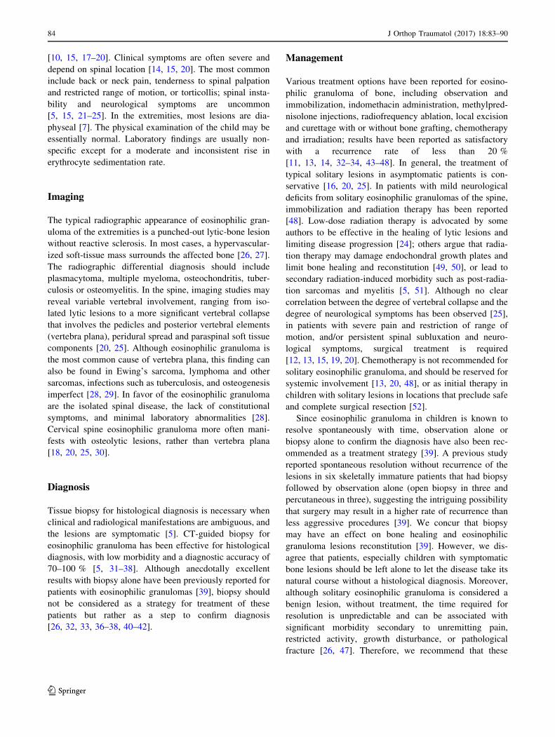
Fig. 1 a Sagittal T2-weighted MRI with fat suppression of the
cervical spine of a 43-year-old woman with a painful osteolytic lesion
of the C7 vertebral body. b CT-guided frozen section biopsy showed
eosinophilic granuloma; intralesional methylprednisolone injection
was performed. c Sagittal T2-weighted MRI with fat suppression.
d Axial CT scan show complete reconstitution of the lesion 4 years
after diagnosis and treatment
J Orthop Traumatol (2017) 18:83–90 85
123
Page 4
involving many organs and the bones. Since this lytic
activity cannot be connected with a neoplastic nature of the
disease, one can reasonably assume that histiocytosis X
cells induce bone resorption in eosinophilic granuloma
through their ability to secrete locally tissue-lytic factors
such as interleukins and prostaglandins [53]. Several
in vitro studies have demonstrated the production of
interleukins (IL) such as IL-1 and prostaglandins (PG) such
as PGE2 and PGD2 by suspensions of Langerhans cells
[53, 54]. Although definitive proof that corticosteroid
injection is responsible for the observed response is diffi-
cult to obtain, the inhibition of IL-1-induced bone resorp-
tion and prostaglandin production by methylprednisolone
[55] may account for its clinical and radiographic effect.
Previous studies on the treatment of certain osteolytic
lesions including bone cysts, aneurysmal cysts, eosino-
philic granulomas and nonossifying fibromas showed that
the results obtained through the introduction of methyl-
prednisolone acetate in crystals were better than those
obtained by using other corticosteroids with topical action
[40]. This is because it is a microcrystalline suspension of
acetate of methylprednisolone that is relatively insoluble
and, therefore, has a prolonged pharmalogical effect [40].
A particular dosage for eosinophilic granulomas cannot be
recommended. The amount of methylprednisolone acetate
injected was established empirically on the basis of the size
of the lesion. A minimum of 40 mg for small lesions
involving less than half the diameter of the involved bone,
and up to 160 mg for large lesions of the pelvis has been
recommended [36–38].
Scaglietti et al. [40] first reported the use of intralesional
methylprednisolone injection for eosinophilic granuloma,
with excellent results, and recommended the injection of
methyl-prednisolone as the treatment of choice. Subse-
quently, similar clinical and radiographic results have been
described in case reports and small series of patients with
solitary and polyostotic lesions involving craniofacial and
long bones [27, 33, 56–58]. The benefit of intralesional
methylprednisolone injection compared with other methods
is that it promotes early relief of pain and pre-
dictable osseous healing [31, 34]. The results of treatment,
either as an adjunct or as primary treatment have been
comparable to other treatments [26, 31–33, 40]. Others
reported that intralesional methylprednisolone injection
adds little in children [33, 39]. However, in patients with
symptomatic lesions, treatment is required. In view of the
usually benign clinical course of the disease, in these
patients a simple, minimally invasive, outpatient treatment
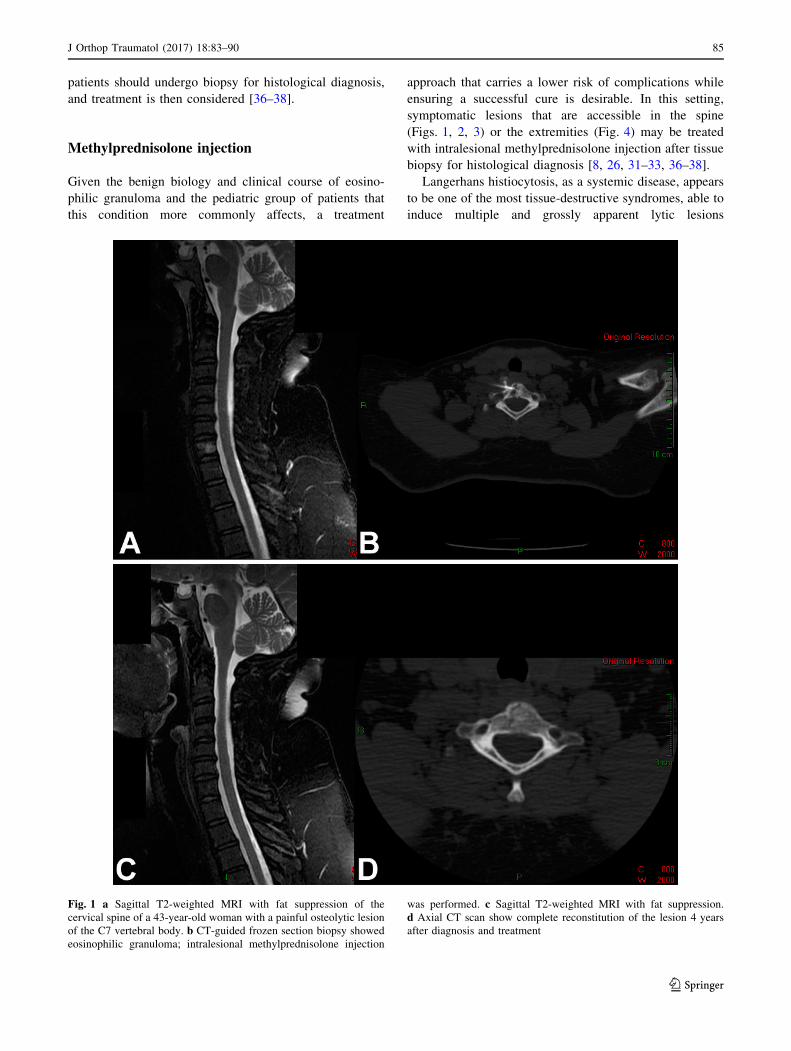
Fig. 2 a Coronal CT scan of the cervical spine of a 5-year-old boy
with a painful osteolytic lesion of the C7 vertebral body. b CT-guided
frozen section biopsy showed eosinophilic granuloma; intralesional
methylprednisolone injection was performed. c, d Sagittal (c)and
axial (d) CT scans showing complete reconstitution of the lesion
5 years after diagnosis and treatment
86 J Orthop Traumatol (2017) 18:83–90
123
Page 5
with a low rate of complications such as CT-guided
intralesional methylprednisolone injection may be consid-
ered the treatment of choice [36–38].
Complications such as femoral osteomyelitis [32] and
obstructive hydrocephalus have been reported after
methylprednisolone injections for eosinophilic granulomas
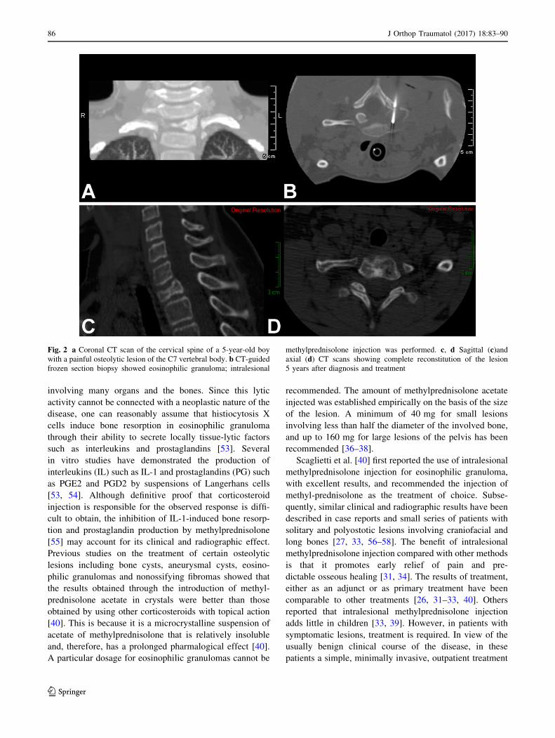
Fig. 3 a Lateral radiograph of the lumbar spine of a 6-year-old girl
with a painful osteolytic lesion of the L2 vertebral body with vertebral
plana deformity. CT-guided frozen section biopsy showed eosino-
philic granuloma; intralesional methylprednisolone injection was
performed. b Lateral radiograph of the lumbar spine shows complete
reconstitution of the lesion 7 years after diagnosis and treatment
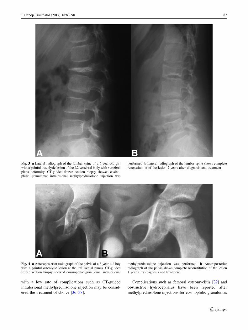
Fig. 4 a Anteroposterior radiograph of the pelvis of a 6-year-old boy
with a painful osteolytic lesion at the left ischial ramus. CT-guided
frozen section biopsy showed eosinophilic granuloma; intralesional
methylprednisolone injection was performed. b Anteroposterior
radiograph of the pelvis shows complete reconstitution of the lesion
1 year after diagnosis and treatment
J Orthop Traumatol (2017) 18:83–90 87
123
Page 6
[33]. However, in general, the morbidity associated with
the procedure has been negligible, even when relatively
inaccessible regions of the spine or pelvis are involved
[31]. The ability of the involved bone to reconstitute after
methylprednisolone injection is believed to be due to the
fact that the disease affects children before skeletal matu-
rity so that the pubertal growth spurt provides sufficient
time for adequate remodeling by the active growth plates
that are spared by the disease process [5, 34]. Some lesions
may fail to respond or are unsuitable for treatment by
injection because of their site, impending fracture, or soft-
tissue invasion [33]. Furthermore, it seems that incomplete
vertebral remodeling usually does not lead to chronic pain
or compromise structural integrity [5, 24, 59, 60].
Authors’ commentary and conclusion
This review summarizes current concepts in the diagnosis
and management of patients with eosinophilic granuloma,
with emphasis on the role of intralesional methylpred-
nisolone injection for the successful cure of patients with
symptomatic lesions. In the past, we planned for observa-
tion alone for patients with imaging evidence of eosino-
philic granuloma, and curettage for the most painful
lesions. It was our initial belief that percutaneous tech-
niques do not provide adequate tissue for definitive diag-
nosis for mesenchymal tumors [61–63]. This belief was
based on the agreement among pathologists that mes-
enchymal tumors are among the most difficult of patholo-
gies to accurately diagnose. We then realized that patients,
especially children with symptomatic bone lesions, should
not be left alone for the disease to take its natural course
without a histological diagnosis. Over the past 15 years, we
have been able to refine the procedures for needle or trocar
and frozen sections biopsy to assess the adequacy of the
biopsy specimen. Nowadays, we believe that histological
diagnosis is necessary for all bone lesions, and recommend
that biopsy should not be considered as a strategy for
treatment of eosinophilic granuloma but rather as a step to
confirm diagnosis. By using CT-guided intralesional
methylprednisolone injection, frozen sections histological
diagnosis can be obtained in all patients. After biopsy,
intralesional injection of methylprednisolone is considered
beneficial [31–33], or at least not harmful. In our practice,
tissue procurement and frozen sections biopsy are usually
diagnostic in all patients with suspected eosinophilic
granuloma. Even if the definite histological diagnosis is
different, intralesional methylprednisolone injection would
not have resulted in any adverse effect, but rather it would
have decreased intralesional edema and provided pain
relief. Our long-term results, (mean follow up, 9 years;
range, 4–23 years) support biopsy and intralesional
methylprednisolone injection as a safe treatment for eosi-
nophilic granulomas of bone with complete resolution of
pain and imaging reconstitution of the lesions.
Compliance with ethical standards
Conflict of interest The authors declare that they have no conflict of
interest. The authors did not receive any outside funding or grants in
support of their research for or preparation of this work. No com-
mercial entity paid or directed, or agreed to pay or direct, any benefits
to any research fund, foundation, division, center, clinical practice, or
other charitable or non-profit organization with which the authors, or
a member of their immediate families, are affiliated or associated.
Open Access This article is distributed under the terms of the
Creative Commons Attribution 4.0 International License (http://crea
tivecommons.org/licenses/by/4.0/), which permits unrestricted use,
distribution, and reproduction in any medium, provided you give
appropriate credit to the original author(s) and the source, provide a
link to the Creative Commons license, and indicate if changes were
made.
References
1. Favara BE, Feller AC, Pauli M, Jaffe ES, Weiss LM, Arico M,
Bucsky P, Egeler RM, Elinder G, Gadner H, Gresik M, Henter JI,
Imashuku S, Janka-Schaub G, Jaffe R, Ladisch S, Nezelof C,
Pritchard J (1997) Contemporary classification of histiocytic
disorders. The WHO Committee On Histiocytic/Reticulum Cell
Proliferations. Reclassification Working Group of the Histiocyte
Society. Med Pediatr Oncol 29(3):157–166
2. Willman CL, Busque L, Griffith BB, Favara BE, McClain KL,
Duncan MH, Gilliland DG (1994) Langerhans’-cell histiocytosis
(histiocytosis X)—a clonal proliferative disease. N Engl J Med
331(3):154–160
3. Chadha M, Agarwal A, Agarwal N, Singh MK (2007) Solitary
eosinophilic granuloma of the radius. An unusual differential
diagnosis. Acta Orthop Belg 73(3):413–417
4. Puzzilli F, Mastronardi L, Farah JO, Ruggeri A, Lunardi P (1998)
Solitary eosinophilic granuloma of the calvaria. Tumori
84(6):712–716
5. Greenlee JD, Fenoy AJ, Donovan KA, Menezes AH (2007)
Eosinophilic granuloma in the pediatric spine. Pediatr Neurosurg
43(4):285–292
6. Kilpatrick SE, Wenger DE, Gilchrist GS, Shives TC, Wollan PC,
Unni KK (1995) Langerhans’ cell histiocytosis (histiocytosis X)
of bone. A clinicopathologic analysis of 263 pediatric and adult
cases. Cancer 76(12):2471–2484
7. Islinger RB, Kuklo TR, Owens BD, Horan PJ, Choma TJ, Mur-
phey MD, Temple HT (2000) Langerhans’ cell histiocytosis in
patients older than 21 years. Clin Orthop Relat Res 379:231–235
8. Azouz EM, Saigal G, Rodriguez MM, Podda A (2005) Langer-
hans’ cell histiocytosis: pathology, imaging and treatment of
skeletal involvement. Pediatr Radiol 35(2):103–115
9. Shimakage M, Sasagawa T, Kimura M, Shimakage T, Seto S,
Kodama K, Sakamoto H (2004) Expression of Epstein-Barr virus
in Langerhans’ cell histiocytosis. Hum Pathol 35:862–868
10. Davidson RI, Shillito J Jr (1970) Eosinophilic granuloma of the
cervical spine in children. Pediatrics 45(5):746–752
11. Sweasey TA, Dauser RC (1989) Eosinophilic granuloma of the
cervicothoracic junction. Case report. J Neurosurg 71:942–944
88 J Orthop Traumatol (2017) 18:83–90
123
Page 7
12. Bilge T, Barut S, Yaymaci Y, Alatli C (1995) Solitary eosino-
philic granuloma of the lumbar spine in an adult. Case report.
Paraplegia 33:485–487
13. Scarpinati M, Artico M, Artizzu S (1995) Spinal cord compres-
sion by eosinophilic granuloma of the cervical spine. Case report
and review of the literature. Neurosurg Rev 18:209–212
14. Brown CW, Jarvis JG, Letts M, Carpenter B (2005) Treatment
and outcome of vertebral Langerhans cell histiocytosis at the
Children’s Hospital of Eastern Ontario. Can J Surg
48(3):230–236
15. Tanaka N, Fujimoto Y, Okuda T, Nakanishi K, Sumida T,
Manabe H, Ochi M (2005) Langerhans cell histiocytosis of the
atlas. A report of three cases. J Bone Joint Surg Am
87(10):2313–2317
16. Metellus P, Gana R, Fuentes S, Eusebio A, Adetchessi A, Dufour
H, Grisoli F (2007) Spinal Langerhans’ cell histiocytosis in a
young adult: case report and therapeutic considerations. Br J
Neurosurg 21(2):228–230
17. Robert H, Dubousset J, Miladi L (1987) Histiocytosis X in the
juvenile spine. Spine 12:167–172
18. Sanchez RL, Llovet J, Moreno A, Galito E (1984) Symptomatic
eosinophilic granuloma of the spine: report of two cases and
review of the literature. Orthopedics 7:1721–1726
19. Dickinson LD, Farhat SM (1991) Eosinophilic granuloma of the
cervical spine. A case report and review of the literature. Surg
Neurol 35:57–63
20. Bertram C, Madert J, Eggers C (2002) Eosinophilic granuloma of
the cervical spine. Spine 27:1408–1413
21. Kumar A (1990) Eosinophilic granuloma of the spine with neu-
rological deficit. Orthopedics 13:1310–1312
22. Villas C, Martınez-Peric R, Barrios RH, Beguiristain JL (1993)
Eosinophilic granuloma of the spine with and without vertebra
plana: longterm follow-up of six cases. J Spinal Disord
6(3):260–268
23. Maggi G, de Sanctis N, Aliberti F, Nunziata Rega A (1966)
Eosinophilic granuloma of C 4 causing spinal cord compression.
Childs Nerv Syst 12(10):630–632
24. Raab P, Hohmann F, Kuhl J, Krauspe R (1998) Vertebral
remodeling in eosinophilic granuloma of the spine: a long-term
follow-up. Spine 23:1351–1354
25. Yeom JS, Lee CK, Shin HY, Lee CS, Han CS, Chang H (1999)
Langerhans’ cell histiocytosis of the spine. Analysis of twenty-
three cases. Spine 24:1740–1749
26. Cohen M, Zornoza J, Cangir A, Murray JA, Wallace S (1980)
Direct injection of methylprednisolone sodium succinate in the
treatment of solitary eosinophilic granuloma of bone: a report of
9 cases. Radiology 136(2):289–293
27. Ruff S, Chapman GK, Taylor TK, Ryan MD (1983) The evolu-
tion of eosinophilic granuloma of bone: a case report. Skelet
Radiol 10(1):37–39
28. O’Donnell J, Brown L, Herkowitz H (1991) Vertebra plana-like
lesions in children: case report with special emphasis on the
differential diagnosis and indications for biopsy. J Spinal Disord
4:480–485
29. Ferguson L, Shapiro CM (1979) Eosinophilic granuloma of the
second cervical vertebra. Surg Neurol 11:435–437
30. Floman Y, Bar-On E, Mosheiff R, Mirovsky Y, Robin GC, Ramu
N (1997) Eosinophilic granuloma of the spine. J Pediatr Orthop B
6(4):260–265
31. Yasko AW, Fanning CV, Ayala AG, Carrasco CH, Murray JA
(1998) Percutaneous techniques for the diagnosis and treatment
of localized Langerhans-cell histiocytosis (eosinophilic granu-
loma of bone). J Bone Joint Surg Am 80(2):219–228
32. Capanna R, Springfield DS, Ruggieri P, Biagini R, Picci P, Bacci
G, Giunti A, Lorenzi EG, Campanacci M (1985) Direct cortisone
injection in eosinophilic granuloma of bone: a preliminary report
on 11 patients. J Pediatr Orthop 5(3):339–342
33. Egeler RM, Thompson RC Jr, Voute PA, Nesbit ME Jr (1992)
Intralesional infiltration of corticosteroids in localized Langer-
hans’ cell histiocytosis. J Pediatr Orthop 12(6):811–814
34. Sessa S, Sommelet D, Lascombes P, Prevot J (1994) Treatment of
Langerhans-cell histiocytosis in children. Experience at the
Children’s Hospital of Nancy. J Bone Joint Surg Am
76(10):1513–1525
35. Elsheikh T, Silverman JF, Wakely PE Jr, Holbrook CT, Joshi VV
(1991) Fine-needle aspiration cytology of Langerhans’ cell his-
tiocytosis (eosinophilic granuloma) of bone in children. Diagn
Cytopathol 7(3):261–266
36. Rimondi E, Mavrogenis AF, Rossi G, Ussia G, Angelini A,
Ruggieri P (2011) CT-guided corticosteroid injection for solitary
eosinophilic granuloma of the spine. Skelet Radiol
40(6):757–764
37. Mavrogenis AF, Rimondi E, Ussia G, Rossi G, Ruggieri P (2011)
Successful treatment of a bifocal eosinophilic granuloma of the
spine with CT-guided corticosteroid injection. Orthopedics
34(3):230
38. Mavrogenis AF, Abati CN, Bosco G, Ruggieri P (2012) Intrale-
sional methylprednisolone for painful solitary eosinophilic
granuloma of the appendicular skeleton in children. J Pediatr
Orthop 32(4):416–422
39. Plasschaert F, Craig C, Bell R, Cole WG, Wunder JS, Alman BA
(2002) Eosinophilic granuloma. A different behaviour in children
than in adults. J Bone Joint Surg Br 84(6):870–872
40. Scaglietti O, Marchetti PG, Bartolozzi P (1982) Final results
obtained in the treatment of bone cysts with methylprednisolone
acetate (depo-medrol) and a discussion of results achieved in
other bone lesions. Clin Orthop Relat Res 165:33–42
41. Nauert C, Zornoza J, Ayala A, Harle TS (1983) Eosinophilic
granuloma of bone: diagnosis and management. Skelet Radiol
10(4):227–235
42. Shabb N, Fanning CV, Carrasco CH, Guo SQ, Katz RL, Ayala
AG, Raymond AK, Cangir A (1993) Diagnosis of eosinophilic
granuloma of bone by fine-needle aspiration with concurrent
institution of therapy: a cytologic, histologic, clinical, and radi-
ologic study of 27 cases. Diagn Cytopathol 9(1):3–12
43. Ladisch S, Gadner H (1994) Treatment of Langerhans cell his-
tiocytosis—evolution and current approaches. Br J Cancer23:S41–S46
44. McLelland J, Broadbent V, Yeomans E, Malone M, Pritchard J
(1990) Langerhans cell histiocytosis: the case for conservative
treatment. Arch Dis Child 65(3):301–303
45. Han I, Suh ES, Lee SH, Cho HS, Oh JH, Kim HS (2009) Man-
agement of eosinophilic granuloma occurring in the appendicular
skeleton in children. Clin Orthop Surg 1(2):63–67
46. King JJ, Melvin JS, Iwenofu OH, Fox EJ (2009) Thigh pain in a
53-year-old woman. Clin Orthop Relat Res 467(6):1652–1657
47. Corby RR, Stacy GS, Peabody TD, Dixon LB (2008) Radiofre-
quency ablation of solitary eosinophilic granuloma of bone. AJR
Am J Roentgenol 190(6):1492–1494
48. Osenbach RK, Youngblood LA, Menezes AH (1990) Atlanto-
axial instability secondary to solitary eosinophilic granuloma of
C2 in a 12-year-old girl. J Spinal Disord 3:408–412
49. Nesbit ME, Kieffer S, D’Angio GJ (1969) Reconstitution of
vertebral height in histiocytosis X: a long-term follow-up. J Bone
Joint Surg Am 51:1360–1368
50. Green NE, Robertson WW Jr, Kilroy AW (1980) Eosinophilic
granuloma of the spine with associated neural deficit: report of
three cases. J Bone Joint Surg Am 62:1198–1202
51. Johansson L, Larsson LG, Damber L (1995) A cohort study with
regard to the risk of haematological malignancies in patients
treated with X-rays for benign lesions in the locomotor system. II.
J Orthop Traumatol (2017) 18:83–90 89
123
Page 8
Estimation of absorbed dose in the red bone marrow. Acta Oncol
34:721–726
52. Levy EI, Scarrow A, Hamilton RC, Wollman MR, Fitz C, Pollack
IF (1999) Medical management of eosinophilic granuloma of the
cervical spine. Pediatr Neurosurg 31(3):159–162
53. Arenzana-Seisdedos F, Barbey S, Virelizier JL, Kornprobst M,
Nezelof C (1986) Histiocytosis X. Purified (T6?) cells from bone
granuloma produce interleukin 1 and prostaglandin E2 in culture.
J Clin Invest 77(1):326–329
54. Gonzalez-Crussi F, Hsueh W, Wiederhold MD (1981) Pros-
taglandins in histiocytosis-X. PG synthesis by histiocytosis-X
cells. Am J Clin Pathol 75(2):243–253
55. Marusic A, Raisz LG (1991) Cortisol modulates the actions of
interleukin-1 alpha on bone formation, resorption, and pros-
taglandin production in cultured mouse parietal bones.
Endocrinology 129(5):2699–2706
56. Chacha PB, Khong BT (1971) Eosinophilic granuloma of bone. A
diagnostic problem. Clin Orthop Relat Res 80:79–88
57. Jones LR, Toth BB, Cangir A (1989) Treatment for solitary
eosinophilic granuloma of the mandible by steroid injection:
report of a case. J Oral Maxillofac Surg 47(3):306–309
58. Wirtschafter JD, Nesbit M, Anderson P, McClain K (1987)
Intralesional methylprednisolone for Langerhans’ cell histiocy-
tosis of the orbit and cranium. J Pediatr Ophthalmol Strabismus
24(4):194–197
59. Seimon LP (1981) Eosinophil granuloma of the spine. J Pediatr
Orthop 1:371–376
60. Ippolito E, Farsetti P, Tudisco C (1984) Vertebra plana: long-
term follow-up in five patients. J Bone Joint Surg Am
66:1364–1368
61. Mankin HJ, Mankin CJ, Simon MA (1996) The hazards of the
biopsy, revisited. Members of the Musculoskeletal Tumor Soci-
ety. J Bone Joint Surg Am 78(5):656–663
62. Lawrence W Jr, Donegan WL, Natarajan N, Mettlin C, Beart R,
Winchester D (1987) Adult soft-tissue sarcomas. A pattern of
care survey of the American College of Surgeons. Ann Surg
205(4):349–359
63. Welker JA, Henshaw RM, Jelinek J, Shmookler BM, Malawer
MM (2000) The percutaneous needle biopsy is safe and recom-
mended in the diagnosis of musculoskeletal masses. Cancer
89(12):2677–2686
90 J Orthop Traumatol (2017) 18:83–90
123