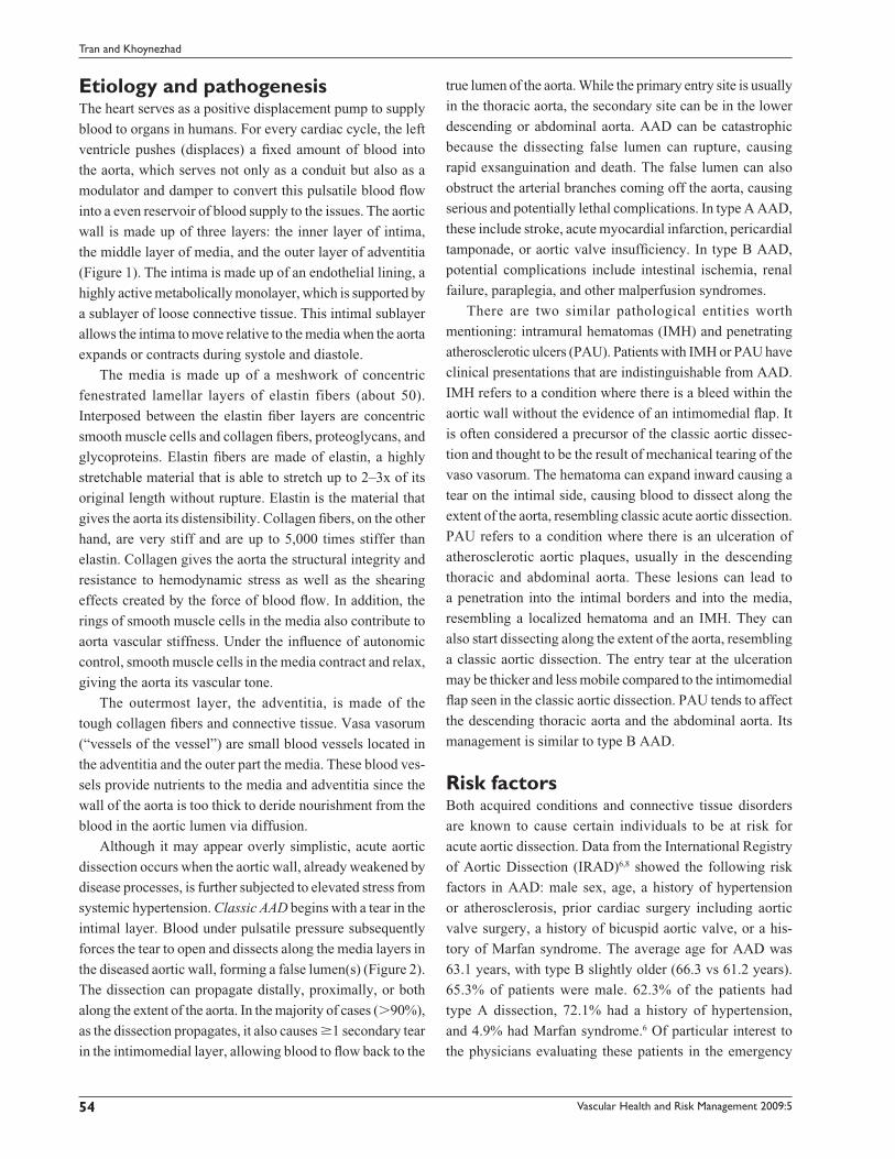
Vascular Health and Risk Management 2009:5 53–63 53
R E V I E W
Current management of type B aortic dissection
T Paul Tran1
Ali Khoynezhad2
1Department of Emergency Medicine; 2Section of Cardiothoracic Surgery, University of Nebraska Medical Center, Omaha, NE, USA
Correspondence: Ali KhoynezhadSection of Cardiothoracic Surgery, University of Nebraska Medical Center, 982315 Nebraska Medical Center,Omaha, NE 68198-2315, USATel +1 402 559 4424Fax +1 402 559 6913Email [email protected]
Abstract: Acute aortic dissection is a life-threatening condition associated with high morbidity
and mortality. In this article, the authors review basic biology of the aorta and aortic dissection,
calcifi cation, and wide mediastinum. Although these classic
radiographic fi ndings are not always present, collectively,
up to 90% of patients with aortic dissection proved to have
some abnormal fi ndings on chest X-rays. Absence of wide
mediastinum and abnormal aortic contour on chest X-ray
lowers the likelihood of aortic dissection (LR 0.3).9
Biochemical markersThe Holy Grail in the workup of acute aortic dissection is
an inexpensive bedside blood test with suffi cient sensitivity
(good negative predictive value) that physicians can use to
rapidly rule out an acute dissection. Of the several biochemi-
cal markers that were evaluated in the last decade, the fol-
lowing three assays deserve further discussion.
Smooth muscle myosin heavy chain, a major component
of the smooth muscle in the aortic medial layer, is released
to the circulation shortly after the onset of dissection. In a
pilot study, the assay (�2.5 microgram/L) had a sensitivity of
90.9% and specifi city of 98% in detecting acute aortic dissec-
tion as compared to healthy volunteers.16 These test numbers
are, however, limited to patients with proximal dissection
who present within 3 hours of onset of symptoms.
Type A Type B
Figure 3B Stanford classifi cation of aortic dissection. Stanford type A includes dissections that involve the ascending aorta, arch, and descending thoracic aorta. Stanford type B includes dissections that originate in the descending (and thoracoab-dominal) aorta, regardless of any retrograde involvement of the arch.
Vascular Health and Risk Management 2009:5 59
Current management of type B aortic dissection
Elastin is another major structural component of the medial
layer of aortic wall. In a retrospective study using the assay
for soluble elastin fragments (sELAF) that was developed
by the authors,17 Shinohara and colleagues demonstrated
that an ELISA measuring sELAF in the serum with the
cutoff set at + 3 SD (standard deviation) above the mean
of age-adjusted healthy subjects had a sensitivity of 88.9%
(16/18) and specifi city of 99.8% (473/474).17 The positive
(PPV) and negative predictive values (NPV) were 94.1% and
98.8%, respectively. Based on the NPV, measuring sELAF
would appear like an excellent test to rule out an acute aortic
dissection. However, since this study was retrospective, data
therein need to be confi rmed in a prospective confi rmatory
multicenter study. More importantly, the ELISA for sELAF
takes 3 hours to perform, a major drawback for a time
sensitive condition such as acute aortic dissection. A positive
test would require a follow-up radiologic confi rmatory test,
which further prolongs the diagnosis.
Plasma D-dimer, a degradation product of cross-linked
fi brin, has emerged as a promising diagnostic marker with
high sensitivity for exclusion of acute aortic dissection. The
rise in D-dimer is thought to result from the thrombogenic-
ity of the false lumen in AAD and the subsequent activa-
tion of the coagulation cascade. As such, D-dimer values
are expected to be lower in proximal AAD (type A). In an
earlier pooled analysis by Sodeck, a d-dimer �0.1 μg/ml
is though to exclude all AAD (sensitivity of 100%).18 In
a more recent pooled sensitivity analysis in EM literature,
using a commonly used threshold for a positive D-dimer
test of �0.5 μg/ml, Marill found that serum d-dimer had
a sensitivity of 94%.19 The authors concluded that serum
d-dimer can be used in patients with a low likelihood of AAD.
Overall, a preponderance of recent studies support drawing
D-dimer in the ED given its high sensitivity, noninvasiveness,
broad availability and low cost.20–24
ImagingThe choice for the diagnostic imaging depends on patient’s
stability, local expertise, and availability. The main goals
in diagnostic imaging are to rapidly confi rm (or exclude)
an AAD, classify the type/extent of the AAD, identify and
locate the intimal tears, confi rm the presence of true/false
lumen and whether a thrombus is present, assess any aortic
side branch involvement, detect any aortic regurgitation, and
discover any extravasation to the pericardium, mediastinum,
or thoracic cavity.
A chest X-ray can detect abnormalities in 60%–90%
of cases of AAD. Abnormalities that may suggest an AAD
include an abnormal aortic knob, deviation of the trachea,
main stem bronchus, or esophagus, wide mediastinum,
pleural cap, or pleural effusion. A normal chest X-ray,
however, can not be used to rule out an AAD. In unstable
patients with strong suspicion of an AAD, chest X-rays may
lead to further delay to defi nitive imaging and therapy and
should be avoided.
Defi nitive imaging techniques are essential in the workup
of AAD. Data from the 2002 IRAD show that computed
tomography angiography (CTA) is used in 63% of cases of
suspected AAD, followed transesophageal echocardiography
(TEE) in 32%, aortography 4%, and magnetic resonance
angiography (MRA) in 1%.25 Compared with the newer
imaging techniques, aortography has a number of serious
disadvantages, including the use of a heavy dose of IV
contrast (1 mg/kg), the risks of an invasive procedure, and
the extended time it takes to complete the procedure (up to
2+ hours). As a result, this imaging method is rarely the fi rst
diagnostic modality in the workup of AAD.
Computed tomography angiography, TEE and MRA
have similar pooled sensitivity (98%–100%) and specifi city
(95%–98%),26 although the pooled positive likelihood ratio
appeared to be higher for MRA (positive likelihood ratio,
25.3; 95% confi dence interval, 11.1–57.1) than for TEE
(14.1; 6.0–33.2) or CTA (13.9; 4.2–46.0). CTA is widely
available and relatively rapid, provides visualization of
the entire aorta down to iliac arteries, and delineates the
involvement of aortic side branches. It is usually the fi rst
choice for imaging in the work up of AAD. Disadvantages
with CTA include the requirement that patients be transported
to the CTA suite, the use of potentially nephrotoxic contrast,
and the inability to assess aortic insuffi ciency.
Overall, CTA of the chest is the preferred fi rst diag-
nostic imaging method in hemodynamically stable
patients (Figure 4). The choice for a second imaging study
includes MRA or TEE, depending on clinical condition and
local environment. MRA is highly accurate and does not
require the use of a contrast dye. It is, however, usually not
available on an emergency basis and requires patients to be
in MRA suite for an extended period of time. Other issues
such as claustrophobia, the use of ventilator, and patient’s
use of metal devices (pacemakers, aneurysmal clips) may
further complicate its routine use.25 TEE is a viable alterna-
tive in patients who are critically ill and/or hemodynamically
unstable. The main advantages of a TEE include speed, good
sensitivity and specifi city, and the fact that it can be per-
formed at the patient’s bedside in the ED. Its main limitations
are lack of widespread expertise and 24/7 availability.
Vascular Health and Risk Management 2009:560
Tran and Khoynezhad
If the diagnosis can not be established using the
aforementioned imaging modalities, the patient should be
taken to the hybrid operating room. An intravascular ultra-
sound and/or aortography performed by the cardiothoracic
surgeon will establish the defi nitive diagnosis (similar to
diagnostic algorithm of traumatic aortic transection).27
Intravascular ultrasound (IVUS) is a novel approach
in diagnosis of type B AAD that has the highest accuracy
of any modality (Figure 5). IVUS has been demonstrated
100% sensitivity and specifi city during endovascular diag-
nosis and treatment of complicated type B aortic dissec-
tion.28 Furthermore, IVUS is a critical imaging modality for
endovascular treatment options for type B AAD.29
ManagementThe modern surgical treatment for AAD, including those for
acute type B aortic dissection, began after the landmark aortic
operation by Drs Cooley and DeBakey in the 1950’s.30 In the
decades following this sentinel event, patients with type B
aortic dissection were mostly treated surgically. The medical
management of acute type B AAD began to gain credence
with the concept of antiimpulsive therapy as described by
Wheat.31 He was one of the fi rst to demonstrate the impor-
tance of the force of contraction (dP/dtmax
) and blood pres-
sure in the propagation of AAD in a dog model.32 Starting
in the early 70’s, medical management of uncomplicated
type B AAD increasingly became the standard of care
due to availability of potent beta-blockers and the lower
mortality compared to surgical approach (for type A, early
mortality rates of 10% vs. 35%, respectively).33 Today, the
antiimpulsive and antihypertensive combination therapy
remains the cornerstone of modern medical management
of type B AAD. Table 2 illustrates the authors’ approach to
the diagnosis and management of an acute aortic dissection
(AAD) in the ED.
At triage, patients who present to the ED with sudden
onset of chest, back, or abdominal pain should be quickly
brought to a room to be examined by a physician. Prompt
diagnosis and treatment of patients with suspected AAD is
essential for improved outcome. The abbreviated history
and physical examination should focus on time of onset,
risk factors for aortic dissection, and fi ndings that are
consistent with AAD (Table 2). Patients should have IV’s
and A-line established, supplemental oxygen, be put on
a cardiac monitor, an EKG, portable chest X-ray, blood
draws, and be typed and crossed for 10 units of PRBC’s.
Surgical consultation should be initiated as soon as the
emergency physician is comfortable with his/her clinical
impression.
Pain should be treated with appropriate analgesics
(eg, morphine). Heart rate and hypertension should be
aggressively controlled with β-blockers.34 An arterial
FALSELUMEN
TRUELUMEN
Figure 5 Intravascular imaging of aortic dissection.Figure 4 Computer tomography with enhanced contrast showing type B dissection.
Vascular Health and Risk Management 2009:5 61
Current management of type B aortic dissection
Table 2 Approach to acute aortic dissections (AAD) in the emergency department
1) Have a high index of suspicion for AAD a) History: i) Sudden onset, severe, sharp or tearing back pain, chest pain, shoulder pain, or abdominal pain ii) Older than 60 years, history of hypertension, aortic dissection or aortic aneurysm (of family history of such), previous cardiac surgery, con-
nective tissue disorder (Bicuspid aortic valve, Marfan syndrome, Ehler-Danlos syndrome, Loeys-Dietz syndrome), or peripartum b) Physical examination: (1) Pulse defi cit, blood pressure differential in various extremities (2) Neurological defi cits (3) Abdominal pain, fl ank pain2) General measures: a) Establish two large bore (�18gauge) IV’s b) Administer supplemental oxygen by nasal cannula or nonrebreather mask c) Put patient on cardiac monitor d) Get an EKG, portable chest X-ray, place a Foley catheter e) Obtain CBC, chemistry panel, coagulation panel, UA, CK, Troponin, d-dimer f) Type and cross 10 units packed red blood cells (PRBC’s) g) Set up an arterial line 3) Early cardiothoracic surgical consultation 4) Defi nitive imaging: a) Computed tomography angiogram (CTA) b) Transesophageal echocardiogram c) Magnetic resonance angiogram (MRA) d) Intravascular ultrasound e) Aortography 5) Blood pressure, heart rate, and pain management a) First line: β-blockers i) Labetalol, bolus (15 mg) ± a drip (5 mg/hour), b) If hypertension persists, add: i) Nicardipine drip (starting dose: 5 mg/h) c) If tachycardia persists, add: i) Esmolol (loading 0.5 mg/kg over 2–5 min, followed by a drip of 10–20 μg/kg/min) ii) Diltiazem drip (loading 0.25 mg/kg over 2–5 min, followed by a drip of 5mg/h) d) Goals: (1) Heart rate �60 beats/min (2) Systolic blood pressure �100 mm Hg e) Morphine (for pain relief) 6) Hemodynamically unstable patients a) Tracheal intubation, mechanical ventilation b) Blood pressure support with crystalloid and colloid (PRBC’s if rupture is suspected) c) TEE at bedside in the Emergency Department or in the OR d) Pericardiocentesis is not recommended (class III)
vasodilator without the refl ex tachycardia (eg, nicardipine),
can be added as second line drugs for refractory hypertension.
β-blockers are the fi rst line drugs because they control the
maximal force of left ventricular contraction (dP/dtmax
) in
addition to controlling HR and BP. This helps reduce further
dissection, branch-vessel malperfusion and weakening of the
aortic wall. Common β-blockers include labetalol, esmolol,
metoprolol, and atenolol. Targeted HR and systolic BP
should be �60 beats/min and �100 mm Hg, respectively. In
patients with potential intolerance to β-blockers (eg, asthma,
heart failure), a test dose of a short acting β-blocker such
as esmolol should be tried.35 If β-blockers can not be used,
calcium channel blockers (eg, nicardipine, clevidipine or
diltiazem) might be alternatives to control blood pressure in
these patients. Hydralazine and sodium nitroprusside have
been used in the past for medical management of type B
AAD. Although these agents reduce blood pressure, they
increase the maximal force of left ventricular contraction
(dP/dtmax
) and cause refl ex tachycardia and are, thus, con-
traindicated in patients with AAD.
Hemodynamically unstable patients with type B AAD are
either in hemorrhagic shock or septic/metabolic shock from
visceral or limb malperfusion. They should be endotracheally
intubated and fl uid resuscitated with crystalloid, PRBC’s,
or other colloid. In these patients, a TEE or CTA are the
preferred diagnostic tools, depending on the hospital setting.
Vascular Health and Risk Management 2009:562
Tran and Khoynezhad
If the diagnosis of a complicated type B AAD is confi rmed,
patients should be immediately brought to a hybrid
operating room for endovascular and/or surgical treatment
options. Endovascular or surgical options may be offered to
hemodynamically stable patients with a complicated type
B AAD. Table 3 lists the indications for endovascular or
surgical intervention in patients with type B AAD.
OutcomeThe short term prognosis for patients with type B AAD is
better than those with type A and the medical management of
type B AAD is associated with less mortality compared to the
open surgical intervention approach. Overall, 89% of patients
with type B AAD survive to hospital discharge, although the
in-hospital survival rates were as low as 29% for the highest
risk group, 64% for the intermediate, and 97% for the lowest
risk group.6,36 Aortic rupture, shock, and malperfusion are the
most important in-hospital risks leading to poor outcome in
up to 20% of these patients.6,35 Of survivors, 80% will develop
aneurysmal dilatation of the false lumen, requiring cardio-
thoracic surgical intervention in one third of the cases.35,37,38
Therefore, although medical management of patients with
type B AAD is the standard of care at the moment, it is far
from being an option with acceptable results to patients with
uncomplicated type B AAD.
The long term prognosis for patients with type B AAD
is poor. Data from IRAD indicated about 1 out of 4 patients
with type B dissection died at the 3-year mark regardless of
the mode of therapy. The 3-year rates of survival were 77.6%,
82.8%, and 76.2% for patients treated medically, surgically,
and with endovascular therapy, respectively.36 Umana and
colleauges reported the outcome of 189 patients with type
B aortic dissection over a 36-year period. Actuarial survival
for all patients was 71%, 60%, 35% and 17% at one, fi ve,
10, and 15 years regardless of medical or surgical therapy.33
A diameter �4 cm, patent false lumen, and a partially throm-
bosed false lumen are independent predictors of poor long
term outcome for type B dissection.39,40
Future developmentGiven the disappointing results of medical and surgical
therapy in uncomplicated type B AAD, there has been inter-
est in use of endovascular stenting along with antiimpulsive
and antihypertensive treatment. The endovascular therapy
is based on the concept that obliteration and thrombosis of
the false lumen may result in improved long-term outcome.
The INSTEAD trial was designed to address this question,
but was associated with methodological fl aws by including
patients with chronic type B aortic dissection.41 Ongoing and
future trials will shed light on this issue. For complicated
type B AAD, endovascular therapies are becoming the
standard of care in the Centers of Excellence, because they
have shown to have a better outcome compared to the open
repair approach.29,42,43
DisclosureThe authors report no confl icts of interest in this work.
References 1. Osler W, Silverman ME, Murray TJ, et al. American College of
Physicians. The quotable Osler. Philadelphia: American College of Physicians, 2003.
2. Coady MA, Rizzo JA, Goldstein LJ, et al. Natural history, pathogenesis, and etiology of thoracic aortic aneurysms and dissections. Cardiol Clin. 1999;17:615–35.
3. DeBakey ME, McCollum CH, Crawford ES, et al. Dissection and dissecting aneurysms of the aorta: twenty-year follow-up of five hundred twenty-seven patients treated surgically. Surgery. 1982;92:1118–34.
4. Fuster V, Andrews P. Medical treatment of the aorta. I. Cardiol Clin. 1999;17:697–715, viii.
5. Lindsay J, Jr., Hurst JW. Clinical features and prognosis in dissecting aneurysm of the aorta. A re-appraisal. Circulation. 1967;35:880–8.
6. Hagan PG, Nienaber CA, Isselbacher EM, et al. The International Registry of Acute Aortic Dissection (IRAD): new insights into an old disease. JAMA. 2000;283:897–903.
7. Suzuki T, Mehta RH, Ince H, et al. Clinical profi les and outcomes of acute type B aortic dissection in the current era: lessons from the International Registry of Aortic Dissection (IRAD). Circulation. 2003;108(Suppl 1):II312–7.
8. Januzzi JL, Isselbacher EM, Fattori R, et al. Characterizing the young patient with aortic dissection: results from the International Registry of Aortic Dissection (IRAD). J Am Coll Cardiol. 2004;43:665–9.
9. Klompas M. Does this patient have an acute thoracic aortic dissection? JAMA. 2002;287:2262–72.
10. McCaig LF, Nawar EW. National Hospital Ambulatory Medical Care Survey: 2004 emergency department summary. Adv Data. 2006;(372):1–29.
11. Meszaros I, Morocz J, Szlavi J, et al. Epidemiology and clinicopathol-ogy of aortic dissection. Chest. 2000;117:1271–8.
12. Sullivan PR, Wolfson AB, Leckey RD, et al. Diagnosis of acute tho-racic aortic dissection in the emergency department. Am J Emerg Med. 2000;18:46–50.
13. Viljanen T. Diagnostic diffi culties in aortic dissection. Retrospec-tive study of 89 surgically treated patients. Ann Chir Gynaecol. 1986;75:328–32.
14. The Associated Press. Winkler Testifi es in John Ritter Lawsuit. The New York Times. 2008;Feb 13.
Table 3 Indications for endovascular or surgical intervention in patients with type B AAD
• Persistent or recurrent pain despite adequate antiimpulsive and anti-hypertensive therapy (at least two parenteral agents at moderate to high dose)
• Acute expansion of the false lumen• Periaortic or mediastinal hematoma (contained rupture)• Visceral, renal or limb malperfusion syndrome
Neurological defi cits (relative indication).
Vascular Health and Risk Management 2009:5 63
Current management of type B aortic dissection
15. Boie ET. Initial evaluation of chest pain. Emerg Med Clin North Am. 2005;23:937–57.
16. Suzuki T, Katoh H, Tsuchio Y, et al. Diagnostic implications of elevated levels of smooth-muscle myosin heavy-chain protein in acute aortic dissection. The smooth muscle myosin heavy chain study. Ann Intern Med. 2000;133:537–41.
17. Shinohara T, Suzuki K, Okada M, et al. Soluble elastin fragments in serum are elevated in acute aortic dissection. Arterioscler Thromb Vasc Biol. 2003;23:1839–44.
18. Sodeck G, Domanovits H, Schillinger M, et al. D-dimer in ruling out acute aortic dissection: a systematic review and prospective cohort study. Eur Heart J. 2007;28:3067–75.
19. Marill KA. Serum D-dimer is a sensitive test for the detection of acute aor-tic dissection: A pooled meta-analysis. J Emerg Med. 2008;34:367–76.
20. Eggebrecht H, Naber CK, Bruch C, et al. Value of plasma fi brin D-dimers for detection of acute aortic dissection. J Am Coll Cardiol. 2004;44:804–9.
21. Hazui H, Fukumoto H, Negoro N, et al. Simple and useful tests for discriminating between acute aortic dissection of the ascending aorta and acute myocardial infarction in the emergency setting. Circ J. 2005;69:677–82.
22. Immer FF. Is there a place for D-dimers in acute type A aortic dissection? Heart. 2006;92:727–8.
23. Ohlmann P, Faure A, Morel O, et al. Diagnostic and prognostic value of circulating D-Dimers in patients with acute aortic dissection. Crit Care Med. 2006;34:1358–64.
24. Ohlmann P, Morel O, Radulescu B, et al. D-Dimer in ruling out acute aortic dissection: sensitivity is not 100%. Eur Heart J. 2008;29:828–9; author reply 829.
25. Moore AG, Eagle KA, Bruckman D, et al. Choice of computed tomog-raphy, transesophageal echocardiography, magnetic resonance imaging, and aortography in acute aortic dissection: International Registry of Acute Aortic Dissection (IRAD). Am J Cardiol. 2002;89:1235–8.
26. Shiga T, Wajima Z, Apfel CC, et al. Diagnostic accuracy of transesopha-geal echocardiography, helical computed tomography, and magnetic resonance imaging for suspected thoracic aortic dissection: systematic review and meta-analysis. Arch Intern Med. 2006;166:1350–6.
27. Khoynezhad A, Donayre CE, Kopchok GE, et al. Use of intravascular ultrasound in endovascular stenting of traumatic rupture of the descend-ing thoracic aorta. Cardiothoracic Surgical Network (CTSNet) [serial on the internet]. 2006 Jun. [cited 2008 Jul 22]. Available from http://www.ctsnet.org/sections/clinicalresources/clinicalcases/article-12.html
28. Lee JT, White RA. Basics of intravascular ultrasound: an essential tool for the endovascular surgeon. Semin Vasc Surg. 2004;17:110–8.
29. Khoynezhad A, Donayre CE, Kopchok GE, et al. Mid-term results of endovascular treatment of complicated acute type B aortic dissection. J Thorac Cardiovasc Surg. 2008;136:424–30.
30. DeBakey ME, Beall AC Jr., Cooley DA, et al. Dissecting aneurysms of the aorta. Surg Clin North Am. 1966;46:1045–55.
31. Wheat MW Jr., Palmer RF, Bartley TD, et al. Treatment of dissecting aneurysms of the aorta without surgery. J Thorac Cardiovasc Surg. 1965;50:364–73.
32. Prokop EK, Palmer RF, Wheat MW Jr. Hydrodynamic forces in dis-secting aneurysms. In-vitro studies in a Tygon model and in dog aortas. Circ Res. 1970;27:121–7.
33. Umana JP, Lai DT, Mitchell RS, et al. Is medical therapy still the opti-mal treatment strategy for patients with acute type B aortic dissections? J Thorac Cardiovasc Surg. 2002;124:896–910.
34. Khoynezhad A, Plestis KA. Managing emergency hypertension in aortic dissection and aortic aneurysm surgery. J Card Surg. 2006;21(Suppl 1):S3–S7.
35. Erbel R, Alfonso F, Boileau C, et al. Diagnosis and management of aortic dissection. Eur Heart J. 2001;22:1642–81.
36. Tsai TT, Fattori R, Trimarchi S, et al. Long-term survival in patients presenting with type B acute aortic dissection: insights from the International Registry of Acute Aortic Dissection. Circulation. 2006;114:2226–31.
37. Fann JI, Smith JA, Miller DC, et al. Surgical management of aortic dissection during a 30-year period. Circulation. 1995;92(9 Suppl):II113–21.
38. Juvonen T, Ergin MA, Galla JD, et al. Prospective study of the natural his-tory of thoracic aortic aneurysms. Ann Thorac Surg. 1997;63:1533–45.
39. Onitsuka S, Akashi H, Tayama K, et al. Long-term outcome and prog-nostic predictors of medically treated acute type B aortic dissections. Ann Thorac Surg. 2004;78:1268–73.
40. Tsai TT, Evangelista A, Nienaber CA, et al. Partial thrombosis of the false lumen in patients with acute type B aortic dissection. N Engl J Med. 2007;357:349–59.
41. Nienaber CA, Zannetti S, Barbieri B, et al. INvestigation of STEnt grafts in patients with type B Aortic Dissection: design of the INSTEAD trial – a prospective, multicenter, European randomized trial. Am Heart J. 2005;149:592–9.
42. Bozinovski J, Coselli JS. Outcomes and survival in surgical treatment of descending thoracic aorta with acute dissection. Ann Thorac Surg. 2008;85:965–70.
43. Eggebrecht H, Lonn L, Herold U, et al. Endovascular stent-graft place-ment for complications of acute type B aortic dissection. Curr Opin Cardiol. 2005;20:477–83.