? Annu. Rev. Biomed. Eng. 2000. 02:315–37 CURRENT METHODS IN MEDICAL IMAGE SEGMENTATION 1 Dzung L. Pham 2,3 , Chenyang Xu 2 , and Jerry L. Prince 2 2 Department of Electrical and Computer Engineering, The Johns Hopkins University, Baltimore, Maryland 21218; e-mail: [email protected], [email protected], [email protected]. 3 Laboratory of Personality and Cognition, National Institute on Aging, Baltimore, Maryland 21224 Key Words medical imaging, image processing, classification, deformable models, magnetic resonance imaging ■ Abstract Image segmentation plays a crucial role in many medical-imaging ap- plications, by automating or facilitating the delineation of anatomical structures and other regions of interest. We present a critical appraisal of the current status of semi- automated and automated methods for the segmentation of anatomical medical images. Terminology and important issues in image segmentation are first presented. Current segmentation approaches are then reviewed with an emphasis on the advantages and disadvantages of these methods for medical imaging applications. We conclude with a discussion on the future of image segmentation methods in biomedical research. CONTENTS INTRODUCTION ................................................ 316 BACKGROUND ................................................. 316 Definitions .................................................... 316 Dimensionality ................................................. 318 Soft Segmentation and Partial-Volume Effects .......................... 318 Intensity Inhomogeneities ......................................... 320 Interaction .................................................... 321 Validation .................................................... 321 METHODS ..................................................... 322 Thresholding .................................................. 322 Region Growing ................................................ 323 Classifiers .................................................... 324 Clustering .................................................... 325 Markov Random Field Models ..................................... 327 Artificial Neural Networks ........................................ 327 1 The US Government has the right to retain a nonexclusive, royalty-free license in and to any copyright covering this paper. 315 Annu. Rev. Biomed. Eng. 2000.2:315-337. Downloaded from arjournals.annualreviews.org by Universitat Pompeu Fabra on 05/20/07. For personal use only.
Transcript
P1: FHY/ftt P2: FDR/FhN/fok/fgg QC: FhN
July 10, 2000 11:13 Annual Reviews AR106-12
?Annu. Rev. Biomed. Eng. 2000. 02:315–37
CURRENT METHODS IN MEDICAL IMAGE
SEGMENTATION1
Dzung L. Pham2,3, Chenyang Xu2, and Jerry L. Prince22Department of Electrical and Computer Engineering, The Johns Hopkins University,Baltimore, Maryland 21218; e-mail: [email protected], [email protected],[email protected] of Personality and Cognition, National Institute on Aging,Baltimore, Maryland 21224
Key Words medical imaging, image processing, classification, deformablemodels, magnetic resonance imaging
■ Abstract Image segmentation plays a crucial role in many medical-imaging ap-plications, by automating or facilitating the delineation of anatomical structures andother regions of interest. We present a critical appraisal of the current status of semi-automated and automated methods for the segmentation of anatomical medical images.Terminology and important issues in image segmentation are first presented. Currentsegmentation approaches are then reviewed with an emphasis on the advantages anddisadvantages of these methods for medical imaging applications. We conclude witha discussion on the future of image segmentation methods in biomedical research.
Diagnostic imaging is an invaluable tool in medicine. Magnetic resonance imaging(MRI), computed tomography (CT), digital mammography, and other imagingmodalities provide an effective means for noninvasively mapping the anatomy ofa subject. These technologies have greatly increased knowledge of normal anddiseased anatomy for medical research and are a critical component in diagnosisand treatment planning.
The growing size and number of these medical images have necessitated the useof computers to facilitate processing and analysis. In particular, computer algo-rithms for the delineation of anatomical structures and other regions of interest arebecoming increasingly important in assisting and automating specific radiologicaltasks. These algorithms, called image segmentation algorithms, play a vital rolein numerous biomedical-imaging applications, such as the quantification of tis-sue volumes (1), diagnosis (2), localization of pathology (3), study of anatomicalstructure (4), treatment planning (5), and computer-integrated surgery (6).
This chapter provides an overview of current methods for computer-assisted orcomputer-automated segmentation of anatomical medical images. Methods andapplications from recent literature are briefly described. A full description of thesecompeting methods is beyond the scope of this chapter, and readers are referred toother references for additional details. We focus instead on providing an introduc-tion to current applications of segmentation in medical imaging and the variousissues that must be confronted. Although we refer to only the most commonly usedradiological modalities for imaging anatomy, most of the concepts described areapplicable to other imaging modalities as well.
BACKGROUND
We first define terminology that is used throughout the review, and we describeimportant issues in the segmentation of medical images.
Definitions
An image is a collection of measurements in two-dimensional (2-D) or three-dimensional (3-D) space. In medical images, these measurements or ‘image in-tensities’ can be radiation absorption in X-ray imaging, acoustic pressure in ultra-sound, or radio frequency (RF) signal amplitude in MRI. If a single measurementis made at each location in the image, then the image is called a scalar image. If
Ann
u. R
ev. B
iom
ed. E
ng. 2
000.
2:31
5-33
7. D
ownl
oade
d fr
om a
rjou
rnal
s.an
nual
revi
ews.
org
by U
nive
rsita
t Pom
peu
Fabr
a on
05/
20/0
7. F
or p
erso
nal u
se o
nly.
P1: FHY/ftt P2: FDR/FhN/fok/fgg QC: FhN
July 10, 2000 11:13 Annual Reviews AR106-12
?IMAGE SEGMENTATION 317
more than one measurement is made (e.g. dual-echo MRI), the image is called avector or multichannel image. Images may be acquired in the continuous domain,such as on X-ray film, or in discrete space as in MRI. In 2-D discrete images, thelocation of each measurement is called a pixel, and in 3-D images, it is called avoxel. For simplicity, we use “pixel” for both the 2-D and 3-D cases.
Classically, image segmentation is defined as the partitioning of an image intononoverlapping, constituent regions that are homogeneous with respect to somecharacteristic such as intensity or texture (7–9). If the domain of the image is givenby�, then the segmentation problem is to determine the setsSk ⊂ �, whose unionis the entire domain�. Thus, the sets that make up a segmentation must satisfy
� =K⋃
k=1
Sk (1)
whereSk ∩ Sj = φ for k 6= j , and eachSk is connected. Ideally, a segmentationmethod finds those sets that correspond to distinct anatomical structures or regionsof interest in the image.
When the constraint that regions be connected is removed, then determiningthe setsSk is called pixel classification, and the sets themselves are called classes.Pixel classification, rather than classical segmentation, is often a desirable goalin medical images, particularly when disconnected regions belonging to the sametissue class require identification. Determination of the total number of classesK in pixel classification can be a difficult problem (10). Often, the value ofK isassumed to be known based on prior knowledge of the anatomy being considered.For example, in the segmentation of magnetic-resonance (MR) brain images, it iscommon to assume that theK = 3, corresponding to gray-matter, white-matter,and cerebrospinal-fluid tissue classes (11).
Labeling is the process of assigning a meaningful designation to each region orclass and can be performed separately from segmentation. It maps the numericalindex k of set Sk to an anatomical designation. In medical imaging, the labelsare often visually obvious and can be determined on inspection by a physician ortechnician. Computer-automated labeling is desirable when labels are not obviousand in automated processing systems. A typical situation involving labeling occursin digital mammography, in which the image is segmented into distinct regionsand the regions are subsequently labeled as healthy or tumorous tissue.
Methods that delineate a structure or structures in an image, including bothclassical segmentation and pixel classification methods, are considered in thisreview. Although we do not discuss specific labeling methods, we do discussseveral techniques that perform both segmentation and labeling simultaneously.Two fields closely related to segmentation that we do not discuss here are featuredetection and motion estimation. Feature detection is concerned with determiningthe presence of some image property, whereas segmentation generally assumesthat the property is already present and attempts to precisely localize areas thatpossess the property. For example, edge detection methods can determine the
Ann
u. R
ev. B
iom
ed. E
ng. 2
000.
2:31
5-33
7. D
ownl
oade
d fr
om a
rjou
rnal
s.an
nual
revi
ews.
org
by U
nive
rsita
t Pom
peu
Fabr
a on
05/
20/0
7. F
or p
erso
nal u
se o
nly.
P1: FHY/ftt P2: FDR/FhN/fok/fgg QC: FhN
July 10, 2000 11:13 Annual Reviews AR106-12
?318 PHAM ■ XU ■ PRINCE
location of edges in an image, but, without further processing, these methodsdo not necessarily extract any region of interest. However, edge detection canbe used in conjunction with other methods to form a segmentation algorithm.Motion estimation methods often consist of applying segmentation algorithms totime sequences of images. We consider this application of segmentation to be aseparate branch of research and do not include it in this review.
Dimensionality
Dimensionality refers to whether a segmentation method operates in a 2-D imagedomain or a 3-D image domain. Methods that rely solely on image intensities areindependent of the image domain. However, certain methods, such as deformablemodels, Markov random fields (MRFs), and region growing (described below),incorporate spatial information and may therefore operate differently depending onthe dimensionality of the image. Generally, 2-D methods are applied to 2-D images,and 3-D methods are applied to 3-D images. In some cases, however, 2-D methodsare applied sequentially to the slices of a 3-D image (12, 13). This may arisebecause of practical reasons such as ease of implementation, lower computationalcomplexity, and reduced memory requirements. In addition, certain structures aremore easily defined along 2-D slices.
A unique situation that occurs in medical-image segmentation is the delineationof regions on a non-Euclidean domain, such as in brain cortex parcellation (14, 15).This is essentially segmentation on a surface of measurements. Because a surfaceis a 2-D object folded in 3-D space, segmentation on a surface cannot be treatedas a standard 2-D or 3-D problem. The modeling of spatial characteristics alonga surface is much more difficult than in a standard imaging plane because ofthe irregular sampling used by mesh representations and because of the need tocompute geodesics (16). This is an emerging area of research, and preliminaryresults have shown great promise particularly for studying brain function andstructure.
Soft Segmentation and Partial-Volume Effects
Partial-volume effects are artifacts that occur where multiple tissue types contributeto a single pixel, resulting in a blurring of intensity across boundaries. Figure 1illustrates how the sampling process can result in partial-volume effects, leadingto ambiguities in structural definitions. In Figure 1b, it is difficult to precisely de-termine the boundaries of the two objects. Partial-volume effects are common inmedical images, particularly for 3-D CT and MRI data, in which the resolution isnot isotropic and, in many cases, is quite poor along one axis of the image. Poor res-olution was often ignored in early work involving the segmentation of MR images,but more recently, improved methods to address partial-volume effects, as well asprogress toward higher-resolution imaging, have helped to alleviate the situation.
The most common approach to addressing partial-volume effects is to producesegmentations that allow regions or classes to overlap, called soft segmentations.
Standard approaches use ‘hard segmentations’ that enforce a binary decision onwhether a pixel is inside or outside the object. Soft segmentations, on the otherhand, retain more information from the original image by allowing for uncertaintyin the location of object boundaries.
In pixel classification methods, the notion of a soft segmentation stems from thegeneralization of a set ‘characteristic function.’ A characteristic function is simplyan indicator function denoting whether a pixel is inside or outside its correspondingset. For a locationj ∈ I , the characteristic functionχk( j ) of the setSk is definedas
χk( j ) ={
1 if j ∈ Sk
0 otherwise(2)
Characteristic functions can be generalized to ‘membership functions’ (17), whichneed not be binary valued. Membership functionsmk( j ) satisfy the followingconstraints:
0≤ mk( j ) ≤ 1, for all j, k (3)
K∑k=1
mk( j ) = 1, for all j (4)
The value of a membership functionmk( j ) can be interpreted as the contribution ofclassk to location j . Thus, wherever membership values are greater than zero fortwo or more classes, those classes are overlapping. Conversely, if the membershipfunction is unity for some value ofj andk, then classk is the only contributingclass at locationj . Membership functions can be derived by using fuzzy clusteringand classifier algorithms (18, 19) or statistical algorithms, in which case the mem-bership functions are probability functions (20, 21), or they can be computed asestimates of partial-volume fractions (22). Soft segmentations based on member-ship functions can be easily converted to hard segmentations by assigning a pixelto its class with the highest membership value.
Ann
u. R
ev. B
iom
ed. E
ng. 2
000.
2:31
5-33
7. D
ownl
oade
d fr
om a
rjou
rnal
s.an
nual
revi
ews.
org
by U
nive
rsita
t Pom
peu
Fabr
a on
05/
20/0
7. F
or p
erso
nal u
se o
nly.
P1: FHY/ftt P2: FDR/FhN/fok/fgg QC: FhN
July 10, 2000 11:13 Annual Reviews AR106-12
?320 PHAM ■ XU ■ PRINCE
Intensity Inhomogeneities
A major difficulty that is specific to the segmentation of MR images is the ‘intensityinhomogeneity artifact’ (23, 24), which causes a shading effect to appear over theimage. The artifact can significantly degrade the performance of methods that as-sume that the intensity value of a tissue class is constant over the image. Althoughimprovements in scanner technology have reduced this artifact somewhat, inhomo-geneities remain a problem particularly in images acquired by using surface coils.Figure 2ashows an axially acquired MR cardiac image taken from a female subjectwith a myocardial infarction. Intensity inhomogeneities are noticeable particularlynear the breasts. Many approaches have been proposed in the literature for per-forming tissue classification in the presence of intensity inhomogeneity artifacts.Some methods suggest a prefiltering operation that attempts to remove the inho-mogeneity before actual segmentation (cf 25–28). Methods that simultaneouslysegment the image and estimate the inhomogeneity, however, offer the advantageof being able to use intermediate information gained from the segmentation.
(a) (b) (c)
(d) (e) ( f )
Figure 2 Example of simultaneous inhomogeneity correction and soft segmentation. (a) Mag-netic resonance heart image acquired with a fast spin echo sequence in a true axial prescription;(b) estimated gain field; (c) hard segmentation into three classes, (d–f) membership functions ofthe three classes (data provided courtesy of C Constantinides).
Ann
u. R
ev. B
iom
ed. E
ng. 2
000.
2:31
5-33
7. D
ownl
oade
d fr
om a
rjou
rnal
s.an
nual
revi
ews.
org
by U
nive
rsita
t Pom
peu
Fabr
a on
05/
20/0
7. F
or p
erso
nal u
se o
nly.
P1: FHY/ftt P2: FDR/FhN/fok/fgg QC: FhN
July 10, 2000 11:13 Annual Reviews AR106-12
?IMAGE SEGMENTATION 321
There are two prevailing approaches for modeling inhomogeneities in methodsthat perform simultaneous segmentation. The first approach assumes that the meanintensity for each tissue class is spatially varied and that these mean intensitiesare independent of one another (11, 29). The second approach models the inho-mogeneities as a multiplicative gain field (18) or additive bias field of the imagelogarithm (21, 30). It is unclear which of these two provides more accurate mod-eling of inhomogeneity effects, although the second approach has the advantageof being computationally less expensive. The second approach can also be usedfor removing inhomogeneities by simple multiplication of the acquired image bythe reciprocal of the estimated gain field.
Figure 2 shows the results of applying the adaptive fuzzyc-means algorithm(18), which performs a soft segmentation while compensating for intensity in-homogeneities. The heart image of Figure 2a was segmented into three classes(roughly corresponding to air, to muscle, and to fat and skin, respectively) andFigure 2d–f corresponds to the membership functions for those three classes.Figure 2bshows the gain field estimated from the original image. The hard segmen-tation in Figure 2c was obtained by using maximum membership classification.Note that the ring artifact present in Figure 2e results from partial-volume effectsthat cause the boundary between fat, skin, and air to have an intensity similar tothat of muscle. This effect is common and is a disadvantage of intensity-basedpixel classification methods.
Interaction
The tradeoff between manual interaction and performance is an important consid-eration in any segmentation application. Manual interaction can improve accuracyby incorporating the prior knowledge of an operator. For large-population stud-ies, however, this can be laborious and time-consuming. The type of interactionrequired by segmentation methods can range from completely manual delineationof an anatomical structure to the selection of a seed point for a region-growingalgorithm (see below). The differences in these types of interaction are the amountsof time and effort required, as well as the amounts of training required by opera-tors. Methods that rely on manual interaction can also be vulnerable to reliabilityissues. However, even automated segmentation methods typically require someinteraction for specifying some initial parameters, whose values can significantlyaffect performance (31).
Validation
To quantify the performance of a segmentation method, validation experimentsare necessary. Validation is typically performed with one of two different typesof truth models. The most straightforward approach to validation is to comparethe automated segmentations with manually obtained segmentations (cf 32). Thisapproach, besides suffering from the drawbacks outlined above, does not guaranteea perfect truth model, because an operator’s performance can also be flawed. The
Ann
u. R
ev. B
iom
ed. E
ng. 2
000.
2:31
5-33
7. D
ownl
oade
d fr
om a
rjou
rnal
s.an
nual
revi
ews.
org
by U
nive
rsita
t Pom
peu
Fabr
a on
05/
20/0
7. F
or p
erso
nal u
se o
nly.
P1: FHY/ftt P2: FDR/FhN/fok/fgg QC: FhN
July 10, 2000 11:13 Annual Reviews AR106-12
?322 PHAM ■ XU ■ PRINCE
other common approach to validating segmentation methods is through the useof physical phantoms (33) or computational phantoms (34). Physical phantomsprovide an accurate depiction of the image acquisition process but typically donot present a realistic representation of anatomy. Computational phantoms canrepresent anatomy realistically, but usually simulate the image acquisition processby using simplified models.
Once a truth model is available, a figure of merit must be defined for quantifyingaccuracy or precision (cf 35). The choice of the figure of merit is dependent on theapplication and can be based on region information, such as the number of pixelsmisclassified, or boundary information, such as distance to the true boundary. Asurvey on this topic has been provided (36).
METHODS
Several common approaches have appeared in the recent literature on medical-image segmentation. We define each method, provide an overview of its imple-mentation, and discuss its advantages and disadvantages. Although each techniqueis described separately, multiple techniques are often used in conjunction for solv-ing different segmentation problems.
We divide segmentation methods into eight categories: (a) thresholding ap-proaches, (b) region growing approaches, (c) classifiers, (d) clustering approaches,(e) Markov random field (MRF) models, (f ) artificial neural networks, (g) de-formable models, and (h) atlas-guided approaches. Other notable methods that donot belong to any of these categories are described at the end of this section. Of themethods discussed in this section, thresholding, classifier, clustering, and MRFapproaches can be considered pixel classification methods.
Several general surveys on image segmentation exist in the literature (7, 9).Several surveys have targeted segmentation of MR images in particular (3, 37, 38).Direct comparisons of different methods for segmenting MR images are also avail-able (39, 40).
Thresholding
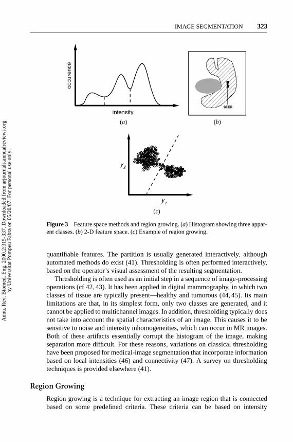
Thresholding approaches segment scalar images by creating a binary partitioningof the image intensities. Figure 3a shows the histogram of a scalar image thatpossesses three apparent classes, corresponding to the three modes. A thresholdingprocedure attempts to determine an intensity value, called the threshold, whichseparates the desired classes. The segmentation is then achieved by grouping allpixels with intensities greater than the threshold into one class and all other pixelsinto another class. Two potential thresholds are shown in Figure 3aat the valleys ofthe histogram. Determination of more than one threshold value is a process calledmultithresholding (41).
Thresholding is a simple yet often effective means for obtaining a segmenta-tion of images in which different structures have contrasting intensities or other
Ann
u. R
ev. B
iom
ed. E
ng. 2
000.
2:31
5-33
7. D
ownl
oade
d fr
om a
rjou
rnal
s.an
nual
revi
ews.
org
by U
nive
rsita
t Pom
peu
Fabr
a on
05/
20/0
7. F
or p
erso
nal u
se o
nly.
P1: FHY/ftt P2: FDR/FhN/fok/fgg QC: FhN
July 10, 2000 11:13 Annual Reviews AR106-12
?IMAGE SEGMENTATION 323
(a) (b)
(c)
Figure 3 Feature space methods and region growing. (a) Histogram showing three appar-ent classes. (b) 2-D feature space. (c) Example of region growing.
quantifiable features. The partition is usually generated interactively, althoughautomated methods do exist (41). Thresholding is often performed interactively,based on the operator’s visual assessment of the resulting segmentation.
Thresholding is often used as an initial step in a sequence of image-processingoperations (cf 42, 43). It has been applied in digital mammography, in which twoclasses of tissue are typically present—healthy and tumorous (44, 45). Its mainlimitations are that, in its simplest form, only two classes are generated, and itcannot be applied to multichannel images. In addition, thresholding typically doesnot take into account the spatial characteristics of an image. This causes it to besensitive to noise and intensity inhomogeneities, which can occur in MR images.Both of these artifacts essentially corrupt the histogram of the image, makingseparation more difficult. For these reasons, variations on classical thresholdinghave been proposed for medical-image segmentation that incorporate informationbased on local intensities (46) and connectivity (47). A survey on thresholdingtechniques is provided elsewhere (41).
Region Growing
Region growing is a technique for extracting an image region that is connectedbased on some predefined criteria. These criteria can be based on intensity
Ann
u. R
ev. B
iom
ed. E
ng. 2
000.
2:31
5-33
7. D
ownl
oade
d fr
om a
rjou
rnal
s.an
nual
revi
ews.
org
by U
nive
rsita
t Pom
peu
Fabr
a on
05/
20/0
7. F
or p
erso
nal u
se o
nly.
P1: FHY/ftt P2: FDR/FhN/fok/fgg QC: FhN
July 10, 2000 11:13 Annual Reviews AR106-12
?324 PHAM ■ XU ■ PRINCE
information and/or edges in the image (7). In its simplest form, region grow-ing requires a seed point that is manually selected by an operator and extracts allpixels connected to the initial seed based on some predefined criteria. For example,one possible criterion might be to grow the region until an edge in the image is met.This is depicted in Figure 3b, in which region growing has been used to isolateone of the structures from Figure 1a.
Like thresholding, region growing is seldom used alone but usually withina set of image-processing operations, particularly for the delineation of small,simple structures such as tumors and lesions (48, 49). The primary disadvantageof region growing is that it requires manual interaction to obtain the seed point.Thus, for each region that needs to be extracted, a seed must be planted. Split-and-merge is an algorithm related to region growing, but it does not require aseed point (50). Region growing can also be sensitive to noise, causing extractedregions to have holes or even become disconnected. Conversely, partial-volumeeffects can cause separate regions to become connected. To help alleviate theseproblems, a homotopic region-growing algorithm has been proposed that preservesthe topology between an initial region and an extracted region (51). Fuzzy analogiesto region growing have also been developed (52).
Classifiers
Classifier methods are pattern recognition techniques that seek to partition a featurespace derived from the image by using data with known labels (37, 53). A featurespace is the range space of any function of the image, with the most commonfeature space being the image intensities themselves. A histogram, as shown inFigure 3a, is an example of a one-dimensional feature space. Figure 3c showsan example of a partitioned 2-D feature space with two apparent classes. Such afeature space might have been generated from a dual-echo MR image, in whichone axis represents the intensities of the proton density-weighted image and theother axis represents the intensities of theT2-weighted image. All pixels with theirassociated features on the left side of the partition would be grouped into one class.
Classifiers are known as supervised methods because they require training datathat are manually segmented and then used as references for automatically seg-menting new data. There are a number of ways in which training data can beapplied in classifier methods. A simple classifier is the nearest-neighbor clas-sifier, in which each pixel is classified in the same class as the training datumwith the closest intensity. Thek-nearest-neighbor classifier is a generalization ofthis approach, in which the pixel is classified into the same class as the major-ity of the k-closest training data. Thek-nearest-neighbor classifier is considereda nonparametric classifier because it makes no underlying assumption about thestatistical structure of the data. Another nonparametric classifier is the Parzenwindow, in which the classification is made by a weighted decision processwithin a predefined window of the feature space, centered at the unlabeled pixelintensity.
Ann
u. R
ev. B
iom
ed. E
ng. 2
000.
2:31
5-33
7. D
ownl
oade
d fr
om a
rjou
rnal
s.an
nual
revi
ews.
org
by U
nive
rsita
t Pom
peu
Fabr
a on
05/
20/0
7. F
or p
erso
nal u
se o
nly.
P1: FHY/ftt P2: FDR/FhN/fok/fgg QC: FhN
July 10, 2000 11:13 Annual Reviews AR106-12
?IMAGE SEGMENTATION 325
A commonly used parametric classifier is the maximum-likelihood or Bayesclassifier. It assumes that the pixel intensities are independent samples from amixture of probability distributions, usually Gaussian. This mixture, called a finite-mixture model, is given by the probability density function
f (yj ; θ, π) =K∑
k=1
πk fk(yj ; θk) (5)
whereyj is the intensity of pixelj, fk is a component probability density functionparameterized byθk, andθ = [θ1, . . . , θK ]. The variablesπk are mixing coeffi-cients that weight the contribution of each density function andπ = [π1, . . . , πK ].Training data are collected by obtaining representative samples from each compo-nent of the mixture model and then estimating eachθk accordingly. For Gaussianmixtures, this means estimatingK -means, covariances, and mixing coefficients.Classification of new data is obtained by assigning each pixel to the class with thehighest posterior probability. When the data truly follow a finite Gaussian mixturedistribution, the maximum-likelihood classifier can perform well and is capable ofproviding a soft segmentation composed of the posterior probabilities. Additionalparametric and nonparametric classifiers are described in Reference 3.
Standard classifiers require that the structures to be segmented possess distinctquantifiable features. Because training data can be labeled, classifiers can trans-fer these labels to new data as long as the feature space sufficiently distinguisheseach label as well. Being noniterative, classifiers are relatively computationallyefficient, and, unlike thresholding methods, they can be applied to multichannelimages (54). A disadvantage of classifiers is that they generally do not performany spatial modeling. This weakness has been addressed in recent work extendingclassifier methods to segmenting images that are corrupted by intensity inhomo-geneities (21). Neighborhood and geometric information was also incorporatedinto a classifier approach in Reference 55. Another disadvantage is the require-ment of manual interaction to obtain training data. Training sets can be acquired foreach image that requires segmenting, but this can be time consuming and labori-ous. On the other hand, use of the same training set for a large number of scans canlead to biased results that do not take into account anatomical and physiologicalvariability between different subjects.
Clustering
Clustering algorithms essentially perform the same function as classifier methodswithout the use of training data. Thus, they are termed unsupervised methods. Tocompensate for the lack of training data, clustering methods iteratatively alternatebetween segmenting the image and characterizing the properties of each class. Ina sense, clustering methods train themselves, using the available data.
Three commonly used clustering algorithms are theK -means or ISODATA al-gorithm (56), the fuzzyc-means algorithm (37), and the expectation-maximization
Ann
u. R
ev. B
iom
ed. E
ng. 2
000.
2:31
5-33
7. D
ownl
oade
d fr
om a
rjou
rnal
s.an
nual
revi
ews.
org
by U
nive
rsita
t Pom
peu
Fabr
a on
05/
20/0
7. F
or p
erso
nal u
se o
nly.
P1: FHY/ftt P2: FDR/FhN/fok/fgg QC: FhN
July 10, 2000 11:13 Annual Reviews AR106-12
?326 PHAM ■ XU ■ PRINCE
(EM) algorithm (33, 57). TheK -means clustering algorithm clusters data by iter-atively computing a mean intensity for each class and segmenting the image byclassifying each pixel in the class with the closest mean (58). Figure 4b showsthe result of applying theK -means algorithm to a slice of an MR brain image inFigure 4a. The number of classes was assumed to be three, representing (fromdark gray to white in Figure 4) cerebrospinal fluid, gray matter, and white matter.The fuzzyc-means algorithm generalizes theK -means algorithm, allowing forsoft segmentations based on fuzzy set theory (17). The EM algorithm appliesthe same clustering principles with the underlying assumption that the data fol-low a Gaussian mixture model (see Equation 5). It iterates between computing
(a) (b)
(c)
Figure 4 Segmentation of a magnetic resonance brain image. (a) Original image. (b)Segmentation using theK -means algorithm. (c) Segmentation using theK -means algorithmwith a Markov random field prior.
Ann
u. R
ev. B
iom
ed. E
ng. 2
000.
2:31
5-33
7. D
ownl
oade
d fr
om a
rjou
rnal
s.an
nual
revi
ews.
org
by U
nive
rsita
t Pom
peu
Fabr
a on
05/
20/0
7. F
or p
erso
nal u
se o
nly.
P1: FHY/ftt P2: FDR/FhN/fok/fgg QC: FhN
July 10, 2000 11:13 Annual Reviews AR106-12
?IMAGE SEGMENTATION 327
the posterior probabilities and computing maximum likelihood estimates of themeans, covariances, and mixing coefficients of the mixture model.
Although clustering algorithms do not require training data, they do requirean initial segmentation (or, equivalently, initial parameters). The EM algorithmhas demonstrated greater sensitivity to initialization than theK -means or fuzzyc-means algorithm (31). Like classifier methods, clustering algorithms do notdirectly incorporate spatial modeling and can therefore be sensitive to noise andintensity inhomogeneities. This lack of spatial modeling, however, can provide sig-nificant advantages for fast computation (59). Work on improving the robustnessof clustering algorithms to intensity inhomogeneities in MR images has demon-strated excellent success (11, 18). Robustness to noise can be incorporated by MRFmodeling as described in the next section.
Markov Random Field Models
MRF modeling itself is not a segmentation method but a statistical model thatcan be used within segmentation methods. MRFs model spatial interactions be-tween neighboring or nearby pixels. These local correlations provide a mecha-nism for modeling a variety of image properties (60). In medical imaging, theyare typically used because most pixels belong to the same class as their neigh-boring pixels. In physical terms, this implies that any anatomical structure thatconsists of only one pixel has a very low probability of occurring under an MRFassumption.
MRFs are often incorporated into clustering segmentation algorithms such astheK -means algorithm under a Bayesian prior model (11, 29, 30). The segmenta-tion is then obtained by maximizing the a posteriori probability of the segmentation,given the image data. This maximization can be achieved by iterative methods suchas iterated conditional modes (61) or simulated annealing (62). Figure 4c showsthe robustness to noise in a segmentation resulting from an MRF prior. The seg-mentation does not exhibit as many small, disconnected regions as the non-MRFresult of Figure 4b.
A difficulty associated with MRF models is proper selection of the parameterscontrolling the strength of spatial interactions (60). A setting that is too high canresult in an excessively smooth segmentation and a loss of important structuraldetails. In addition, MRF methods usually require computationally intensive al-gorithms. Despite these disadvantages, MRFs are widely used not only to modelsegmentation classes, but also to model intensity inhomogeneities that can occurin MR images (30) and textural properties, which is useful in the segmentation ofdigital mammograms (63).
Artificial Neural Networks
Artificial neural networks (ANNs) are parallel networks of processing elementsor nodes that simulate biological learning. Each node in an ANN is capable ofperforming elementary computations. Learning is achieved through the adaptation
Ann
u. R
ev. B
iom
ed. E
ng. 2
000.
2:31
5-33
7. D
ownl
oade
d fr
om a
rjou
rnal
s.an
nual
revi
ews.
org
by U
nive
rsita
t Pom
peu
Fabr
a on
05/
20/0
7. F
or p
erso
nal u
se o
nly.
P1: FHY/ftt P2: FDR/FhN/fok/fgg QC: FhN
July 10, 2000 11:13 Annual Reviews AR106-12
?328 PHAM ■ XU ■ PRINCE
of weights assigned to the connections between nodes. A thorough treatment onneural networks can be found in References 64 and 65.
ANNs represent a paradigm for machine learning and can be used in a varietyof ways for image segmentation. The most widely applied use in medical imagingis as a classifier (40, 66), in which the weights are determined by using trainingdata and the ANN is then used to segment new data. ANNs can also be used inan unsupervised fashion as a clustering method (37, 67), as well as for deformablemodels (68). Because of the many interconnections used in a neural network,spatial information can be easily incorporated into its classification procedures.Although ANNs are inherently parallel, their processing is usually simulated on astandard serial computer, thus reducing this potential computational advantage.
Deformable Models
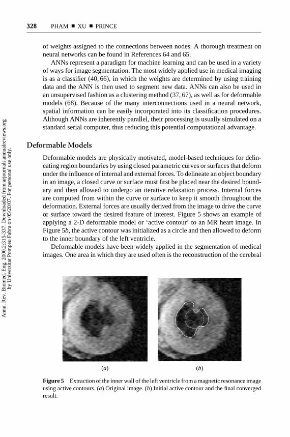
Deformable models are physically motivated, model-based techniques for delin-eating region boundaries by using closed parametric curves or surfaces that deformunder the influence of internal and external forces. To delineate an object boundaryin an image, a closed curve or surface must first be placed near the desired bound-ary and then allowed to undergo an iterative relaxation process. Internal forcesare computed from within the curve or surface to keep it smooth throughout thedeformation. External forces are usually derived from the image to drive the curveor surface toward the desired feature of interest. Figure 5 shows an example ofapplying a 2-D deformable model or ‘active contour’ to an MR heart image. InFigure 5b, the active contour was initialized as a circle and then allowed to deformto the inner boundary of the left ventricle.
Deformable models have been widely applied in the segmentation of medicalimages. One area in which they are used often is the reconstruction of the cerebral
(a) (b)
Figure 5 Extraction of the inner wall of the left ventricle from a magnetic resonance imageusing active contours. (a) Original image. (b) Initial active contour and the final convergedresult.
Ann
u. R
ev. B
iom
ed. E
ng. 2
000.
2:31
5-33
7. D
ownl
oade
d fr
om a
rjou
rnal
s.an
nual
revi
ews.
org
by U
nive
rsita
t Pom
peu
Fabr
a on
05/
20/0
7. F
or p
erso
nal u
se o
nly.
P1: FHY/ftt P2: FDR/FhN/fok/fgg QC: FhN
July 10, 2000 11:13 Annual Reviews AR106-12
?IMAGE SEGMENTATION 329
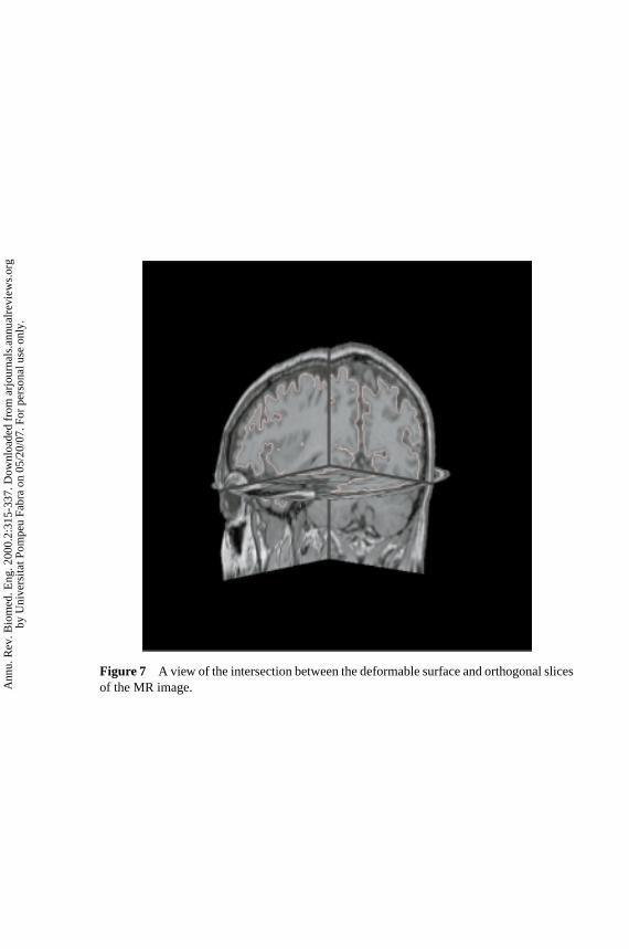
cortex from MR images (69–71). An example of using a deformable surface modelfor this application is shown in Figure 6 (see color insert). A view of the intersectionbetween this surface and orthogonal slices of the MR image volume is givenin Figure 7 (see color insert). Deformable models have also been used in thesegmentation of cardiac images (72), bone in CT images (73), and ultrasound(74). The dynamic nature of deformable models make tham especially well suitedto motion-tracking tasks, which are common in ultrasound imaging.
The main advantages of deformable models are their ability to directly gener-ate closed parametric curves or surfaces from images and their incorporation ofa smoothness constraint that provides robustness to noise and spurious edges. Adisadvantage is that they require manual interaction to place an initial model andchoose appropriate parameters. Reducing sensitivity to initialization has been atopic of research that has demonstrated excellent success (75–78). Standard de-formable models can also exhibit poor convergence to concave boundaries. Thisdifficulty can be alleviated somewhat through the use of pressure forces (75) andother modified external-force models (78). Another important extension of de-formable models is the adaptivity of model topology by using an implicit repre-sentation rather than an explicit parameterization (76, 77, 79). A general review ondeformable models in medical image analysis can be found in reference 80.
Atlas-Guided Approaches
Atlas-guided approaches are a powerful tool for medical-image segmentation whena standard atlas or template is available. The atlas is generated by compiling infor-mation on the anatomy that requires segmenting. This atlas is then used as a ref-erence frame for segmenting new images. Conceptually, atlas-guided approachesare similar to classifiers except that they are implemented in the spatial domain ofthe image rather than in a feature space.
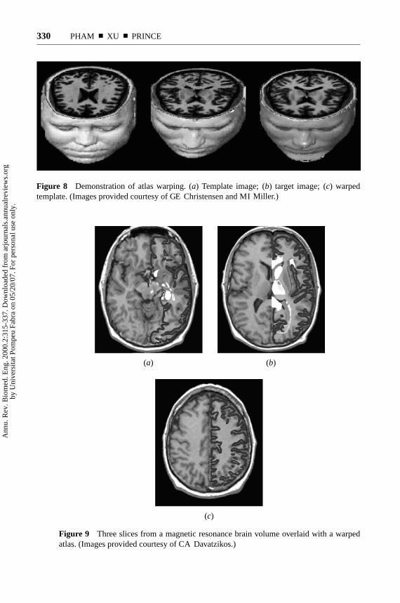
The standard atlas-guided approach treats segmentation as a registration prob-lem (see 81 for a detailed survey on registration techniques). It first finds a one-to-one transformation that maps a presegmented atlas image to the target imagethat requires segmenting. This process is often referred to as ‘atlas warping.’ Thewarping can be performed with linear (82–84) transformations, but, because ofanatomical variability, a sequential application of linear and nonlinear (15, 85–87)transformations is often used. An example of atlas warping for an MR head scanis shown in Figure 8 (87). Because the atlas is already segmented, all structuralinformation is transferred to the target image. This is shown in Figure 9, in whichthe Talairach brain atlas (82) has been mapped to an MR image (86).
Atlas-guided approaches have been applied mainly in MR brain imaging forsegmentation of various structures (85), as well as for extracting the brain volumefrom head scans (88). An advantage of atlas-guided approaches is that labelsare transferred as well as the segmentation. They also provide a standard systemfor studying morphometric properties (89, 90). Even with nonlinear registrationmethods, however, finding accurate segmentations of complex structures is difficult
Ann
u. R
ev. B
iom
ed. E
ng. 2
000.
2:31
5-33
7. D
ownl
oade
d fr
om a
rjou
rnal
s.an
nual
revi
ews.
org
by U
nive
rsita
t Pom
peu
Fabr
a on
05/
20/0
7. F
or p
erso
nal u
se o
nly.
P1: FhN/FGI P2: FPX/FOK QC: FHN/fgm T1: FhN
July 31, 2000 4:55 Annual Reviews AR106-01
?Figure 6 An example of using a deformable surface in the reconstruction of the cerebralcortex.A
nnu.
Rev
. Bio
med
. Eng
. 200
0.2:
315-
337.
Dow
nloa
ded
from
arj
ourn
als.
annu
alre
view
s.or
gby
Uni
vers
itat P
ompe
u Fa
bra
on 0
5/20
/07.
For
per
sona
l use
onl
y.
P1: FhN/FGI P2: FPX/FOK QC: FHN/fgm T1: FhN
July 31, 2000 4:55 Annual Reviews AR106-01
?Figure 7 A view of the intersection between the deformable surface and orthogonal slicesof the MR image.A
nnu.
Rev
. Bio
med
. Eng
. 200
0.2:
315-
337.
Dow
nloa
ded
from
arj
ourn
als.
annu
alre
view
s.or
gby
Uni
vers
itat P
ompe
u Fa
bra
on 0
5/20
/07.
For
per
sona
l use
onl
y.
P1: FHY/ftt P2: FDR/FhN/fok/fgg QC: FhN
July 10, 2000 11:13 Annual Reviews AR106-12
?330 PHAM ■ XU ■ PRINCE
Figure 8 Demonstration of atlas warping. (a) Template image; (b) target image; (c) warpedtemplate. (Images provided courtesy of GE Christensen and MI Miller.)
Figure 9 Three slices from a magnetic resonance brain volume overlaid with a warpedatlas. (Images provided courtesy of CA Davatzikos.)
(a) (b)
(c)
Ann
u. R
ev. B
iom
ed. E
ng. 2
000.
2:31
5-33
7. D
ownl
oade
d fr
om a
rjou
rnal
s.an
nual
revi
ews.
org
by U
nive
rsita
t Pom
peu
Fabr
a on
05/
20/0
7. F
or p
erso
nal u
se o
nly.
P1: FHY/ftt P2: FDR/FhN/fok/fgg QC: FhN
July 10, 2000 11:13 Annual Reviews AR106-12
?IMAGE SEGMENTATION 331
owing to anatomical variability. This is shown in Figure 9, in which the cerebralcortex is not segmented as accurately as shown in Figure 4. Thus, atlas-guidedapproaches are generally better suited for segmentation of structures that are stableover the population of study. One method that helps model anatomical variability isthe use of probabilistic atlases (89), but these require additional time and interactionto accumulate data. Another method is the use of manually selected landmarks toconstrain transformation (86).
Other Approaches
Model-fitting is a segmentation method that typically fits a simple geometric shapesuch as an ellipse or parabola to the locations of extracted image features in animage (91). This technique is specialized to the structure being segmented but iseasily implemented and can provide good results when the model is appropriate.A more general approach is to fit spline curves or surfaces (92) to the features.The main difficulty with model-fitting is that image features must first be extractedbefore the fitting can take place.
The watershed algorithm uses concepts from edge detection and mathematicalmorphology (8) to partition images into homogeneous regions (93). The methodcan suffer from oversegmentation, which occurs when the image is segmented intoan unnecessarily large number of regions. Thus, watershed algorithms in medicalimaging are usually followed by a post-processing step to merge separate regionsthat belong to the same structure (94).
Figure 10 shows an example in which a mammogram is initially oversegmentedby using a watershed algorithm. A statistical classifier (95) is then used to deter-mine which regions contain microcalcifications. This classification step is typicallyperformed based on textural properties. Note that a perfect delineation of microcal-cifications and masses in mammograms is difficult but not often necessary becausedetection is the primary goal.
CONCLUSION
Future research in the segmentation of medical images will strive toward improv-ing the accuracy, precision, and computational speed of segmentation methods,as well as reducing the amount of manual interaction. Accuracy and precisioncan be improved by incorporating prior information from atlases and by combin-ing discrete and continuous spatial-domain segmentation methods. For increasingcomputational efficiency, multiscale processing (cf 96) and parallelizable methodssuch as neural networks are promising approaches. Computational efficiency willbe particularly important in real-time processing applications.
Possibly the most important question surrounding the use of image segmen-tation is its application in clinical settings. Computerized segmentation methods
Ann
u. R
ev. B
iom
ed. E
ng. 2
000.
2:31
5-33
7. D
ownl
oade
d fr
om a
rjou
rnal
s.an
nual
revi
ews.
org
by U
nive
rsita
t Pom
peu
Fabr
a on
05/
20/0
7. F
or p
erso
nal u
se o
nly.
P1: FHY/ftt P2: FDR/FhN/fok/fgg QC: FhN
July 10, 2000 11:13 Annual Reviews AR106-12
?332 PHAM ■ XU ■ PRINCE
(a) (b)
(c)
Figure 10 Segmentation in digital mammography. (a) Digitized mammogram and radi-ologist’s boundary for biopsy-proven malignant tumor. (b) Result of watershed algorithm.(c) Suspicious regions determined by automated method. (Images provided courtesy ofCE Priebe.)
have already demonstrated their utility in research applications and are now gar-nering increased use for computer-aided diagnosis and radiotherapy planning.For segmentation methods to gain acceptance in routine clinical applications,extensive validation is required on the particular methods in question. Further-more, one must be able to demonstrate some significant performance advan-tage (e.g. more accurate diagnosis or earlier detection of pathology) over tra-ditional methods to warrant the training and equipment costs associated withusing computerized methods. It is unlikely that automated segmentation meth-ods will ever replace physicians, but they will likely become crucial elements ofmedical-image analysis. Segmentation methods will be particularly valuable in ar-eas such as image-guided surgery, in which visualization of the anatomy is a criticalcomponent.
Ann
u. R
ev. B
iom
ed. E
ng. 2
000.
2:31
5-33
7. D
ownl
oade
d fr
om a
rjou
rnal
s.an
nual
revi
ews.
org
by U
nive
rsita
t Pom
peu
Fabr
a on
05/
20/0
7. F
or p
erso
nal u
se o
nly.
P1: FHY/ftt P2: FDR/FhN/fok/fgg QC: FhN
July 10, 2000 11:13 Annual Reviews AR106-12
?IMAGE SEGMENTATION 333
Visit the Annual Reviews home page at www.AnnualReviews.org
LITERATURE CITED
1. Larie SM, Abukmeil SS. 1998. Brain ab-normality in schizophrenia: a systematicand quantitative review of volumetric mag-netic resonance imaging studies.J. Psy-chol.172:110–20
2. Taylor P. 1995. Invited review: com-puter aids for decision-making in diagnos-tic radiology—a literature review.Br. J. Ra-diol. 68:945–57
3. Zijdenbos AP, Dawant BM. 1994. Brainsegmentation and white matter lesion de-tection in MR images.Crit. Rev. Biomed.Eng.22:401–65
4. Worth AJ, Makris N, Caviness VS,Kennedy DN. 1997. Neuroanatomical seg-mentation in MRI: technological objec-tives.Int. J. Pattern Recognit. Artif. Intell.11:1161–87
5. Khoo VS, Dearnaley DP, Finnigan DJ,Padhani A, Tanner SF, Leach MO. 1997.Magnetic resonance imaging (MRI): con-siderations and applications in radiothera-phy treatment planning.Radiother. Oncol.42:1–15
6. Grimson WEL, Ettinger GJ, Kapur T, Lev-enton ME, Wells WM, et al. 1997. Utiliz-ing segmented MRI data in image-guidedsurgery.Int. J. Pattern Recognit. Artif. In-tell. 11:1367–97
9. Pal NR, Pal SK. 1993. A review on imagesegmentation techniques.Pattern Recog-nit. 26:1277–94
10. Langan DA, Modestino JW, Zhang J.1998. Cluster validation for unsupervisedstochastic model-based image segmenta-tion. IEEE Trans. Image Process.7:180–95
11. Rajapakse JC, Giedd JN, Rapoport JL.1997. Statistical approach to segmenta-tion of single-channel cerebral MR images.IEEE Trans. Med. Imaging16:176–86
12. Pham DL, Prince JL, Dagher AP, Xu C.1997. An automated technique for statis-tical characterization of brain tissues inmagnetic resonance imaging.Int. J. PatternRecognit. Artif. Intell.11(8):1189–211
13. Ge Y, Fitzpatrick JM, Dawant BM, Bao J,Kessler RM, Margolin R. 1996. Accuratelocalization of cortical convolutions in MRbrain images.IEEE Trans. Med. Imaging15:418–28
14. Rademacher J, Galaburda AM, KennedyDN, Filipek PA, Caviness VS. 1992. Hu-man cerebral cortex: localization, parcel-lation and morphometry with magnetic res-onance imaging.J. Cogn. Neurosci.4:352–74
15. Sandor S, Leahy R. 1997. Surface-basedlabeling of cortical anatomy using a de-formable atlas.IEEE Trans. Med. Imaging16:41–54
16. Khaneja N, Miller MI, Grenander U.1998. Dynamic programming generationof curves on brain surfaces.IEEE Trans.Pattern Anal. Mach. Intell.20:1260–65
18. Pham DL, Prince JL. 1999. An adap-tive fuzzy c-means algorithm for imagesegmentation in the presence of intensityinhomogeneities.Pattern Recognit. Lett.20:57–68
19. Herndon RC, Lancaster JL, Toga AW, FoxPT. 1996. Quantification of white matterand gray matter volumes from T1 paramet-ric images using fuzzy classifiers.J. Magn.Reson. Imaging6:425–35
20. Liang Z. 1993. Tissue classification andsegmentation of MR images.IEEE Eng.Med. Biol.12:81–85
32. Wust P, Gellermann J, Beier J, Wegner S,Tilly W, et al. 1998. Evaluation of seg-mentation algorithms for generation of pa-tient models in radiofrequency hyperther-mia. Phys. Med. Biol.43:3295–307
33. Lei T, Sewchand W. 1992. Statistical ap-proach to X-ray CT imaging and its ap-plications in image analysis. II. A newstochastic model-based image segmenta-tion technique for X-ray CT image.IEEETrans. Med. Imaging11(1):62–69
34. Collins DL, Zijdenbos AP, Kollokian V,Sled JG, Kabani NJ, et al. 1998. De-sign and construction of a realistic digitalbrain phantom.IEEE Trans. Med. Imaging17:463–68
35. Chalana V, Kim Y. 1997. A methodologyfor evaluation of boundary detection algo-rithms on medical images.IEEE Trans.Med. Imaging16:642–52
36. Zhang YJ. 1996. A survey of evaluationmethods for image segmentation.PatternRecognit. Lett.29:1335–46
37. Bezdek JC, Hall LO, Clarke LP. 1993.Review of MR image segmentation tech-niques using pattern recognition.Med.Phys.20:1033–48
38. Clarke LP, Velthuizen RP, Camacho MA,Heine JJ, Vaidyanathan M, et al. 1995.MRI segmentation: methods and applica-tions. Magn. Reson. Imaging13:343–68
39. Vaidyanathan M, Clarke LP, Velthuizen RP,Phuphanich S, Bensaid AM, et al. 1995.Comparison of supervised MRI segmenta-tion methods for tumor volume determina-tion during therapy.Magn. Reson. Imaging13:719–28
40. Hall LO, Bensaid AM, Clarke LP,Velthuizen RP, Silbiger MS, Bezdek JC.1992. A comparison of neural network andfuzzy clustering techniques in segmentingmagnetic resonance images of the brain.IEEE Trans. Neural Netw.3:672–82
41. Sahoo PK, Soltani S, Wong AKC. 1988. Asurvey of thresholding techniques.Com-put. Vis. Graph. Image Proc.41:233–60
43. Gordon CL, Webber CE, Adachi JD,Christoforou N. 1996. In vivo assessmentof trabecular bone structure at the distalradius from high-resolution computed to-mography images.Phys. Med. Biol.41:495–508
44. Polakowski WE, Cournoyer DA, RogersSK, DeSimio MP, Ruck DW, et al. 1997.Computer-aided breast cancer detectionand diagnosis of masses using differenceof Gaussians and derivative-based fea-ture saliency.IEEE Trans. Med. Imaging16:811–19
45. Cheng H, Lui YM, Freimanis RI. 1998.A novel approach to microcalcification de-tection using fuzzy logic technique.IEEETrans. Med. Imaging17:442–50
46. Li HD, Kallergi M, Clarke LP, Jain VK,Clark RA. 1995. Markov random field fortumor detection in digital mammography.IEEE Trans. Med. Imaging14:565–76
47. Lee C, Hun S, Ketter TA, Unser M. 1998.Unsupervised connectivity-based thresh-olding segmentation of midsaggital brainMR images.Comput. Biol. Med.28:309–38
48. Gibbs P, Buckley DL, Blackband SJ, Hors-man A. 1996. Tumour volume detectionfrom MR images by morphological seg-mentation.Phys. Med. Biol.41:2437–46
49. Pohlman S, Powell KA, Obuchowski NA,Chilcote WA, Broniatowski SG. 1996.Quantitative classification of breast tumorsin digitized mammograms.Med. Phys.23:1337–45
50. Manousakas IN, Undrill PE, Cameron GG,Redpath TW. 1998. Split-and-merge seg-mentation of magnetic resonance medi-cal images: performance evaluation andextension to three dimensions.Comput.Biomed. Res.31:393–412
51. Mangin JF, Frouin V, Bloch I, Regis J,Krahe J, Lopez. 1995. From 3D mag-
netic resonance images to structural rep-resentations of the cortex topography us-ing topology preserving deformations.J.Math. Imaging Vis.5:297–318
52. Udupa JK, Samarasekera S. 1996. Fuzzyconnectedness and object definition: the-ory, algorithms and applications in imagesegmentation.Graph. Models Image Pro-cess.58(3):246–61
53. Schalkoff RJ. 1992.Pattern Recognition:Statistical, Structural and Neural Ap-proaches. New York: Wiley & Sons. 364pp.
54. Vannier MW, Butterfield RL, Jordan D,Murphy WA, Levitt RG, Gado M. 1985.Multispectral analysis of magnetic reso-nance images.Radiology154:221–24
55. Kapur T, Grimson WEL, Kikinis R, WellsWM. 1998. Enhanced spatial priors for seg-mentation of magnetic resonance imageryIn Proc. Int. Conf. Med. Image Comput.Comp. Assist. Interv., 1st, Cambridge, MA,pp. 457–68. Berlin: Springer-Verlag
56. Coleman GB, Andrews HC. 1979. Imagesegmentation by clustering.Proc. IEEE5:773–85
57. Liang Z, MacFall JR, Harrington DP. 1994.Parameter estimation and tissue segmenta-tion from multispectral MR images.IEEETrans. Med. Imaging13:441–49
59. Hebert TJ. 1997. Fast iterative segmen-tation of high resolution medical images.IEEE Trans. Nucl. Sci.44:1363–67
60. Li SZ. 1995.Markov Random Field Model-ing in Computer Vision.Berlin/New York:Springer-Verlag. 264 pp.
61. Besag J. 1986. On the statistical analysisof dirty pictures.CVGIP: Image Underst.57:359–72
62. Geman S, Geman D. 1984. Stochasticrelaxation, Gibbs distributions, and theBayesian restoration of images.IEEETrans. Pattern Anal. Mach. Intell.6:721–41
Ann
u. R
ev. B
iom
ed. E
ng. 2
000.
2:31
5-33
7. D
ownl
oade
d fr
om a
rjou
rnal
s.an
nual
revi
ews.
org
by U
nive
rsita
t Pom
peu
Fabr
a on
05/
20/0
7. F
or p
erso
nal u
se o
nly.
P1: FHY/ftt P2: FDR/FhN/fok/fgg QC: FhN
July 10, 2000 11:13 Annual Reviews AR106-12
?336 PHAM ■ XU ■ PRINCE
63. Chen CH, Lee GG. 1997. On digitalmammogram segmentation and microcal-cification detection using multiresolutionwavelet analysis.Graph. Models ImageProcess.59:349–64
64. Clark JW. 1991. Neural network mod-elling. Phys. Med. Biol.36:1259–317
65. Haykin S. 1994.Neural Networks: AComprehensive Foundation. New York:Macmillan. 696 pp.
66. Gelenbe E, Feng Y, Krishnan KRR. 1996.Neural network methods for volumetricmagnetic resonance imaging of the humanbrain.Proc. IEEE84:1488–96
67. Reddick WE, Glass JO, Cook EN, ElkinTD, Deaton RJ. 1997. Automated segmen-tation and classification of multispectralmagnetic resonance images of brain us-ing artificial neural networks.IEEE Trans.Med. Imaging16:911–18
68. Vilarino DL, Brea VM, Cabello D, PardoJM. 1998. Discrete-time CNN for imagesegmentation by active contours.PatternRecognit. Lett.19:721–34
69. Davatzikos C, Bryan RN. 1996. Using a de-formable surface model to obtain a shaperepresentation of the cortex.IEEE Trans.Med. Imaging15:785–95
70. McInerney T, Terzopoulos D. 1997. Med-ical image segmentation using topologi-cally adaptable surfaces.Lect. Notes Com-put. Sci.1205:23–32
71. Xu C, Pham DL, Prince JL, Etemad ME,Yu D. 1998. Reconstruction of the centrallayer of the human cerebral cortex fromMR images. InProc. Int. Conf. Med. ImageComput. Comp. Assist. Interv., 1st, Cam-bridge, MA, pp. 482–88
72. Bardinet E, Cohen LD, Ayache N. 1998.A parametric deformable model to fit un-structured 3D data.Comput. Vis. ImageUnderst.71:39–54
73. Neumann A, Lorenz C. 1998. Statisticalshape model based segmentation of med-ical images.Comput. Med. Image Graph.22:133–43
74. Mikic I, Krucinski S, Thomas JD. 1998.
Segmentation and tracking in echocardio-graphic sequences: active contours guidedby optical flow estimates. IEEE Trans.Med. Imaging17:274–84
75. Cohen LD. 1991. On active contour mod-els and balloons.CVGIP: Image Underst.53:211–18
76. Caselles V, Catte F, Coll T, Dibos F. 1993.A geometric model for active contours.Nu-mer. Math.66:1–31
77. Malladi R, Sethian JA, Vemuri BC. 1995.Shape modeling with front propagation: alevel set approach.IEEE Trans. PatternAnal. Mach. Intell.17:158–75
80. McInerney T, Terzopoulos D. 1996. De-formable models in medical image anal-ysis: a survey. Med. Image Anal.1:91–108
81. Maintz JBA, Viergever MA. 1998. A sur-vey of medical image registration.Med.Image Anal.2:1–36
82. Talairach J, Tournoux P. 1988.Co-PlanarStereotaxic Atlas of the Human Brain.3-Dimensional Proportional System: AnApproach to Cerebral Imaging. Stuttgart,Ger. Thieme. 122 pp.
83. Lancaster JL, Rainey LH, Summerlin JL,Freitas CS, Fox PT, et al. 1997. Automatedlabeling of the human brain: a prelimi-nary report on the development and evalua-tion of a forward-transform method.Hum.Brain Mapp.5:238–42
84. Rajarethinam NC, Andreasen R, CizadloT, Arndt S, Swayze VW, et al. 1996. Au-tomatic atlas-based volume estimation ofhuman brain regions from MR images.J.Comput. Assist. Tomogr.20:98–106
86. Davatzikos C. 1996. Spatial normalizationof 3D images using deformable models.J.Comput. Assist. Tomogr.20:656–65
87. Christensen GE, Joshi SC, Miller MI.1997. Volumetric transformation of brainanatomy.IEEE Trans. Med. Imag.16:864–77
88. Aboutanos GB, Dawant BM. 1997. Au-tomatic brain segmentation and valida-tion: image-based versus atlas-based de-formable models. InSPIE Proc. Med.Imag. 3034:299–310
89. Thompson P, Toga AW. 1997. Detection,visualization and animation of abnormalanatomic structure with a probabilisticbrain atlas based on random vector fieldtransformations.Med. Image Anal.1:271–94
90. Joshi SC, Miller MI, Grenander U. 1997.On the geometry and shape of brain sub-manifolds.Int. J. Pattern Recognit. Artif.Intell. 11:1317–43
tal ultrasound guided prostate cancer ther-apy. IEEE Trans. Med. Imaging17:762–71
92. Bae KT, Giger ML, Chen C, Kahn CE.1993. Automatic segmentation of liverstructure in CT images.Med. Phys.20:71–78
93. Vincent L, Soille P. 1991. Watersheds indigital spaces: an efficient algorithm basedon immersion simulation. IEEE Trans.Pattern Anal. Mach. Intell.13:583–98
94. Sijbers J, Scheunders P, Verhoye M, VanDer Linden A, Van Dyck D, et al. 1997.Watershed-based segmentation of 3D MRdata for volume quantization.Magn. Re-son. Imag.15:679–88
95. Priebe CE, Marchette DJ, Rogers GW.1997. Segmentation of random fieldsvia borrowed strength density estimation.IEEE Trans. Pattern Anal. Mach. Intell.19:494–99
Annual Review of Bimedical Engineering Volume 2, 2000
CONTENTS
PIERRE M. GALLETTI: A Personal Reflection, Robert M. Nerem 1
PHYSICOCHEMICAL FOUNDATIONS AND STRUCTURAL DESIGN OF HYDROGELS IN MEDICINE AND BIOLOGY, N. A. Peppas, Y. Huang, M. Torres-Lugo, J. H. Ward, J. Zhang 9
BIOENGINEERING MODELS OF CELL SIGNALING, Anand R. Asthagiri, Douglas A. Lauffenburger 31
FUNDAMENTALS OF IMPACT BIOMECHANICS: Part I - Biomechanics of the Head, Neck, and Thorax, Albert I. King 55INJURY AND REPAIR OF LIGAMENTS AND TENDONS, Savio L.-Y. Woo, Richard E. Debski, Jennifer Zeminski, Steven D. Abramowitch, Serena S. Chan Saw, MS, James A. Fenwick 83
ELECTROPHYSIOLOGICAL MODELING OF CARDIAC VENTRICULAR FUNCTION: From Cell to Organ, R. L. Winslow, D. F. Scollan, A. Holmes, C. K. Yung, J. Zhang, M. S. Jafri 119
CRYOSURGERY, Boris Rubinsky 157CELL MECHANICS: Mechanical Response, Cell Adhesion, and Molecular Deformation, Cheng Zhu, Gang Bao, Ning Wang 189
MICROENGINEERING OF CELLULAR INTERACTIONS, Albert Folch, Mehmet Toner 227
QUANTITATIVE MEASUREMENT AND PREDICTION OF BIOPHYSICAL RESPONSE DURING FREEZING IN TISSUES, John C. Bischof 257
MICROFABRICATED MICRONEEDLES FOR GENE AND DRUG DELIVERY, Devin V. McAllister, Mark G. Allen, Mark R. Prausnitz 289CURRENT METHODS IN MEDICAL IMAGE SEGMENTATION, Dzung L. Pham, Chenyang Xu, Jerry L. Prince 315
ANTIBODY ENGINEERING, Jennifer Maynard, George Georgiou 339
NEW CURRENTS IN ELECTRICAL STIMULATION OF EXCITABLE TISSUES, Peter J. Basser, Bradley J. Roth 377
TWO-PHOTON EXCITATION FLUORESCENCE MICROSCOPY, Peter T. C. So, Chen Y. Dong, Barry R. Masters, Keith M. Berland 399IMAGING THREE-DIMENSIONAL CARDIAC FUNCTION, W. G. O'Dell, A. D. McCulloch 431
THREE-DIMENSIONAL ULTRASOUND IMAGING, Aaron Fenster, Donal B. Downey 457
BIOPHYSICAL INJURY MECHANISMS IN ELECTRICAL SHOCK TRAUMA, Raphael C. Lee, Dajun Zhang, Jurgen Hannig 477
WAVELETS IN TEMPORAL AND SPATIAL PROCESSING OF BIOMEDICAL IMAGES, Andrew F. Laine 511
Ann
u. R
ev. B
iom
ed. E
ng. 2
000.
2:31
5-33
7. D
ownl
oade
d fr
om a
rjou
rnal
s.an
nual
revi
ews.
org
by U
nive
rsita
t Pom
peu
Fabr
a on
05/
20/0
7. F
or p
erso
nal u
se o
nly.
MICRODEVICES IN MEDICINE, Dennis L. Polla, Arthur G. Erdman, William P. Robbins, David T. Markus, Jorge Diaz-Diaz, Raed Rizq, Yunwoo Nam, Hui Tao Brickner, Amy Wang, Peter Krulevitch 551NEUROENGINEERING MODELS OF BRAIN DISEASE, Leif H. Finkel 577
EXTRACORPOREAL TISSUE ENGINEERED LIVER-ASSIST DEVICES, Emmanouhl S. Tzanakakis, Donavon J. Hess, Timothy D. Sielaff, Wei-Shou Hu 607
MAGNETIC RESONANCE STUDIES OF BRAIN FUNCTION AND NEUROCHEMISTRY, Kâmil Ugurbil, Gregor Adriany, Peter Andersen, Wei Chen, Rolf Gruetter, Xiaoping Hu, Hellmut Merkle, Dae-Shik Kim, Seong-Gi Kim, John Strupp, Xiao Hong Zhu, Seiji Ogawa 633
INTERVENTIONAL AND INTRAOPERATIVE MAGNETIC RESONANCE IMAGING, J. Kettenbach, D. F. Kacher, S. K. Koskinen, Stuart G. Silverman, A. Nabavi, Dave Gering, Clare M. C. Tempany, R. B. Schwartz, R. Kikinis, P. M. Black, F. A. Jolesz 661CARTILAGE TISSUE REMODELING IN RESPONSE TO MECHANICAL FORCES, Alan J. Grodzinsky, Marc E. Levenston, Moonsoo Jin, Eliot H. Frank 691
IN VIVO NEAR-INFRARED SPECTROSCOPY, Peter Rolfe 715