Surface plasmon resonance-based biosensors: From the development of different SPR structures to novel surface functionalization strategies Edy Wijaya a , Cédric Lenaerts b , Sophie Maricot a , Juriy Hastanin b , Serge Habraken c , Jean-Pierre Vilcot a,⇑ , Rabah Boukherroub a,d , Sabine Szunerits a,d,⇑ a Institut d’Electronique, de Microélectronique, et de Nanotechnologie (IEMN), UMR 8520, Université Lille Nord de France, Avenue Poincaré, BP 60069, 59652 Villeneuve d’Ascq Cedex, France b Centre Spatial de Liège, Université de Liège, Avenue du Pré Aily, 4031 Angleur, Belgium c Université de Liège, Hololab, Allée du 6 Août, 17, 4000 Liège, Belgium d Institut de Recherche Interdisciplinaire (IRI), USR-3078, Université Lille Nord de France, Parc de la Haute Borne, 50 Avenue de Halley, BP 70478, 59658 Villeneuve d’Ascq, France article info Article history: Received 21 December 2010 Accepted 14 May 2011 Available online 7 June 2011 Keywords: Surface plasmon resonance SPR Biosensors Surface functionalization abstract Surface plasmon resonance (SPR)-based biosensors are very powerful tools for the study of biomolecular interactions, chemical detection and immunoassays. This paper reviews the performance of various SPR structures and detection schemes focusing on propagating surface plasmons generated in planar struc- tures. Some aspects of their surface functionalization, the key element which imparts biofunctionality to these structures and hence transforming them into biosensors, will also be discussed accordingly. The ultimate performance of SPR-based biosensors will thus be determined by both their inherent optical performance and suitable surface functionalization. Ó 2011 Elsevier Ltd. All rights reserved. 1. Introduction The first documented observation of surface plasmons dates back as early as 1902 when R.W. Wood reported unexplained narrow dark bands in the diffracted spectrum of metallic gratings illuminated with polychromatic light [1]. This anomalous phenom- enon, referred to as Wood’s anomaly, was then explained in terms of surface plasmon resonance (SPR) in 1968 [2]. In the same year, optical excitation of surface plasmons by attenuated total reflec- tion was introduced by Otto [3] and Kretschmann and Raether [4]. The application of surface plasmon resonance for gas detection and biosensing was later demonstrated in 1983 [5]. Since then, the great potential of SPR sensor technology for the detection of chem- ical and biological substances has been receiving growing interest from the scientific community. Today, SPR-based biosensors are increasingly employed, not only in gas sensing [6], but also in many other important applications in food safety, biology, and medical diagnostics. To cite just some examples: detection of low levels of Escherichia coli in fresh spinach by SPR spectroscopy [7], serotyping of Salmonella by SPR [8], and sensing of living cells [9]. We begin this review with a brief discussion of some physical fundamentals of surface plasmons and their optical generation scheme. The concept of SPR-based biosensors and their perfor- mance characteristics are then highlighted. Finally, advances in both optical performance of SPR sensors and their surface function- alization are discussed. 2. Physics of surface plasmons Surface plasmons, also often known in the literature as surface plasmon polaritons (SPPs) or surface plasma waves (SPWs), are electromagnetic excitations in the form of charge density oscilla- tions propagating at the interface between a dielectric and a metal, evanescently confined in the perpendicular direction (Fig. 1). It can be shown that surface plasmons are longitudinal waves with magnetic vector perpendicular to the plane of incidence (i.e. transverse-magnetic (TM) or p-polarized) whose dispersion rela- tion is given by: k SP ¼ x c e m e d e m þ e d 1=2 ð1Þ where k SP is the propagation constant of the surface plasmons, x is the angular frequency, c is the speed of light in vacuum, and e m and e d are the dielectric constants of the metal and dielectric, 1359-0286/$ - see front matter Ó 2011 Elsevier Ltd. All rights reserved. doi:10.1016/j.cossms.2011.05.001 ⇑ Corresponding authors. Address: Institut de Recherche Interdisciplinaire (IRI), USR-3078, Université Lille Nord de France, Parc de la Haute Borne, 50 Avenue de Halley, BP 70478, 59658 Villeneuve d’Ascq, France. Tel.: +33 3 62 53 17 25; fax: +33 3 62 53 17 01 (S. Szunerits), tel.: +33 3 20 19 79 65; fax: +33 3 20 19 79 66 (J.-P. Vilcot). E-mail addresses: [email protected](J.-P. Vilcot), sabine. [email protected](S. Szunerits). Current Opinion in Solid State and Materials Science 15 (2011) 208–224 Contents lists available at ScienceDirect Current Opinion in Solid State and Materials Science journal homepage: www.elsevier.com/locate/cossms
Transcript
Current Opinion in Solid State and Materials Science 15 (2011) 208–224
Contents lists available at ScienceDirect
Current Opinion in Solid State and Materials Science
Surface plasmon resonance-based biosensors: From the development of differentSPR structures to novel surface functionalization strategies
Edy Wijaya a, Cédric Lenaerts b, Sophie Maricot a, Juriy Hastanin b, Serge Habraken c, Jean-Pierre Vilcot a,⇑,Rabah Boukherroub a,d, Sabine Szunerits a,d,⇑a Institut d’Electronique, de Microélectronique, et de Nanotechnologie (IEMN), UMR 8520, Université Lille Nord de France, Avenue Poincaré, BP 60069,59652 Villeneuve d’Ascq Cedex, Franceb Centre Spatial de Liège, Université de Liège, Avenue du Pré Aily, 4031 Angleur, Belgiumc Université de Liège, Hololab, Allée du 6 Août, 17, 4000 Liège, Belgiumd Institut de Recherche Interdisciplinaire (IRI), USR-3078, Université Lille Nord de France, Parc de la Haute Borne, 50 Avenue de Halley, BP 70478, 59658 Villeneuve d’Ascq, France
a r t i c l e i n f o
Article history:Received 21 December 2010Accepted 14 May 2011Available online 7 June 2011
1359-0286/$ - see front matter � 2011 Elsevier Ltd. Adoi:10.1016/j.cossms.2011.05.001
⇑ Corresponding authors. Address: Institut de RechUSR-3078, Université Lille Nord de France, Parc de laHalley, BP 70478, 59658 Villeneuve d’Ascq, France. Tel3 62 53 17 01 (S. Szunerits), tel.: +33 3 20 19 79 65;Vilcot).
E-mail addresses: [email protected]@iri.univ-lille1.fr (S. Szunerits).
a b s t r a c t
Surface plasmon resonance (SPR)-based biosensors are very powerful tools for the study of biomolecularinteractions, chemical detection and immunoassays. This paper reviews the performance of various SPRstructures and detection schemes focusing on propagating surface plasmons generated in planar struc-tures. Some aspects of their surface functionalization, the key element which imparts biofunctionalityto these structures and hence transforming them into biosensors, will also be discussed accordingly.The ultimate performance of SPR-based biosensors will thus be determined by both their inherent opticalperformance and suitable surface functionalization.
� 2011 Elsevier Ltd. All rights reserved.
1. Introduction
The first documented observation of surface plasmons datesback as early as 1902 when R.W. Wood reported unexplainednarrow dark bands in the diffracted spectrum of metallic gratingsilluminated with polychromatic light [1]. This anomalous phenom-enon, referred to as Wood’s anomaly, was then explained in termsof surface plasmon resonance (SPR) in 1968 [2]. In the same year,optical excitation of surface plasmons by attenuated total reflec-tion was introduced by Otto [3] and Kretschmann and Raether[4]. The application of surface plasmon resonance for gas detectionand biosensing was later demonstrated in 1983 [5]. Since then, thegreat potential of SPR sensor technology for the detection of chem-ical and biological substances has been receiving growing interestfrom the scientific community. Today, SPR-based biosensors areincreasingly employed, not only in gas sensing [6], but also inmany other important applications in food safety, biology, andmedical diagnostics. To cite just some examples: detection of low
ll rights reserved.
erche Interdisciplinaire (IRI),Haute Borne, 50 Avenue de
levels of Escherichia coli in fresh spinach by SPR spectroscopy [7],serotyping of Salmonella by SPR [8], and sensing of living cells [9].
We begin this review with a brief discussion of some physicalfundamentals of surface plasmons and their optical generationscheme. The concept of SPR-based biosensors and their perfor-mance characteristics are then highlighted. Finally, advances inboth optical performance of SPR sensors and their surface function-alization are discussed.
2. Physics of surface plasmons
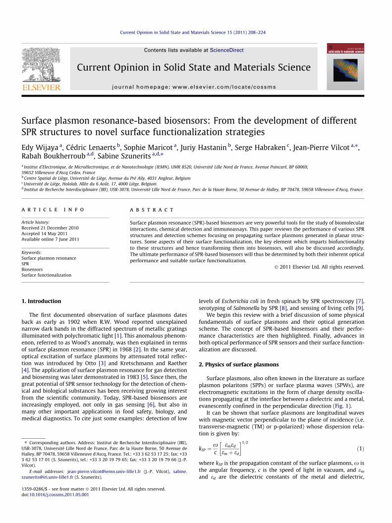
Surface plasmons, also often known in the literature as surfaceplasmon polaritons (SPPs) or surface plasma waves (SPWs), areelectromagnetic excitations in the form of charge density oscilla-tions propagating at the interface between a dielectric and a metal,evanescently confined in the perpendicular direction (Fig. 1).
It can be shown that surface plasmons are longitudinal waveswith magnetic vector perpendicular to the plane of incidence (i.e.transverse-magnetic (TM) or p-polarized) whose dispersion rela-tion is given by:
kSP ¼xc
emed
em þ ed
� �1=2
ð1Þ
where kSP is the propagation constant of the surface plasmons, x isthe angular frequency, c is the speed of light in vacuum, and em
and ed are the dielectric constants of the metal and dielectric,
Fig. 1. Schematic illustration of charge density oscillations at a metal-dielectric interface propagating in the x direction.
E. Wijaya et al. / Current Opinion in Solid State and Materials Science 15 (2011) 208–224 209
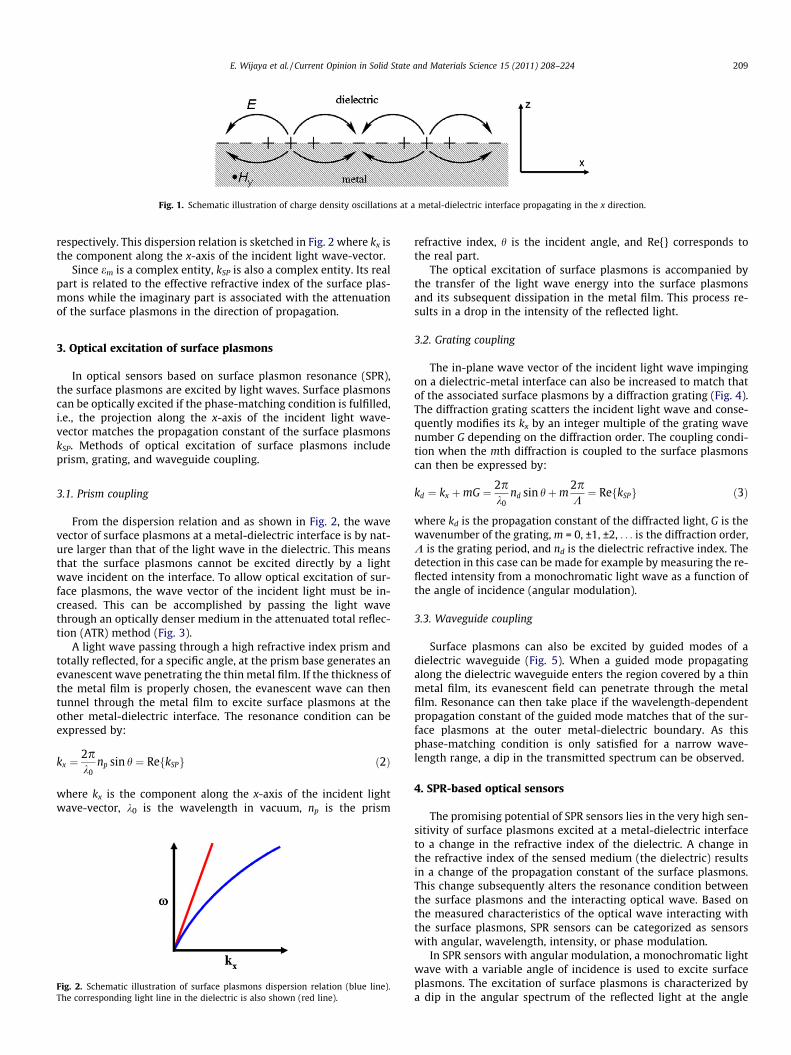
respectively. This dispersion relation is sketched in Fig. 2 where kx isthe component along the x-axis of the incident light wave-vector.
Since em is a complex entity, kSP is also a complex entity. Its realpart is related to the effective refractive index of the surface plas-mons while the imaginary part is associated with the attenuationof the surface plasmons in the direction of propagation.
3. Optical excitation of surface plasmons
In optical sensors based on surface plasmon resonance (SPR),the surface plasmons are excited by light waves. Surface plasmonscan be optically excited if the phase-matching condition is fulfilled,i.e., the projection along the x-axis of the incident light wave-vector matches the propagation constant of the surface plasmonskSP. Methods of optical excitation of surface plasmons includeprism, grating, and waveguide coupling.
3.1. Prism coupling
From the dispersion relation and as shown in Fig. 2, the wavevector of surface plasmons at a metal-dielectric interface is by nat-ure larger than that of the light wave in the dielectric. This meansthat the surface plasmons cannot be excited directly by a lightwave incident on the interface. To allow optical excitation of sur-face plasmons, the wave vector of the incident light must be in-creased. This can be accomplished by passing the light wavethrough an optically denser medium in the attenuated total reflec-tion (ATR) method (Fig. 3).
A light wave passing through a high refractive index prism andtotally reflected, for a specific angle, at the prism base generates anevanescent wave penetrating the thin metal film. If the thickness ofthe metal film is properly chosen, the evanescent wave can thentunnel through the metal film to excite surface plasmons at theother metal-dielectric interface. The resonance condition can beexpressed by:
kx ¼2pk0
np sin h ¼ Re kSPf g ð2Þ
where kx is the component along the x-axis of the incident lightwave-vector, k0 is the wavelength in vacuum, np is the prism
Fig. 2. Schematic illustration of surface plasmons dispersion relation (blue line).The corresponding light line in the dielectric is also shown (red line).
refractive index, h is the incident angle, and Re{} corresponds tothe real part.
The optical excitation of surface plasmons is accompanied bythe transfer of the light wave energy into the surface plasmonsand its subsequent dissipation in the metal film. This process re-sults in a drop in the intensity of the reflected light.
3.2. Grating coupling
The in-plane wave vector of the incident light wave impingingon a dielectric-metal interface can also be increased to match thatof the associated surface plasmons by a diffraction grating (Fig. 4).The diffraction grating scatters the incident light wave and conse-quently modifies its kx by an integer multiple of the grating wavenumber G depending on the diffraction order. The coupling condi-tion when the mth diffraction is coupled to the surface plasmonscan then be expressed by:
kd ¼ kx þmG ¼ 2pk0
nd sin hþm2pK¼ Re kSPf g ð3Þ
where kd is the propagation constant of the diffracted light, G is thewavenumber of the grating, m = 0, ±1, ±2, . . . is the diffraction order,K is the grating period, and nd is the dielectric refractive index. Thedetection in this case can be made for example by measuring the re-flected intensity from a monochromatic light wave as a function ofthe angle of incidence (angular modulation).
3.3. Waveguide coupling
Surface plasmons can also be excited by guided modes of adielectric waveguide (Fig. 5). When a guided mode propagatingalong the dielectric waveguide enters the region covered by a thinmetal film, its evanescent field can penetrate through the metalfilm. Resonance can then take place if the wavelength-dependentpropagation constant of the guided mode matches that of the sur-face plasmons at the outer metal-dielectric boundary. As thisphase-matching condition is only satisfied for a narrow wave-length range, a dip in the transmitted spectrum can be observed.
4. SPR-based optical sensors
The promising potential of SPR sensors lies in the very high sen-sitivity of surface plasmons excited at a metal-dielectric interfaceto a change in the refractive index of the dielectric. A change inthe refractive index of the sensed medium (the dielectric) resultsin a change of the propagation constant of the surface plasmons.This change subsequently alters the resonance condition betweenthe surface plasmons and the interacting optical wave. Based onthe measured characteristics of the optical wave interacting withthe surface plasmons, SPR sensors can be categorized as sensorswith angular, wavelength, intensity, or phase modulation.
In SPR sensors with angular modulation, a monochromatic lightwave with a variable angle of incidence is used to excite surfaceplasmons. The excitation of surface plasmons is characterized bya dip in the angular spectrum of the reflected light at the angle
Fig. 3. (a) Excitation of surface plasmons by the attenuated total reflection (ATR) method. (b) The light line in the denser medium (green line) is tilted to the right relative tothat in the dielectric (red dashed line) resulting in an increase of the incident light’s wave vector. For a given wavelength, resonance takes place at a specific angle of incidencewhen this light line intersects the dispersion curve of the surface plasmons (blue line).
Fig. 4. (a) Excitation of surface plasmons by the diffraction grating method. (b)Dispersion diagram illustrating the phase-matching condition. The original x-component of the wave vector of the incident photons kx is increased by G throughm = +1 diffraction order to match that of the surface plasmons.
Fig. 6. Illustration of angular and wavelength modulation. The wavelength or angleof resonance shifts as the refractive index of the sensed dielectric changes from n(solid line) to n + Dn (dashed line).
210 E. Wijaya et al. / Current Opinion in Solid State and Materials Science 15 (2011) 208–224
of resonance. The shift of the angle of resonance is monitored asthe refractive index change of the sensed medium (Fig. 6);typically, this is the working scheme of configurations using prismcoupling. In SPR sensors using wavelength modulation, a polychro-matic optical wave is employed and the resonance wavelength cor-responding to the surface plasmons excitation is monitored; this is,for example, the case for waveguide coupling configurations. Final-ly, intensity and phase shift of the interacting optical wave at fixedwavelength and angle of incidence is monitored in SPR sensorswith intensity and phase modulation, respectively. Of these differ-ent modulation schemes, the angular and wavelength modulationsare the most commonly employed in SPR sensors owing to theirhigh resolution and relatively simple instrumentation. But inten-sity modulation can be also used, mainly in systems devoted to
Fig. 5. (a) Excitation of surface plasmons by waveguide coupling. Dark blue layer indicatblue layer indicates dielectric. (b) Schematic illustration of the transmitted spectrum sh
multiple analyses, under the SPR-i (surface plasmon resonanceimaging) acronym.
5. SPR-based biosensors
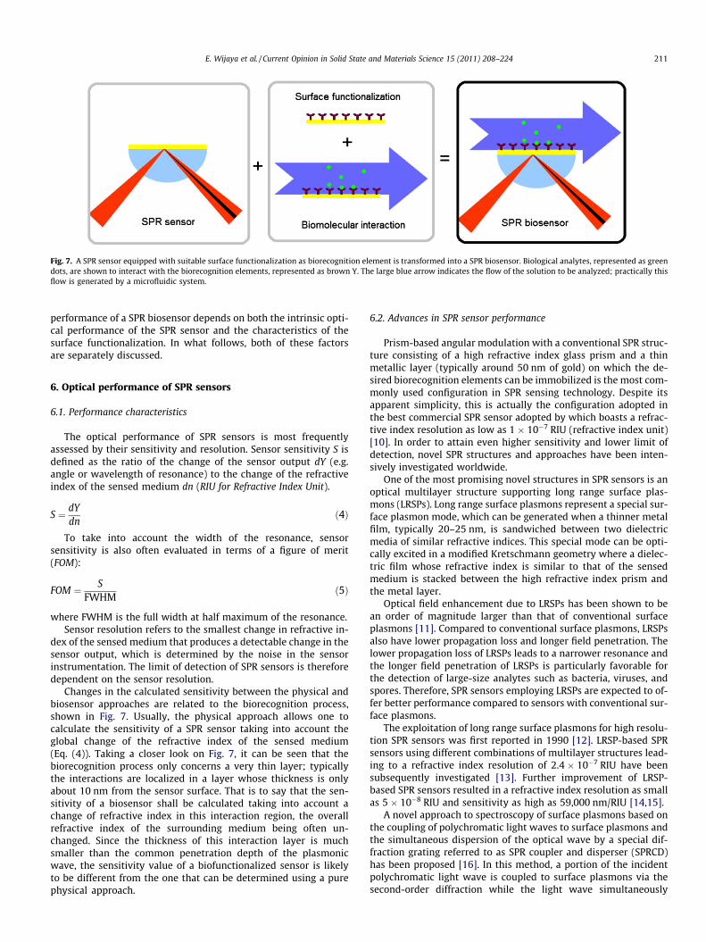
A SPR-based biosensor is made up of a SPR sensor and suitablesurface functionalization acting as the biorecognition element(Fig. 7). When a biomolecular interaction (e.g. specific binding ofanalytes) takes place, the refractive index near the surface is al-tered. This modification of refractive index can then be detectedby the SPR sensor.
As a SPR sensor and appropriate surface functionalization formthe building blocks of a SPR biosensor, it is clear that the overall
es substrate, gray layer indicates waveguide, yellow layer indicates metal film, lightowing a resonance dip.
Fig. 7. A SPR sensor equipped with suitable surface functionalization as biorecognition element is transformed into a SPR biosensor. Biological analytes, represented as greendots, are shown to interact with the biorecognition elements, represented as brown Y. The large blue arrow indicates the flow of the solution to be analyzed; practically thisflow is generated by a microfluidic system.
E. Wijaya et al. / Current Opinion in Solid State and Materials Science 15 (2011) 208–224 211
performance of a SPR biosensor depends on both the intrinsic opti-cal performance of the SPR sensor and the characteristics of thesurface functionalization. In what follows, both of these factorsare separately discussed.
6. Optical performance of SPR sensors
6.1. Performance characteristics
The optical performance of SPR sensors is most frequentlyassessed by their sensitivity and resolution. Sensor sensitivity S isdefined as the ratio of the change of the sensor output dY (e.g.angle or wavelength of resonance) to the change of the refractiveindex of the sensed medium dn (RIU for Refractive Index Unit).
S ¼ dYdn
ð4Þ
To take into account the width of the resonance, sensorsensitivity is also often evaluated in terms of a figure of merit(FOM):
FOM ¼ SFWHM
ð5Þ
where FWHM is the full width at half maximum of the resonance.Sensor resolution refers to the smallest change in refractive in-
dex of the sensed medium that produces a detectable change in thesensor output, which is determined by the noise in the sensorinstrumentation. The limit of detection of SPR sensors is thereforedependent on the sensor resolution.
Changes in the calculated sensitivity between the physical andbiosensor approaches are related to the biorecognition process,shown in Fig. 7. Usually, the physical approach allows one tocalculate the sensitivity of a SPR sensor taking into account theglobal change of the refractive index of the sensed medium(Eq. (4)). Taking a closer look on Fig. 7, it can be seen that thebiorecognition process only concerns a very thin layer; typicallythe interactions are localized in a layer whose thickness is onlyabout 10 nm from the sensor surface. That is to say that the sen-sitivity of a biosensor shall be calculated taking into account achange of refractive index in this interaction region, the overallrefractive index of the surrounding medium being often un-changed. Since the thickness of this interaction layer is muchsmaller than the common penetration depth of the plasmonicwave, the sensitivity value of a biofunctionalized sensor is likelyto be different from the one that can be determined using a purephysical approach.
6.2. Advances in SPR sensor performance
Prism-based angular modulation with a conventional SPR struc-ture consisting of a high refractive index glass prism and a thinmetallic layer (typically around 50 nm of gold) on which the de-sired biorecognition elements can be immobilized is the most com-monly used configuration in SPR sensing technology. Despite itsapparent simplicity, this is actually the configuration adopted inthe best commercial SPR sensor adopted by which boasts a refrac-tive index resolution as low as 1 � 10�7 RIU (refractive index unit)[10]. In order to attain even higher sensitivity and lower limit ofdetection, novel SPR structures and approaches have been inten-sively investigated worldwide.
One of the most promising novel structures in SPR sensors is anoptical multilayer structure supporting long range surface plas-mons (LRSPs). Long range surface plasmons represent a special sur-face plasmon mode, which can be generated when a thinner metalfilm, typically 20–25 nm, is sandwiched between two dielectricmedia of similar refractive indices. This special mode can be opti-cally excited in a modified Kretschmann geometry where a dielec-tric film whose refractive index is similar to that of the sensedmedium is stacked between the high refractive index prism andthe metal layer.
Optical field enhancement due to LRSPs has been shown to bean order of magnitude larger than that of conventional surfaceplasmons [11]. Compared to conventional surface plasmons, LRSPsalso have lower propagation loss and longer field penetration. Thelower propagation loss of LRSPs leads to a narrower resonance andthe longer field penetration of LRSPs is particularly favorable forthe detection of large-size analytes such as bacteria, viruses, andspores. Therefore, SPR sensors employing LRSPs are expected to of-fer better performance compared to sensors with conventional sur-face plasmons.
The exploitation of long range surface plasmons for high resolu-tion SPR sensors was first reported in 1990 [12]. LRSP-based SPRsensors using different combinations of multilayer structures lead-ing to a refractive index resolution of 2.4 � 10�7 RIU have beensubsequently investigated [13]. Further improvement of LRSP-based SPR sensors resulted in a refractive index resolution as smallas 5 � 10�8 RIU and sensitivity as high as 59,000 nm/RIU [14,15].
A novel approach to spectroscopy of surface plasmons based onthe coupling of polychromatic light waves to surface plasmons andthe simultaneous dispersion of the optical wave by a special dif-fraction grating referred to as SPR coupler and disperser (SPRCD)has been proposed [16]. In this method, a portion of the incidentpolychromatic light wave is coupled to surface plasmons via thesecond-order diffraction while the light wave simultaneously
212 E. Wijaya et al. / Current Opinion in Solid State and Materials Science 15 (2011) 208–224
dispersed via the first-order diffraction is projected onto a posi-tion-sensitive detector. The need for a spectrometer in this systemis therefore effectively eliminated making this design attractive forsimple and low-cost SPR sensors. Subsequent experimental dem-onstration of this approach shows a refractive index sensitivity ofaround 620 nm/RIU and resolution as low as 3 � 10�7 RIU [17].
In addition to higher sensitivity and lower limit of detection,compactness is also a desirable feature in SPR sensor technology.Therefore, different designs of miniaturized or integrated SPR sen-sors have been actively explored in recent years.
Optical fiber-based SPR sensors offer a possibility for sensorminiaturization and are extremely attractive for in vivo applica-tions. Miniaturization of SPR sensors based on optical fibers witha refractive index resolution of 4 � 10�5 RIU has been proposed[18]. In these sensors, the cladding layer of the optical fiber islocally removed and subsequently covered by a thin gold film toenable the excitation of surface plasmons. A theoretical analysisshows that by using a Bragg grating, the refractive index resolutionof optical fiber SPR sensors could be lowered to 2 � 10�6 RIU [19].Tapering of the fiber core has also been theoretically shown to in-crease the sensitivity of optical fiber SPR sensors by a factor of 15and this improved sensitivity could be further enhanced by a factor4 by introducing a Teflon layer between the core and the metallayer to excite long-range surface plasmons [20].
Integrated optical waveguide SPR sensors are particularlypromising candidates for the development of miniaturized multi-channel sensing devices on a single chip. A SPR sensor based onan integrated optical waveguide with a sensitivity of 2100 nm/RIU and a resolution of 1.2 � 10�6 RIU has been reported [21].The waveguide was fabricated by a K+
M Na+ ion-exchange methodon BK7 glass substrate. The original operating range of the sensoraround a refractive index of 1.44 was then optimized for an aque-ous environment (refractive index around 1.33) relevant to biosen-sing by using a tantalum pentoxide overlayer. A SPR sensor basedon a germanium-doped silicon dioxide waveguide on silicon sub-strate with slightly higher sensitivity (2500 nm/RIU) has also beendemonstrated [22]. The waveguide was fabricated by plasma-en-hanced chemical vapor deposition (PECVD) method, which allowsprecise control of the refractive index difference between the coreand the cladding layer.
In view of developing high-throughput fabrication with lowercost, a single-mode SPR waveguide sensor fabricated by polymerimprinting technique has been proposed [23]. This sensor is basedon intensity modulation with a refractive index resolution ofapproximately 3.8 � 10�4 RIU. An optical waveguide SPR sensorwith dual light-emitting diodes and a photodiode based on thedetection of differential intensity shift at two different wave-lengths has also been reported [24]. The refractive index resolutionof this sensor is estimated to be around 2.3 � 10�5 RIU. The rela-tively poor resolution of these sensors is rather characteristic ofsensors based on intensity modulation whose typical resolutionis on the order of 10�5 RIU [25].
Lastly, combining the conventional Kretschmann geometrywith metamaterials can potentially improve the performance ofSPR sensors as well. The use of an array of gold nanorods to replacethe continuous gold film in the Kretschmann geometry resulting inenhanced SPR performance with refractive index sensitivity ashigh as 32,000 nm/RIU has been experimentally demonstrated[26,27].
7. Commonly used and newly developed surfacefunctionalization strategies
One of the first steps in any SPR-based protocol concerns theway the receptor molecules are anchored onto the SPR surface.
The technique of SPR becomes indeed interesting when themetallic film, supporting the propagation of charge density waves(the plasmons), is chemically modified. The bioreceptors are eitherphysisorbed or chemically attached onto the sensor surface. Cova-lent attachment is often preferred, as it provides strong and stablebinding of the receptor to the SPR surface. This allows conse-quently easy regeneration of the sensor interface using conditions,which can remove the analyte from the surface, but not the at-tached ligand itself. The covalent immobilization strategies includechemical reactions such as amine, aldehyde or thiol coupling onpreviously formed functional self-assembled monolayers. Themost widely used approach for the introduction of functionalgroups onto thin gold SPR films is based on thiolated organic com-pounds, which spontaneously form self-assembled monolayers(SAMs) on gold surfaces. The use of polymers is an alternative withassociated advantages and disadvantages. The selection of one overthe other depends on the application sought after. In general, poly-meric layers such as carboxylated dextran (CM-dextran) thin filmsallow molecules to move freely on the surface, thus reducing someproblems with binding interference. In addition, they provide moreattachment points than a monolayer and act as a buffer to reducenonspecific binding to the surface. However, it is more difficult formolecules to diffuse through such a hydrogel than through SAMs,which can potentially alter the measured kinetics. While mono-layer films do not show such diffusion problems, they are proneto be less sensitive if not properly formed. However, this problemwas addressed in the literature showing that, in fact, there is nodifference in binding kinetics between monolayers and a dextanhydrogel [28]. Thus, whether the coating is a monolayer or a thinfilm, the selection of the immobilization procedure is mainly appli-cation driven. Generally, flat surfaces with SAMs are beneficialcompared to polymeric layers both when the analytes of interestare larger than molecules, such as cells and viruses, and for kineticparameter determination, when a low amount of non-specificbinding is required and a low level of immobilization is recom-mended [29]. The achievement of negligible non-specific bindingto the SPR sensor interface is an important factor contributing tothe success of the sensor applications. Non-specific binding contri-butions lead to errors in concentration determination and in calcu-lation of kinetic constants. Reduction of non-specific binding canbe achieved by creating more hydrophilic interfaces or by includ-ing compounds such as polyethylene glycol derivatives in theimmobilization steps or incorporated into the ligand to be attachedonto the SPR surface [30].
Next to the selected immobilization strategy to bind the recep-tors to the SPR chip, the choice of the metal film is critical for sen-sitive SPR sensing. Gold is the substrate of choice for SPRmeasurements for mainly two reasons: it is relatively stable inaqueous environments required for monitoring biomolecularinteractions and can be easily functionalized through the forma-tion of alkanethiols SAMs. However, gold is not the best candidatefor achieving high-sensitivity SPR sensing. Theoretical modeling ofSPR in conducting metal oxide thin films has been performed byFranzen et al. who suggested that ITO could be a better suited sub-strate [31]. However, this would require excitation and detectionin the infrared range. In the conventional visible range, silver sub-strates appear to be the most appealing, because plasmon couplingexhibits a sharper angular resonance as compared to that on gold,yielding an increased sensitivity [32]. Silver substrates can be func-tionalized similarly to gold ones, using thiol or disulfide moleculeswhich adsorb from solution and self-assemble into densely packedmonolayers or by depositing polymers. However, silver suffersfrom a poor chemical stability, which hampers its wide use forSPR sensing. One strategy to circumvent this limitation is to usebimetallic silver/gold layers [33,34]. In this case, the usual thiol-on-gold chemistry can be performed for coupling probes to the
E. Wijaya et al. / Current Opinion in Solid State and Materials Science 15 (2011) 208–224 213
sensor surface. Alternatively, lamellar structures, in which a thinlayer of a dielectric is deposited onto the surface plasmon activemetal thin film, were developed [35–47]. These overlayers allowan efficient protection of the underlying silver film and at the sametime open the scope for new surface functionalization schemes,which can be employed for anchoring ligands to the SPR sensorchip. Currently, the investigated dielectric layers are either oxidebased [35,37–45], where the attachment of ligands to the surfaceis mainly achieved through silanization of the surface hydroxylgroups. The other approach consists on the deposition of carbon[36] and amorphous silicon–carbon alloys [46,47]. These thin car-bon-based films allow covalent immobilization of biomolecules ofinterest using well-developed and robust chemistry based on theattachment of alkene-containing molecules to the substratethrough carbon–carbon or Si–C bonds. The different concepts willbe presented in the following with additional highlights of newlydeveloped surface functionalization routes, without beingexhaustive.
7.1. Thiolated functional groups
While new strategies for the immobilization of bioreceptorshave been developed in the last ten years, the most commonlyused strategy for the introduction of surface functional groups ontoSPR chips is still based on the attachment of sulfur-containingligands. Gold–sulfur (Au–S) bonds are readily formed on goldenabling in an easy and fast manner the direct attachment ofreceptors to the gold surface by the formation of self-assembledmonolayers. Several reviews are devoted to this topic and the read-er is referred to them [48,49]. The Au–S chemistry has thus madepossible the routine analysis of aqueous binding events to immobi-lize molecules at nearly neutral pH and moderate temperature.
Two strategies are employed for making bioreceptor SAMs ongold. The first is based on the separate synthesis of the bioreceptorderivative with a pendant alkanethiol group and subsequent for-mation of the SAM [50–52]. This is often used in the case ofDNA, where a thiol (SH) group can be easily incorporated ontothe 50 end. The drawback of this strategy arises from the highersynthetic effort, especially for more complex bioreceptors. Further-more, as the complexity of tethered bioreceptors increases, there isno guarantee that the molecules will pack to form a structurallywell-defined monolayer. To control the surface density, the useof mixed monolayers by diluting with short chain thiols (e.g. mer-captohexanol) is thus often preferred [50].
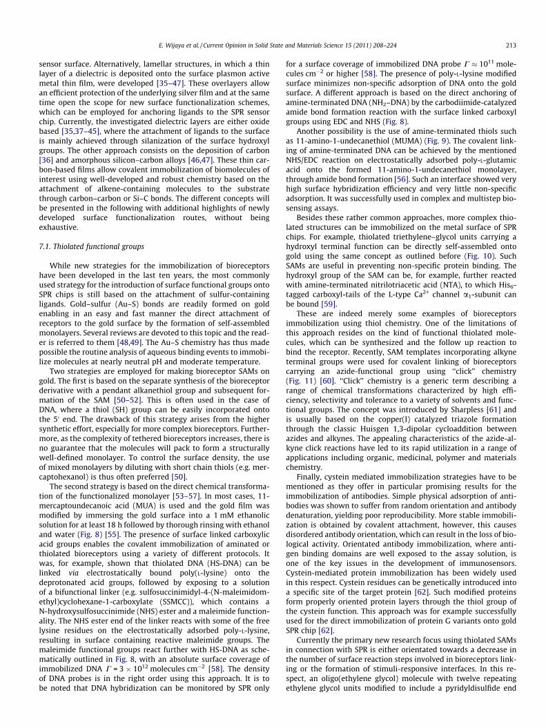
The second strategy is based on the direct chemical transforma-tion of the functionalized monolayer [53–57]. In most cases, 11-mercaptoundecanoic acid (MUA) is used and the gold film wasmodified by immersing the gold surface into a 1 mM ethanolicsolution for at least 18 h followed by thorough rinsing with ethanoland water (Fig. 8) [55]. The presence of surface linked carboxylicacid groups enables the covalent immobilization of aminated orthiolated bioreceptors using a variety of different protocols. Itwas, for example, shown that thiolated DNA (HS-DNA) can belinked via electrostatically bound poly(L-lysine) onto thedeprotonated acid groups, followed by exposing to a solutionof a bifunctional linker (e.g. sulfosuccinimidyl-4-(N-maleimidom-ethyl)cyclohexane-1-carboxylate (SSMCC)), which contains aN-hydroxysulfosuccinimide (NHS) ester and a maleimide function-ality. The NHS ester end of the linker reacts with some of the freelysine residues on the electrostatically adsorbed poly-L-lysine,resulting in surface containing reactive maleimide groups. Themaleimide functional groups react further with HS-DNA as sche-matically outlined in Fig. 8, with an absolute surface coverage ofimmobilized DNA C = 3 � 1012 molecules cm�2 [58]. The densityof DNA probes is in the right order using this approach. It is tobe noted that DNA hybridization can be monitored by SPR only
for a surface coverage of immobilized DNA probe C � 1011 mole-cules cm�2 or higher [58]. The presence of poly-L-lysine modifiedsurface minimizes non-specific adsorption of DNA onto the goldsurface. A different approach is based on the direct anchoring ofamine-terminated DNA (NH2–DNA) by the carbodiimide-catalyzedamide bond formation reaction with the surface linked carboxylgroups using EDC and NHS (Fig. 8).
Another possibility is the use of amine-terminated thiols suchas 11-amino-1-undecanethiol (MUMA) (Fig. 9). The covalent link-ing of amine-terminated DNA can be achieved by the mentionedNHS/EDC reaction on electrostatically adsorbed poly-L-glutamicacid onto the formed 11-amino-1-undecanethiol monolayer,through amide bond formation [56]. Such an interface showed veryhigh surface hybridization efficiency and very little non-specificadsorption. It was successfully used in complex and multistep bio-sensing assays.
Besides these rather common approaches, more complex thio-lated structures can be immobilized on the metal surface of SPRchips. For example, thiolated triethylene–glycol units carrying ahydroxyl terminal function can be directly self-assembled ontogold using the same concept as outlined before (Fig. 10). SuchSAMs are useful in preventing non-specific protein binding. Thehydroxyl group of the SAM can be, for example, further reactedwith amine-terminated nitrilotriacetic acid (NTA), to which His6-tagged carboxyl-tails of the L-type Ca2+ channel a1-subunit canbe bound [59].
These are indeed merely some examples of bioreceptorsimmobilization using thiol chemistry. One of the limitations ofthis approach resides on the kind of functional thiolated mole-cules, which can be synthesized and the follow up reaction tobind the receptor. Recently, SAM templates incorporating alkyneterminal groups were used for covalent linking of bioreceptorscarrying an azide-functional group using ‘‘click’’ chemistry(Fig. 11) [60]. ‘‘Click’’ chemistry is a generic term describing arange of chemical transformations characterized by high effi-ciency, selectivity and tolerance to a variety of solvents and func-tional groups. The concept was introduced by Sharpless [61] andis usually based on the copper(I) catalyzed triazole formationthrough the classic Huisgen 1,3-dipolar cycloaddition betweenazides and alkynes. The appealing characteristics of the azide-al-kyne click reactions have led to its rapid utilization in a range ofapplications including organic, medicinal, polymer and materialschemistry.
Finally, cystein mediated immobilization strategies have to bementioned as they offer in particular promising results for theimmobilization of antibodies. Simple physical adsorption of anti-bodies was shown to suffer from random orientation and antibodydenaturation, yielding poor reproducibility. More stable immobili-zation is obtained by covalent attachment, however, this causesdisordered antibody orientation, which can result in the loss of bio-logical activity. Orientated antibody immobilization, where anti-gen binding domains are well exposed to the assay solution, isone of the key issues in the development of immunosensors.Cystein-mediated protein immobilization has been widely usedin this respect. Cystein residues can be genetically introduced intoa specific site of the target protein [62]. Such modified proteinsform properly oriented protein layers through the thiol group ofthe cystein function. This approach was for example successfullyused for the direct immobilization of protein G variants onto goldSPR chip [62].
Currently the primary new research focus using thiolated SAMsin connection with SPR is either orientated towards a decrease inthe number of surface reaction steps involved in bioreceptors link-ing or the formation of stimuli-responsive interfaces. In this re-spect, an oligo(ethylene glycol) molecule with twelve repeatingethylene glycol units modified to include a pyridyldisulfide end
5 mMNaHCO3Gold
(CH2)7
S
CHO O
poly-L-lysine Gold
(CH2)7
S
CO O
HN
HN
O
O
H2N
NH3
1. SSMCC
2. HS-DNAGold
(CH2)7
S
CO O
HN
HN
O
O
HN
NH3
O
CH2
NO
O
S-DNA
Gold
(CH2)7
HS
CHO O
ethanol
Gold
(CH2)7
S
CHN O
1. NHS/EDC
2. NH2-DNA
DNA
Fig. 8. Formation of 11-mercaptoundecanoic acid (MUA) on gold followed by linking of thiolated DNA or amine-terminated DNA molecules.
Gold1. NHS/EDC
2. NH2-DNAL-glutamic acid
-OO
H2NO
-O
HS
H2N
GoldS
H2N
GoldS
+H3N
HNO
H2NO
-O
GoldS
+H3N
DNA
Fig. 9. Schematic illustration of gold functionalization with 11-amino-1-undecanethiol and subsequent linking of amine-functional DNA molecules.
214 E. Wijaya et al. / Current Opinion in Solid State and Materials Science 15 (2011) 208–224
group generated an interface highly responsive to temperaturechanges in the vicinity of the physiological temperature, 37 �C[30]. Such a SPR interface exhibits switchable physical properties,
which are highly interesting for biomedical applications. It hasbeen shown that such an interface allows controlling the affinitybinding of streptavidin to biotin-tethered surface [30].
(CH2)12
HS
CO
O
O
O
HO
(CH2)12
S
CO
O
O
O
ONHO
N
OHO
OHO
Gold ethanol
(CH2)12
S
CO
O
O
O
HO
Gold2. COOH-NTA
Gold
OH
O
Fig. 10. Immobilization of thiolated triethylene–glycol units onto gold substrate followed by linking of nitrilotriacetic acid (NTA).
E. Wijaya et al. / Current Opinion in Solid State and Materials Science 15 (2011) 208–224 215
However, independent from the intensive use of thiolatedcompounds for the immobilization of bioreceptors, the susceptibil-ity of the gold–sulfur bond to oxidation and photodecomposition isa real challange for thiol chemistry. Other functionalizationschemes based on hydrogels and conducting polymers have beeninvestigated in parallel over the years and will be discussed inmore details in the following section.
7.2. Carboxymethylated dextran layers: the Biacore chip
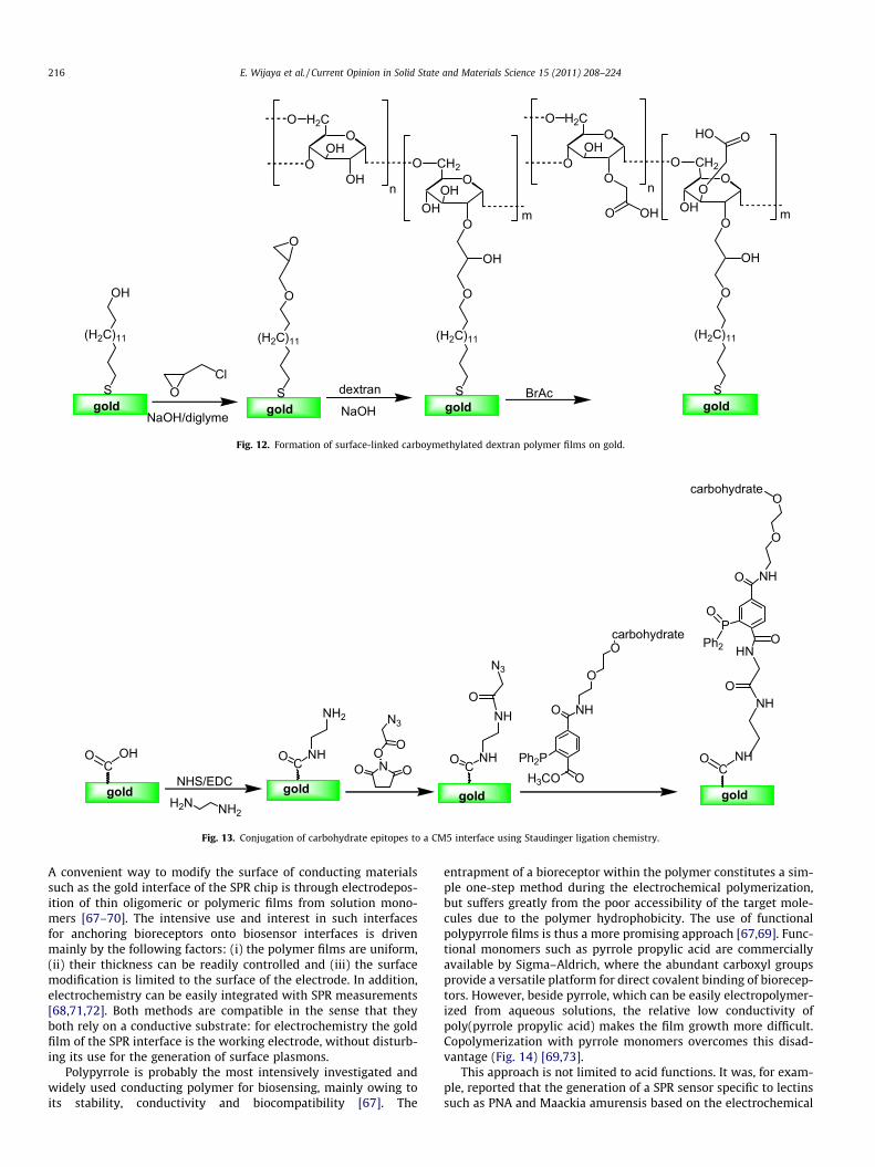
Next to the formation of Au–S bonds, one of the most commonstrategies to immobilize bioreceptors is to use activated carbo-xymethylated dextran (CM-dextran). Dextrans are hydrophilic,non-charged neutral polymeric carbohydrates, soluble in water inany proportion forming highly hydrated hydrogels. Due to theseproperties, dextrans display very low non-specific interactionswith bioreceptors. Owing to the high concentration of hydroxylgroups in the dextran matrix, chemical modification is possiblewithout significantly affecting its hydrophilicity. The essentiallynon-branched polymer chains are highly flexible and ligandsimmobilized in dextran matrices are thus well accessible. Intro-duced by Lofas and Johnsoon [63], it is one of the commerciallyavailable SPR chips commercialized by Biacore. The matrix is con-structed by self-assembly of 1,x-hydroxyalkythiol (16-mercapto-hexadecane-1-ol) onto gold, followed by covalently linking of thedextran polymer by activation of the hydroxyl groups with
epichlorohydrin under basic condition to yield epoxides, whichby further reaction with bromoacetic acid results in the formationof surface linked carboxymethylated dextran films (Fig. 12) [64].The thickness of the polymer matrix is about 100 nm and thecarboxylic acid groups can be further used to covalently linkamine-terminated or thiol-terminated bioreceptors as discussedbefore hand for acid terminated SAMs [65].
The references which use this type of substrate are countless, asthis kind of interface, known as CM5 chip, is commercialized byBiacore. More recently, carbohydrate epitopes were also immobi-lized on CM5 SPR interface using Staudinger ligation chemistry[66]. This was accomplished by first introducing azide functional-ities to the CM5 interface, followed by reaction with phosphane-modified carbohydrate ligands (Fig. 13). The advantage of thisapproach is that the chemistry employed is extremely mild andcan be easily adapted to any biosystem.
7.3. Polymer films
7.3.1. Conducting polymersOne important aspect when constructing biosensors is the sup-
pression of non-specific adsorption of the bioreceptor on the sens-ing surface. In most cases, the degree of non-specific adsorptiondetermines the sensitivity and specificity of a biosensor. Some ef-fects such as hydrophobicity, surface charge and pH have beenidentified for being essential to decrease non-specific adsorption.
dextran BrAcOCl
NaOH/diglyme
(H2C)11
S
O
NaOHgold
O
(H2C)11
S
O
gold
OH
(H2C)11
S
OH
gold
O
OH2C
OOH
OH
O
O
OHOH
CH2
m
O
n
(H2C)11
S
O
gold
OH
O
OH2C
OOH
O
O
O
OHO
CH2
m
O
n
OHO
OHO
Fig. 12. Formation of surface-linked carboymethylated dextran polymer films on gold.
COHO
NHS/EDC
H2N NH2
CNHO
NH2
NO OO
N3
O
CNHO
NH
N3
O
Ph2PH3CO O
NH
O
O
O
carbohydrate
gold gold gold
HN
P
O NH
O
Ocarbohydrate
O
Ph2O
NHO
CNHO
gold
Fig. 13. Conjugation of carbohydrate epitopes to a CM5 interface using Staudinger ligation chemistry.
216 E. Wijaya et al. / Current Opinion in Solid State and Materials Science 15 (2011) 208–224
A convenient way to modify the surface of conducting materialssuch as the gold interface of the SPR chip is through electrodepos-ition of thin oligomeric or polymeric films from solution mono-mers [67–70]. The intensive use and interest in such interfacesfor anchoring bioreceptors onto biosensor interfaces is drivenmainly by the following factors: (i) the polymer films are uniform,(ii) their thickness can be readily controlled and (iii) the surfacemodification is limited to the surface of the electrode. In addition,electrochemistry can be easily integrated with SPR measurements[68,71,72]. Both methods are compatible in the sense that theyboth rely on a conductive substrate: for electrochemistry the goldfilm of the SPR interface is the working electrode, without disturb-ing its use for the generation of surface plasmons.
Polypyrrole is probably the most intensively investigated andwidely used conducting polymer for biosensing, mainly owing toits stability, conductivity and biocompatibility [67]. The
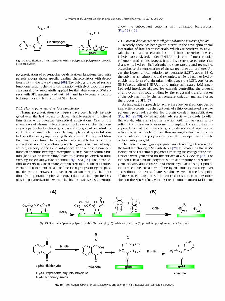
entrapment of a bioreceptor within the polymer constitutes a sim-ple one-step method during the electrochemical polymerization,but suffers greatly from the poor accessibility of the target mole-cules due to the polymer hydrophobicity. The use of functionalpolypyrrole films is thus a more promising approach [67,69]. Func-tional monomers such as pyrrole propylic acid are commerciallyavailable by Sigma–Aldrich, where the abundant carboxyl groupsprovide a versatile platform for direct covalent binding of biorecep-tors. However, beside pyrrole, which can be easily electropolymer-ized from aqueous solutions, the relative low conductivity ofpoly(pyrrole propylic acid) makes the film growth more difficult.Copolymerization with pyrrole monomers overcomes this disad-vantage (Fig. 14) [69,73].
This approach is not limited to acid functions. It was, for exam-ple, reported that the generation of a SPR sensor specific to lectinssuch as PNA and Maackia amurensis based on the electrochemical
N N
OHO
Electropolym.N N N N
O OH
+
gold
Fig. 14. Modification of SPR interfaces with a polypyrrole/poly(pyrrole propylicacid) copolymer.
E. Wijaya et al. / Current Opinion in Solid State and Materials Science 15 (2011) 208–224 217
polymerization of oligosaccharide derivatives functionalized withpyrrole groups shows specific binding characteristics with detec-tion limits in the low nM range [68]. The polypyrrole based surfacefunctionalization scheme in combination with electrospotting pro-cess can also be successfully applied for the fabrication of DNA ar-rays with SPR imaging read out [74], and has become a routinetechnique for the fabrication of SPR chips.
7.3.2. Plasma polymerized surface modificationPlasma polymerization techniques have been largely investi-
gated over the last decade to deposit highly reactive, functionalthin films with potential biomedical applications. One of theadvantages of plasma polymerization techniques is that the den-sity of a particular functional group and the degree of cross-linkingwithin the polymer network can be largely tailored by careful con-trol over the energy input during the deposition. The types of filmsthat have been found to be particularly suitable for biosensingapplications are those containing reactive groups such as carbonyl,amines, carboxylic acids and anhydrides. For example, amine-ter-minated or amine bearing bioreceptors such as bovine serum albu-min (BSA) can be irreversibly linked to plasma-polymerized filmscarrying maleic anhydride functions (Fig. 15A) [75]. The introduc-tion of esters has been more complicated due to the difficultiesencountered to retain the active functional groups during the plas-ma deposition. However, it has been shown recently that thinfilms from pentafluorophenyl methacrylate can be deposited viaplasma polymerization, where the highly reactive ester groups
(A)
R-NH2
OO O OO O O OOH NH
R
O OOH NH
R F
OF
Fig. 15. Reaction of plasma polymerized thin films carrying (A) ma
H
O
H
O
R1-SH OH
S
H
O
R1
o-phtaldialdehyde thioacetal
R1-SH represents any thiol moleculeR2-NH2 primary amine
Fig. 16. The reaction between o-phthalialdehyde and
allow the subsequent coupling with aminated bioreceptors(Fig. 15B) [76].
7.3.3. Recent developments: intelligent polymeric materials for SPRRecently, there has been great interest in the development and
integration of intelligent materials, which are sensitive to physi-cal, chemical and/or electrical stimuli into biosensing devices.Poly(N-isopropylacrylamide) (PNIPAAm) is one of most popularpolymers used in this respect. It is a heat-sensitive polymer thatchanges its hydrophilic/hydrophobic state rapidly and reversibly,according to the temperature of the surrounding atmosphere. Un-der the lowest critical solution temperature (LCST), about 32 �C,the polymer is hydrophilic and extended, while it becomes hydro-phobic in a form of a shrunken helix above the LCST. AnchoringNHS-functionalized PNIPAAm onto amine-terminated SAM modi-fied gold interfaces allowed for example controlling the amountof anti-biotin antibody binding by the structural transformationof the polymer film by the temperature variation and monitoringthe process by SPR [77].
An innovative approach for achieving a low level of non-specificinteractions consists on the synthesis of a thiol-terminated reactivepolymer, polythiol, suitable for protein covalent immobilization(Fig. 16) [29,78]. O-Phthaldialdehyde reacts with thiols to offerthioacetals, which in a further reaction with primary amines re-sults in the formation of an isoindole complex. The interest in thisapproach is that the thioacetal groups do not need any specificactivation to react with proteins, thus making it attractive for sens-ing. In addition, the polymer contains thiol groups that promoteself-assembly on gold.
The same research group proposed an interesting alternative forthe local structuring of SPR interfaces [79]. It is based on the in situformation of a functional polymer film using the energy of the eva-nescent wave generated on the surface of a SPR device [79]. Themethod is based on the polymerization of a mixture of N,N-meth-ylene-bis-acrylamide (MAA) and methacrylic acid using a photo-initiator couple consisting of methylene blue (sensitizing dye)and sodium p-toluenesulfinate as reducing agent at the focal pointof the SPR. No polymerization occurred in solution or any othersites on the SPR surface. Varying the monomer concentration and
(B) F
FO
FF F
FOO
F FR-NH2
RHN
O
RHN
O
leic anhydride or (B) pentafluorophenyl active ester functions.
R2-NH2N
S
R2HO
S
H
O
isoindole
gold gold
S
thiol to yield thioacetal and isoindole derivatives.
(A) (B)
0
0.2
0.4
0.6
0.8
1
60 65 70 75 80 85 90
nor
mal
ized
inte
nsity
/ a.
u.
angle / °
Fig. 18. (A) Schematic illustration of lamellar structure of dielectric-on-goldsubstrate, (B) Theoretical influence of silica thickness (nSiOx = 1.48) on the SPRsignal. The SPR chip consists of 5 nm titanium adhesion layer (n = 2.36 + i3.47) and50 nm gold layer (n = 0.197 + i3.67) using a prism with n = 1.52: 0 nm (black), 5 nm(gray), 10 nm (bright gray), 20 nm (blue), 40 nm (magenta), 60 nm (red) thick silicalayer.
218 E. Wijaya et al. / Current Opinion in Solid State and Materials Science 15 (2011) 208–224
exposure time allowed a precise control over the polymer thick-ness (20–200 nm), while standard coupling chemistry using EDC/NHS was used for the immobilization of Protein G [79].
Very recently, polysilsesquioxanes have been deposited as thinfilms onto SPR chips by spin coating and post-curing [80]. Poly-silsesquioxanes are an emerging class of hybrid (organic–inorganic) nanostructured polymers containing nanosized cages.Chemical pre-functionalization of the polysilsesquioxane withpentafluorophenyl acrylate units allows the immobilization ofamine-terminated bioreceptors by simple dipping into the solutionof the analyte without the need of activation process (Fig. 17).
7.4. Lamellar SPR structures based on dielectric overcoatings
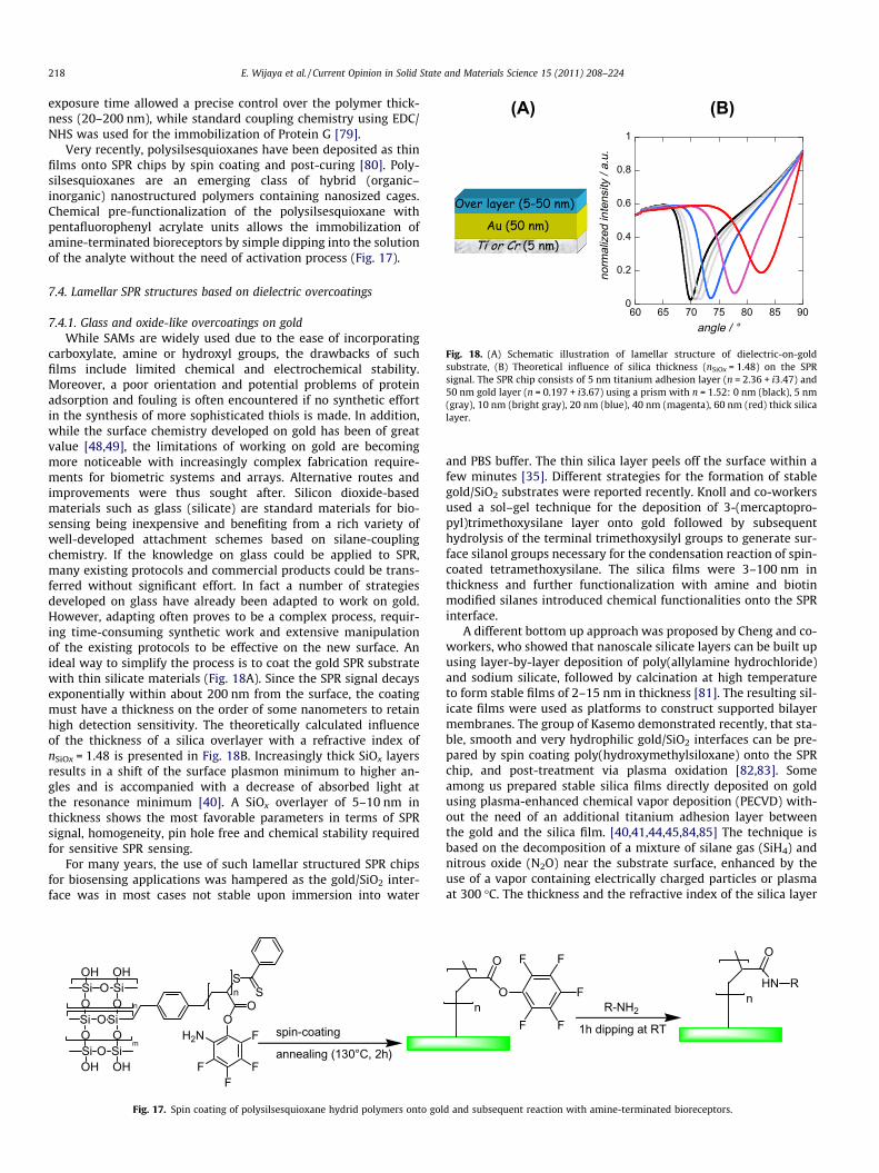
7.4.1. Glass and oxide-like overcoatings on goldWhile SAMs are widely used due to the ease of incorporating
carboxylate, amine or hydroxyl groups, the drawbacks of suchfilms include limited chemical and electrochemical stability.Moreover, a poor orientation and potential problems of proteinadsorption and fouling is often encountered if no synthetic effortin the synthesis of more sophisticated thiols is made. In addition,while the surface chemistry developed on gold has been of greatvalue [48,49], the limitations of working on gold are becomingmore noticeable with increasingly complex fabrication require-ments for biometric systems and arrays. Alternative routes andimprovements were thus sought after. Silicon dioxide-basedmaterials such as glass (silicate) are standard materials for bio-sensing being inexpensive and benefiting from a rich variety ofwell-developed attachment schemes based on silane-couplingchemistry. If the knowledge on glass could be applied to SPR,many existing protocols and commercial products could be trans-ferred without significant effort. In fact a number of strategiesdeveloped on glass have already been adapted to work on gold.However, adapting often proves to be a complex process, requir-ing time-consuming synthetic work and extensive manipulationof the existing protocols to be effective on the new surface. Anideal way to simplify the process is to coat the gold SPR substratewith thin silicate materials (Fig. 18A). Since the SPR signal decaysexponentially within about 200 nm from the surface, the coatingmust have a thickness on the order of some nanometers to retainhigh detection sensitivity. The theoretically calculated influenceof the thickness of a silica overlayer with a refractive index ofnSiOx = 1.48 is presented in Fig. 18B. Increasingly thick SiOx layersresults in a shift of the surface plasmon minimum to higher an-gles and is accompanied with a decrease of absorbed light atthe resonance minimum [40]. A SiOx overlayer of 5–10 nm inthickness shows the most favorable parameters in terms of SPRsignal, homogeneity, pin hole free and chemical stability requiredfor sensitive SPR sensing.
For many years, the use of such lamellar structured SPR chipsfor biosensing applications was hampered as the gold/SiO2 inter-face was in most cases not stable upon immersion into water
spin-coating
annealing (130°C, 2h)
S
OH2N
F
F
F
OS
F
n
SiO
OOSi
Si
Si
O
O
Si
Si
OOH OH
OOH OH
m
n
Fig. 17. Spin coating of polysilsesquioxane hydrid polymers onto gol
and PBS buffer. The thin silica layer peels off the surface within afew minutes [35]. Different strategies for the formation of stablegold/SiO2 substrates were reported recently. Knoll and co-workersused a sol–gel technique for the deposition of 3-(mercaptopro-pyl)trimethoxysilane layer onto gold followed by subsequenthydrolysis of the terminal trimethoxysilyl groups to generate sur-face silanol groups necessary for the condensation reaction of spin-coated tetramethoxysilane. The silica films were 3–100 nm inthickness and further functionalization with amine and biotinmodified silanes introduced chemical functionalities onto the SPRinterface.
A different bottom up approach was proposed by Cheng and co-workers, who showed that nanoscale silicate layers can be built upusing layer-by-layer deposition of poly(allylamine hydrochloride)and sodium silicate, followed by calcination at high temperatureto form stable films of 2–15 nm in thickness [81]. The resulting sil-icate films were used as platforms to construct supported bilayermembranes. The group of Kasemo demonstrated recently, that sta-ble, smooth and very hydrophilic gold/SiO2 interfaces can be pre-pared by spin coating poly(hydroxymethylsiloxane) onto the SPRchip, and post-treatment via plasma oxidation [82,83]. Someamong us prepared stable silica films directly deposited on goldusing plasma-enhanced chemical vapor deposition (PECVD) with-out the need of an additional titanium adhesion layer betweenthe gold and the silica film. [40,41,44,45,84,85] The technique isbased on the decomposition of a mixture of silane gas (SiH4) andnitrous oxide (N2O) near the substrate surface, enhanced by theuse of a vapor containing electrically charged particles or plasmaat 300 �C. The thickness and the refractive index of the silica layer
O
O F F
F
FFn R-NH2
HN R
O
n
1h dipping at RT
d and subsequent reaction with amine-terminated bioreceptors.
E. Wijaya et al. / Current Opinion in Solid State and Materials Science 15 (2011) 208–224 219
are controlled by the reaction time and stoichiometry of the film.Silica coatings as thin as 7 nm exhibited very good stability in bothorganic and aqueous solutions as well as in a piranha solution at80 �C. This harsh treatment did also not induce any thickness orSPR response changes and produced considerable amounts of Si–OH, which allows chemical modification with functional silanemolecules (Fig. 19). Amine-terminated oligonucleotides can be,for example, grafted on such an interface using a standard proce-dure developed for glass, including the following sequences: (i)reaction of the silicon oxide layer with 3-aminopropyltrimethoxy-silane (APTES) to produce amine terminal groups on the surface,(ii) transformation of the amine to aldehyde termination by chem-ical coupling with a bifunctional linker such as glutaraldehyde andimmobilization of ODNs bearing a terminal amine group [39].
One limitation of the silica coating concerns its insulating char-acter, limiting its use for electrochemistry-SPR (E-SPR). A lamellarstructure with 7 nm thick SiOx showed charge transfer, which wasrather sluggish [39,41].
Antimony-doped tin oxide (SnO2:Sb) and indium tin oxide (ITO)thin films are a possible alternative to SiOx layers for E-SPR, display-ing a resistivity in the order of 10�4–10�2 X cm [37,42,43]. On theother hand, while for a 10 nm SiOx overlayer the SPR signal is notsignificantly changed, in the case of SnO2:Sb the signal is largelydeteriorated (Fig. 20A) [37,38]. This is linked to the higher imaginaryrefractive index of Sb-doped SnO2 (n = i0.249), which confers theAu–SnO2:Sb composite interfaces a yellow color and an additionallight absorption. This problem is less severe in the case of a 10 nmITO overlayer. While only a shift to larger angles is observed, the
OHOHOH OOOSi
H
H
gold gold
H2N
SiC2H5O OC2H5
OC2H5
H2N
Fig. 19. Schematic outline of the covalent linking of amine-
(A)
0
0,2
0,4
0,6
0,8
1
60 65 70 75 80 85 90
nor
mal
ized
inte
nsity
/ a.
u.
angle / °
-1
Fig. 20. (A) Influence of the refractive index of 10 nm thick dielectric overcoatings on(gray),+SnO2:Sb (2%) (n = 1.91 + i0.249) (red), ITO (n = 2.0 + i0.001) (blue); (B) Experimebefore and after coating with different dielectric overlayers of SiOx (d = 7 nm), ITO (d = 8
ITO films display several interesting advantages such as opticaltransparency, electrical conductivity, and excellent adhesionproperties to metals. However, for the development of such novellamellar SPR interfaces one important characteristic is the finalrefractive index sensitivity, S, of the interface (Eq. (4)). Fig. 20Bdepicts the experimentally determined refractive index sensitivityin the form of a bar diagram for a classical gold SPR interface andfor different experimentally constructed lamellar surfaces wherethe film thickness was between 5 and 8.5 nm. The sensitivity ofgold/SiOx (7 nm) and gold/ITO (8.5 nm) are about the same and low-er than the pure gold interface. The decrease is most drasticallyencountered on a gold/SnO2:Sb (2%) (5.5 nm) interface.
Bioreceptors can be integrated on ITO and SnO2:Sb (2%) inter-faces in the same way as for silica-based lamellar structures.Fig. 21 shows how the silanization reaction can be used to incorpo-rate surface carboxyl groups. There are indeed several advantageslinked to the presence of carboxylic groups on the interface towhich amine-terminated alkyl-chain silanes can be readily at-tached. In addition using longer functional alkyl chain silanes, suchas undecenyltrichlorosilane (UETS), limits the formation of poly-condensated side products of the hydrolyzed silane reagent as wellas vertical polymerization [86]. This side reaction is unavoidableusing aminopropyltrimethoxysilane. Also some bifunctional link-ers, especially glutaraldehyde, besides being toxic, are prone toform polymers. The resulting imine bond is also chemically unsta-ble and requires an additional reduction step using NaBH4 solution.This is not the case if using EDC/NHS chemistry of acid terminatedinterfaces.
O
ONH2-DNA
NaBH4
OOOSi
gold
N
O
H
OOOSi
gold
HN
NH-DNA
terminated bioreceptors to silica coated SPR interfaces.
(B)
0
5
10
15
20
25
30
35
40
sens
itivi
ty /
RIU
interface
gold
gold
/ITO
gold
/ S
nO2:S
b
gold
/SiO
x
the SPR signal using a prism with n = 1.52: naked gold (black), +SiOx (n = 1.48)ntally determined refractive index sensitivities S for a classical gold SPR interface.5 nm), SnO2:Sb (2%) (d = 5.5 nm).
OH OHOHCl3Si
O OOSi
KMnO4/NaIO4O OO
OH
Si
O
(1) EDC/NHS
(2)
O OO
NH
Si
O
R
NH2-Rgold gold gold gold
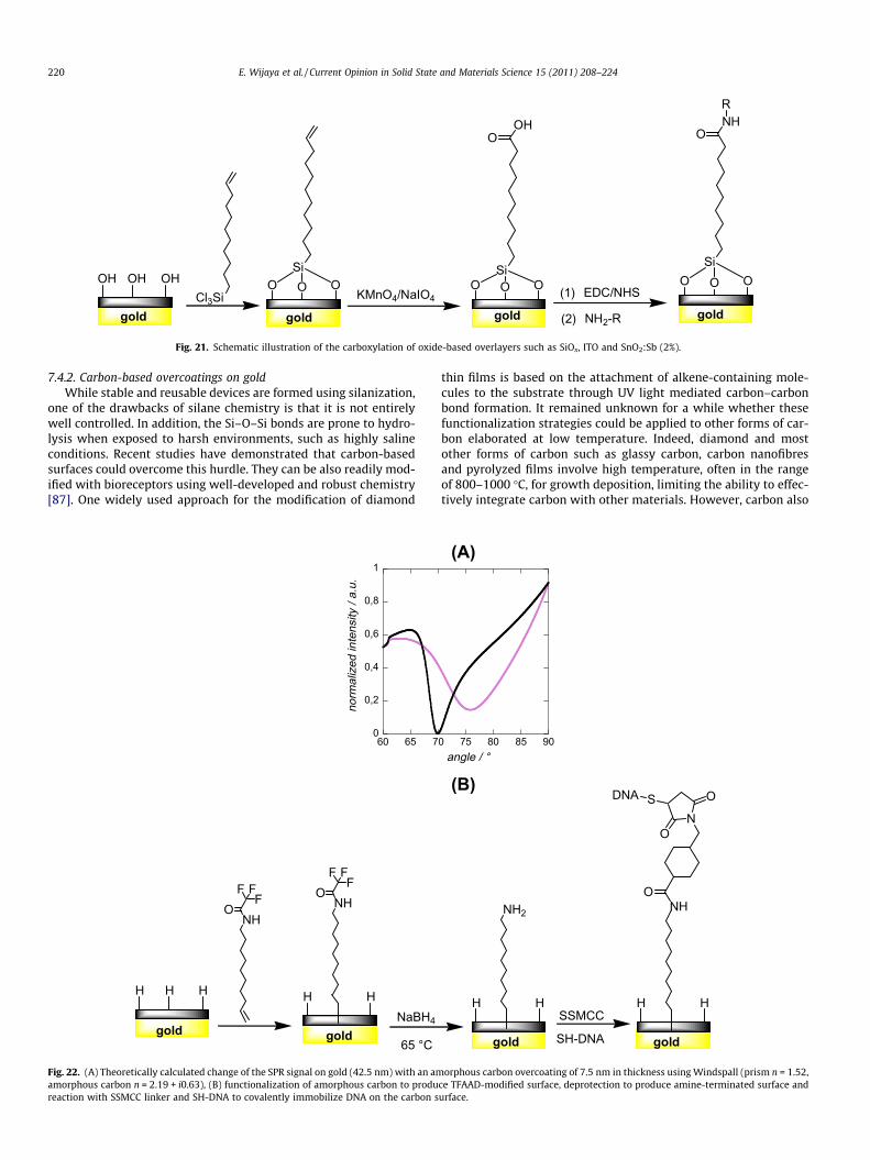
Fig. 21. Schematic illustration of the carboxylation of oxide-based overlayers such as SiOx, ITO and SnO2:Sb (2%).
220 E. Wijaya et al. / Current Opinion in Solid State and Materials Science 15 (2011) 208–224
7.4.2. Carbon-based overcoatings on goldWhile stable and reusable devices are formed using silanization,
one of the drawbacks of silane chemistry is that it is not entirelywell controlled. In addition, the Si–O–Si bonds are prone to hydro-lysis when exposed to harsh environments, such as highly salineconditions. Recent studies have demonstrated that carbon-basedsurfaces could overcome this hurdle. They can be also readily mod-ified with bioreceptors using well-developed and robust chemistry[87]. One widely used approach for the modification of diamond
0
0,2
0,4
0,6
0,8
1
60 65 7
nor
mal
ized
inte
nsity
/ a.
u.
H HH
NHO
FF
F
H H
NHO
FF
F
NaBH4
65 °Cgold gold
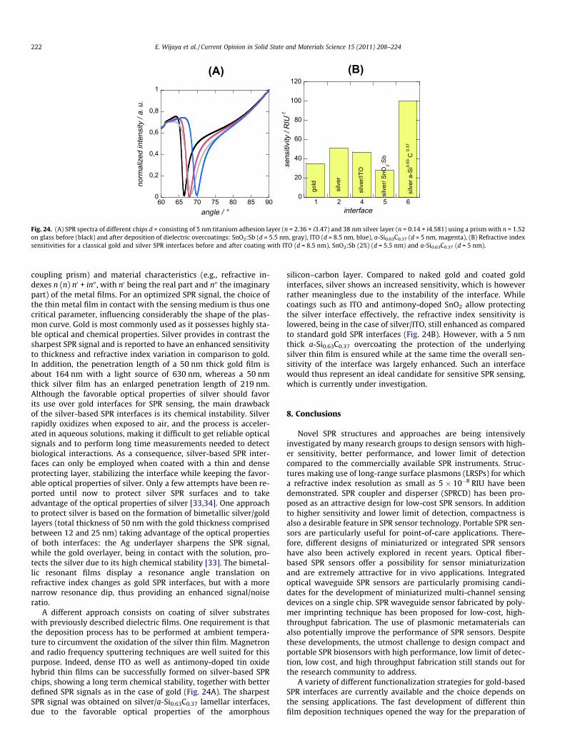
Fig. 22. (A) Theoretically calculated change of the SPR signal on gold (42.5 nm) with an amamorphous carbon n = 2.19 + i0.63), (B) functionalization of amorphous carbon to produreaction with SSMCC linker and SH-DNA to covalently immobilize DNA on the carbon s
thin films is based on the attachment of alkene-containing mole-cules to the substrate through UV light mediated carbon–carbonbond formation. It remained unknown for a while whether thesefunctionalization strategies could be applied to other forms of car-bon elaborated at low temperature. Indeed, diamond and mostother forms of carbon such as glassy carbon, carbon nanofibresand pyrolyzed films involve high temperature, often in the rangeof 800–1000 �C, for growth deposition, limiting the ability to effec-tively integrate carbon with other materials. However, carbon also
0 75 80 85 90angle / °
(B)
(A)
H H
NH2
SSMCC
SH-DNA
H H
dlogdlog
NH
N
O
O
OSDNA
orphous carbon overcoating of 7.5 nm in thickness using Windspall (prism n = 1.52,ce TFAAD-modified surface, deprotection to produce amine-terminated surface andurface.
E. Wijaya et al. / Current Opinion in Solid State and Materials Science 15 (2011) 208–224 221
forms a range of less-crystalline materials, including diamond-likecarbon (DLC) and other forms of amorphous carbon (a-C:H) films.Amorphous carbon is particularly an interesting material as itcan be deposited at room temperature [88,89]. It can also be hydro-gen-terminated using an inductively coupled hydrogen plasma,allowing the use of the same surface functionalization schemesdeveloped for diamond. There are currently only few reports onthe integration of amorphous carbon with gold-based SPR chips.Lockett et al. recently showed that thin layers of amorphous carbon(0–20 nm) can be deposited onto a surface plasmon active goldfilm using a sputtering technique [36]. A decrease of the photon-plasmon coupling efficiency and a broadening of the angle scan-ning SPR curves are ascribed to the complex dielectric function ofamorphous carbon (n = 2.19 + i0.63). Theoretical determination ofthe refractive index sensitivity gave S = 15, which is significantlylower than that of the previously described interfaces (Fig. 22A).The loss in sensitivity caused by the amorphous carbon film couldbe slightly reduced by altering the thickness of the surface plasmonactive gold film; a S = 18 was obtained in this case. The interfacewas further investigated for the in situ synthesis of oligonucleotidearrays utilizing photochemically protected oligonucleotide build-ing blocks (Fig. 22B). This is in fact not possible with traditionalgold SPR substrates, as extended exposure to ultraviolet light andoxidizing chemical conditions leads to the degradation of the goldthin film [36].
In a related approach, different a-C:H layers (nitrogenated andfluorinated) were deposited onto a gold SPR interface by radio-frequency sputtering from a graphite target in the presence ofargon and an additional gas plasma of nitrogen or tetrafluorome-thane [89]. Here, the resistance to protein adsorption was studied.
We and others have found that the sensitivity can remain thesame as on gold by choosing amorphous silicon-carbon alloys
(
0
5
10
15
20
25
30
35
40
sens
itivi
ty /
RIU
-1
in
gold
HF vaporH
gold gold
OHO
Fig. 23. (A) Experimentally determined refractive index sensitivities S for a classical gohydrogenation of a-Si1�xCx:H thin film and subsequent functionalization with undecylen
(abbreviated as a-Si1�xCx:H) with the right chemical composition(Fig. 23A) [47]. Amorphous silicon–carbon alloys can be depositedas thin films, and changing the carbon content of the film allowsfor fine tuning the material properties. Increasing the carbon con-tent leads to the optical band gap enlargement and to a transparentmaterial, and at the same time decreasing the refractive index,which is beneficial for the fabrication of lamellar SPR interfaces.Stable hydrogenated coatings of about 5 nm in thickness ofa-Si0.63C0.37:H film were thus formed on gold using PECVD in the‘‘low-power’’ regime. Surface hydrogenated a-Si0.63C0.37:H can beconveniently functionalized by stable organic layers throughrobust Si–C covalent bonds in a similar way as crystalline silicon.Fig. 23B shows one possibility to introduce acid terminal functiononto such a dielectric layer. Immersing surface-hydrogenateda-Si0.63C0.37:H films into undecylenic acid followed by UV irradia-tion leads to the formation of an organic monolayer covalentlyattached to the surface through Si–C bonds. By making a quantita-tive infrared data analysis, a molecular density of linked carbo-xydecyl groups of NA � 2 � 1013 mol cm�2 was found [47]. Thisvalue is lower than that on crystalline silicon, as expected for amaterial incorporating a significant amount of methyl groups.The acid function can be further converted to an activated estergroup using EDC/NHS chemistry, to which amine-terminatedbioreceptors can be easily anchored.
7.4.3. Overcoatings on silverFor sensing applications, it is mandatory that the width, posi-
tion, and height of the resulting SPR signal to be highly sensitiveto any variation in the refractive index of the dielectric mediumin the vicinity. For a SPR set up in the Kretschmann–Raether ATRconfiguration, reflectance depends strongly on the experimentalconditions (e.g., polarization, wavelength of the coupled radiation,
A)
terface
gold
/ Si
0.63
C0.
37:H
(B)
2. NH2-R
1. EDC/NHSgold
OHO
gold
OR-HN
ld SPR interface without and with 5 nm thick a-Si0.63C0.37 coating layer, (B) Surfaceic acid.
(A) (B)
0
0,2
0,4
0,6
0,8
1
60 65 70 75 80 85 90
norm
aliz
ed in
tens
ity /
a. u
.
angle / °
0
20
40
60
80
100
120
1 2 4 5 6
sens
itivi
ty /
RIU
-1
interface
gold
silv
er
silv
er/IT
O
silv
er/ S
nO2:S
b
silv
er a
-Si0.
63 C
0.37
Fig. 24. (A) SPR spectra of different chips d = consisting of 5 nm titanium adhesion layer (n = 2.36 + i3.47) and 38 nm silver layer (n = 0.14 + i4.581) using a prism with n = 1.52on glass before (black) and after deposition of dielectric overcoatings: SnO2:Sb (d = 5.5 nm, gray), ITO (d = 8.5 nm, blue), a-Si0.63C0.37 (d = 5 nm, magenta), (B) Refractive indexsensitivities for a classical gold and silver SPR interfaces before and after coating with ITO (d = 8.5 nm), SnO2:Sb (2%) (d = 5.5 nm) and a-Si0.63C0.37 (d = 5 nm).
222 E. Wijaya et al. / Current Opinion in Solid State and Materials Science 15 (2011) 208–224
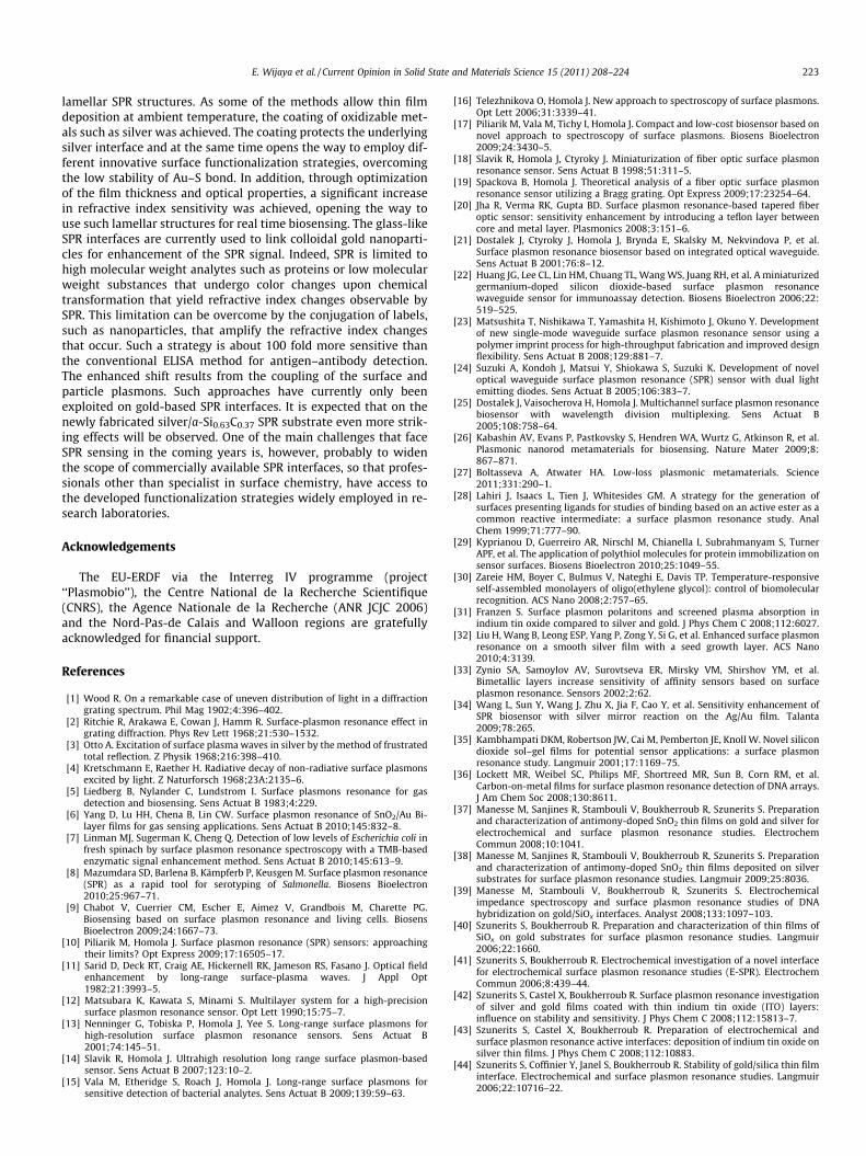
coupling prism) and material characteristics (e.g., refractive in-dexes n (n) n0 + in00, with n0 being the real part and n00 the imaginarypart) of the metal films. For an optimized SPR signal, the choice ofthe thin metal film in contact with the sensing medium is thus onecritical parameter, influencing considerably the shape of the plas-mon curve. Gold is most commonly used as it possesses highly sta-ble optical and chemical properties. Silver provides in contrast thesharpest SPR signal and is reported to have an enhanced sensitivityto thickness and refractive index variation in comparison to gold.In addition, the penetration length of a 50 nm thick gold film isabout 164 nm with a light source of 630 nm, whereas a 50 nmthick silver film has an enlarged penetration length of 219 nm.Although the favorable optical properties of silver should favorits use over gold interfaces for SPR sensing, the main drawbackof the silver-based SPR interfaces is its chemical instability. Silverrapidly oxidizes when exposed to air, and the process is acceler-ated in aqueous solutions, making it difficult to get reliable opticalsignals and to perform long time measurements needed to detectbiological interactions. As a consequence, silver-based SPR inter-faces can only be employed when coated with a thin and denseprotecting layer, stabilizing the interface while keeping the favor-able optical properties of silver. Only a few attempts have been re-ported until now to protect silver SPR surfaces and to takeadvantage of the optical properties of silver [33,34]. One approachto protect silver is based on the formation of bimetallic silver/goldlayers (total thickness of 50 nm with the gold thickness comprisedbetween 12 and 25 nm) taking advantage of the optical propertiesof both interfaces: the Ag underlayer sharpens the SPR signal,while the gold overlayer, being in contact with the solution, pro-tects the silver due to its high chemical stability [33]. The bimetal-lic resonant films display a resonance angle translation onrefractive index changes as gold SPR interfaces, but with a morenarrow resonance dip, thus providing an enhanced signal/noiseratio.
A different approach consists on coating of silver substrateswith previously described dielectric films. One requirement is thatthe deposition process has to be performed at ambient tempera-ture to circumvent the oxidation of the silver thin film. Magnetronand radio frequency sputtering techniques are well suited for thispurpose. Indeed, dense ITO as well as antimony-doped tin oxidehybrid thin films can be successfully formed on silver-based SPRchips, showing a long term chemical stability, together with betterdefined SPR signals as in the case of gold (Fig. 24A). The sharpestSPR signal was obtained on silver/a-Si0.63C0.37 lamellar interfaces,due to the favorable optical properties of the amorphous
silicon–carbon layer. Compared to naked gold and coated goldinterfaces, silver shows an increased sensitivity, which is howeverrather meaningless due to the instability of the interface. Whilecoatings such as ITO and antimony-doped SnO2 allow protectingthe silver interface effectively, the refractive index sensitivity islowered, being in the case of silver/ITO, still enhanced as comparedto standard gold SPR interfaces (Fig. 24B). However, with a 5 nmthick a-Si0.63C0.37 overcoating the protection of the underlyingsilver thin film is ensured while at the same time the overall sen-sitivity of the interface was largely enhanced. Such an interfacewould thus represent an ideal candidate for sensitive SPR sensing,which is currently under investigation.
8. Conclusions
Novel SPR structures and approaches are being intensivelyinvestigated by many research groups to design sensors with high-er sensitivity, better performance, and lower limit of detectioncompared to the commercially available SPR instruments. Struc-tures making use of long-range surface plasmons (LRSPs) for whicha refractive index resolution as small as 5 � 10�8 RIU have beendemonstrated. SPR coupler and disperser (SPRCD) has been pro-posed as an attractive design for low-cost SPR sensors. In additionto higher sensitivity and lower limit of detection, compactness isalso a desirable feature in SPR sensor technology. Portable SPR sen-sors are particularly useful for point-of-care applications. There-fore, different designs of miniaturized or integrated SPR sensorshave also been actively explored in recent years. Optical fiber-based SPR sensors offer a possibility for sensor miniaturizationand are extremely attractive for in vivo applications. Integratedoptical waveguide SPR sensors are particularly promising candi-dates for the development of miniaturized multi-channel sensingdevices on a single chip. SPR waveguide sensor fabricated by poly-mer imprinting technique has been proposed for low-cost, high-throughput fabrication. The use of plasmonic metamaterials canalso potentially improve the performance of SPR sensors. Despitethese developments, the utmost challenge to design compact andportable SPR biosensors with high performance, low limit of detec-tion, low cost, and high throughput fabrication still stands out forthe research community to address.
A variety of different functionalization strategies for gold-basedSPR interfaces are currently available and the choice depends onthe sensing applications. The fast development of different thinfilm deposition techniques opened the way for the preparation of
E. Wijaya et al. / Current Opinion in Solid State and Materials Science 15 (2011) 208–224 223
lamellar SPR structures. As some of the methods allow thin filmdeposition at ambient temperature, the coating of oxidizable met-als such as silver was achieved. The coating protects the underlyingsilver interface and at the same time opens the way to employ dif-ferent innovative surface functionalization strategies, overcomingthe low stability of Au–S bond. In addition, through optimizationof the film thickness and optical properties, a significant increasein refractive index sensitivity was achieved, opening the way touse such lamellar structures for real time biosensing. The glass-likeSPR interfaces are currently used to link colloidal gold nanoparti-cles for enhancement of the SPR signal. Indeed, SPR is limited tohigh molecular weight analytes such as proteins or low molecularweight substances that undergo color changes upon chemicaltransformation that yield refractive index changes observable bySPR. This limitation can be overcome by the conjugation of labels,such as nanoparticles, that amplify the refractive index changesthat occur. Such a strategy is about 100 fold more sensitive thanthe conventional ELISA method for antigen–antibody detection.The enhanced shift results from the coupling of the surface andparticle plasmons. Such approaches have currently only beenexploited on gold-based SPR interfaces. It is expected that on thenewly fabricated silver/a-Si0.63C0.37 SPR substrate even more strik-ing effects will be observed. One of the main challenges that faceSPR sensing in the coming years is, however, probably to widenthe scope of commercially available SPR interfaces, so that profes-sionals other than specialist in surface chemistry, have access tothe developed functionalization strategies widely employed in re-search laboratories.
Acknowledgements
The EU-ERDF via the Interreg IV programme (project‘‘Plasmobio’’), the Centre National de la Recherche Scientifique(CNRS), the Agence Nationale de la Recherche (ANR JCJC 2006)and the Nord-Pas-de Calais and Walloon regions are gratefullyacknowledged for financial support.
References
[1] Wood R. On a remarkable case of uneven distribution of light in a diffractiongrating spectrum. Phil Mag 1902;4:396–402.
[2] Ritchie R, Arakawa E, Cowan J, Hamm R. Surface-plasmon resonance effect ingrating diffraction. Phys Rev Lett 1968;21:530–1532.
[3] Otto A. Excitation of surface plasma waves in silver by the method of frustratedtotal reflection. Z Physik 1968;216:398–410.
[4] Kretschmann E, Raether H. Radiative decay of non-radiative surface plasmonsexcited by light. Z Naturforsch 1968;23A:2135–6.
[5] Liedberg B, Nylander C, Lundstrom I. Surface plasmons resonance for gasdetection and biosensing. Sens Actuat B 1983;4:229.
[6] Yang D, Lu HH, Chena B, Lin CW. Surface plasmon resonance of SnO2/Au Bi-layer films for gas sensing applications. Sens Actuat B 2010;145:832–8.
[7] Linman MJ, Sugerman K, Cheng Q. Detection of low levels of Escherichia coli infresh spinach by surface plasmon resonance spectroscopy with a TMB-basedenzymatic signal enhancement method. Sens Actuat B 2010;145:613–9.
[8] Mazumdara SD, Barlena B, Kämpferb P, Keusgen M. Surface plasmon resonance(SPR) as a rapid tool for serotyping of Salmonella. Biosens Bioelectron2010;25:967–71.
[9] Chabot V, Cuerrier CM, Escher E, Aimez V, Grandbois M, Charette PG.Biosensing based on surface plasmon resonance and living cells. BiosensBioelectron 2009;24:1667–73.
[10] Piliarik M, Homola J. Surface plasmon resonance (SPR) sensors: approachingtheir limits? Opt Express 2009;17:16505–17.
[12] Matsubara K, Kawata S, Minami S. Multilayer system for a high-precisionsurface plasmon resonance sensor. Opt Lett 1990;15:75–7.
[13] Nenninger G, Tobiska P, Homola J, Yee S. Long-range surface plasmons forhigh-resolution surface plasmon resonance sensors. Sens Actuat B2001;74:145–51.
[14] Slavik R, Homola J. Ultrahigh resolution long range surface plasmon-basedsensor. Sens Actuat B 2007;123:10–2.
[15] Vala M, Etheridge S, Roach J, Homola J. Long-range surface plasmons forsensitive detection of bacterial analytes. Sens Actuat B 2009;139:59–63.
[16] Telezhnikova O, Homola J. New approach to spectroscopy of surface plasmons.Opt Lett 2006;31:3339–41.
[17] Piliarik M, Vala M, Tichy I, Homola J. Compact and low-cost biosensor based onnovel approach to spectroscopy of surface plasmons. Biosens Bioelectron2009;24:3430–5.
[18] Slavik R, Homola J, Ctyroky J. Miniaturization of fiber optic surface plasmonresonance sensor. Sens Actuat B 1998;51:311–5.
[19] Spackova B, Homola J. Theoretical analysis of a fiber optic surface plasmonresonance sensor utilizing a Bragg grating. Opt Express 2009;17:23254–64.
[20] Jha R, Verma RK, Gupta BD. Surface plasmon resonance-based tapered fiberoptic sensor: sensitivity enhancement by introducing a teflon layer betweencore and metal layer. Plasmonics 2008;3:151–6.
[21] Dostalek J, Ctyroky J, Homola J, Brynda E, Skalsky M, Nekvindova P, et al.Surface plasmon resonance biosensor based on integrated optical waveguide.Sens Actuat B 2001;76:8–12.
[22] Huang JG, Lee CL, Lin HM, Chuang TL, Wang WS, Juang RH, et al. A miniaturizedgermanium-doped silicon dioxide-based surface plasmon resonancewaveguide sensor for immunoassay detection. Biosens Bioelectron 2006;22:519–525.
[23] Matsushita T, Nishikawa T, Yamashita H, Kishimoto J, Okuno Y. Developmentof new single-mode waveguide surface plasmon resonance sensor using apolymer imprint process for high-throughput fabrication and improved designflexibility. Sens Actuat B 2008;129:881–7.
[24] Suzuki A, Kondoh J, Matsui Y, Shiokawa S, Suzuki K. Development of noveloptical waveguide surface plasmon resonance (SPR) sensor with dual lightemitting diodes. Sens Actuat B 2005;106:383–7.
[25] Dostalek J, Vaisocherova H, Homola J. Multichannel surface plasmon resonancebiosensor with wavelength division multiplexing. Sens Actuat B2005;108:758–64.
[26] Kabashin AV, Evans P, Pastkovsky S, Hendren WA, Wurtz G, Atkinson R, et al.Plasmonic nanorod metamaterials for biosensing. Nature Mater 2009;8:867–871.
[27] Boltasseva A, Atwater HA. Low-loss plasmonic metamaterials. Science2011;331:290–1.
[28] Lahiri J, Isaacs L, Tien J, Whitesides GM. A strategy for the generation ofsurfaces presenting ligands for studies of binding based on an active ester as acommon reactive intermediate: a surface plasmon resonance study. AnalChem 1999;71:777–90.
[29] Kyprianou D, Guerreiro AR, Nirschl M, Chianella I, Subrahmanyam S, TurnerAPF, et al. The application of polythiol molecules for protein immobilization onsensor surfaces. Biosens Bioelectron 2010;25:1049–55.
[30] Zareie HM, Boyer C, Bulmus V, Nateghi E, Davis TP. Temperature-responsiveself-assembled monolayers of oligo(ethylene glycol): control of biomolecularrecognition. ACS Nano 2008;2:757–65.
[31] Franzen S. Surface plasmon polaritons and screened plasma absorption inindium tin oxide compared to silver and gold. J Phys Chem C 2008;112:6027.
[32] Liu H, Wang B, Leong ESP, Yang P, Zong Y, Si G, et al. Enhanced surface plasmonresonance on a smooth silver film with a seed growth layer. ACS Nano2010;4:3139.
[33] Zynio SA, Samoylov AV, Surovtseva ER, Mirsky VM, Shirshov YM, et al.Bimetallic layers increase sensitivity of affinity sensors based on surfaceplasmon resonance. Sensors 2002;2:62.
[34] Wang L, Sun Y, Wang J, Zhu X, Jia F, Cao Y, et al. Sensitivity enhancement ofSPR biosensor with silver mirror reaction on the Ag/Au film. Talanta2009;78:265.
[35] Kambhampati DKM, Robertson JW, Cai M, Pemberton JE, Knoll W. Novel silicondioxide sol–gel films for potential sensor applications: a surface plasmonresonance study. Langmuir 2001;17:1169–75.
[36] Lockett MR, Weibel SC, Philips MF, Shortreed MR, Sun B, Corn RM, et al.Carbon-on-metal films for surface plasmon resonance detection of DNA arrays.J Am Chem Soc 2008;130:8611.
[37] Manesse M, Sanjines R, Stambouli V, Boukherroub R, Szunerits S. Preparationand characterization of antimony-doped SnO2 thin films on gold and silver forelectrochemical and surface plasmon resonance studies. ElectrochemCommun 2008;10:1041.
[38] Manesse M, Sanjines R, Stambouli V, Boukherroub R, Szunerits S. Preparationand characterization of antimony-doped SnO2 thin films deposited on silversubstrates for surface plasmon resonance studies. Langmuir 2009;25:8036.
[39] Manesse M, Stambouli V, Boukherroub R, Szunerits S. Electrochemicalimpedance spectroscopy and surface plasmon resonance studies of DNAhybridization on gold/SiOx interfaces. Analyst 2008;133:1097–103.
[40] Szunerits S, Boukherroub R. Preparation and characterization of thin films ofSiOx on gold substrates for surface plasmon resonance studies. Langmuir2006;22:1660.
[41] Szunerits S, Boukherroub R. Electrochemical investigation of a novel interfacefor electrochemical surface plasmon resonance studies (E-SPR). ElectrochemCommun 2006;8:439–44.
[42] Szunerits S, Castel X, Boukherroub R. Surface plasmon resonance investigationof silver and gold films coated with thin indium tin oxide (ITO) layers:influence on stability and sensitivity. J Phys Chem C 2008;112:15813–7.
[43] Szunerits S, Castel X, Boukherroub R. Preparation of electrochemical andsurface plasmon resonance active interfaces: deposition of indium tin oxide onsilver thin films. J Phys Chem C 2008;112:10883.
[44] Szunerits S, Coffinier Y, Janel S, Boukherroub R. Stability of gold/silica thin filminterface. Electrochemical and surface plasmon resonance studies. Langmuir2006;22:10716–22.
224 E. Wijaya et al. / Current Opinion in Solid State and Materials Science 15 (2011) 208–224
[45] Szunerits S, Nunes-Kirchner C, Wittstock G, Boukherroub R, Chantal G.Electrochemical investigation of the influence of thin SiOx films depositedon gold on charge transfer characteristics. Electrochim Acta 2008;53:7805–7914.
[46] Touahir L, Jenkins ATA, Boukherroub R, Gouget-Laemmel AC, Chazalviel J-N,Peretti J, et al. Surface plasmon-enhanced fluorescence spectroscopy on silverbased SPR substrates. J Phys Chem C 2010;114:22582.
[47] Touahir L, Niedziółka-Jönsson J, Galopin E, Boukherroub R, Gouget-LaemmelAC, Solomon I, et al. Surface plasmon resonance on gold and silver films coatedwith thin layers of amorphous silicon–carbon alloys. Langmuir 2010;26:6058.
[48] Love JC, Estroff LA, Kriebel JK, Nuzzo RG, Whitesides GM. Thiolates on metalsas a form of nanotechnology. Chem Rev 2005;105:1103.
[49] Ulman A. Formation and structure of self-assembled monolayers. Chem Rev1996;96:1533.
[50] Peeters S, Stakenborg T, Reekmans G, Laureyn W, Lagae L, Van Aerschot A, et al.Impact of spacers on the hybridization efficiency of mixed self-assembledDNA/alkanethiol films. Biosens Bioelectron 2008;24:72.
[51] Malic L, Veres T, Tabrizian M. Biochip functionalization using electrowetting-on-dielectric digital microfluidics for surface plasmon resonance imagingdetection of DNA hybridization. Biosens Bioelectron 2009;24:2218.
[52] Ananthanawat C, Vilaivan T, Mekboonsonglarp W, Hoven VP. Thiolatedpyrrolidinyl peptide nucleic acids for the detection of DNA hybridizationusing surface plasmon resonance. Biosens Bioelectron 2009;24:3544.
[53] Wang J, Munir A, Li Z, Shou HS. Aptamer-Au NPs conjugates-enhanced SPRsensing for the ultrasensitive sandwich immunoassay. Biosens Bioelectron2010;25:124.
[54] Frasconi M, Tel-Vered R, Riskin M, Willner I. Surface plasmon resonanceanalysis of antibodies using imprinted boronic acid-functionalized goldnanoparticle composites. Anal Chem 2010;82:2512–9.
[55] Liu Y, Dong Y, Jauw J, Linman MJ, Cheng Q. Highly sensitive detection ofprotein toxins by surface plasmon resonance with biotinylation-based inlineatom transfer radical polymerization amplification. Anal Chem2010;82:3679–85.
[56] Chen Y, Nguyen A, Niu L, Corn RM. Fabrication of DNA microarrays with poly(L-glutamic acid) monolayers on gold substrates for SPR imaging measurements.Langmuir 2009;25:5054–60.
[57] Hoa XD, Kirk AG, Tabrizian M. Enhanced SPR response from patternedimmobilization of surface bioreceptors on nano-gratings. Biosens Bioelectron2009;24:3043.
[58] Jordan CE, Frutos AG, Thiel AJ, Corn RM. Surface plasmon resonance imagingmeasurements of DNA hybridization adsorption and streptavidin/DNAmultilayer formation at chemically modified gold surfaces. Anal Chem1997;69:4939–47.
[59] Gamsjaeger R, Wimmer B, Kahr H, Tinazli A, Picuric S, Lata S, et al. Orientedbinding of the His6-tagged carboxyl-tail of the L-type Ca2+ channel a-subunitto a new NTA-functionalized self assembled monolayer. Langmuir2004;20:5885.
[60] Zhang Y, Luo S, Tang Y, Yu L, Hou K-Y, Cheng J-P, et al. Carbohydrate–proteininteractions by ‘‘clicked’’ f carbohydrate self-assembled monolayers. AnalChem 2006;78:2001–8.
[61] Kolb HC, Finn MG, Sharpless KB. Chemistry: diverse chemical. Function from afew good reactions. Angew Chem Int Ed 2001;40:2004–21.
[62] Lee J-M, Park HK, Jung YS, Kim JK, Jung SO, Chung BH. Direct immobilization ofprotein G variants with various numbers of cysteine residues on a gold surface.Anal Chem 2007;79:2680–7.
[63] Lofas S, Johnsoon BJ. A novel hydrogel matrix on gold surface plasmonresonance sensors. Chem Soc, Chem Commun 1990;21:1526.
[64] Lofas S, Johnsson B, Tegendal K, Ronnberg I. Dextran modified gold surfaces forsurface plasmon resonance sensors: immunoreactivity of immobilizedantibiotics and antibody-surface interaction studies. Colloids Surf B1993;1:83–9.
[65] Jung S-H, Jung J-W, Suh I-B, Yuk J-S, Kim W-J, Choi E-Y, et al. Analysis of C-reactive protein on amide-linked N-hydroxysuccinimide-dextran arrays with aspectral surface plasmon resonance biosensor for serodiagnosis. Anal Chem2007;79:5703–10.
[66] Loka RS, Cairo CW. Immobilization of carbohydrate epitopes for surfaceplasmon resonance using Staudinger ligation chemistry. Carbohydr Res,accepted for publication, doi:10.1016/j.carrs.2010.1009.1020.
[67] Le HQA, Sauriat-Dorizon H, Korri-Youssoufi H. Investigation of SPR andelectrochemical detection of antigen with polypyrrole functionalized bybiotinylated single-chain antibody: a review. Anal Chim Acta 2010;674:1.
[68] Gondran C, Dubois M-P, Fort S, Cosnier S, Szunerits S. Detection ofcarbohydrate-binding proteins by oligosaccharide-modified polypyrroleinterfaces using electrochemical surface plasmon resonance. Analyst2008;133:206.
[69] Dong H, Cao X, Li CM, Hu W. An in situ electrochemical surface plasmonresonance immunosensor with polypyrrole propylic acid film: comparisonsbetween SPR and electrochemical responses from polymer formation toprotein immunosensing. Biosens Bioelectron 2008;23:1055.
[70] Su X, Teh H, Aung KMM, Zong Y, Gao Z. Femtomol SPR detection of DNA–PNAhybridization with the assistance of DNA-guided polyaniline deposition.Biosens Bioelectron 2008;23:1715.
[71] Zhang N, Schweiss R, Zong Y, Knoll W. Electrochim Acta 2007;52:2869.[72] Szunerits S, Knorr N, Calemczuk R, Livache T. A new approach to writing and
simultaneous reading of micropatterns: combining surface plasmon resonanceimaging (SPR-i) with scanning electrochemical microscopy (SECM). Langmuir2004;20:9236–41.
[73] Hu W, Li M, Cui X, Zhou Q. In situ studies of protein adsorption onpoly(pyrrole-co-pyrrole propylic acid) films by electrochemical surfaceplasmon resonance. Langmuir 2007;23:2761.
[74] Guedon P, Livache T, Martin F, Lesbre F, Roget A, Bidan G, et al.Characterisation and optimisation of a real time parallel, label free,polypyrrole based DNA sensor by SPRi. Anal Chem 2000;72:6003.
[75] Liu S, Vareiro MMLM, Fraser S, Jenkins ATA. Control of attachment of bovineserum albumin to pulse plasma-polymerized maleic anhydride by variation ofpulse conditions. Langmuir 2005;21:8572.
[76] Duque L, Menges B, Borros S, Forch R. Immobilization of Biomolecules toplasma polymerized pentafluorophenyl methacrylate. Biomacromolecules2010; xxx.
[77] Song SY, Choi HG, Hong JW, Kim BW, Sim SJ, Yoo HC. Selective antigen–antibody recognition on SPR sensor based on the heat-sensitiveconformational change of poly(N-isopropylacrylamide). Colloid Surf A:Physicochem Eng Aspects 2008;313–314:504.
[78] Kyprianou D, Guerreiro AR, Chianella I, Piletska EV, Fowler SA, Karim K, et al.New reactive polymer for protein immobilization on sensor surfaces. BiosensBioelectron 2009;24:1365.
[79] Chegel V, Whitcombe MJ, Turner NW, Piletsky SA. Deposition of functionalizedpolymer layers in surface plasmon resonance immunosensors by in-situpolymerization in the evanescent wave field. Biosens Bioelectron2009;24:1270.
[80] Kessler D, Roth P, Theato P. Reactive surface coatings based onpolysilsesquioxanes: controlled functionalization for specific proteinimmobization. Langmuir 2010;25:10068.
[81] Phillips KS, Han J, Martinez M, Wang Z, Carter D, Cheng Q. Nanoscaleglassification of gold substrates for surface plasmon resonance analysis ofprotein toxins with supported lipid membranes. Anal Chem 2006;78:596.
[82] Satriano C, Edvardsson M, Ohlsson G, Wang G, Svedhem S, Kasemo B. Plasmaoxidized polyhydroxymethylsiloxane – a new smooth surface for supportedlipid bilayer formation. Langmuir 2010;26:5715–25.
[83] Satriano C, Marletta G, Kasemo B. Oxygen plasma-induced conversion ofpolysiloxane into hydrophilic and smooth SiOx surfaces. Surf Interf Anal2008;40:649.
[84] Zawica I, Wittstock G, Boukherroub R, Szunerits S. Polarization modulationinfrared reflection absorption spectroscopy investigations of thin silica filmsdeposited on gold. 2. Structural analysis of 1,2-dimyristoyl-sn-glycero-3-phosphocholine bilayer. Langmuir 2008;24:3922–9.
[85] Zawica I, Wittstock G, Boukherroub R, Szunerits S. PM IRRAS investigation ofthin silica films deposited on gold. Part 1. Theory and proof of concept.Langmuir 2007;23:9303–9.
[86] Fadeev AY, McCarthy TJ. Self-assembly is not the only reaction possiblebetween alkyltrichlorosilanes and surfaces: monomolecular and oligomericcovalently attached layers of dichloro- and trichloroalkylsilanes on silicon.Langmuir 2000;16:7268.
[87] Szunerits S, Boukherroub R. Review: chemical, photochemical andelectrochemical surface modification of diamond. J Solid State Electrochem2008;12:1205–18.
[88] Sun B, Colavita PE, Kim H, Lockett M, Marus MS, Smith LM, et al. Covalentphotochemical functionalization of amorphous carbon thin films forintegrated real-time biosensing. Langmuir 2006;22:9598.
[89] Akasaka H, Gawazawa N, Suzuki T, Nakano M, Ohshio S, Saitoh H. Evaluation ofprotein adsorption on hydrogenated amorphous carbon films by surfaceplasmon resonance phenomenon. Diamond Relat Mater 2010;19:1235.