Microbial Production of Nanoparticles and their Applications Chapter 2 Current Research in Microbiology Pragati Sahai 1 ; Nabeel Ahmad 2 ; Vimlendu B Sinha 1 ; Rajiv Dutta* 1 Department of Biotechnology, School of Engineering & Technology, Sharda University, Greater Noi- da-201306 (U.P.) India. 2 Department of Biotechnology, School of Engineering & Technology, IFTM University, NH-24, Delhi Road, Moradabad-244001 (U.P.), India. *Correspondence to: Rajiv Dutta, Department of Biotechnology, College of Engineering & Technology, IILM- AHL, Greater Noida-201306 (U.P.), India. Email: [email protected]Abstract Nanoparticles are particles that have size of 100 nm or less with one or more dimensions has gained larger attention due to their characteristic and unique proper- ties apart from wide range of applications over their other counterparts. The physi- cal, chemical, biological and hybrid ways of synthesis of nanoparticles is dependent on the requirement and type of nanoparticles however for clinical and biological application the chemical methods have proven to be toxic to the living system there- fore better and safer alternatives are chosen like biological methods of production of nanoparticles. In biological methods the use of microorganism for production of nanoparticles is gaining lots of attention for being economical, rapid and safer alternative to physical and chemical methods. The wide range of microorganism and their potential to adapt in different environment gives them an edge over other ways. The microbial production of nanoparticles is the part of microbial growth that involves two processes: reduction process and precipitation process. The latter is further achieved by either nucleation or crystal growth. The entire production is controlled by controlling the growth parameter of the microbes. Thus the process is simpler and economical but is slow and time consuming as compared to chemical ways, however the quality and quantity of the nanoparticle is far better in biological methods than in chemical methods.
Transcript
Microbial Production of Nanoparticles and their Applications
Chapter 2
Current Research in Microbiology
Pragati Sahai1; Nabeel Ahmad2; Vimlendu B Sinha1; Rajiv Dutta*1Department of Biotechnology, School of Engineering & Technology, Sharda University, Greater Noi-
da-201306 (U.P.) India.2Department of Biotechnology, School of Engineering & Technology, IFTM University, NH-24, Delhi
Road, Moradabad-244001 (U.P.), India.
*Correspondence to: Rajiv Dutta, Department of Biotechnology, College of Engineering & Technology, IILM-
Nanoparticles are particles that have size of 100 nm or less with one or more dimensions has gained larger attention due to their characteristic and unique proper-ties apart from wide range of applications over their other counterparts. The physi-cal, chemical, biological and hybrid ways of synthesis of nanoparticles is dependent on the requirement and type of nanoparticles however for clinical and biological application the chemical methods have proven to be toxic to the living system there-fore better and safer alternatives are chosen like biological methods of production of nanoparticles. In biological methods the use of microorganism for production of nanoparticles is gaining lots of attention for being economical, rapid and safer alternative to physical and chemical methods. The wide range of microorganism and their potential to adapt in different environment gives them an edge over other ways. The microbial production of nanoparticles is the part of microbial growth that involves two processes: reduction process and precipitation process. The latter is further achieved by either nucleation or crystal growth. The entire production is controlled by controlling the growth parameter of the microbes. Thus the process is simpler and economical but is slow and time consuming as compared to chemical ways, however the quality and quantity of the nanoparticle is far better in biological methods than in chemical methods.
Current Research in Microbiology
2
ww
w.openaccessebooks.com
Dut
ta R
1. Introduction
Nanoparticles are those entities of matter that have one or more dimensions ranging from 100 nm or even less. These particles have higher surface area but smaller size making them a better alternative for application than their bulk counterparts [1,2]. The physical, chem-ical, biological and hybrid ways of synthesis of nanoparticles is dependent on the requirement and type of nanoparticles [3-6].
Since physical and chemical methods are more fastidious and give high yield of nano-particles, they are most popular ways for nanoparticle synthesis, however the toxicity in the living system due to use of chemicals greatly limits their biomedical applications, particularly in clinical use. Secondly it was found that biogenic nanoparticles had greater potentials to include wider varieties and different shapes, compositions, coatings and structures of nano-particles with special properties as compared to their chemical counterparts [7]. Thirdly it was reported that even if synthetic nanoparticles are not used directly to the living system yet their accumulation was found because of use of certain daily products like consumer products which contains trace amount of nanoparticles that can lead to their accumulation into the living system which is harmful for both prokaryotic and eukaryotic system [8-9]. By using micro-organisms for synthesis of nanoparticles, a reliable, nontoxic and eco-friendly methods is de-signed that is of utmost importance to expand the biomedical applications of nanoparticles and also keeping in mind the environmental hazard the accumulation of synthetic nanoparticles can lead to.
Biological entities of matter have tremendous property to produce variety of potential nanoparticles. If fully understood and deciphered, these entities can be used for large scale production of almost all types of nanoparticles at industrial level manufacturing. The biologi-cally aided synthesis not only decreases the consumption of energy and toxic chemicals but also opens the path for environmentally friendly green manufacturing [10].
The use of bacteria among all biological systems for production of metal and metal oxide nanoparticles of various sizes, compositions and properties are well documented. For example the use of Bacillus sp. for reduction of Tellurium to Rosette- aggregated rod shape nanoparticles of size approximately 30x200 nm and Selenium to 200 nm spherical nanopar-ticles [11,12]. Another example is of Shewanella oneidensis, a specialized bacterium with a property of reducing metals like Tellerium to spherical nanoparticles of size 50-80 nm [13] and Magnetospirillum magneticum that produces magnetic nanoparticles of 30-120 nm [14].
Despiteofthefactthattherearesufficientexamplesofdifferenttypesofbiologicalenti-ties that can produce variety of nanoparticles of varying properties, yet there is a huge knowl-edge gap in understanding the mechanism behind the formation of those nanoparticles and the mechanismtocontrolthefinalproductisstillunclear.Thereisstillnotsufficientinformation
Current Research in Microbiology
3
thatcanleadtostandardizationoftheprocessofformationofnanoparticleswithspecificde-sired properties, concentration and size. Similarly there is no possible information that can providethewaystostandardizethefinalproductwhenthenanoparticlesareusedinaprocess.This knowledge gap refrains the use of biological agents for manufacturing of nanoparticles at industrial level. The bacterial based nano-manufacturing for mass production is precluded becauseofinsufficientknowledge.
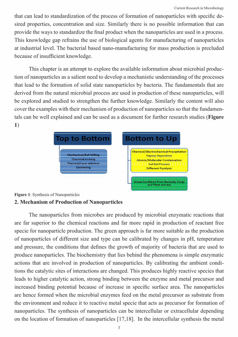
This chapter is an attempt to explore the available information about microbial produc-tion of nanoparticles as a salient need to develop a mechanistic understanding of the processes that lead to the formation of solid state nanoparticles by bacteria. The fundamentals that are derived from the natural microbial process are used in production of these nanoparticles, will be explored and studied to strengthen the further knowledge. Similarly the content will also cover the examples with their mechanism of production of nanoparticles so that the fundamen-tals can be well explained and can be used as a document for further research studies (Figure 1)
Figure 1: Synthesis of Nanoparticles
2. Mechanism of Production of Nanoparticles
The nanoparticles from microbes are produced by microbial enzymatic reactions that are far superior to the chemical reactions and far more rapid in production of reactant free specie for nanoparticle production. The green approach is far more suitable as the production of nanoparticles of different size and type can be calibrated by changes in pH, temperature andpressure,theconditionsthatdefinesthegrowthofmajorityofbacteriathatareusedtoproduce nanoparticles. The biochemistry that lies behind the phenomena is simple enzymatic actions that are involved in production of nanoparticles. By calibrating the ambient condi-tions the catalytic sites of interactions are changed. This produces highly reactive species that leads to higher catalytic action, strong binding between the enzyme and metal precursor and increased binding potential because of increase in specific surface area.The nanoparticlesare hence formed when the microbial enzymes feed on the metal precursor as substrate from the environment and reduce it to reactive metal specie that acts as precursor for formation of nanoparticles. The synthesis of nanoparticles can be intercellular or extracellular depending on the location of formation of nanoparticles [17,18]. In the intercellular synthesis the metal
Current Research in Microbiology
4
substrate from the environment is transported into the cell where microbial enzymes acts on it and reduce it to reactive specie leading to formation of nanoparticle inside the cell whereas in extracellular the metal substrate is trapped on the cell surface and the enzyme is transported or excreted out of the cell where it reacts with the metal ion on the cell surface to reduce it to reactive species thereby the nanoparticle is formed on the cell surface [19]. In general the use of microorganisms leads to the nanoparticle formation by two distinct approaches. First is: Bottom up approach in which the supersaturated solution is made to saturate more till it settles down in some phase and nanoparticles of particular size is produced. Second is: Top down approach in which the organic polymer produced by microorganism leads to the nucleation offirstreactivespeciealsocalledasnanoparticleseed.Theseorganicpolymersmodifythenucleation process of the nanoparticle seed by either favouring it or by inhibiting it, either way it can stabilize the molecule to produce the nanoparticle of particular size.
The bacterial production of metallic nanoparticles is by two processes:
a. Reduction Process
b. Precipitation Process
The precipitation process is further achieved by two ways
a. Nucleation
b. Crystal growth
From the above two processes, the reduction process is most studied and documented than precipitation process by nucleation or by crystal growth.
2.1. Reduction process of nanoparticle synthesis by microbes
The microbes use their reducing agents in formation of nanoparticles from its precursor molecule. These reducing equivalents can be taken by inorganic compounds as in lithotrophs or by organic compound as in organotrophs thus these precursors of nanoparticles act as sub-strates for reducing agents. The reduction of metals to their corresponding sulphides by metal reducing bacteria is an example of such reduction mechanism. The variety of metal nanoparti-cles produced by microorganisms are deposited in the cytoplasm, periplasm, and extracellular area or on the cell surface. These nanoparticles are produced either by energy conserving metal reduction dissimilation process or by cell building assimilation or by both as in co- metabo-lism. Such nanoparticles generally helped in remediation process as most of them lowered the concentration of toxic compounds. Usually such reduction processes occur in growth phase of microbial culture but some of them were reported in stationary phase too or can be produced extracellular by isolated microbial enzyme from the growing culture [15]. With the biological
Current Research in Microbiology
5
chemical reduction method the M+ stage is converted to M0 active stage or formation of free radicle is there that initiates the further production of large amount of nanoparticles in low cost and less time. Secondly it is easier to tune the formation of nanoparticles of varying size and tunebyjustchangingthereducingagent,thedispersingagent,temperatureandtime.Howeverin case of microbial production of nanoparticles no reducing agent is added from outside as the biological entity itself has reducing agent in large amount in growing microbial culture, that are highly reactive and capable of producing the nanoparticles in less time. However the dispersing agent can be added to give the desired size to the nanoparticles. Since no chemi-cal agent like reducing agent is added from outside the amount of impurity is lesser than in chemical production of nanoparticles. So this process is used in biological manufacturing of nanoparticles. Once the nanoparticles are formed they are then precipitated by crystal growth or by nucleation (Figure 2).
Figure 2: Example of Microbial Synthesis of Nanoparticles
2.2. Precipitation of nanoparticles
There are many ways of separation of Nanoparticles like poly condensation of sol to gel or gel to sol state sedimentation as seen in metallic nanoparticles. The aggregation of crystals can be by simple drying after the liquid phase sedimentation. In order to form mono disperse nanoparticle of particular size it is important that crystal grows at very slow and steady rate from the rapidly generating seed particles. Once the desired nanoparticles is formed then dis-persing agent is added to avoid further aggregation like addition of citrate as dispersing agent in formation of gold nanoparticles. Once the desired size nanoparticles is achieved then it is separated by various methods like the sedimentation method of nanoparticles by co-precipita-tionoralkalineprecipitation,bygelfiltration,bygelelectrophoresisandbycentrifugalsepara-tion.
On the basis of fact that precipitation of nanoparticles is most fruitful method for sedi-mentation of desired nanoparticles from colloid with varying sizes of nanoparticles, new tech
Current Research in Microbiology
6
niques are being used in this process. When the precipitation is done under high gravity con-ditions, it produces larger amount of nanoparticles and that too in low cost. This technique is findingitsapplicationforindustrialproductionofnanoparticlesbecauseofitslowcostandenvironment friendly green approach. This technique is called HIGH GRAVITY REACTIVE PRECIPITATION (HGRP) [16].
3. Production of Nanoparticles by Microbes
The microbes are those biological entities that are well studied for their constant envi-ronment interactions. Therefore they have wide range of energy producing processes ranging from organotrophy to lithotrophy in presence of oxygen or in absence of it, thus using different bio-mechanism as per the environmental need. This basic fact led to the use of microbes for production of nanoparticles by lithotrophy in presence of air or in absence of it. The common-est nanoparticles produced by microbes are discussed here in details to give an insight of the biosynthesis of nanoparticles by the microbes (Table 1). The nanoparticles are divided under four categories: Metallic nanoparticles (Au, Ag, Alloy, and metal nanoparticles), Oxide nano-particles (metallic and non-metallic oxide nanoparticles), Sulphide nanoparticles and other miscellaneous nanoparticles. Each type when studied in details helped in understanding the mechanism behind the production of nanoparticles by microbes.
3.1. Metallic nanoparticles
The metallic nanoparticles show different optical property that is of the fundamental at-traction and characteristics of nanoparticle. In general these properties are size dependent rang-ing from 1-100 nm. The metallic nanoparticles have different physical and chemical properties than bulk metals and so they exhibit optical characteristics for example 20 nm Au-Np exhibit wine red colour, Ag-Np exhibit yellowish grey and Pd-Np; Pt-Np exhibit black colour.
The metallic nanoparticles are produced by microbes as a result of reduction of metal to free radicle specie that aggregates to form nanoparticles. Thus M+ is converted to M0 in pres-ence of reducing agent of microbial origin or system.
The environmental toxicity is also largely due to accumulation of heavy metals that are toxic to microbes as well, however there are certain microbes that are resistant to these heavy metals and can use them as substrate in one or the other biochemical reaction for generation ofenergyasioneffluxfromthecellbymembraneproteinsthatfunctioneitherasATPaseoraschemiosmoticcationorprotonantitransportersthatcauseschemicaldetoxification.Thisled to the research behind production of nanoparticles from heavy metals as well for example production of Palladium, Mercury, Platinum etc. nanoparticles apart from gold and silver or their alloy nanoparticles.
Shewanella oneidensis Fe3O4 28 40–50Rectangular and
Extracellular
Saccharomyces cer-evisiae
Sb2O3 25–60 2–10Spherical and Intra-
cellular
Lactobacillus sp. TiO2 25 8–35Spherical and Ex-
tracellular
Fusarium oxysporum BaTiO3 25 4-5Spherical and Ex-
tracellular
Brevibacterium caseiPHB (Poly hy-droxybutyrate)
37 100–125 Intracellular
E. coli CdS 25 2–5Wurtzite crystal and Intracellular
Rhodobacter sphaeroi-des
ZnS Unknown 10.5+/-0.15Spherical and Ex-
tracellular
Desulfobacteraceae FeS Unknown 2Spherical and Ex-
tracellular
Brevibacterium casei Au, Ag 37 10–50Spherical and Intra-
cellular
Table 1: Common examples of Nanoparticles produced from microbes
The extracellular synthesis of gold nanoparticles by fungus Fusarium oxysporum and actinomycete Thermomonospora sp. and the intracellular synthesis of gold nanoparticles by fungus Verticillium sp.hasbeenreportedbyMukherjee,Sastryandco-workers [20,21,22].Similarly it has been demonstrated that gold nanoparticles can be produced intercellularly inside the bacterial cell when it is incubated in media with Au3+ ions [23]. The study was also done in microbial synthesis of monodisperse Au nanoparticles from alkali tolerant Rho-dococcus sp. in alkaline environment under bit high temperature [24]. The synthesis of Au nanoparticles in different structure was reported by Lengke et al. They claimed to produce Au nanoparticlesofdifferentstructures likespherical,cubicalandoctahedralfromfilamentouscyanobacteria by using Au(I)- thiosulfate and Au(III) chloride complexes [25,26]. Similarly Lactate degrading bacteria, Lactobacillus was reported to produce nanocrystals and nanoal-loys at the time of microbial log phase of growth by Nair and Pradeep [27].
The other metallic nanoparticles that are being produced rapidly by microbes are silver nanoparticles that has more importance in biomedical sector as they have antimicrobial activi-ties that led to the development of biomimetic approach for their production. Since Vedic ages, silver is known for its antimicrobial activities and so the use of silver utensils for eating was very common and application of silver vark on sweets to prevent bacterial and fungal growth on them were common practices. It has been proved that they not only show antimicrobial activity against Gram negative and Gram positive bacteria but also against highly tolerant and multi resistant strains like methicillin resistant Staphylococcus aureus [28]. Various mi-
Current Research in Microbiology
8
crobes are known to reduce the Ag+ ions to form silver nanoparticles mostly spherical in shape [29–31].Theresearchgrouphasalsoreportedtoproducenanoparticlesfromaspecificstrainisolated from a silver mineof Pseudomon asstutzeri which they tagged as AG259. This strain produced silver nanoparticles within periplasmic space when placed in concentrated aqueous solution of Silver Nitrate. The bacterium produced free reactive Ag0 species from silver nitrate solution by reduction of Ag+. It was found that the Ag nanoparticles so formed were deposited in periplasmic space in bacterial cell [32]. On other hand when fungi, Verticillium, Fusarium oxysporum, or Aspergillus flavus, were employed, the synthesis of Silver nanoparticles were intheformofafilmortheywerereleasedinsolutionortheyaggregatedonthecellsurface[33].
After the production of silver and gold nanoparticles, nanoparticles in form of alloy hold numerousapplicationinthefieldofelectronics,alloycoatings,ascatalystinreactionsandasoptical material for communication etc. [34].
Moving ahead with alloy nanoparticles, Senapati et al. reported that fungi F.oxysporum can synthesise hybrid alloy of Ag-Au in presence of Co-factor NADH secreted indigenously by the microbe that even decides the composition of the alloy [35]. Similar hybrid alloy of Ag-Au is also reported to be synthesised by yeast cells by Zheng et al. that after the synthesis of al-loy,didthecharacterisationbyfluorescencemicroscopeandtransmissionelectronmicroscopethat indicated that the alloy is produced extracellularly in form of polygons. Similarly by the samegrouptheelectrochemicalstudystatedthatthevanillinsensorwasamodifiedglasscar-bon electrode with Ag-Au alloy coatings that enhanced the electrochemical activity of vanillin byfivefolds[36].AfterthereportofsynthesisofpolygonalhybridalloyofAg-Au,therewerereports of synthesis of core shell alloy nanoparticles of Ag-Au that were synthesised by fungus Fusarium semitectum and these nanoparticles were found to be highly stable in suspension for many weeks. This study was done by Sawle et al. [37].
In the genre of metallic nanoparticles is the new edition of heavy metal nanoparticles synthesised by metal resistant microbes. The use of metal ion-reducing bacterium Shewanella algae for production of Platinum nanoparticles in periplasm of 5nm size is a microbial bio-chemistry of reduction of Platinum chloride to Platinum free radicles at room temperature and pH within an hour in presence of Lactate as electron donor [38]. Similarly Mercury nano-particles of size 2-5 nm were prepared by Enterobactersp. at slightly alkaline pH of 8 and lowerconcentrationofmercuryleadtoincreaseinchemicaldetoxification[39].Thepalladiumnanoparticles could be synthesized by the sulphate reducing bacterium, Desulfovibrio desul-furicans, and metal ion-reducing bacterium, S. Oneidensis mentioned earlier [40]. Similarly the metal resistant bacteria that use Hydrogen as electron donor are capable of reducing large amount of heavy metals like Chromium, Uranium, and Cobalt etc. [41].
Current Research in Microbiology
9
3.2. Magnetic and Non- magnetic Oxide Nanoparticles
Magnetic oxide nanoparticles and nonmagnetic oxide nanoparticles are important type of compound nanoparticles that are synthesized by microbes. The magnetic Nanoparticles havegainedsomuchimportancebecauseoftheiruniquemicroconfigurationandsuperpara-magnetic properties. Biocompatible magnetic nanoparticles like Fe3O4 (magnetite) and Fe2O3 (Maghemite) are found to be clinically safe. Since they are biocompatible, they can be used for clinical application as in targeted cancer treatment, sorting and manipulation of stem cell, sitedirecteddrugdelivery,targetedgenetherapy,targetedDNAanalysis,andidentificationbymagnetic resonance imaging (MRI).
The microbes used for production of such nanoparticles are Magnetotactic bacteria. These bacteria are capable of synthesizing intracellular magnetic particles that comprises of ironoxide,ironsulfides,orevenboth[42,43].Thesemagneticparticlesbeingofmicrobialori-gin are enveloped by phospholipids and proteins organic membranes that can easily disperse them in aqueous solutions. Furthermore, an individual nanoparticle or magnetite is a mini magnet that contains a single magnetic domain that yields higher magnetic properties [44]. The members of the family Magnetospirillaceae are the bacteria that are found to produce the maximum number of magnetic nanoparticles or to say it this way that to date the maximum number of magnetotactic bacteria belong to this family. These bacteria are found in fresh wa-ter sediments and they were segregated from other fresh water bacteria by differential growth medium and magnetic isolation techniques. The bacteria can be chemoorganotroph or chemo-lithotroph.ThefirstisolatedbacteriaofthisfamilywasMagnetospirillum magnetotacticum, identifiedasstrainMS-1[45].Mostlyculturedmagnetotacticbacteriaaremesophilicandtendnot to grow much above 30oC however uncultured magnetotactic bacteria were mostly at or below 30oC with only few reports describing thermophilic magnetotactic bacteria. These bac-teria tend to form magnet aggregates lined in form of chain along the geometric north of the earth and often cluster in periplasm or intercellular spaces that help the bacteria to move in oxygengradientundertheinfluenceofEarth’smagneticfield.ItwasreportedthatmagneticFe3O4 nanomaterials with mesoporous structure were synthesized by co-precipitation method using yeast cells as a template [46,47] that led to precipitate out the magnetic oxide nanopar-ticle from the growing bacteria without its lysis.
Beside magnetic nanoparticles, large number of nanoparticles were produced from non-magnetic elements too. The mechanism of production remains the same. It was reported by Jha and co-workers that biosynthesis of Sb2O3 nanoparticles can be mediated by Saccharomy-cescerevisiae and this green process is economical and reproducible [48]. Similarly Bansal et al. used F. oxysporum (Fungus) to produce SiO2 and TiO2 nanoparticles from aqueous anionic complexes SiF6
2−and TiF62−, respectively [49].
Current Research in Microbiology
10
3.3. Sulphide and other Nanoparticles
Thenextinthegenerationofnanoparticlesusedextensivelyinbiomedicalfieldsascelllabelling agents, for protein targeting and for developing quantum dots as they exhibit novel electronic and optical properties [50]. These are Sulphide nanoparticles of CdS nanoparticle is the commonest example that act as quantum dots in technical applications apart from labelling agent. These quantum dots were formed by the reaction of Cd2+ ions with sulphide ions which were produced by the enzymatic reduction of sulphate ions to sulphide ions (SO4
2- to S2-, Cd2+ S2-CdS). The sulphate reducing bacteria use sulphur as electron donor that act as reducing agents to reduce metal sulphates to their corresponding metal sulphides.
It was found that Clostridium thermoaceticum could precipitate CdS from CdCl2 on the cell surface as well as in the medium in the presence of Cysteine Hydrochloride in the growth medium as Sulphide source [51]. Similarly Klebsiella pneumonia and E. Coli were reported to form CdS on cell surface when grown in media with Cd2+ ions [52]. The production of CdS and other commonly produced nanoparticles of ZnS and PbS were synthesised from Rhodobacter sphaeroides and Desulfobacteraceae and the diameter of the nanoparticles were controlled by the culture time [53-55]. Production of magnetic nanoparticles like Fe3S4 or FeS nanoparticle from uncultured magneto tactic sulphate reducing bacteria was also reported [56,57]. The sulphide nanoparticles can also be generated extracellular by the fungus Fusarium oxysporum when exposed to aqueous solution of metal sulphate [58].
Other Nanoparticles: In nature the compounds are never in free form they are always bound to one another for stability and better interaction, such compounds are called biopolymers that can be synthesised by other biopolymers like proteins or by using microbes for example Pb-CO3, CdCO3, SrCO3, PHB, Zn3(PO4)2, and CdSe nanoparticles were reported to be synthesized by microbes like Fusarium oxysporum [59,60] and Yeast [61] that can form nanoparticles in form of crystals or in form of powder.
4. Microbial Biochemistry of Production of Important Nanoparticles
The microorganism are the biological entities that have more than one mechanism for livingandtheycanusemanydifferentwaystoproducenanoparticles.Themetalionsarefirsttrappedmetalionsarefirsttrappedonthesurfaceorinsideofthemicrobialcellsthatarethenreduced to nanoparticles in the presence of enzymes. The exact mechanism of intracellular formation of nanoparticles is not well understood, however the presence of silver and gold nanoparticles on the surface of the algal mycelia supports the theory. The precursor ions of these nanoparticles are found to be trapped in surface of microbial cell via electrostatic inter-action between positive charge on ions and negative charge on microbial cell surface where the enzymes reduces the metal ions to form gold and silver radicles that further forms nuclei and grow through further reduction and accumulation. Some workers speculated that the syn-
Current Research in Microbiology
11
thesis of silver nanoparticles in B. licheniformis is mediated by nitrate reductase enzyme. The possible mechanism involving this enzyme could be reduction of silver ions to reactive silver specie because of electron activity due to reduction of nitrate ions. This generates Co factor NADH, a powerful reducing agent that further reduce silver ions [62]. It has to be noted that the synthesis of metal nanoparticles in presence of enzyme reductase is directly dependent on NADH and if not in presence of enzyme then only NADH in system acts as an important fac-tor.
The formation of heavy metallic nanoparticles from heavy metal ions like Hg2+, Cd2+, Ag+, Co2+,Cu2+, Ni2+, Pb2+, and Zn2+ as discussed earlier is due to the metallophilic microbes that have potential to synthesise heavy metal nanoparticles in presence of toxic heavy metals. These bacteria develop heavy metal resistance in order to survive in the heavy metal toxicity. The microbes were thus well adapted in heavy metal environment and developed metal ho-meostasis gradually. This generated a unique genetic and proteomic responses in this bacteria to toxic environment like mines, waste rock piles, metal processing plants drains or natural mineralized zone of earth. Such responses were uncommon in bacteria inhabiting the normal surrounding [63,64,65].
The formation or bio mineralization of bacterial Magnetic Nanoparticles and its mo-lecular mechanism is hypothesized to be a multistep process as following:
1. The mechanism of vesicle formation: It is proposed that the process vesicle formation resembles the process of formation of mesosomes in eukaryotes that is an energy dependent process and utilises GTPase enzymes at time of invagination. Similarly the invagination of cytoplasmic membrane forms vesicle in presence of GTPase enzyme. These vesicles are seeds to bacterial magnetic nanoparticles that are surrounded by phospholipids and protein organic membranes because of invaginations of cytoplasmic membranes.
2. The linear arrangement of vesicle: the arrangement of formed vesicles is in the linear formalongwithcytoskeletalfilaments.Thisformlinearchainsofsmallmagnetssurroundedby organic membranes called as magnetosomes.
3. Accumulation of iron ions: The accumulation of ferrous ions occurs into the vesicles with the help of iron transporters that are transmembrane proteins or by siderophores, main-taining the external ion concentration by the process called biomineralization, however the internal ion concentration is maintained by simple cellular oxidation-reduction system.
4. Nucleation: This process is the final stage. Themagnetosomes bounded by organicmembranes form magnetite crystals because of the process of nucleation. There are various proteins associated with the bacterial magnetic particle membrane that play functional roles involved in magnetite generation. The last step involves accumulation of supersaturating iron
Current Research in Microbiology
12
concentrations inside the cell. The high concentration causes partial reduction and dehydration in case ferrohydrite is used for production of magnetite crystalsor else it is the maintenance of reductive conditions and the oxidation of iron to induce mineralization to magnetite.
The formation of magnetic nanoparticles from bacteria like Shewanella oneidensis has already been discussed earlier, however the mechanism that involves the production of mag-netites consists of both passive and active mechanisms. It involves following two steps:
1. Production of Fe2+: The utilization of ferrohydrite by bacteria as a terminal electron acceptor for active production of Fe2+ and the pH value surrounding the cells rises probably due to the bacterial metabolism of amino acids.
2. Localization of Iron ions: The localized concentration of Fe2+ and Fe3+ at the net negatively charged cell wall, cell structures, and/or cell debris is through a passive mechanism that in-duces a local rise of supersaturation of the system with respect to magnetite, causing the mag-netite phase to precipitate. Thus the precipitation of nanoparticle crystals by simple low cost andefficientmethodisonlyfeasiblebecauseofmicrobialinteraction.
Next in the line are quantum dots or CdS nanoparticles produced from sulphur reducing bacteria that follows the following steps:
1. Breakage of Cysteine bridges on Cell surface of bacteria: The proposed mechanism can be because of disulphide (cysteine) bridges in cell structure of the bacteria and may be because of cleavage of S–H bond and formation of a new bond, that is, –S–Cd bond where Cd is from Cd-thiolate (Cd–S–CH2COOH) causing the nanoparticle production on the surface.
2. Interaction of Cd-thiolate group: It has to be noted that the –COOH groups from the cadmium-thiolate complexes do not react with the –NH2 groups of protein on the cell surface of the bacteria because of electronegative potentials but interact with hydrogen bond.
3. Capping of CdS Nanoparticles: The capping the CdS nanoparticles is because they are bonded to –NH2 groups by hydrogen bond [66] and one of the oxygen atoms of the carboxylic group (–COOH) forms the coordinate bond between the oxygen atomand Cd2+ ions [67], thus on grounds of electric potentials it competes with the thiol group of cysteine bridges to assem-ble onto the surfaces of the CdS nanoparticles causing its capping. This leads to accumulation of CdS nanoparticles on the surface of bacteria.
4. Application of Nanoparticles Produced from Microbes and Future Prospects
There are many applications of nanoparticles but when it comes to biomedical appli-cation the nanoparticles risk analysis needs to be done. To overcome this problem the green production of nanoparticles was done. As a result the use of nanoparticles from such biologi-
Current Research in Microbiology
13
cal producers makes them a safer option in nanomedicines and nanotherapeutics involving safedeliveryofdrugs,proteinsortargetingofoncogenesorimmunesystemsetc.Thefieldofnanomedicines in diagnosis and treatment, primarily of human diseases is an upcoming avenue inthefieldofresearchthrivingforcontinuousimprovementandstandardization.Thebiosyn-thesisofnanoparticlesbymicrobesmakesitasaferoptioninthisfieldofnanomedicinesasthe green chemistry procedure is found to be clean, biocompatible, nontoxic and environmen-tally safe. The production of desired nanoparticle can be manipulated as to its intracellular and extracellular synthesis that employs the use of microbes, right from bacteria to actinomycetes depending upon the location where the nanoparticles to be formed.
The second property of these green nanoparticles is that by simple manipulations of pH, temperature and other growth conditions like substrate concentration and exposure time, the rate of intercellular production of nanoparticles and their sizes can be calibrated. Some chang-es occur in exponential phase and some occur in lag and few in stationary phase of microbial growth (Figure 4).
Thus these green particles have some chief applications as mentioned below;
1. In Cancer targeted treatment: The use of Iron nanoparticles like Magnetite and Maghemite for targeted cancer treatment as already been reported as they are biocompatible and has role gene therapy and DNA analysis. They also hold application in MRI imaging and stem cell sort-ing that further helps in tracking the oncogenesity. It was also found that when these magneto-somes were used on mammalian immune system then they showed neutral behaviour without altering the immunology of the host [68]. In another experiment directly the drug duboroxin, an anti-tumour drug was loaded on magnetosomes and it was found to effectively target and kill the tumour cells without effecting the normal cells [69]. Thus proving that these magne-tosomes can be effective carriers of drug, gene or any other therapeutic for cancer treatment. Similarly Silver nanoparticles were found to be anti angiogenic and exhibited caspase depen-dent apoptosis of the tumour cell line. Thus it can be seen that these green nanoparticles are capable of acting in more than one way.
Current Research in Microbiology
14
Figure 4: Application of biologically synthesised nanoparticles
2. The targeted drug delivery: The sulphide nanoparticles, the iron nanoparticles were re-ported to be the best drug delivery carriers as they bear all the properties of a good carrier, be-ing smallest in size, bear large surface area, are biocompatible and inert and most importantly cancrossthebloodbrainbarrierandsurfaceepithelialjunctionswithoutbeingrejected.Thesecarriers had potential to distribute the drug at the targeted site without causing its accumulation elsewhere and the probability of drug toxicity is reduced. They have improved pharmacoki-netics and biodistribution of therapeutic agents. The use of magnetosomes and the bacterial cell as whole with magnetosomes as carriers for drug delivery has been used extensively. The bacterialcellswithmagnetosomescanbederivedtothetargetedareaundertheinfluenceofmagneticfield,howeverforthisMRIimagingisveryimportantthatcanshowthemovementof bacteria to the targeted site. Once the bacteria reaches the targeted site then the magne-tosomes on its surface deliver the drug to the target and the treatment of the disease is there [70]. Similarly the use of gold as therapeutic agent has been since a very long time and their nanoparticles are far more effective than the compound because of smaller size, high surface to volume ratio, unique optical and electronic properties and are tuneable. These gold nanoparti-clescanbeeasilymodifiedbybindingligandsthatexhibitgoldaffinitylikethiols,aminesandphosphines that increase the reactivity of these particles. This has made them more promising fordrugandgenedelivery.Thusthenanoparticle-mediatedtargeteddeliveryofdrugsmodifieschemotherapybyreducingthedosageoflessspecificandhighlytoxicanticancerdrugsandbyusingchemodrugswithbetterspecificitythatenhancestheefficacyoftherapyandcauseslow toxicity in the system. The process will be less expensive and fastidious. Secondly it will bebiocompatiblesochancesof rejection isalso ruledout.Thus theupcoming trends isof
Current Research in Microbiology
15
nanomedicines to solve the problems in cancer therapy that arises due to heterogeneity, non-targeted therapy and development of drug resistance in cancer patients.
3. Antimicrobial agent: The use of silver nanoparticle as antimicrobial agent is already know. However the only concern of use of these silver nanoparticles were toxicity that can result in their accumulation so the biomedical application of these chemically synthesised silver nano-particles was restricted. But when the green chemistry approach of synthesis of silver nano-particles from fungi like F. oxysporum was studied, it was found that these nanoparticles are highly reactive but biocompatible so the risk of toxicity was reduced. The silver nanoparticles alsoactedascarriersformajorantibioticslikeampicillin,kanamycin,chloramphenicolanderythromycin highly reactive to Gram positive and Gram negative bacteria. When these antibi-otics were loaded with Silver nanoparticles the antimicrobial activity enhanced without bring-ing any change in media. Recently a new type of work was done by researchers where they in-corporated these green nanoparticles in a textile to prevent it from Staphylococcus aureus [71] infection. Similarly the beauty products also have these biocompatible silver nanoparticles
4. Biosensors:Theopticalandelectronicpropertiesofnanoparticlesmakethemanefficientbiosensors. The single ion reactivity can be detected making these biosensors highly sensitive. It was reported that when conventional glucose biosensor was compared with gold nanopar-ticle based biosensor then the activity of glucose oxidase for smaller amount of sample was increased by folds, making the sensor highly sensitive to even a drop of the sample. Thus the use of such glucose sensor is now common in biomedical applications [72]. Similarly the use ofGold Silver alloy nanoparticles inmodified glassy carbon electrodewhose commercialapplication is as Vanillin biosensor in testing purity and amount of vanilla extract or vanillin fromvanillabeansorvanillatea[73].Themodificationswerealsodoneinconventionalfirstenzyme based biosensor with enzyme Horseradish Peroxidase, making it more sensitive and highlyspecific.ThemodifiedHorseradishPeroxidasebiosensorcontainsSeleniumnanopar-ticles produced from Bacillus species. These H2O2biosensorshadhighsensitivityandaffin-ity for H2O2. The highly reactive Se- NP, with large surface to volume ratio, is stable at room temperature and has good adhesive ability, and biocompatibility that led to enhancement of the HRP- biosensor. These sensors exhibited good electrocatalytic activity towards the reduction of H2O2 due to the good adhesive ability, and biocompatibility of Se-NP [74]. Another effec-tive biosensor is that of Gold nanoparticles being largely used in cancer targeting [75] because of its surface plasmon resonance properties of light scattering.
5. As reducing and catalytic agents: The nanoparticles being a highly reactive species act as effective reductants and catalyst in many chemical reactions. Their high surface to volume ratio and electronic properties facilitate the chemical process. It has been reported earlier the use of silver nanoparticles with antibiotics to enhance the antimicrobial activity. Similarly the magnetosomes capping on bacteria or their formation enhance the microbial activity like
Current Research in Microbiology
16
enhancement of desulphurisation of complex polymer by Pseudomonas sp. when coated with magnetite[76]orenhancementindetoxificationofheavymetalsbythemagnetotacticbacte-ria with magnetosomes. The magnetic nanoparticles bearing high surface energy caused their strongadsorptiononthecellswheretheybehaveascatalystandjustlikeenzymescanbepro-curedbacksimilarlyinpresenceofanexternalmagneticfieldtheseparticleswerealwaysinsuspended form in the solution and can be collected back thus the cells with nanoparticles can be used several times making the use of nanoparticles as reductants or catalyst for any chemi-cal reactions more economical affair.
6. As a tracer and imaging particle: The optical and electronic properties of nanoparticles makethemandefficienttracermoleculeindetectionofcomplexbiochemicalpathways.Theseparticles exhibit different light scattering patterns at different sizes like gold nanoparticles exhibit optical activity at different sizes and it is this property that was exploited for biomo-lecular recognition with help of single gold nanoparticle functionalised with biotin to which when streptavidin binds. The reactivity of gold nanoparticles produces high light scattering wherever the binding of biotin with streptavidin will take place and the biomolecule can be recognised [77]. Similarly as discussed earlier that iron nanoparticles since are magnetic in properties,inpresenceofmagneticfieldwhentaggedwithabiomoleculehelpsinknowingthe bioassay of that biomolecule and so they act as effective biological label. Competitive chemiluminescence, enzyme immunoassays using antibodies immobilized onto bacterial mag-neticparticles,modifiedbiosensors,andweredevelopedfortherapidandsensitivedetectionof small molecules, such as environmental pollutants, hormone, and toxic detergents [78]. Apartfrommagneticparticlesactinginpresenceofmagneticfieldtherearecertainspecialisednanoparticles like that of gold quantum dots of Au67 that can trace DNA directly in one step processunderinfluenceofmagneticfield[79].
The MRI imaging in presence of magnetic particles has proven to be more effective than conventional imaging of cancer targeted treatment. Similarly Cadmium Sulphide nanopar-ticle tags are extensively used in DNA hybridisation experiments in electrochemical stripping method [80]. The nanoparticle tracers are also being used for environmental concerns. The tracers are the most direct ways of diagnosing environmental problems of groundwater con-taminationsorforknowledgeofnaturalgasandoilproductionsbytracingthesubsurfacefluidflowpathway.Thenanoparticletracersaremoreeffectiveastheyarepathsensitiveandhighlyspecificsotheyneverdiffuseoutofthespecifiedflowchannelandthetimetakentocoverthedistance between the two points is very less. The green nanoparticle tracers are tuneable and so the chances of their aggregation or sticking to the narrow porous channels is greatly reduced and far more avoidable [81].
modifying the existing processes or by producing more varieties of nanoparticles employing green technologies. The recent advances focus around manipulations at microbial molecular levelinvolvingalterationsatgenomicandproteomiclevelstoproducehighlyefficientnano-particle that can be used extensively for biomedical application. Secondly the manipulations at molecular level can help in standardizing the process so that the large and commercial scale production of nanomedicines can be facilitated as boon in health care sector.
Apart from the prospective applications of the nanoparticles from microbes still there are certain consequences that need to be overcome so that the microbial production of nano-particles becomes the best commercial process that can be used in large scale. The microbial production of nanoparticles is still less rapid and slow process as compared to physical or chemical ways of production of nanoparticles. Secondly lot of effort is required to improve the synthesisefficiencyandefforttocontroltheparticlesizeandmorphology.Thereductionofsynthesis time and making the process tuneable will make this biosynthesis route much more attractive. The desired particlesize and the nature of nanoparticles are two important issues in the evaluation of monodisperse nanoparticle synthesis. This requires an effective dispers-ing agent along with microbial reductants. To identify more and more dispersing agents that are eco-friendly is the area of study. Thirdly it was seen that the shelf life of the nanoparticles produced by microbes was very intangible as such the decomposition rate was nearly rapid and after certain time, they decomposed. Thus, the tangibility of nanoparticles production by biological means needs extensive study and standardization. It has already been seen that the control of particle size is in physical and chemical ways is easily feasible however with biolog-ical ways the control of particle size can be by varying parameters like the type of microbes, their stage of microbial growth, pH, substrate concentration, temperature, the concentration of source of target nanoparticles, the reaction time and the capping or coatings with different nanoparticles or by adding an untargeted ion that can act as dispersing agents, can lead to con-trol of particle size and monodispersity. Sometimes the coating with lipids and proteins also confer the physiological stability of the nanoparticles making them more biocompatible and with longer shelf life, that is important for biomedical applications
The research is currently revolving around manipulating cells at the genomic and pro-teomic levels, because that will help in creating microbes that can produce stable and biocom-patible nanoparticles with longer shelf life. With a better understanding of the mechanism at thecellularandmolecularlevel,theisolationandidentificationofcompounds,betterreduc-tants and production conditions could be explored. This further helps in reducing the reaction timeandincreasingtheefficacyoftheprocessandtheproductthatisnanoparticle,importantfor biomedical applications. The microbial approach to production of biocompatible nanopar-ticles that are economical, nontoxic and safer to the environmentfurther strengthen the nano-medicines mediated therapeutics.
Current Research in Microbiology
18
As it is said what you give to the nature comes back to you so if we give a healthier green approach to the environment then environment will also keep us healthy. The latest technologiesandresearchshouldmowfocusmoreongreenapproacheslikeuseoffloraandfauna for innovations rather than using the consumables for research that will exhaust one day and lead to the accumulation of toxic substance. Therefore to conclude it can be said that the tiny factories (microbes) are harbours of most skilled technicians (nanoparticles) for Dynamos (energyefficientprocess/products) that isbyemployingmicrobes thenanoparticlescanbeproduced and these nanoparticles can be used for different applications that are necessary for the environment wellbeing.
5. References
1. Daniel MC, Astruc D. Gold nanoparticles: assembly, supramolecular chemistry, quantum-size-related properties, and applications toward biology, catalysis, and nanotechnology. Chemical reviews. 2004; 104(1): 293-346.
2. Kato H. In vitro assays: tracking nanoparticles inside cells. Nature Nanotechnology. 2011; 6(3): 139-140.
3. Liu J, Qiao SZ, Hu QH. Magnetic nanocomposites with mesoporous structures: synthesis and applications small. 2011; 7(4): 425-443.
4. Luechinger NA, Grass RN, Athanassiou EK, Stark WJ. Bottom-up fabrication of metal/metal nanocomposites from nanoparticles of immiscible metals. Chemistry of Materials. 2009; 22(1): 155-160.
5. Tiwari DK, Behari J, Sen P. Time and dose-dependent antimicrobial potential of Ag nanoparticles synthesized by top-down approach. Current Science. 2008; 647-655.
6. Mohanpuria P, Rana NK, Yadav SK. Biosynthesis of nanoparticles: technological concepts and future applications. Journal of Nanoparticle Research. 2008; 10(3): 507-517.
7. Rai M, Gade A, Yadav A. Biogenic nanoparticles: an introduction to what they are, how they are synthesized and their applications. InMetal nanoparticles in microbiology 2011 (pp. 1-14). Springer Berlin Heidelberg.
8. Gottschalk F, Nowack B. The release of engineered nanomaterials to the environment. Journal of Environmental Monitoring. 2011; 13(5): 1145-1155.
9. Wiesner MR, Lowry GV, Jones KL, Hochella, Jr MF, Di Giulio RT, Casman E, Bernhardt ES. Decreasing uncertain-ties in assessing environmental exposure, risk, and ecological implications of nanomaterials.
10. Pearce CI, Coker VS, Charnock JM, Pattrick RA, Mosselmans JF, Law N, Beveridge TJ, Lloyd JR. Microbial manu-facture of chalcogenide-based nanoparticles via the reduction of selenite using Veillonella atypica: an in situ EXAFS study. Nanotechnology. 2008; 19(15): 155603.
11.OremlandRS,HerbelMJ,BlumJS,LangleyS,BeveridgeTJ,AjayanPM,SuttoT,EllisAV,CurranS.Structuralandspectral features of selenium nanospheres produced by Se-respiring bacteria. Applied and environmental microbiology. 2004; 70(1): 52-60.
12. Baesman SM, Bullen TD, Dewald J, Zhang D, Curran S, Islam FS, Beveridge TJ, Oremland RS. Formation of tel-lurium nanocrystals during anaerobic growth of bacteria that use Te oxyanions as respiratory electron acceptors. Applied and environmental microbiology. 2007; 73(7): 2135-2143.
13. Klonowska A, Heulin T, Vermeglio A. Selenite and tellurite reduction by Shewanella oneidensis. Applied and envi-ronmental microbiology. 2005; 71(9): 5607-5609.
Current Research in Microbiology
19
14. Lang C, Schüler D. Biogenic nanoparticles: production, characterization, and application of bacterial magneto-somes. Journal of Physics: Condensed Matter. 2006; 18(38): S2815.
15. Narayanan KB, Sakthivel N. Biological synthesis of metal nanoparticles by microbes. Advances in colloid and in-terface science. 2010; 156(1): 1-3.
16. Chen JF, Wang YH, Guo F, Wang XM, Zheng C. Synthesis of nanoparticles with novel technology: high-gravity reactive precipitation. Industrial & engineering chemistry research. 2000; 39(4): 948-954.
17. Simkiss K, Wilbur KM. Biomineralization. Elsevier; 2012.
18. Mann S. Biomineralization: principles and concepts in bioinorganic materials chemistry. Oxford University Press on Demand; 2001.
19. Zhang X, Yan S, Tyagi RD, Surampalli RY. Synthesis of nanoparticles by microorganisms and their application in enhancing microbiological reaction rates. Chemosphere. 2011; 82(4): 489-494.
20.MukherjeeP,SenapatiS,MandalD,AhmadA,KhanMI,KumarR,SastryM.Extracellularsynthesisofgoldnano-particles by the fungus Fusarium oxysporum. Chem Bio Chem. 2002 May 3; 3(5): 461-463.
21. Ahmad A, Senapati S, Khan MI, Kumar R, Sastry M. Extracellular biosynthesis of monodisperse gold nanoparticles by a novel extremophilic actinomycete, Thermomonospora sp. Langmuir. 2003; 19(8): 3550-3553.
22.MukherjeeP,AhmadA,MandalD,SenapatiS,SainkarSR,KhanMI,RamaniR,ParischaR,AjayakumarPV,AlamM,SastryM.BioreductionofAuCl4−ionsbythefungus,Verticilliumsp.andsurfacetrappingofthegoldnanoparticlesformed. Angewandte Chemie International Edition. 2001; 40(19): 3585-3588.
23. Southam G, Beveridge TJ. The occurrence of sulfur and phosphorus within bacterially derived crystalline and pseudocrystalline octahedral gold formed in vitro. Geochimica et Cosmochimica Acta. 1996; 60(22): 4369-4376.
24. Ahmad A, Senapati S, Khan MI, Kumar R, Ramani R, Srinivas V, Sastry M. Intracellular synthesis of gold nanopar-ticles by a novel alkalotolerant actinomycete, Rhodococcus species. Nanotechnology. 2003; 14(7): 824.
27. Nair B, Pradeep T. Coalescence of nanoclusters and formation of submicron crystallites assisted by Lactobacillus strains. Crystal Growth & Design. 2002; 2(4): 293-298.
28.PanáčekA,KvítekL,PrucekR,KolářM,VečeřováR,PizúrováN,SharmaVK,NevěčnáTJ,ZbořilR.Silvercol-loid nanoparticles: synthesis, characterization, and their antibacterial activity. J. Phys. Chem. B. 2006; 110(33): 16248-16253.
29.MukherjeeP,AhmadA,MandalD,SenapatiS,SainkarSR,KhanMI,ParishchaR,AjaykumarPV,AlamM,KumarR, Sastry M. Fungus-mediated synthesis of silver nanoparticles and their immobilization in the mycelial matrix: a novel biological approach to nanoparticle synthesis. Nano Letters. 2001; 1(10): 515-519.
30. Syed A, Ahmad A. Extracellular biosynthesis of platinum nanoparticles using the fungus Fusarium oxysporum. Col-loids and Surfaces B: Biointerfaces. 2012; 97: 27-31.
31. Krishna GU, Kumar SS, Pranitha VA, Alha MA, Charaya S. Biogenic synthesis of silver nanoparticles and their synergisticeffectwithantibiotics:AstudyagainstsalmonellaSP.InternationaljournalofpharmacyandpharmaceuticalSciences. 2015; 7(11): 84-88.
Current Research in Microbiology
20
32. Klaus T, Joerger R, Olsson E, Granqvist CG. Silver-based crystalline nanoparticles, microbially fabricated. Proceed-ings of the National Academy of Sciences. 1999; 96(24): 13611-13614.
34. Babu MG, Gunasekaran P. Production and structural characterization of crystalline silver nanoparticles from Bacil-lus cereus isolate. Colloids and surfaces B: Biointerfaces. 2009; 74(1): 191-195.
35. Senapati S, Ahmad A, Khan MI, Sastry M, Kumar R. Extracellular biosynthesis of bimetallic Au–Ag alloy nanopar-ticles. Small. 2005; 1(5): 517-520.
36. Zheng D, Hu C, Gan T, Dang X, Hu S. Preparation and application of a novel vanillin sensor based on biosynthesis of Au–Ag alloy nanoparticles. Sensors and Actuators B: Chemical. 2010; 148(1): 247-252.
37. Sawle BD, Salimath B, Deshpande R, Bedre MD, Prabhakar BK, Venkataraman A. Biosynthesis and stabilization of Au and Au–Ag alloy nanoparticles by fungus, Fusarium semitectum. Science and technology of advanced materials. 2008; 9(3): 035012.
38. Konishi Y, Ohno K, Saitoh N, Nomura T, Nagamine S, Hishida H, Takahashi Y, Uruga T. Bioreductive deposition of platinum nanoparticles on the bacterium Shewanella algae. Journal of biotechnology. 2007; 128(3): 648-653.
39. Sinha A, Khare SK. Mercury bioaccumulation and simultaneous nanoparticle synthesis by Enterobacter sp. cells. Bioresource Technology. 2011; 102(5): 4281-4284.
40.KashefiK,LovleyDR.ReductionofFe(III),Mn(IV),andtoxicmetalsat100CbyPyrobaculumislandicum.Ap-plied and Environmental Microbiology. 2000; 66(3): 1050-1056.
41. Windt WD, Aelterman P, Verstraete W. Bioreductive deposition of palladium (0) nanoparticles on Shewanella one-idensis with catalytic activity towards reductive dechlorination of polychlorinated biphenyls. Environmental Microbiol-ogy. 2005; 7(3): 314-325.
42. Bazylinski DA, Garratt-Reed AJ, Frankel RB. Electron microscopic studies of magnetosomes in magnetotactic bac-teria. Microscopy research and technique. 1994; 27(5): 389-401.
43. Bazylinski DA, Frankel RB, Heywood BR, Mann S, King JW, Donaghay PL, Hanson AK. Controlled Biominer-alization of Magnetite (Fe (inf3) O (inf4)) and Greigite (Fe (inf3) S (inf4)) in a Magnetotactic Bacterium. Applied and Environmental Microbiology 1995; 61(9): 3232-3239.
44. Thornhill RH, Burgess JG, Matsunaga T. PCR for direct detection of indigenous uncultured magnetic cocci in sedimentandphylogeneticanalysisofamplified16SribosomalDNA.Appliedandenvironmentalmicrobiology.1995;61(2): 495-500.
45. Spring S, Schleifer KH. Diversity of magnetotactic bacteria. Systematic and Applied Microbiology. 1995; 18(2): 147-53.
46. Zhou W, He W, Zhong S, Wang Y, Zhao H, Li Z, Yan S. Biosynthesis and magnetic properties of mesoporous Fe 3 O 4 composites. Journal of Magnetism and Magnetic Materials. 2009; 321(8): 1025-1028.
47. Zhou W, He W, Zhang X, Yan S, Sun X, Tian X, Han X. Biosynthesis of iron phosphate nanopowders. Powder Technology. 2009; 194(1): 106-108.
48. Jha AK, Prasad K, Prasad K. A green low-cost biosynthesis of Sb 2 O 3 nanoparticles. Biochemical engineering journal.2009;43(3):303-306.
49. Bansal V, Rautaray D, Bharde A, Ahire K, Sanyal A, Ahmad A, Sastry M. Fungus-mediated biosynthesis of silica and titania particles. Journal of Materials Chemistry. 2005; 15(26): 2583-2589.
Current Research in Microbiology
21
50. Yang H, Sentra S, Holloway PH. Syntheses and applications of MN-doped II-VI semiconductor Nano crystals. Jour-nal of nanosciences and nanotechnology. 2005; 5(9): 1364-1375.
51. Cunningham DP, Lundie LL. Precipitation of cadmium by Clostridium thermoaceticum. Applied and Environmental Microbiology. 1993; 59(1): 7-14.
54. Labrenz M, Druschel GK, Thomsen-Ebert T, Gilbert B, Welch SA, Kemner KM, Logan GA, Summons RE, De Sta-sioG,BondPL,LaiB.Formationofsphalerite(ZnS)depositsinnaturalbiofilmsofsulfate-reducingbacteria.Science.2000; 290(5497): 1744-1747.
56.WatsonJH,EllwoodDC,SoperAK,CharnockJ.Nanosizedstrongly-magneticbacterially-producedironsulfidematerials. Journal of Magnetism and Magnetic Materials. 1999; 203(1): 69-72.
57. Bazylinski DA, Frankel RB, Heywood BR, Mann S, King JW, Donaghay PL, Hanson AK. Controlled Biominer-alization of Magnetite (Fe (inf3) O (inf4)) and Greigite (Fe (inf3) S (inf4)) in a Magnetotactic Bacterium. Applied and Environmental Microbiology.1995; 61(9): 3232-3239.
58.MukherjeeP,SenapatiS,MandalD,AhmadA,KhanMI,KumarR,SastryM.Extracellularsynthesisofgoldnano-particles by the fungus Fusarium oxysporum. ChemBioChem. 2002; 3(5): 461-463.
59. Rautaray D, Sanyal A, Adyanthaya SD, Ahmad A, Sastry M. Biological synthesis of strontium carbonate crystals using the fungus Fusarium oxysporum. Langmuir. 2004; 20(16): 6827-6833.
60. Kumar SA, Ansary AA, Ahmad A, Khan MI. Extracellular biosynthesis of CdSe quantum dots by the fungus, Fusar-ium oxysporum. Journal of Biomedical Nanotechnology. 2007; 3(2): 190-194.
61. Yan S, He W, Sun C, Zhang X, Zhao H, Li Z, Zhou W, Tian X, Sun X, Han X. The biomimetic synthesis of zinc phosphate nanoparticles. Dyes and Pigments. 2009; 80(2): 254-258.
62. Kalishwaralal K, Deepak V, Ramkumarpandian S, Nellaiah H, Sangiliyandi G. Extracellular biosynthesis of silver nanoparticles by the culture supernatant of Bacillus licheniformis. Materials letters. 2008; 62(29): 4411-4413.
63. Reith F, Lengke MF, Falconer D, Craw D, Southam G. The geomicrobiology of gold. The ISME Journal. 2007; 1(7): 567.
64. Nies DH. Microbial heavy-metal resistance. Applied microbiology and biotechnology. 1999; 51(6): 730-50.
65. Mergeay M, Monchy S, Vallaeys T, Auquier V, Benotmane A, Bertin P, Taghavi S, Dunn J, van der Lelie D, Wattiez R.Ralstoniametallidurans,abacteriumspecificallyadaptedtotoxicmetals:towardsacatalogueofmetal-responsivegenes. FEMS microbiology reviews. 2003; 27(2-3): 385-410.
66. Tang H, Yan M, Zhang H, Xia M, Yang D. Preparation and characterization of water-soluble CdS nanocrystals by surfacemodificationofethylenediamine.Materialsletters.2005;59(8):1024-1027.
67. Løver T, Henderson W, Bowmaker GA, Seakins JM, Cooney RP. Functionalization and capping of a cds nanocluster: a study of ligand exchange by electrospray mass spectrometry. Chemistry of materials. 1997; 9(8): 1878-1886.
69. Sun JB, Duan JH, Dai SL, Ren J, Zhang YD, Tian JS, Li Y. In vitro and in vivo antitumor effects of doxorubicin loaded with bacterial magnetosomes (DBMs) on H22 cells: the magnetic bio-nanoparticles as drug carriers. Cancer let-ters. 2007; 258(1): 109-117.
70. Felfoul O, Mohammadi M, Martel S. Magnetic resonance imaging of Fe 3 O 4 nanoparticles embedded in living magnetotactic bacteria for potential use as carriers for in vivo applications. InEngineering in Medicine and Biology So-ciety, 2007. EMBS 2007. 29th Annual International Conference of the IEEE 2007 Aug 22 (pp. 1463-1466). IEEE.
71. Durán N, Marcato PD, De Souza GI, Alves OL, Esposito E. Antibacterial effect of silver nanoparticles produced by fungalprocessontextilefabricsandtheireffluenttreatment.Journalofbiomedicalnanotechnology.2007;3(2):203-208.
72. Zheng B, Qian L, Yuan H, Xiao D, Yang X, Paau MC, Choi MM. Preparation of gold nanoparticles on eggshell membrane and their biosensing application. Talanta. 2010; 82(1): 177-183.
73. Zheng D, Hu C, Gan T, Dang X, Hu S. Preparation and application of a novel vanillin sensor based on biosynthesis of Au–Ag alloy nanoparticles. Sensors and Actuators B: Chemical. 2010; 148(1): 247-52.
74. Wang T, Yang L, Zhang B, Liu J. Extracellular biosynthesis and transformation of selenium nanoparticles and ap-plication in H 2 O 2 biosensor. Colloids and Surfaces B: Biointerfaces. 2010; 80(1): 94-102.
75. El-Sayed IH, Huang X, El-Sayed MA. Surface plasmon resonance scattering and absorption of anti-EGFR antibody conjugatedgoldnanoparticlesincancerdiagnostics:applicationsinoralcancer.Nanoletters.2005;5(5):829-834.
76. Shan G, Xing J, Zhang H, Liu H. Biodesulfurization of dibenzothiophene by microbial cells coated with magnetite nanoparticles. Applied and environmental microbiology. 2005; 71(8): 4497-4502.
77. Raschke G, Kowarik S, Franzl T, Sönnichsen C, Klar TA, Feldmann J, Nichtl A, Kürzinger K. Biomolecular recogni-tion based on single gold nanoparticle light scattering. Nano letters. 2003; 3(7): 935-938.
78.TanakaT,TakedaH,UekiF,ObataK,TajimaH,TakeyamaH,GodaY,FujimotoS,MatsunagaT.Rapidandsensi-tivedetectionof17β-estradiolinenvironmentalwaterusingautomatedimmunoassaysystemwithbacterialmagneticparticles. Journal of biotechnology. 2004; 108(2): 153-159.
79.PumeraM,CastanedaMT,PividoriMI,EritjaR,MerkoçiA,AlegretS.Magneticallytriggeddirectelectrochemicaldetection of DNA hybridization using Au67 quantum dot as electrical tracer. Langmuir. 2005; 21(21): 9625-9629.