*Corresponding author email: [email protected]Symbiosis Group Symbiosis www.symbiosisonline.org www.symbiosisonlinepublishing.com Cutaneous Leishmaniasis Following Local Trauma: A Case Report Marcela ST. Mendes 1 , Daniel G 2 , Jefferson BP. Ribeiro 2 , Adriana OS. Alfani 2 , Beatriz D. Lima 3 , Carmen DR. Paula 1 , Ciro M. Gomes 1 and Raimunda NR. Sampaio 2 * 1 Hospital Universitário de Brasília, Universidade de Brasília, Brasília, Brazil 2 Departamento de Ciências Médicas, Universidade de Brasília, Brasília, Brazil 5 Departamento de Biologia Celular, Universidade de Brasília, Brasília, Brazil Clinical Research in Dermatology: Open Access Open Access Case Report Introduction Considered to be an emergent disease, Cutaneous Leishmaniasis (CL) happens every 20 seconds in the world. In Brazil an average of 30,000 cases are notified annually [1]. Even though, the certain about its pathogenesis is not enough yet [2]. Gender, skin temperature [3], polymorphisms of the species [2,4], host’s immune response [4], site of inoculation [5] have already being implicated, but poorly understood [5]. Thus, this study aims to report a case of Cutaneous Leishmaniasis triggered after local trauma. Case Report A Thirty year-old information analyst, female patient, from Brasília, Brazil, presented at our centre in November 7 th , 2012, with an erythematous ulcerated plaque with crusts in the lower third of her right limb (Figure 1). The lesion started as a necrotic patch a day after a Glass laser ® session for hair removal in July 7 th , 2012. She denied any recent trips before the event, but informed that she usually visits the local botanic park. She was treated with cephalexin and bacitracin ointment without improvement. During the investigation, she was submitted to a skin biopsy that showed amastigotes inside histiocytes and inflammatory process (Figure 2). Smear and culture were positive. The Leishmania identified as Leishmania (Viannia) braziliensis was performed by Polymerase Chain Reaction (PCR), RFLP and sequencing of ribosomal DNA from region ITS1 [6]. The HIV serology was negative and the patient was not taking any immunosuppressant drugs. The investigation for fungi, mycobacteria and cutaneous tuberculosis was negative. She was treated with 3 (three) doses of intramuscular pentamidine injections, 4 mg/kg/day. This treatment’s choice was based on a clinical trial in course at our centre, after the patient’s informed consent. As no improvement was obtained after three months of follow up, the patient was retreated with N-methyl glucamine 20 mgSbV/kg/day - 20 days, with complete healing of the ulcer (Figure 3). Discussion Several reports in literature describe primary or secondary Abstract In Brazil there is an average of 30,000 cases of Cutaneous Leishmaniasis reported annually, and around the world it happens for about each 20 seconds. Although there are still opportunities to contribute with studies about this disease; supporting the medical community, especially dermatologists, mainly because of the necessity of knowing that a relatively simple procedure can result such a relevant trauma. Female patients presented erythematous plaque in the limb. Lesion appeared days after laser sessions for hair removal. Biopsy showed amastigotes forms and positive culture. After unsuccessful treatment it was managed with N-methyl glucamine 20 mgSbV/kg/day, during 20 days, there was significant improvement of the clinical picture. This study aims to present a case report of Cutaneous Leishmaniasis initiated after local trauma. Literature reports describe primary or secondary lesions of Cutaneous Leishmaniasis elicited after a local trauma. The mechanism used in order to explain these events was the migration of infected macrophages induced by cytokines. Similar events have also been reported as part of the locus minoris resistentiae concept that comprises situations in which microorganisms have a tendency to settle at places of weakened resistance. Considering that Leishmaniasis lesions are usually developed in promastigote forms are inoculated by the Phlebotominae, in this case it was noted that the infection has been favored by the local trauma. Keywords: Public Health; Dermatology; Cutaneous leishmaniasis; Leishmania; Local trauma; Lesion; Skin Received: 23 August, 2014; Accepted: 06 October, 2014; Published: 15 October 2014 *Corresponding author: Raimunda Nonata Ribeiro Sampaio, Professor, SHIS QI 25, conj 02, casa 01, Brasília, Distrito Federal, 71660-220, Tel: +55-61- 8121-6100; E-mail: [email protected]Figure 1: Erythematous ulcerated plaque with crusts in the lower third of the right limb.
Cutaneous Leishmaniasis Following Local Trauma: A Case Report
Marcela ST. Mendes1, Daniel G2, Jefferson BP. Ribeiro2, Adriana OS. Alfani2, Beatriz D. Lima3, Carmen DR. Paula1, Ciro M. Gomes1 and Raimunda NR. Sampaio2*
1Hospital Universitário de Brasília, Universidade de Brasília, Brasília, Brazil2Departamento de Ciências Médicas, Universidade de Brasília, Brasília, Brazil5Departamento de Biologia Celular, Universidade de Brasília, Brasília, Brazil
Clinical Research in Dermatology: Open Access Open AccessCase Report
IntroductionConsidered to be an emergent disease, Cutaneous
Leishmaniasis (CL) happens every 20 seconds in the world. In Brazil an average of 30,000 cases are notified annually [1]. Even though, the certain about its pathogenesis is not enough yet [2]. Gender, skin temperature [3], polymorphisms of the species
[2,4], host’s immune response [4], site of inoculation [5] have already being implicated, but poorly understood [5]. Thus, this study aims to report a case of Cutaneous Leishmaniasis triggered after local trauma.
Case ReportA Thirty year-old information analyst, female patient, from
Brasília, Brazil, presented at our centre in November 7th, 2012, with an erythematous ulcerated plaque with crusts in the lower third of her right limb (Figure 1). The lesion started as a necrotic patch a day after a Glass laser® session for hair removal in July 7th, 2012. She denied any recent trips before the event, but informed that she usually visits the local botanic park. She was treated with cephalexin and bacitracin ointment without improvement.
During the investigation, she was submitted to a skin biopsy that showed amastigotes inside histiocytes and inflammatory process (Figure 2). Smear and culture were positive. The Leishmania identified as Leishmania (Viannia) braziliensis was performed by Polymerase Chain Reaction (PCR), RFLP and sequencing of ribosomal DNA from region ITS1 [6]. The HIV serology was negative and the patient was not taking any immunosuppressant drugs. The investigation for fungi, mycobacteria and cutaneous tuberculosis was negative.
She was treated with 3 (three) doses of intramuscular pentamidine injections, 4 mg/kg/day. This treatment’s choice was based on a clinical trial in course at our centre, after the patient’s informed consent. As no improvement was obtained after three months of follow up, the patient was retreated with N-methyl glucamine 20 mgSbV/kg/day - 20 days, with complete healing of the ulcer (Figure 3).
DiscussionSeveral reports in literature describe primary or secondary
AbstractIn Brazil there is an average of 30,000 cases of Cutaneous
Leishmaniasis reported annually, and around the world it happens for about each 20 seconds. Although there are still opportunities to contribute with studies about this disease; supporting the medical community, especially dermatologists, mainly because of the necessity of knowing that a relatively simple procedure can result such a relevant trauma. Female patients presented erythematous plaque in the limb. Lesion appeared days after laser sessions for hair removal. Biopsy showed amastigotes forms and positive culture. After unsuccessful treatment it was managed with N-methyl glucamine 20 mgSbV/kg/day, during 20 days, there was significant improvement of the clinical picture. This study aims to present a case report of Cutaneous Leishmaniasis initiated after local trauma. Literature reports describe primary or secondary lesions of Cutaneous Leishmaniasis elicited after a local trauma. The mechanism used in order to explain these events was the migration of infected macrophages induced by cytokines. Similar events have also been reported as part of the locus minoris resistentiae concept that comprises situations in which microorganisms have a tendency to settle at places of weakened resistance. Considering that Leishmaniasis lesions are usually developed in promastigote forms are inoculated by the Phlebotominae, in this case it was noted that the infection has been favored by the local trauma.
Keywords: Public Health; Dermatology; Cutaneous leishmaniasis; Leishmania; Local trauma; Lesion; Skin
*Corresponding author: Raimunda Nonata Ribeiro Sampaio, Professor, SHIS QI 25, conj 02, casa 01, Brasília, Distrito Federal, 71660-220, Tel: +55-61-8121-6100; E-mail: [email protected]
Figure 1: Erythematous ulcerated plaque with crusts in the lower third of the right limb.
Page 2 of 3Citation: Mendes MST, Daniel G, Ribeiro JBP, Alfani AOS, Lima BD, et al. (2014) Cutaneous Leishmaniasis Following Local Trauma: A Case Report. Clin Res Dermatol Open Access 1(1): 1-3.
lesions of CL elicited after local trauma [7-10]. In two experimental models described, involving hamsters [3] and BALB/c mice
[11], it was observed an earlier and more frequent onset of Leishmaniasis lesions where trauma was previously induced. The mechanism used to explain these events would be the migration of infected macrophages induced by inflammatory cytokines
[3,8,11]. The profile of these cytokines is also important, since the tissue growth factor beta (TGFβ) is increased during tissue repair. TGFβ is also implicated in macrophage inactivation, which could favor the progression and recurrence of the disease [3,5].
Similar events have also been reported as part of the locus minoris resistentiae concept that comprises situations in which microorganisms have a tendency to settle at places of weakened resistance, such as sites of trauma [12]. In other words, trauma does not inoculate the parasite, but favors its fixation at that site [13]. These reports identified this mechanism during paracoccidioidomycosis [13], Pott’s disease [14], parvovirus B19 [12], atypical mycobacteria [15] and Tricophyton mentagrophytes infections [16].
On the reported case, an immunocompetent patient, that resides in an endemical area for CL (Distrito Federal, Brazil), and who did not have any clinical signs of the disease, is described. The patient developed CL lesion at the same site of the induced trauma, with short incubation period, as stated in literature [3,11]. The Leishmania species responsible for such lesions is already expected to be Leishmania (Viannia) braziliensis, since it is the most prevalent species in Brazil and in Distrito Federal
[4,17].
Leishmaniasis is dynamic disease, as its transmission is continually changed in relation to environmental, demographic and human behavioural factors [18]. Considering that Leishmaniasis lesions are usually developed where promastigote forms are inoculated by the Phlebotominae [4]. The finding of Leishmaniasis lesions following trauma may contribute to a greater comprehension of the metastasis-like formation process during Leishmania infection. It may also alert dermatologists to the possibility of this event on such a frequent practice.
tropical infectious diseases: report of the Disease Control Priorities in Developing Countries Project. Clin Infect Dis. 2004; 38 (6): 871-8.
2. Carvalho LP, Passos S, Schriefer A, Carvalho EM. Protective and pathologic immune responses in human tegumentary leishmaniasis. Frontiers in Immunology, Microbial Immunology. 2012; 3 (301). doi: 10.3389/fimmu.2012.00301.
3. Travi BL, Osorio Y, Saravia NG. The inflammatory response promotes cutaneous metastasis in hamsters infected with Leishmania (Viannia) panamensis. J Parasitol. 1996; 82 (3): 454-7.
4. de Assis Souza M, de Castro MC, de Oliveira AP, de Almeida AF, de Almeida TM, Reis LC, et al. Cytokines and NO in American tegumentary leishmaniasis patients: Profiles in active disease, after therapy and in self-healed individuals, Microb Pathog. 2013; dx.doi.org/10.1016/j.micpath.2013.02.004.
5. Osorio Y, Melby PC, Pirmez C, Chandrasekar B, Guarín N, Travi BL. The site of cutaneous infection influences the immunological response and clinical outcome of hamsters infected with Leishmania panamensis. Parasite Immunol. 2003; 25(3):139-48.
6. de Oliveira JP, Fernandes F, Cruz AK, Trombela V, Monteiro E, Camargo AA, et al. Genetic diversity of Leishmania amazonensis strains isolated in northeastern Brazil as revealed by DNA sequencing, PCR-based analyses and molecular karyotyping. Kinetoplastid biology and disease. 2007; (6): 5.
7. Mulvaney P, Aram G, Maggiore PR, Kutzner H, Carlson JA. Delay in diagnosis: trauma-and coinfection-related cutaneous leishmaniasis because of Leishmania guyanensis infection. J Cutan Pathol. 2009; 36(1): 53-60.
8. Wortmann GW, Aronson NE, Miller RS, Blazes D, Oster CN. Cutaneous leishmaniasis following local trauma: a clinical pearl. Clin Infec Disease. 2000; 31(1): 199-201.
9. Ozdemir M, Cimen K, Mevlitoğlu I. Post-traumatic erysipeloid cutaneous leishmaniasis. Int J Dermatol. 2007; 46 (12): 1292-3.
10. Barrio J, Lecona M, Cosin J, Olalquiaga FJ, Hernanz JM, Soto J. Leishmania infection occurring in herpes zoster lesions in an HIV-positive patient. Br J Dermatol. 1996; 134 (1): 164-6.
11. Bertho AL, Santiago MA, Coutinho SG. An experimental model of the production of metastases in murine cutaneous leishmaniasis. J Parasitol. 1994; 80 (1): 93-9.
12. Shah P, Dawn A, Yan AC. Picture of the month. Parvovirus-associated papular-purpuric “gloves and socks” eruption, with atypical unilateral facial involvement in locus minoris resistentiae. Arch Pediatr Adolesc Med. 2010; 164 (11): 1065-6.
13. Rosario Filho NA, Telles Filho FQ, Costa O, Marinoni LP. Paracoccidioidomycosis in children with different skeletal involvement. Rev Inst Med Trop Sao Paulo. 1985; 27 (6): 337-40.
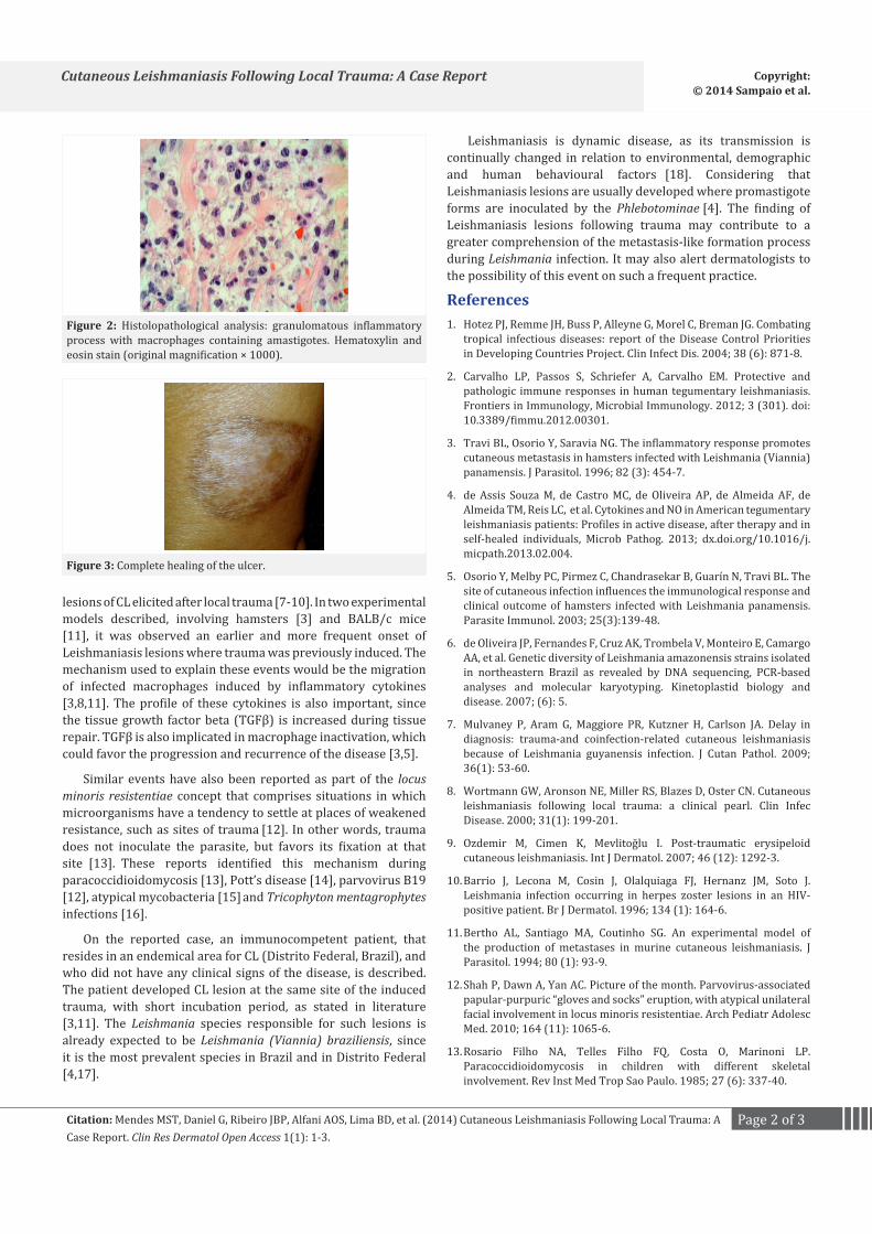
Figure 2: Histolopathological analysis: granulomatous inflammatory process with macrophages containing amastigotes. Hematoxylin and eosin stain (original magnification × 1000).
Page 3 of 3Citation: Mendes MST, Daniel G, Ribeiro JBP, Alfani AOS, Lima BD, et al. (2014) Cutaneous Leishmaniasis Following Local Trauma: A Case Report. Clin Res Dermatol Open Access 1(1): 1-3.
14. Bouvresse S, Chiras J, Bricaire F, Bossi P. Pott’s disease occurring after percutaneous vertebroplasty: an unusual illustration of the principle of locus minoris resistentiae. J Infect. 2006; 53(6): 251-3.
15. Chan ED, Kong PM, Fennelly K, Dwyer AP, Iseman MD. Vertebral osteomyelitis due to infection with nontuberculous Mycobacterium species after blunt trauma to the back: 3 examples of the principle of locus minoris resistentiae. Clin Infect Dis. 2001; 32(10): 1506-10.
16. Sander CS, Sander O, Khatib A, Berger T. Tinea barbae spreading to locus minoris resistentiae. Eur J Dermatol. 2009; 19 (2):173-4doi:
10.1684/ejd.2008.0588.
17. Dos Santos, G, Santos W, Sampaio RNR. Leishmania (Viannia) braziliensis is the main species causing cutaneous leishmaniasis in the Federal District of Brazil (1975). J VAT 2012 (Online); 18: 340-3.doi: 10.1590/S1678-91992012000300012.
18. Gramiccia M, Gradoni L. The current status of zoonotic leishmaniases and approaches to disease control. Int J Parasitol. 2005; 35 (11-12): 1169-80.