CX3CL1/CX3CR1 axis attenuates early braininjury via promoting the delivery ofexosomal microRNA-124 from neuron tomicroglia after subarachnoid hemorrhageXiao Chen1†, Ming Jiang1†, Haiying Li1†, Yang Wang1,2, Haitao Shen1, Xiang Li1, Yunhai Zhang3, Jiang Wu1,Zhengquan Yu1* and Gang Chen1*
Abstract
Background: Microglial activation-mediated neuroinflammation is a major contributor to early brain injury (EBI)after subarachnoid hemorrhage (SAH). MicroRNA-124 (miR-124) is the most abundant miRNAs in the centralnervous system (CNS) and plays a vital role in microglial activation by targeting protein CCAAT-enhancer-bindingprotein α (C/EBPα). It has been reported that the CX3CL1/CX3CR1 axis is involved in the delivery of miR-124 fromneurons to microglia.
Methods: An experimental rat SAH model was established by injecting autologous arterial blood into the prechiasmaticcistern, and cultured primary neurons and microglia were exposed to oxyhemoglobin to mimic SAH in vitro. We additionallyexploited specific expression plasmids encoding CX3CL1 and CX3CR1.
Results:We observed significant decreases in CX3CL1 and CX3CR1 in the brain tissues of SAH patients. We also observeddecreases in the levels of CX3CL1 in neurons and CX3CR1 in microglia after SAH in rats. Moreover, microglia exhibited anactivated phenotype with macrophage-like morphology and high levels of CD45 and major histocompatibility complex(MHC) class II after SAH. After overexpression of CX3CL1/CX3CR1, the level of CD45 and MHC class II and the release ofinflammatory factors tumor necrosis factor α, interleukin 1α and complement 1q were significantly decreased. There was alsoincreased neuronal degeneration and behavior dysfunction after SAH, both of which were inhibited by CX3CL1/CX3CR1overexpression. Additionally, we found that the delivery of exosomal miR-124 from neurons to microglia was significantlyreduced after SAH, accompanied by an increase in C/EBPα expression, and was inhibited by CX3CL1/CX3CR1 overexpression.In conclusion, the CX3CL1/CX3CR1 axis may play protective roles after SAH by promoting the delivery of exosomal miR-124to microglia and attenuate microglial activation and neuroinflammation.
Conclusions: CX3CL1/CX3CR1 axis may be a potential intervention target for the inhibition of SAH-induced EBIby promoting exosome transport of miR-124 to microglia.
* Correspondence: [email protected]; [email protected]†Xiao Chen, Ming Jiang and Haiying Li contributed equally to this work.1Department of Neurosurgery & Brain and Nerve Research Laboratory, TheFirst Affiliated Hospital of Soochow University, 188 Shizi Street, Suzhou215006, ChinaFull list of author information is available at the end of the article
Chen et al. Journal of Neuroinflammation (2020) 17:209 https://doi.org/10.1186/s12974-020-01882-6
BackgroundSubarachnoid hemorrhage (SAH) is a relatively seriousacute nervous system disease with high morbidity andmortality [1, 2]. There have been a large number of studiesfor seeking simpler and more efficient treatment methods,but the results are not satisfactory. Early brain injury(EBI), a critical window for determining disease progres-sion, has also received increasing attention but has not yetbeen fully explored [3, 4]. Neuroinflammation is one ofthe main pathological processes in EBI, which is closelyrelated to the activation of microglia [5]. Microglia areimportant immune cells, accounting for about 10% of thetotal number of cells in the central nervous system (CNS).They are the first and most important line of defenseagainst CNS insults [6]. After SAH, a large number ofmicroglia are activated and release inflammatory factors,which would lead to inflammatory responses and aggra-vate the neurological deficit [7–9].MicroRNAs (miRNAs) are a class of small, non-coding
RNA molecules and involved in the regulation of gene ex-pression at the post-transcriptional level [10–12]. Emer-ging research shows that miRNAs can be secreted anddelivered into recipient cells to inhibit the translation oftarget genes and thereby affecting the activities of cells[13]. MicroRNA-124 (miR-124) is currently the mostabundant miRNA found in neurons. It has been reportedthat miR-124 participates in regulating microglial activa-tion [14–16]. Exosomes, also referred to as intraluminalvesicles, are secreted by all cell types [17]. Exosomes aremicrovesicles with a diameter of 30–150 nm that originatefrom multivesicular bodies and can participate in intercel-lular communication by transmitting intracellular pro-teins, messenger RNAs, miRNAs, and long non-codingRNAs [18–20]. Recent studies have shown that exosomesplay an important role in the delivery of miR-124 [16, 21,22]. Therefore, exosomal miR-124 from neurons may playa role in microglia after SAH.CX3CL1 (also called fractalkine) is a class of chemo-
kine and the only member of the CX3C group [23].CX3CL1 is a unique chemokine that binds to its only re-ceptor, CX3CR1 [24]. In the normal CNS, CX3CL1 andits receptor CX3CR1 form signaling network betweenneurons and microglia [25, 26]. One study reveals thatthe CX3CL1/CX3CR1 axis facilitates exosome transportof miR-124 from neurons to microglia [16]. Moreover,many studies have confirmed that the CX3CL1/CX3CR1axis plays an important role in regulating microglial acti-vation and neuroinflammation [26, 27].These findings suggest that targeting the CX3CL1/
CX3CR1 axis may provide new insights into the inhib-ition of SAH-induced EBI. However, the relationship be-tween CX3CL1/CX3CR1 axis, microglia, and miR-124 inSAH remain obscure. Therefore, one of the purposes ofthis study was to elucidate the effect of the CX3CL1/
CX3CR1 axis on SAH. Moreover, we aim to examinethe role of the CX3CL1/CX3CR1 axis in the delivery ofmiR-124 and to provide a new direction for the treat-ment of SAH.
Materials and methodsPatientsThe study protocol was reviewed and approved by theEthics Committee of the First Affiliated Hospital of Soo-chow University. Brain tissue samples were obtainedfrom eight patients aged from 40 to 90 years who werewilling to provide written informed consent. Five non-SAH brain samples were obtained from patients with abrain tumor and without any history of SAH. Duringneurosurgical operations for tumor treatment, normalcortical tissue that was removed to gain access to thetumor was collected. Histological examination showedno tumor infiltration in the tissue (data not shown).SAH brain tissues were obtained from three post-SAHpatients. The clinical parameters and medical images ofthe patients are shown in Supplemental Figure 1 andSupplemental Table 1. Both brain tumor and SAH werediagnosed by neurosurgeons and radiologists based onphysical examination and neuroimaging. Due to ethicalconsiderations, the operators of subsequent studies onthe excised tissue did not include neurosurgeons in-volved in the diagnosis and surgical treatment. The brainsamples were stored in liquid nitrogen for western blotdetection.
Experimental animalsMale SD rats weighing 300–350 g were purchased fromthe Zhaoyan New Drug Research Center (Suzhou,China) Co., LTD. They were housed in a suitable livingenvironment with a 12-h dark-light cycle and givenadequately qualified feed and drinking water. The studywas approved by the Ethics Committee of the First Affil-iated Hospital of Suzhou University.
SAH model and gradeIn this experiment, the SAH model was established byinjection of autologous arterial blood into the prechias-matic cistern. First, after being anesthetized, the rat wasfixed on the stereotaxic frame (Anhui Zhenghua Bio-logical Equipment Co., Ltd., Anhui, China). After disin-fection, a median incision was made to expose theperiosteum. Then, a hole was drilled 7.5 mm anterior tothe bregma and 3 mm beside the midline. The needlewas advanced 11–12 mm into the prechiasmatic cisternat an angle of 45 in the sagittal plane. Then, 300 μl ofautologous arterial blood collected from the femoral ar-tery was injected in 20 s. After SAH model operations,the rats were immediately injected with 5 ml physio-logical saline solution. The rats in the sham group were
Chen et al. Journal of Neuroinflammation (2020) 17:209 Page 2 of 15
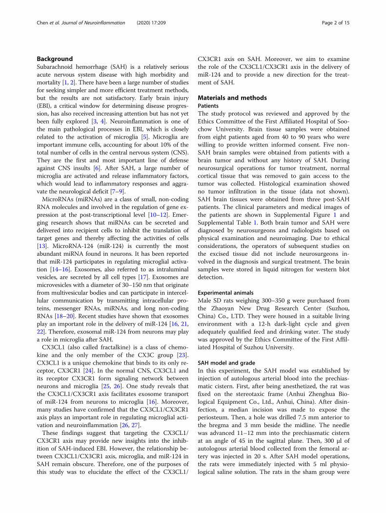
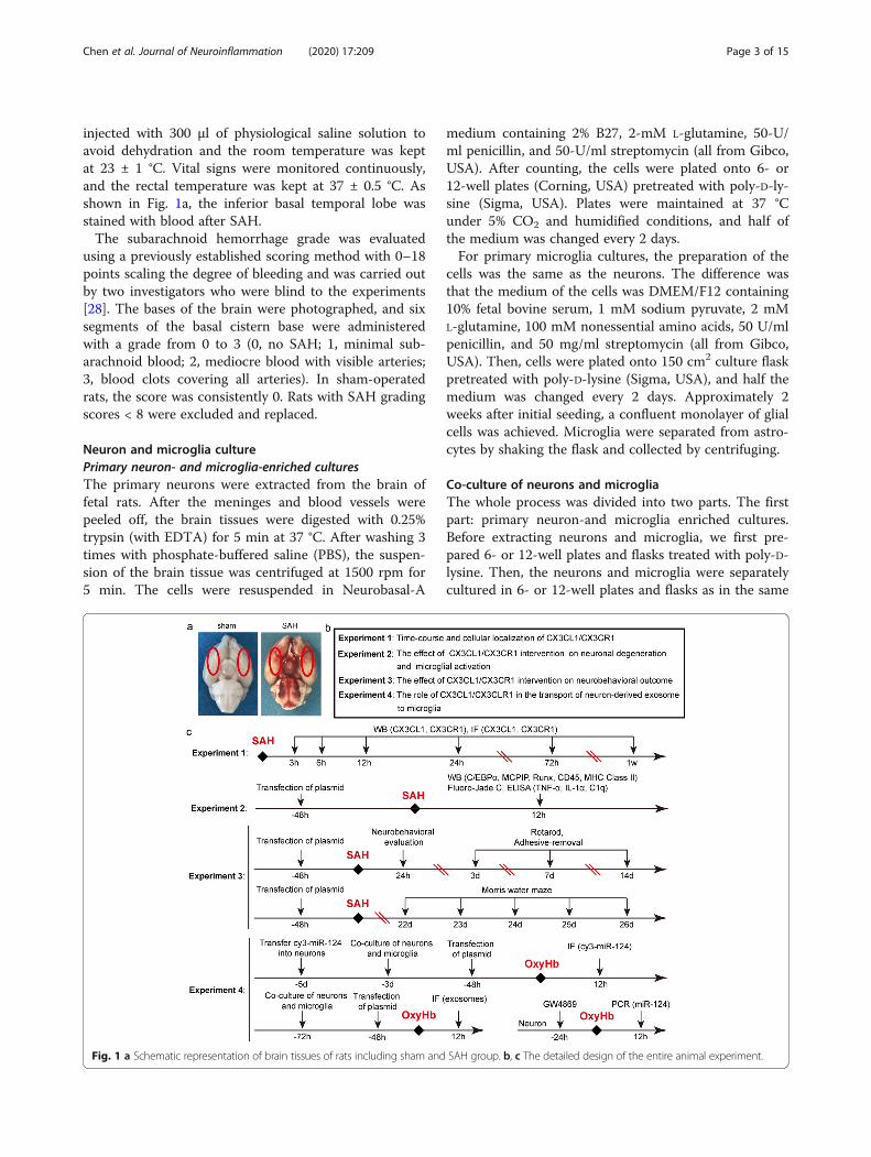
injected with 300 μl of physiological saline solution toavoid dehydration and the room temperature was keptat 23 ± 1 °C. Vital signs were monitored continuously,and the rectal temperature was kept at 37 ± 0.5 °C. Asshown in Fig. 1a, the inferior basal temporal lobe wasstained with blood after SAH.The subarachnoid hemorrhage grade was evaluated
using a previously established scoring method with 0–18points scaling the degree of bleeding and was carried outby two investigators who were blind to the experiments[28]. The bases of the brain were photographed, and sixsegments of the basal cistern base were administeredwith a grade from 0 to 3 (0, no SAH; 1, minimal sub-arachnoid blood; 2, mediocre blood with visible arteries;3, blood clots covering all arteries). In sham-operatedrats, the score was consistently 0. Rats with SAH gradingscores < 8 were excluded and replaced.
Neuron and microglia culturePrimary neuron- and microglia-enriched culturesThe primary neurons were extracted from the brain offetal rats. After the meninges and blood vessels werepeeled off, the brain tissues were digested with 0.25%trypsin (with EDTA) for 5 min at 37 °C. After washing 3times with phosphate-buffered saline (PBS), the suspen-sion of the brain tissue was centrifuged at 1500 rpm for5 min. The cells were resuspended in Neurobasal-A
medium containing 2% B27, 2-mM L-glutamine, 50-U/ml penicillin, and 50-U/ml streptomycin (all from Gibco,USA). After counting, the cells were plated onto 6- or12-well plates (Corning, USA) pretreated with poly-D-ly-sine (Sigma, USA). Plates were maintained at 37 °Cunder 5% CO2 and humidified conditions, and half ofthe medium was changed every 2 days.For primary microglia cultures, the preparation of the
cells was the same as the neurons. The difference wasthat the medium of the cells was DMEM/F12 containing10% fetal bovine serum, 1 mM sodium pyruvate, 2 mML-glutamine, 100 mM nonessential amino acids, 50 U/mlpenicillin, and 50 mg/ml streptomycin (all from Gibco,USA). Then, cells were plated onto 150 cm2 culture flaskpretreated with poly-D-lysine (Sigma, USA), and half themedium was changed every 2 days. Approximately 2weeks after initial seeding, a confluent monolayer of glialcells was achieved. Microglia were separated from astro-cytes by shaking the flask and collected by centrifuging.
Co-culture of neurons and microgliaThe whole process was divided into two parts. The firstpart: primary neuron-and microglia enriched cultures.Before extracting neurons and microglia, we first pre-pared 6- or 12-well plates and flasks treated with poly-D-lysine. Then, the neurons and microglia were separatelycultured in 6- or 12-well plates and flasks as in the same
Fig. 1 a Schematic representation of brain tissues of rats including sham and SAH group. b, c The detailed design of the entire animal experiment.
Chen et al. Journal of Neuroinflammation (2020) 17:209 Page 3 of 15
manner described above. The second part: After themicroglia in the flask were shaken, they were directlyplated onto the 6- or 12-well plates of neurons after cen-trifuging and counting.
Experimental designFirst, we examined the expression of CX3CL1 andCX3CR1 in the brain tissues of the human. Then, the ani-mal experiment was carried out. In order to ensure the ac-curacy of the experimental results, we first successfullyestablished the SAH models (Fig. 1a). The animal experi-ment was divided into 4 parts (Fig. 1b, c). We used Ran-dom Number Generator (Stat Trek) to select randomsamples, which was available on the website: http://stat-trek.com/statistics/random-number-generator.aspx. In ex-periment 1, 84 rats (102 rats were used, 88 rats survivedafter the surgery, and 4 rats were excluded) were dividedinto 7 groups, with 12 rats per group: sham group and sixexperimental groups were arranged by time 3 h, 6 h, 12 h,24 h, 72 h, and 7 days after SAH. When the set time wasreached, the rats were sacrificed and the brain tissue wascollected for the time course study. In experiment 2, weexploited the time period at 12 h after SAH based on theresults of experiment 1. A total of 48 rats (62 rats wereused, 52 rats survived after the surgery, and 4 rats were ex-cluded) were divided into 4 groups of 12 rats each: shamgroup, SAH group, and SAH + Vector group, SAH +Over-CX3CL1/CX3CR1 group. All rats were sacrificed 12h after SAH, and the brain tissue and serum were separ-ately collected for western-blotting, fluoro-Jade C (FJC)staining, and enzyme-linked immunosorbent assay(ELISA). Experiment 3 was aimed to test the role ofCX3CL1/CX3CR1 on the behavioral ability of rats. A totalof 40 rats (51 rats were used, 44 rats survived after SAH, 4rats were excluded) were divided into 4 groups: shamgroup, SAH group, SAH + Vector group, and SAH +Over-CX3CL1/CX3CR1 group. The rats in each groupwere used for ethological testing, including neurobehav-ioral scores, adhesive-removal test, rotarod test, and Mor-ris water maze test. In experiment 4, primary-culturedneurons and microglia were used to verify potentialunderlying mechanisms. In addition to primary neuron-and microglia-enriched cultures, we also created an envir-onment in which neurons and microglia were co-culturedto make the two symbiotic. Here, we simulated the SAHenvironment by adding oxyhemoglobin (OxyHb; 10 μM)to the medium. After a series of interventions, changes inexosome-mediated transport of miR-124 between neuronsand microglia were observed by immunofluorescencestaining and polymerase chain reaction (PCR).
Drug administrationBased on the former study, the GW4869 (D1692, Sigma,USA) is a commonly used drug that inhibits the
production of exosomes [29]. GW4869 is dissolved inDMSO (Beyotime, China) and then diluted in culturesupernatant to achieve concentration at 20 μM in a cul-ture medium. The culture neurons were treated withGW4869 at 37 °C for 24 h, then the culture supernatantswere harvested for collecting exosomes.
Transfection of the plasmid in vivoIn this process, we used two plasmids: the plasmid ofCX3CL1 and the plasmid of CX3CR1. We co-transfectedthem into the rat brains through Entranster—in vivoDNA transfection reagent (Engreen, China). According tothe manufacturer’s instructions, 5 μl plasmid (2.5 μl ofCX3CL1 plasmid and 2.5 μl CX3CR1 plasmid) or emptyVector was dissolved in 10 μl Entranster—in vivo DNAtransfection reagent. After standing at room temperaturefor 15 min, the mixed 15 μl of the solution was injectedintracerebroventricularly at 48 h before SAH.
Transfection of the cy3-miR-124 in vitroTransfection of the cy3-miR-124 into neurons was per-formed using lipofectamine 3000 Transfection Reagent(L3000-015, Invitrogen). After 48 h, the neurons werefurther processed.
ReagentsAnti-CX3CL1 antibody (ab25088), anti-CX3CR1 antibody(ab8021), anti-CX3CL1 antibody (ab25088), anti-CX3CR1antibody (ab8020), anti-CD45 antibody (ab8216), anti-MHC class II antibody (ab23990), anti-Runx1 antibody(ab23980), anti-Iba1 antibody (ab5076), anti-NSE antibody(ab53025), and anti-NeuN (ab104224) were from Abcam(USA). Anti-MCPIP antibody (sc-515275) and β-tubulin(sc-9014) were obtained from Santa Cruz Biotechnology(USA). Anti-C/EBPα antibody (2295), anti-rabbit-IgG-HRP (7074s), and anti-mouse-IgG-HRP (7076s) were ob-tained from the Cell Signaling Technology (USA). AlexaFluor-488 donkey anti-rabbit IgG antibody (A21206),Alexa Fluor-555 donkey anti-mouse IgG antibody(A31570), Alexa Fluor-555 donkey anti-goat IgG antibody(A21432), and Alexa Fluor-633 donkey anti-goat IgG anti-body (A21082) were from the Life Technologies.RBFOX3/NeuN antibody [Alexa Fluor-405] (NBP1-77686AF405) was from Novus (USA).
Isolation and collection of exosomesThis detection was performed according to the manufac-turer’s instructions (EXOQ20A-1, System Biosciences,USA). Supernatants from cultured neurons were col-lected and centrifuged at 3000g for 15 min to removeany cells and cell debris, and then supernatants weretransferred to a fresh tube. Then, according to the in-structions, the exosome isolation reagent was added tothe supernatants and allowed to stand at 4 °C overnight
Chen et al. Journal of Neuroinflammation (2020) 17:209 Page 4 of 15
(at least 12 h). Finally, the mixture was centrifuged at1500g for 30 min.
Determination of exosomal miRNA abundanceExosomal miR-124 abundance was determined by real-time quantitative PCR (RT-qPCR). First, the total RNAwas extracted from the precipitated exosomes obtainedabove by TRIzol. According to the manufacturer’s in-structions (ZK00805, ShineGene Molecular Biotech,China), the RNA was reverse transcribed to complemen-tary DNA (cDNA), and RT-PCR was performed.GAPDH served as loading controls. Primers used in RT-qPCR were obtained from the ShineGene Molecular Bio-tech (China). The qPCR amplification reaction was per-formed with a volume of 50 μl, containing 25 μl 2 ×Hotstart Fluo-PCR mix, 1 μl each primer (25 pmol/l),0.5 μl probe (25 pmol/l), 8 μl DEPC water, and 1 μlcDNA. The PCR amplification was as follows: denatur-ation at 94 °C for 4 min, followed by 40 cycles of 94 °Cfor 20 s and 60 °C for 30 s with continuous fluorescencemeasurement. Quantification was performed by using acomparative CT method (2−ΔΔCT). All samples were ana-lyzed in triplicate [30, 31].
Western-blotting analysisAfter lavaging of the brain tissue with PBS, the temporalbase brain tissues were taken out. After lysis and standing,the brain tissue was centrifuged (12000 rpm, 5 min, 4 °C).The supernatant protein concentration was measuredusing the 96-well Cell Culture Cluster and enhanced BCAProtein Assay Kit (P0010S, Beyotime, China). After addinga loading buffer to the trimmed protein sample, it washeated at 100 °C for 5 min. Then, the protein sampleswere loaded on SDS-polyacrylamide gels and were thenseparated and electrophoretically transferred topolyvinylidene-difluoride membranes (IPVH00010, Milli-pore Corporation, USA). After blocking with 5% non-fatmilk at room temperature for 1 h, the membranes wereincubated overnight at 4 °C with primary antibodies. β-tubulin was used as a loading control. After washing 3times with PBST (PBS + 0.1%Tween-20), the membraneswere incubated at room temperature for 1.5 h with sec-ondary antibodies against mouse or rabbit. Finally, theprotein bands were visualized using an Enhanced Chemi-luminescence (ECL) Kit (Clinx, China) and digitalizedwith a ChemiScope 5300 Chemiluminescence imaging
system (Clinx). Blots were imaged and quantified usingthe ImageJ software (NIH, Bethesda, MD, USA) [32, 33].
Immunofluorescent analysisAfter lavaging with PBS, the total coronal sections con-taining the temporal base brain tissue were fixed with4% paraformaldehyde, embedded in paraffin, and sec-tioned. After heating and dewaxing, the sections were in-cubated overnight at 4 °C with primary antibodies. Forcells, they could be incubated as long as they were fixedby 4% paraformaldehyde. After washing 3 times withPBST, the sections were incubated with secondary anti-bodies at 37 °C for 1 h. After washing three times withPBST, the sections were sealed with 4′,6-diamidino-2-phenylindole (DAPI) Fluoromount-G@ (Southern Bio-tech, USA). Finally, sections were observed by fluores-cent microscope (OLYMPUS BX50/BX-FLA/DP70;Olympus Co., Japan), laser scanning confocal micro-scope (ZEISS LSM 880, Carl Zeiss AG, Germany) andstimulated emission depletion microscopy (Jiangsu KeyLaboratory of Medical Optics, Suzhou Institute of Bio-medical Engineering and Technology, China) [34].
FJC stainingThe first step was the same as that for immunofluores-cent analysis. After heating and dewaxing, the sectionswere incubated in 80% alcohol with 20% sodium hydrox-ide for 5 min, 70% alcohol for 2 min, distilled water for2 min, 0.06% K permanganate for 10 min and 0.0004%FJC-working solution for 20 min, and were then dried inan incubator (50–60 °C) for 15–30 min. After drying,the sections were incubated in xylene for 2 min. Then,they were sealed with neutral gum. Finally, sections wereobserved by a fluorescent microscope (OLYMPUSBX50/BX-FLA/DP70; Olympus Co., Japan).
ELISAAccording to the manufacturer’s instructions, the levels oftumor necrosis factor α (TNF-α), interleukin 1α (IL-1α),and Complement 1q (C1q) in the serum were measuredusing a specific ELISA Kit (Bio-Swamp, China).
Neurobehavioral scoresAt 24 h after SAH, the rats were examined for behav-ioral impairment using an established scoring system.This scoring system consisted of three parts: appetite,activity, and neurologic defects [35].
Adhesive-removal testThis test was used to measure motor coordination andsensory neglect after SAH. First, we placed the rat in aglass box for a period of time and attached a circularsticker to the palm of each forepaw. The time at whichthe rat removed all the stickers was recorded. All rats
Chen et al. Journal of Neuroinflammation (2020) 17:209 Page 5 of 15
were trained daily for 3 days prior to modeling. Then,the test was carried out 1 day before modeling and onthe 1st, 3rd, 7th, and 14th days after the SAH.
Rotarod testThis test was to assess the locomotor ability of the ratsby a rota-rod cylinder (ZH-300B, Anhui Zhenghua Bio-logical Equipment Co., Ltd, China). The rat was placedon the horizontal axis that had been set at a constantrate from 4 to 30 rpm within 1 min. When the ratdropped or gripped the device around for two revolu-tions, the test was finished and the time animalsremained on the rotarod was recorded. As withadhesive-removal test, all rats were trained 3 days priorto modeling. The test was also carried out 1 day beforemodeling and on the 1st, 3rd, 7th, and 14th days afterthe SAH.
Morris water mazeThe method of the Morris water maze test has been pre-viously described [36]. The test was carried out in a cir-cular tank of 180-cm diameter and 50-cm depth. Acircular platform with the diameter of 12 cm was placed2 cm below the surface of the water. All rats weretrained for 4 days from the 18th day after modeling, 3times a day. Each training lasted 1 min and was sepa-rated by 5 min every two times. If the rat could boardthe platform within 1 min, it would be allowed to stayfor 15 s; on the contrary, it would be guided to boardthe platform. The testing phase took place from the22nd day to the 26th day after SAH.
Statistical analysisGraphPad Prism 7.0 software (GraphPad, USA) was usedfor statistical analysis. Neurobehavioral scoring is shownas the median with the interquartile range, and theMann-Whitney U test was used to compare scoresamong groups. All other data are reported as the mean± SD. One-way or two-way ANOVA was used for mul-tiple comparisons, and Bonferroni’s or Tukey’s post hoctest was used for comparison between two pairs in mul-tiple groups. P < 0.05 indicated a statistically significantdifference. Specific statistics are shown in SupplementalTable 2.
ResultsGeneral observationThroughout the study, the mortality rate of rats in thesham group was 0% (0/34 rats) and was 17.1% (31/181rats) in the SAH groups (Supplemental Table 3).
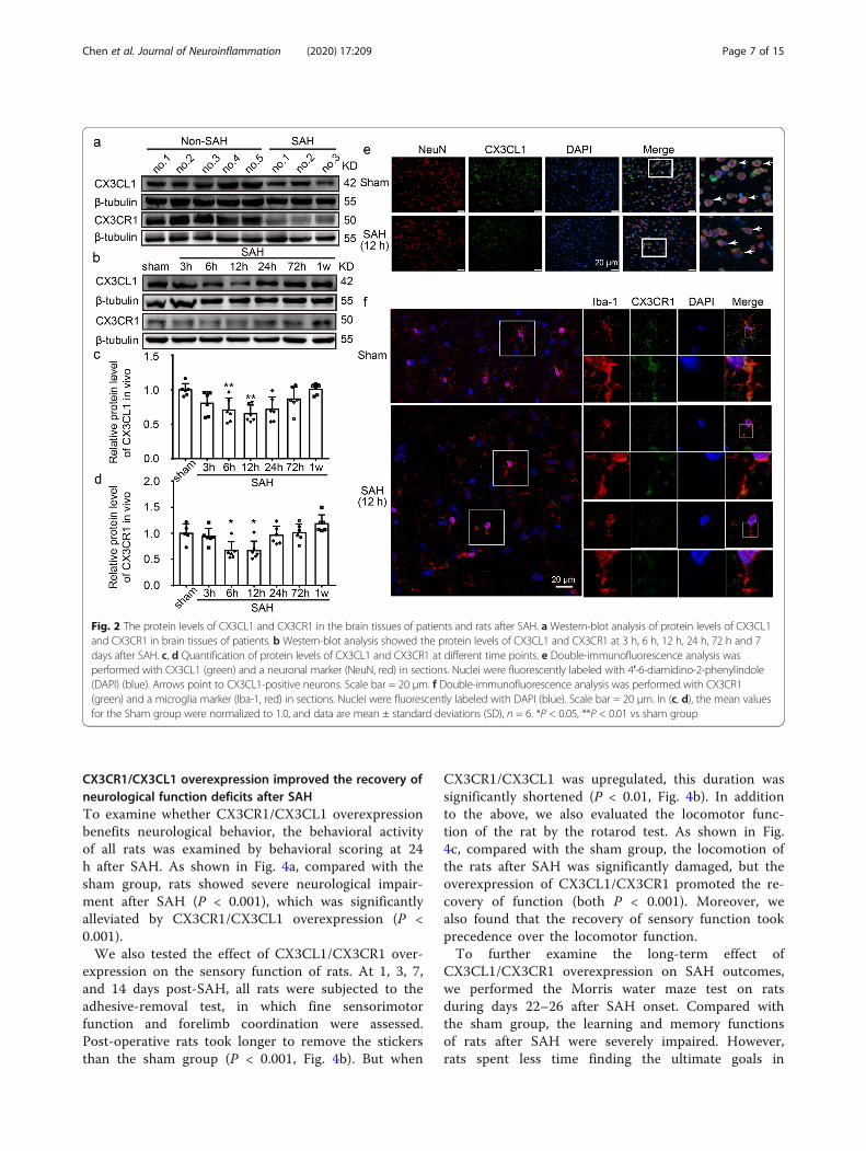
CX3CL1 and CX3CR1 protein levels decreased in the braintissues of patients after SAHTo detect the protein levels of CX3CL1 and CX3CR1after SAH, western blot analysis of protein samples fromthe brain tissues of patients was performed (Fig. 2a). Theresults showed that both CX3CL1 and CX3CR1 wereexpressed in the brain tissues of non-SAH and SAH pa-tients. Compared with non-SAH group, the proteinlevels of CX3CL1 and CX3CR1 were reduced in SAHpatients.
CX3CL1 and CX3CR1 protein levels decreased in the braintissues of rats after SAHAfter observing the changes in proteins CX3CL1 andCX3CR1 in the human brain, we further examined theirlevels in the rat brain. Western blot analysis showed that,compared with the sham group, the protein level of CX3CL1was significantly decreased at 12 h after SAH (P < 0.01, Fig.2b, c). Similarly, the protein level of CX3CR1 also decreasedand reached the lowest level at 12 h (P < 0.05, Fig. 2b, d).Previous study has demonstrated that, in the CNS, CX3CL1was expressed predominantly in neurons, and its receptorCX3CR1 was expressed solely on microglia [37]. Double im-munofluorescence was performed to distinguish the changesin cell type-specificity expression of CX3CL1 and CX3CR1after SAH. Compared with the sham group, the proteinlevels of CX3CL1 in neurons and CX3CR1 in microgliashowed significant decreases at 12 h after SAH (Fig. 2e, f). Inaddition to the expression of target proteins, we alsoobserved a significant change in the morphology of micro-glia. After SAH, microglia lost processes and increased in size(Fig. 2f).
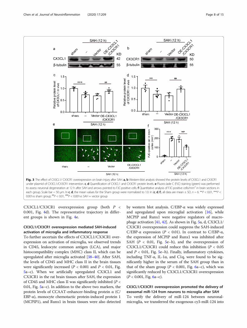
CX3CR1/CX3CL1 overexpression inhibited SAH-inducedneuronal degenerationTo investigate the role of CX3CL1 and CX3CR1 onEBI induced by SAH, we used FJC staining to detectthe effect of CX3CL1 and CX3CR1 overexpression onneuronal degeneration in the brain at 12 h after SAH.We simultaneously upregulated CX3CL1 and CX3CR1in the brain tissues by plasmid transfection, and thetransfection efficiency of plasmid was verified by west-ern blotting analysis (Fig. 3a, b). CX3CL1 and CX3CR1protein levels reduced at 12 h after SAH comparedwith the sham group (P < 0.001 and P < 0.01, Fig. 3c,d), and they were significantly upregulated by overex-pression of CX3CL1 and CX3CR1 (P < 0.01, Fig. 3c,d). Compared with the sham group, the number ofFJC-positive cells significantly increased in the SAHgroup (P < 0.001, Fig. 3e, f), which was significantlydecreased after CX3CR1/CX3CL1 overexpression (P <0.001, Fig. 3e, f).
Chen et al. Journal of Neuroinflammation (2020) 17:209 Page 6 of 15
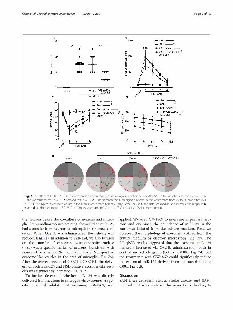
CX3CR1/CX3CL1 overexpression improved the recovery ofneurological function deficits after SAHTo examine whether CX3CR1/CX3CL1 overexpressionbenefits neurological behavior, the behavioral activityof all rats was examined by behavioral scoring at 24h after SAH. As shown in Fig. 4a, compared with thesham group, rats showed severe neurological impair-ment after SAH (P < 0.001), which was significantlyalleviated by CX3CR1/CX3CL1 overexpression (P <0.001).We also tested the effect of CX3CL1/CX3CR1 over-
expression on the sensory function of rats. At 1, 3, 7,and 14 days post-SAH, all rats were subjected to theadhesive-removal test, in which fine sensorimotorfunction and forelimb coordination were assessed.Post-operative rats took longer to remove the stickersthan the sham group (P < 0.001, Fig. 4b). But when
CX3CR1/CX3CL1 was upregulated, this duration wassignificantly shortened (P < 0.01, Fig. 4b). In additionto the above, we also evaluated the locomotor func-tion of the rat by the rotarod test. As shown in Fig.4c, compared with the sham group, the locomotion ofthe rats after SAH was significantly damaged, but theoverexpression of CX3CL1/CX3CR1 promoted the re-covery of function (both P < 0.001). Moreover, wealso found that the recovery of sensory function tookprecedence over the locomotor function.To further examine the long-term effect of
CX3CL1/CX3CR1 overexpression on SAH outcomes,we performed the Morris water maze test on ratsduring days 22–26 after SAH onset. Compared withthe sham group, the learning and memory functionsof rats after SAH were severely impaired. However,rats spent less time finding the ultimate goals in
Fig. 2 The protein levels of CX3CL1 and CX3CR1 in the brain tissues of patients and rats after SAH. a Western-blot analysis of protein levels of CX3CL1and CX3CR1 in brain tissues of patients. b Western-blot analysis showed the protein levels of CX3CL1 and CX3CR1 at 3 h, 6 h, 12 h, 24 h, 72 h and 7days after SAH. c, d Quantification of protein levels of CX3CL1 and CX3CR1 at different time points. e Double-immunofluorescence analysis wasperformed with CX3CL1 (green) and a neuronal marker (NeuN, red) in sections. Nuclei were fluorescently labeled with 4′-6-diamidino-2-phenylindole(DAPI) (blue). Arrows point to CX3CL1-positive neurons. Scale bar = 20 μm. f Double-immunofluorescence analysis was performed with CX3CR1(green) and a microglia marker (Iba-1, red) in sections. Nuclei were fluorescently labeled with DAPI (blue). Scale bar = 20 μm. In (c, d), the mean valuesfor the Sham group were normalized to 1.0, and data are mean ± standard deviations (SD), n = 6. *P < 0.05, **P < 0.01 vs sham group
Chen et al. Journal of Neuroinflammation (2020) 17:209 Page 7 of 15
CX3CL1/CX3CR1 overexpression group (both P <0.001, Fig. 4d). The representative trajectory in differ-ent groups is shown in Fig. 4e.
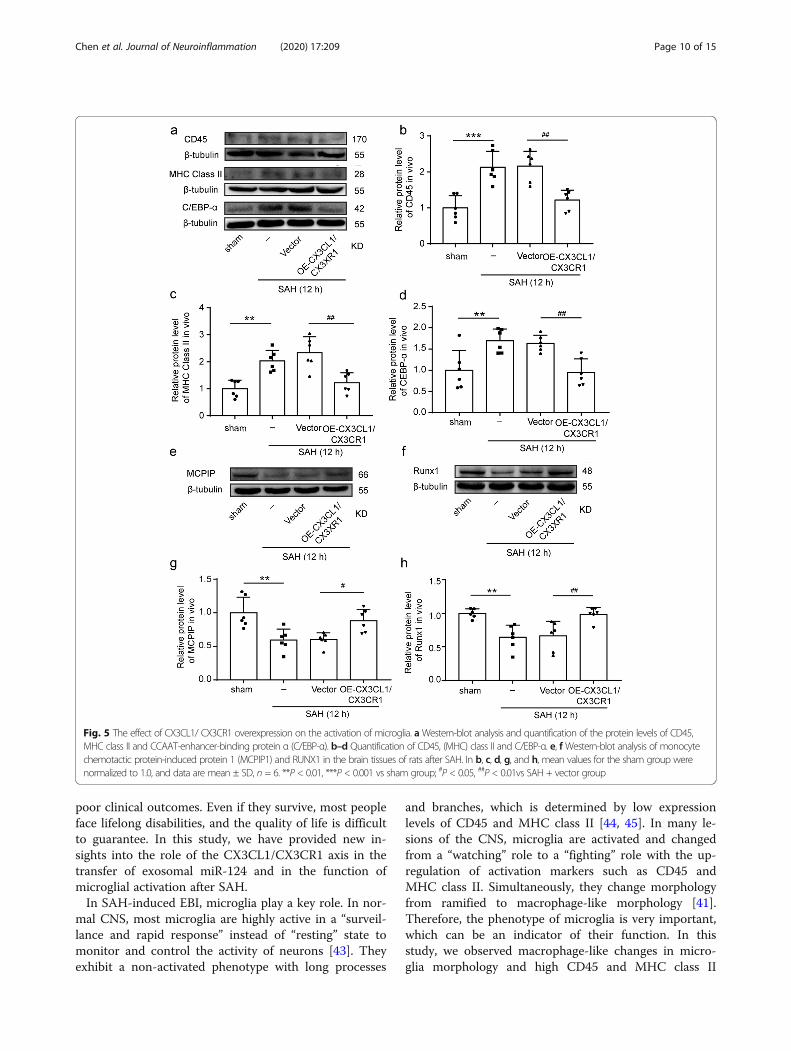
CX3CL1/CX3CR1 overexpression mediated SAH-inducedactivation of microglia and inflammatory responseTo further ascertain the effects of CX3CL1/CX3CR1 over-expression on activation of microglia, we observed trendsin CD45, leukocyte common antigen (LCA), and majorhistocompatibility complex (MHC) class II, which can beupregulated after microglia activated [38–40]. After SAH,the levels of CD45 and MHC class II in the brain tissueswere significantly increased (P < 0.001 and P < 0.01, Fig.5a–c). When we artificially upregulated CX3CL1 andCX3CR1 in the rat brain tissues after SAH, the expressionof CD45 and MHC class II was significantly inhibited (P <0.01, Fig. 5a–c). In addition to the above two markers, theprotein levels of CCAAT-enhancer-binding protein α (C/EBP-α), monocyte chemotactic protein-induced protein 1(MCPIP1), and Runx1 in brain tissues were also detected
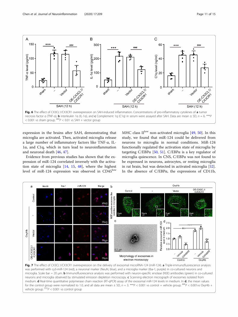
by western blot analysis. C/EBP-α was widely expressedand upregulated upon microglial activation [16], whileMCPIP and Runx1 were negative regulators of macro-phage activation [41, 42]. As shown in Fig. 5a, d, CX3CL1/CX3CR1 overexpression could suppress the SAH-inducedC/EBP-α expression (P < 0.01). In contrast to C/EBP-α,the expression of MCPIP and Runx1 was inhibited afterSAH (P < 0.01, Fig. 5e–h), and the overexpression ofCX3CL1/CX3CR1 could reduce this inhibition (P < 0.05and P < 0.01, Fig. 5e–h). Finally, inflammatory cytokines,including TNF-α, IL-1α, and C1q, were found to be sig-nificantly higher in the serum of the SAH group than inthat of the sham group (P < 0.001, Fig. 6a–c), which wassignificantly reduced by CX3CL1/CX3CR1 overexpression(P < 0.001, Fig. 6a–c).
CX3CL1/CX3CR1 overexpression promoted the delivery ofexosomal miR-124 from neurons to microglia after SAHTo verify the delivery of miR-124 between neuronal-microglia, we transferred the exogenous cy3-miR-124 into
Fig. 3 The effect of CX3CL1/ CX3CR1 overexpression on brain injury after SAH. a, bWestern-blot analysis showed the protein levels of CX3CL1 and CX3CR1under plasmid of CX3CL1/CX3CR1 intervention. c, d Quantification of CX3CL1 and CX3CR1 protein levels. e Fluoro-Jade C (FJC) staining (green) was performedto assess neuronal degeneration at 12 h after SAH and arrows pointed to FJC-positive cells. f Quantitative analysis of FJC-positive cells/mm2 in brain sections ineach group. Scale bar = 50 μm. In c, d, the mean values for the Sham group were normalized to 1.0. In (c, d, f), all data are mean ± SD, n = 6. **P < 0.01, ***P <0.001vs sham group; ##P < 0.01, ###P < 0.001vs SAH + vector group
Chen et al. Journal of Neuroinflammation (2020) 17:209 Page 8 of 15
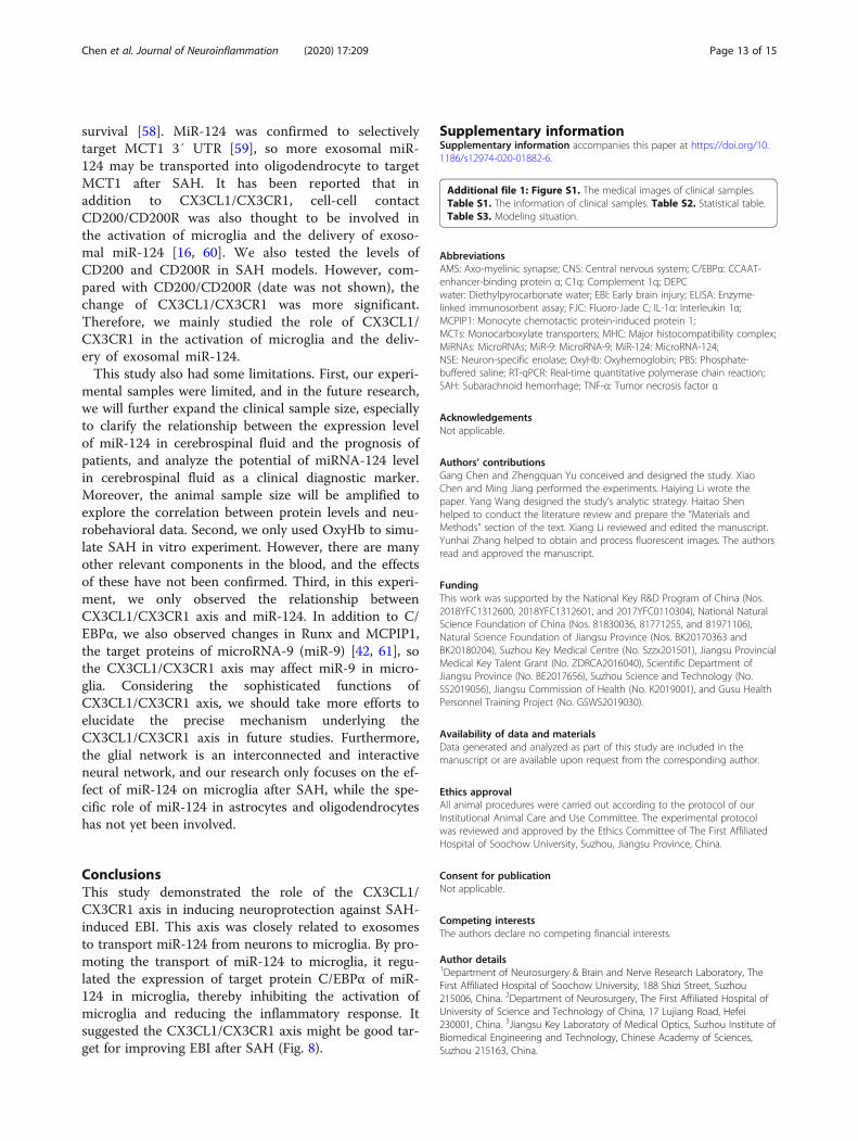
the neurons before the co-culture of neurons and micro-glia. Immunofluorescence staining showed that miR-124had a transfer from neurons to microglia in a normal con-dition. When OxyHb was administered, the delivery wasreduced (Fig. 7a). In addition to miR-124, we also focusedon the transfer of exosome. Neuron-specific enolase(NSE) was a specific marker of neurons. Consistent withneuron-derived miR-124, there were fewer NSE-positiveexosome-like vesicles in the area of microglia (Fig. 7b).After the overexpression of CX3CL1/CX3CR1, the deliv-ery of both miR-124 and NSE-positive exosome-like vesi-cles was significantly increased (Fig. 7a, b).To further determine whether miR-124 was directly
delivered from neurons to microglia via exosomes, a spe-cific chemical inhibitor of exosome, GW4869, was
applied. We used GW4869 to intervene in primary neu-rons and examined the abundance of miR-124 in theexosomes isolated from the culture medium. First, weobserved the morphology of exosomes isolated from theculture medium by electron microscopy (Fig. 7c). TheRT-qPCR results suggested that the exosomal miR-124markedly increased via OxyHb administration both incontrol and vehicle group (both P < 0.001, Fig. 7d), butthe treatments with GW4869 could significantly reducethe exosomal miR-124 derived from neurons (both P <0.001, Fig. 7d).
DiscussionSAH is an extremely serious stroke disease, and SAH-induced EBI is considered the main factor leading to
Fig. 4 The effect of CX3CL1/ CX3CR1 overexpression on recovery of neurological function of rats after SAH. a Neurobehavioral scores, n = 10. bAdhesive-removal test, n = 10. c Rotarod test, n = 10. d Time to reach the submerged platform in the water maze from 22 to 26 days after SAH,n = 6. e The typical swim path of rats in the Morris water maze test at 26 days after SAH. In a, the data are median and interquartile range; in b,c, and d, all data are mean ± SD. ***P < 0.001 vs sham group; ##P < 0.01, ###P < 0.001 vs SAH + vector group
Chen et al. Journal of Neuroinflammation (2020) 17:209 Page 9 of 15
poor clinical outcomes. Even if they survive, most peopleface lifelong disabilities, and the quality of life is difficultto guarantee. In this study, we have provided new in-sights into the role of the CX3CL1/CX3CR1 axis in thetransfer of exosomal miR-124 and in the function ofmicroglial activation after SAH.In SAH-induced EBI, microglia play a key role. In nor-
mal CNS, most microglia are highly active in a “surveil-lance and rapid response” instead of “resting” state tomonitor and control the activity of neurons [43]. Theyexhibit a non-activated phenotype with long processes
and branches, which is determined by low expressionlevels of CD45 and MHC class II [44, 45]. In many le-sions of the CNS, microglia are activated and changedfrom a “watching” role to a “fighting” role with the up-regulation of activation markers such as CD45 andMHC class II. Simultaneously, they change morphologyfrom ramified to macrophage-like morphology [41].Therefore, the phenotype of microglia is very important,which can be an indicator of their function. In thisstudy, we observed macrophage-like changes in micro-glia morphology and high CD45 and MHC class II
Fig. 5 The effect of CX3CL1/ CX3CR1 overexpression on the activation of microglia. aWestern-blot analysis and quantification of the protein levels of CD45,MHC class II and CCAAT-enhancer-binding protein α (C/EBP-α). b–d Quantification of CD45, (MHC) class II and C/EBP-α. e, fWestern-blot analysis of monocytechemotactic protein-induced protein 1 (MCPIP1) and RUNX1 in the brain tissues of rats after SAH. In b, c, d, g, and h, mean values for the sham group werenormalized to 1.0, and data are mean ± SD, n = 6. **P < 0.01, ***P < 0.001 vs sham group; #P < 0.05, ##P < 0.01vs SAH + vector group
Chen et al. Journal of Neuroinflammation (2020) 17:209 Page 10 of 15
expression in the brains after SAH, demonstrating thatmicroglia are activated. Then, activated microglia releasea large number of inflammatory factors like TNF-α, IL-1α, and C1q, which in turn lead to neuroinflammationand neuronal death [46, 47].Evidence from previous studies has shown that the ex-
pression of miR-124 correlated inversely with the activa-tion state of microglia [14, 15, 48], where the highestlevel of miR-124 expression was observed in CD45low
MHC class IIlow non-activated microglia [49, 50]. In thisstudy, we found that miR-124 could be delivered fromneurons to microglia in normal conditions. MiR-124functionally regulated the activation state of microglia bytargeting C/EBPα [50, 51]. C/EBPα is a key regulator ofmicroglia quiescence. In CNS, C/EBPα was not found tobe expressed in neurons, astrocytes, or resting microgliain rat brain, but was detected in activated microglia [52].In the absence of C/EBPα, the expressions of CD11b,
Fig. 6 The effect of CX3CL1/CX3CR1 overexpression on SAH-induced inflammation. Concentrations of pro-inflammatory cytokines of a tumornecrosis factor α (TNF-α), b interleukin 1α (IL-1α), and c Complement 1q (C1q) in serum were assayed after SAH. Data are mean ± SD, n = 6. ***P< 0.001 vs sham group; ###P < 0.01 vs SAH + vector group
Fig. 7 The effect of CX3CL1/CX3CR1 overexpression on the delivery of exosomal microRNA-124 (miR-124). a Triple-immunofluorescence analysiswas performed with cy3-miR-124 (red), a neuronal marker (NeuN, blue), and a microglia marker (Iba-1, purple) in co-cultured neurons andmicroglia. Scale bar = 20 μm. b Immunofluorescence analysis was performed with neuron-specific enolase (NSE) antibodies (green) in co-culturedneurons and microglia observed by stimulated emission depletion microscopy. c Scanning electron micrograph of exosomes isolated frommedium. d Real-time quantitative polymerase chain reaction (RT-qPCR) assay of the exosomal miR-124 levels in medium. In d, the mean valuesfor the control group were normalized to 1.0, and all data are mean ± SD, n = 3. ***P < 0.001 vs control + vehicle group; ###P < 0.001vs OxyHb +vehicle group; $$$P < 0.001 vs control group
Chen et al. Journal of Neuroinflammation (2020) 17:209 Page 11 of 15
MHC class II, and CD86 were decreased [50]. Herein,we further demonstrated that the neuron-derived miR-124 was delivered via exosomes. Therefore, exosomalmiR-124 from neurons could bind C/EBPα to regulatethe phenotype of microglia after SAH.As previously reported, cell-cell contacts in the
CX3CL1/CX3CR1 axis were involved in the transfer ofmiR-124 [16], and this conjugation kept microglia “qui-escent” [53, 54]. In CNS, CX3CL1 mainly expressed inneurons and had two different forms: one is amembrane-bound glycoprotein providing direct inter-action with CX3CR1, which expressed in microglia; theother is a soluble form working as an extracellular medi-ator [25]. First, we found that the protein levels ofCX3CL1 and CX3CR1 were significantly reduced inSAH patients’ brain. We further tested their expressionsin the rat model. Consistently, the protein levels ofCX3CL1 and CX3CR1 were also significantly reduced at12 h after SAH. These results suggested that this conju-gation was reduced after SAH. As shown in Figs. 5 and7, C/EBPα, CD45, and MHC class II levels were upregu-lated after SAH, which were correlated with the reduc-tion of exosomal miR-124 in microglia. When werestored the junction of CX3CL1/CX3CR1 by plasmidtransfection after SAH, exosomal miR-124 could be nor-mally delivered from neurons to microglia to target C/EBPα, thereby inhibiting the activation of microglia.These results indicated that the CX3CL1/CX3CR1 axiswas involved in the signaling pathways of exosomalmiR-124-C/EBPα to promote microglia quiescence.
In addition, we found that the upregulation ofCX3CL1/CX3CR1 could reduce TNF-α, IL-1α, and C1qsecretion under SAH conditions. Moreover, we also ob-served that the overexpression of CX3CL1/CX3CR1 re-duced neuronal degeneration and improved short- andlong-term neurological functions after SAH. These re-sults suggested that the CX3CL1/CX3CR1 axis couldplay anti-inflammatory and neuroprotective roles inSAH models. These results were consistent with severalprevious observations. Cipriani et al. [55] found thatCX3CL1 could reduce infarct volume and improve be-havioral outcomes for middle cerebral artery occlusionrats. Additionally, the lacking of CX3CR1 was cytotoxicin models of amyotrophic lateral sclerosis, Parkinson’sdisease, and Alzheimer’s disease [56, 57]. Therefore,CX3CL1/CX3CR1 may be a potential intervention targetfor SAH patients; thus, promoting the delivery of miR-124 to microglia may be a novel approach for ameliorat-ing SAH-induced EBI.As shown in Fig. 7d, more neuron-derived miR-124
was released via exosomes after SAH. However, theloss of the CX3CL1/CX3CR1 junction caused exoso-mal miR-124 not to be delivered to microglia. So,where did it go? Recently, studies have emerged thataxo-myelinic synapse (AMS) composed of neuronsand oligodendrocytes could transport lactic acid toaxons via monocarboxylate transporters (MCTs). Inaddition, the axonal injury but not oligodendrocytedeath after oligodendrocyte-specific removal of MCT1suggested that this transport could be vital for axon
Fig. 8 Roles of the CX3CL1/CX3CR1 axis in SAH-induced early brain injury. MicroRNA-124 (miR-124) plays a vital role in microglial activation bytargeting protein CCAAT-enhancer-binding protein α (C/EBPα). CX3CL1/CX3CR1 axis is involved in the delivery of miR-124 from neurons tomicroglia. The protein levels of CX3CL1/CX3CR1 were significantly reduced after SAH, accompanied by an increase in C/EBPα expression. CX3CL1/CX3CR1 axis may play a protective role after SAH by promoting the delivery of exosomal miR-124 from neurons to microglia to attenuatemicroglial activation and neuroinflammation
Chen et al. Journal of Neuroinflammation (2020) 17:209 Page 12 of 15
survival [58]. MiR-124 was confirmed to selectivelytarget MCT1 3′ UTR [59], so more exosomal miR-124 may be transported into oligodendrocyte to targetMCT1 after SAH. It has been reported that inaddition to CX3CL1/CX3CR1, cell-cell contactCD200/CD200R was also thought to be involved inthe activation of microglia and the delivery of exoso-mal miR-124 [16, 60]. We also tested the levels ofCD200 and CD200R in SAH models. However, com-pared with CD200/CD200R (date was not shown), thechange of CX3CL1/CX3CR1 was more significant.Therefore, we mainly studied the role of CX3CL1/CX3CR1 in the activation of microglia and the deliv-ery of exosomal miR-124.This study also had some limitations. First, our experi-
mental samples were limited, and in the future research,we will further expand the clinical sample size, especiallyto clarify the relationship between the expression levelof miR-124 in cerebrospinal fluid and the prognosis ofpatients, and analyze the potential of miRNA-124 levelin cerebrospinal fluid as a clinical diagnostic marker.Moreover, the animal sample size will be amplified toexplore the correlation between protein levels and neu-robehavioral data. Second, we only used OxyHb to simu-late SAH in vitro experiment. However, there are manyother relevant components in the blood, and the effectsof these have not been confirmed. Third, in this experi-ment, we only observed the relationship betweenCX3CL1/CX3CR1 axis and miR-124. In addition to C/EBPα, we also observed changes in Runx and MCPIP1,the target proteins of microRNA-9 (miR-9) [42, 61], sothe CX3CL1/CX3CR1 axis may affect miR-9 in micro-glia. Considering the sophisticated functions ofCX3CL1/CX3CR1 axis, we should take more efforts toelucidate the precise mechanism underlying theCX3CL1/CX3CR1 axis in future studies. Furthermore,the glial network is an interconnected and interactiveneural network, and our research only focuses on the ef-fect of miR-124 on microglia after SAH, while the spe-cific role of miR-124 in astrocytes and oligodendrocyteshas not yet been involved.
ConclusionsThis study demonstrated the role of the CX3CL1/CX3CR1 axis in inducing neuroprotection against SAH-induced EBI. This axis was closely related to exosomesto transport miR-124 from neurons to microglia. By pro-moting the transport of miR-124 to microglia, it regu-lated the expression of target protein C/EBPα of miR-124 in microglia, thereby inhibiting the activation ofmicroglia and reducing the inflammatory response. Itsuggested the CX3CL1/CX3CR1 axis might be good tar-get for improving EBI after SAH (Fig. 8).
Supplementary informationSupplementary information accompanies this paper at https://doi.org/10.1186/s12974-020-01882-6.
Additional file 1: Figure S1. The medical images of clinical samples.Table S1. The information of clinical samples. Table S2. Statistical table.Table S3. Modeling situation.
Authors’ contributionsGang Chen and Zhengquan Yu conceived and designed the study. XiaoChen and Ming Jiang performed the experiments. Haiying Li wrote thepaper. Yang Wang designed the study’s analytic strategy. Haitao Shenhelped to conduct the literature review and prepare the “Materials andMethods” section of the text. Xiang Li reviewed and edited the manuscript.Yunhai Zhang helped to obtain and process fluorescent images. The authorsread and approved the manuscript.
FundingThis work was supported by the National Key R&D Program of China (Nos.2018YFC1312600, 2018YFC1312601, and 2017YFC0110304), National NaturalScience Foundation of China (Nos. 81830036, 81771255, and 81971106),Natural Science Foundation of Jiangsu Province (Nos. BK20170363 andBK20180204), Suzhou Key Medical Centre (No. Szzx201501), Jiangsu ProvincialMedical Key Talent Grant (No. ZDRCA2016040), Scientific Department ofJiangsu Province (No. BE2017656), Suzhou Science and Technology (No.SS2019056), Jiangsu Commission of Health (No. K2019001), and Gusu HealthPersonnel Training Project (No. GSWS2019030).
Availability of data and materialsData generated and analyzed as part of this study are included in themanuscript or are available upon request from the corresponding author.
Ethics approvalAll animal procedures were carried out according to the protocol of ourInstitutional Animal Care and Use Committee. The experimental protocolwas reviewed and approved by the Ethics Committee of The First AffiliatedHospital of Soochow University, Suzhou, Jiangsu Province, China.
Consent for publicationNot applicable.
Competing interestsThe authors declare no competing financial interests.
Author details1Department of Neurosurgery & Brain and Nerve Research Laboratory, TheFirst Affiliated Hospital of Soochow University, 188 Shizi Street, Suzhou215006, China. 2Department of Neurosurgery, The First Affiliated Hospital ofUniversity of Science and Technology of China, 17 Lujiang Road, Hefei230001, China. 3Jiangsu Key Laboratory of Medical Optics, Suzhou Institute ofBiomedical Engineering and Technology, Chinese Academy of Sciences,Suzhou 215163, China.
Chen et al. Journal of Neuroinflammation (2020) 17:209 Page 13 of 15
3. Zhao C, Ma J, Wang Z, et al. Mfsd2a attenuates blood-brain barrierdisruption after sub-arachnoid hemorrhage by inhibiting caveolae-mediatedtranscellular transport in rats. Transl Stroke Res. 2020.
4. Wan W, Ding Y, Xie Z, et al. PDGFR-b modulates vascular smooth musclecell phenotype via IRF-9/SIRT-1/NF-κB pathway in subarachnoidhemorrhage rats. J Cereb Blood Flow Metab. 2019;39(7):1369–80.
5. Liu W, Li R, Yin J, et al. Mesenchymal stem cells alleviate the early braininjury of subarachnoid hemorrhage partly by suppression of Notch1-dependent neuroinflammation: involvement of Botch. J Neuroinflammation.2019;16(1):8.
6. Wang J, Zhao D, Pan B, et al. Toll-like receptor 2 deficiency shifts PrP106-126-induced microglial activation from a neurotoxic to a neuroprotectivephenotype. J Mol Neurosci. 2015;55(4):880–90.
7. Suzuki H. Inflammation: a good research target to improve outcomes of poor-grade subarachnoid hemorrhage. Transl Stroke Res. 2019;10(6):597–600.
8. Zheng ZV, Lyu H, Lam SYE, et al. The dynamics of microglial polarizationreveal the resident neuroinflammatory responses after subarachnoidhemorrhage. Transl Stroke Res. 2019.
9. Zuo Y, Huang L, Enkhjargal B, et al. Activation of retinoid X receptor bybexarotene attenuates neuroinflammation via PPARgamma/SIRT6/FoxO3apathway after subarachnoid hemorrhage in rats. J Neuroinflammation. 2019;16(1):47.
10. Cheng X, Ander BP, Jickling GC, et al. MicroRNA and their target mRNAschange expression in whole blood of patients after intracerebralhemorrhage. J Cereb Blood Flow Metab. 2020;40(4):775–86.
11. Kittelmann S, McGregor AP. Modulation and evolution of animaldevelopment through microRNA regulation of gene expression. Genes(Basel). 2019;10(4).
12. Tabet F, Lee S, Zhu W, et al. MicroRNA-367-3p regulation of GPRC5A issuppressed in ischemic stroke. J Cereb Blood Flow Metab. 2019:271678X19858637.
14. Fang M, Zhong L, Jin X, et al. Effect of inflammation on the process of strokerehabilitation and poststroke depression. Front Psychiatry. 2019;10:184.
15. Hamzei Taj S, Kho W, Riou A, et al. MiRNA-124 induces neuroprotection andfunctional improvement after focal cerebral ischemia. Biomaterials. 2016;91:151–65.
16. Ponomarev ED, Veremeyko T, Weiner HL. MicroRNAs are universal regulatorsof differentiation, activation, and polarization of microglia and macrophagesin normal and diseased CNS. Glia. 2013;61(1):91–103.
17. Doyle LM, Wang MZ. Overview of extracellular vesicles, their origin,composition, purpose, and methods for exosome isolation and analysis.Cells. 2019;8(7).
18. Cheng L, Sharples RA, Scicluna BJ, Hill AF. Exosomes provide a protectiveand enriched source of miRNA for biomarker profiling compared tointracellular and cell-free blood. J Extracell Vesicles. 2014;3.
19. Thery C. Exosomes: secreted vesicles and intercellular communications.F1000 Biol Rep. 2011;3:15.
20. Otero-Ortega L, Laso-Garcia F, Gomez-de Frutos M, et al. Role of exosomesas a treatment and potential biomarker for stroke. Transl Stroke Res. 2019;10(3):241–9.
21. Venkat P, Chen J, Chopp M. Exosome-mediated amplification ofendogenous brain repair mechanisms and brain and systemic organinteraction in modulating neurological outcome after stroke. J Cereb BloodFlow Metab. 2018;38(12):2165–78.
22. Yang Y, Ye Y, Kong C, et al. MiR-124 Enriched exosomes promoted the M2polarization of microglia and enhanced hippocampus neurogenesis aftertraumatic brain injury by inhibiting TLR4 pathway. Neurochem Res. 2019;44(4):811–28.
23. Luo P, Chu SF, Zhang Z, et al. Fractalkine/CX3CR1 is involved in the cross-talk between neuron and glia in neurological diseases. Brain Res Bull. 2019;146:12–21.
24. Poniatowski LA, Wojdasiewicz P, Krawczyk M, et al. Analysis of the role ofCX3CL1 (Fractalkine) and its receptor CX3CR1 in traumatic brain and spinalcord injury: insight into recent advances in actions of neurochemokineagents. Mol Neurobiol. 2017;54(3):2167–88.
25. Pandur E, Tamasi K, Pap R, et al. Fractalkine induces hepcidin expression ofBV-2 microglia and causes iron accumulation in SH-SY5Y cells. Cell MolNeurobiol. 2019;39(7):985–1001.
26. Finneran DJ, Nash KR. Neuroinflammation and fractalkine signaling inAlzheimer’s disease. J Neuroinflammation. 2019;16(1):30.
27. Liu Y, Wu XM, Luo QQ, et al. CX3CL1/CX3CR1-mediated microglia activationplays a detrimental role in ischemic mice brain via p38MAPK/PKC pathway.J Cereb Blood Flow Metab. 2015;35(10):1623–31.
28. Sugawara T, Ayer R, Jadhav V, et al. A new grading system evaluatingbleeding scale in filament perforation subarachnoid hemorrhage rat model.J Neurosci Methods. 2008;167(2):327–34.
29. Essandoh K, Yang L, Wang X, et al. Blockade of exosome generation withGW4869 dampens the sepsis-induced inflammation and cardiacdysfunction. Biochim Biophys Acta. 2015;1852(11):2362–71.
30. Pan Y, Sun L, Wang J, et al. STI571 protects neuronal cells from neurotoxicprion protein fragment-induced apoptosis. Neuropharmacology. 2015;93:191–8.
31. Pan B, Yang L, Wang J, et al. C-Abl tyrosine kinase mediates neurotoxicprion peptide-induced neuronal apoptosis via regulating mitochondrialhomeostasis. Mol Neurobiol. 2014;49(2):1102–16.
32. Pan B, Zhang H, Cui T, et al. TFEB activation protects against cardiacproteotoxicity via increasing autophagic flux. J Mol Cell Cardiol. 2017;113:51–62.
33. Zhang H, Pan B, Wu P, et al. PDE1 inhibition facilitates proteasomaldegradation of misfolded proteins and protects against cardiacproteinopathy. Sci Adv. 2019;5(5):eaaw5870.
34. Wang Y, Zhao D, Pan B, et al. Death receptor 6 and caspase-6 regulateprion peptide-induced axonal degeneration in rat spinal neurons. J MolNeurosci. 2015;56(4):966–76.
35. Tian X, Sun L, Feng D, et al. HMGB1 promotes neurovascular remodeling viarage in the late phase of subarachnoid hemorrhage. Brain Res. 2017;1670:135–45.
36. Li X, Li J, Qian J, et al. Loss of ribosomal RACK1 (receptor for activatedprotein kinase C 1) induced by phosphorylation at T50 alleviates cerebralischemia-reperfusion injury in rats. Stroke. 2018;Strokeaha118022404.
37. Harrison JK, Jiang Y, Chen S, et al. Role for neuronally derived fractalkine inmediating interactions between neurons and CX3CR1-expressing microglia.Proc Natl Acad Sci U S A. 1998;95(18):10896–901.
38. Tan J, Town T, Mullan M. CD45 inhibits CD40L-induced microglial activationvia negative regulation of the Src/p44/42 MAPK pathway. J Biol Chem. 2000;275(47):37224–31.
39. Finneran DJ, Morgan D, Gordon MN, et al. CNS-Wide over expression offractalkine improves cognitive functioning in a tauopathy model. JNeuroImmune Pharmacol. 2019;14(2):312–25.
40. Kaufer C, Chhatbar C, Broer S, et al. Chemokine receptors CCR2 and CX3CR1regulate viral encephalitis-induced hippocampal damage but not seizures.Proc Natl Acad Sci U S A. 2018;115(38):E8929–e8938.
41. Zusso M, Methot L, Lo R, et al. Regulation of postnatal forebrain amoeboidmicroglial cell proliferation and development by the transcription factorRunx1. J Neurosci. 2012;32(33):11285–98.
42. Yao H, Ma R, Yang L, et al. MiR-9 promotes microglial activation bytargeting MCPIP1. Nat Commun. 2014;5:4386.
43. Salter MW, Beggs S. Sublime microglia: expanding roles for the guardians ofthe CNS. Cell. 2014;158(1):15–24.
44. Lynch MA. The multifaceted profile of activated microglia. Mol Neurobiol.2009;40(2):139–56.
45. Ponomarev ED, Shriver LP, Maresz K, et al. Microglial cell activation andproliferation precedes the onset of CNS autoimmunity. J Neurosci Res. 2005;81(3):374–89.
47. Zhang J, Liu Y, Liu X, et al. Dynamic changes of CX3CL1/CX3CR1 axis duringmicroglial activation and motor neuron loss in the spinal cord of ALSmouse model. Transl Neurodegener. 2018;7:35.
Chen et al. Journal of Neuroinflammation (2020) 17:209 Page 14 of 15
48. Pan Y, Liu R, Terpstra E, et al. Dysregulation and diagnostic potential ofmicroRNA in Alzheimer’s disease. J Alzheimers Dis. 2016;49(1):1–12.
49. Fernandes A, Miller-Fleming L, Pais TF. Microglia and inflammation:conspiracy, controversy or control? Cell Mol Life Sci. 2014;71(20):3969–85.
50. Ponomarev ED, Veremeyko T, Barteneva N, et al. MicroRNA-124 promotesmicroglia quiescence and suppresses EAE by deactivating macrophages viathe C/EBP-alpha-PU.1 pathway. Nat Med. 2011;17(1):64–70.
51. Yu A, Zhang T, Duan H, et al. MiR-124 contributes to M2 polarization ofmicroglia and confers brain inflammatory protection via the C/EBP-alphapathway in intracerebral hemorrhage. Immunol Lett. 2017;182:1–11.
52. Ejarque-Ortiz A, Tusell JM, Serratosa J, et al. CCAAT/enhancer bindingprotein-alpha is down-regulated by toll-like receptor agonists in microglialcells. J Neurosci Res. 2007;85(5):985–93.
53. He HY, Ren L, Guo T, et al. Neuronal autophagy aggravates microglialinflammatory injury by downregulating CX3CL1/fractalkine after ischemicstroke. Neural Regen Res. 2019;14(2):280–8.
54. McMillin M, Grant S, Frampton G, et al. Elevated circulating TGFbeta1 duringacute liver failure activates TGFbetaR2 on cortical neurons and exacerbatesneuroinflammation and hepatic encephalopathy in mice. JNeuroinflammation. 2019;16(1):69.
55. Cipriani R, Villa P, Chece G, et al. CX3CL1 is neuroprotective in permanentfocal cerebral ischemia in rodents. J Neurosci. 2011;31(45):16327–35.
56. Lee S, Xu G, Jay TR, et al. Opposing effects of membrane-anchored CX3CL1on amyloid and tau pathologies via the p38 MAPK pathway. J Neurosci.2014;34(37):12538–46.
57. Cardona AE, Pioro EP, Sasse ME, et al. Control of microglial neurotoxicity bythe fractalkine receptor. Nat Neurosci. 2006;9(7):917–24.
58. Micu I, Plemel JR, Caprariello AV, et al. Axo-myelinic neurotransmission: anovel mode of cell signalling in the central nervous system. Nat RevNeurosci. 2017;19(1):58.
59. Pullen TJ, da Silva XG, Kelsey G, et al. miR-29a and miR-29b contribute topancreatic beta-cell-specific silencing of monocarboxylate transporter 1(Mct1). Mol Cell Biol. 2011;31(15):3182–94.
60. Feng Z, Ye L, Klebe D, et al. Anti-inflammation conferred by stimulation ofCD200R1 via Dok1 pathway in rat microglia after germinal matrixhemorrhage. J Cereb Blood Flow Metab. 2019;39(1):97–107.
61. Fu L, Shi J, Liu A, et al. A minicircuitry of microRNA-9-1 and RUNX1-RUNX1T1 contributes to leukemogenesis in t(8;21) acute myeloid leukemia.Int J Cancer. 2017;140(3):653–61.
Publisher’s NoteSpringer Nature remains neutral with regard to jurisdictional claims inpublished maps and institutional affiliations.
Chen et al. Journal of Neuroinflammation (2020) 17:209 Page 15 of 15