Research ArticleCxcl8b and Cxcr2 Regulate Neutrophil Migration throughBloodstream in Zebrafish
Constanza Zuñiga-Traslaviña,1,2 Karina Bravo,1,2 Ariel E. Reyes,1,2 and Carmen G. Feijóo1,2
1Departamento de Ciencias Biologicas, Facultad de Ciencias Biologicas, Universidad Andrés Bello, Republica 217,8370146 Santiago, Chile2Interdisciplinary Center for Aquaculture Research (INCAR), Concepción, Chile
Correspondence should be addressed to Carmen G. Feijóo; [email protected]
Received 1 February 2017; Revised 30 March 2017; Accepted 11 April 2017; Published 31 May 2017
Neutrophils play an essential role during an inflammatory response, which is dependent on their rapid recruitment from the bonemarrow to the vasculature. However, there is no information about the molecular signals that regulate neutrophil entry tocirculation during an inflammatory process in humans. This is mainly due to the lack of a suitable model of study that containssimilar set of molecules and that allows in vivo analyses. In this study, we used the zebrafish to assess the role of Cxcl8a, Cxcl8b,and Cxcr2 in neutrophil migration to blood circulation after injury. Using Tg(BACmpx:GFP)i114 transgenic embryos and twodamage models (severe and mild), we developed in vivo lack of function assays. We found that the transcription levels of cxcl8a,cxcl8b, and cxcr2 were upregulated in the severe damage model. In contrast, only cxcr2 and cxcl8a mRNA levels were increasedduring mild damage. After knocking down Cxcl8a, neutrophil quantity decreased at the injury site, while Cxcl8b decreasedneutrophils in circulation. When inhibiting Cxcr2, we observed a decrease in neutrophil entry to the bloodstream. Inconclusion, we identified different functions for both Cxcl8 paralogues, being the Cxcl8b/Cxcr2 axis that regulates neutrophilentry to the bloodstream, while Cxcl8a/Cxcr2 regulates the migration to the affected area.
1. Introduction
Neutrophils are the most abundant types of leukocytes andneutrophil migration represents the hallmark of inflamma-tion. Under homeostatic conditions, in humans as well asin other mammals, the great majority of neutrophils areretained in the bone marrow and only a small fraction ispresent in peripheral blood [1]. Under a stress condition,when an inflammatory process is triggered, this fractionrapidly increase ensuring proper response [2]. On the otherhand, in several human inflammatory diseases, such aschronic obstructive pulmonary disease, cystic fibrosissyndrome, rheumatoid arthritis, and atherosclerosis, theexcessive accumulation of neutrophils in the blood vesselscan have deleterious effects. Therefore, it is crucial toprecisely control neutrophil levels in the blood to ensureefficiency during wound or infection but at the same timeprevent an enhanced response that could damage tissue
which would worsen the situation. Although neutrophilmigration by circulation is a critical step during an inflam-matory process, there is no detailed information about themolecular signals that regulate this process in humans.
In mice, during homeostatic conditions, bone marrowneutrophil retention signals are favored because theCXCL12/CXCR4 pathway is dominant to the promigratorypathway mediated by CXCL1-CXCL2/CXCR2 [3–7]. Onthe other hand, when an aggression is produced, the levelsof promigratory cytokines CXCL1 and CXCL2 increase,displacing the balance towards the migration, therebyincreasing the amount of neutrophils that travel from thehematopoietic tissue to the bloodstream. In humans, the pri-mary ligand of CXCR2 is CXCL8, which gene is not presentin the mouse genome. Also, humans have a second CXCL8receptor, CXCR1, absent in mice neutrophils [7, 8]. There-fore, the difference between humans and rodents regardingCXCL8 represents a considerable obstacle, especially when
HindawiJournal of Immunology ResearchVolume 2017, Article ID 6530531, 11 pageshttps://doi.org/10.1155/2017/6530531
considering that CXCL8 greatly contributes to severalchronic diseases in which neutrophils are involved [9–12].Consequently, it is of utmost importance to identify a suit-able biological model that contains the CXCL8/CXCR2 axisand that allows in vivo analyses at the cellular and molecularlevels to better understand the molecular signals that regulateinflammation in humans.
In the last decade, zebrafish (Danio rerio) have beenincreasingly used to study innate immunity, particularly inregard to neutrophil functions. As in humans, this teleost fishcontains Cxcr1, Cxcr2, and Cxcl8 (found as paraloguesCxcl8a and Cxcl8b) [13, 14]. Also, under normal conditions,the majority of neutrophils are present at the hematopoietictissue; they are immobile and retained there by the actionof the Cxcl12a-Cxcr4 signaling pathway [15]. Therefore, zeb-rafish may represent a suitable model for understandingwhich chemokines regulate neutrophil migration by thebloodstream, a process likely to overlap with that present inhumans. Previously, we determined that the inflammatoryprocess triggered by severe damage, such a caudal fin transec-tion, differs in several aspects frommild damage, such as a fincut [16]. For example, in a severe damage model, first-responding neutrophils migrate across the interstitial tissueto reach the wound. Later, neutrophils start to migrate tothe damaged tissue by circulation [16]. On the contrary, ina mild damage model, neutrophils only migrate to theaffected area through the interstitial tissue. Likewise, theGcsf-Chr19 cytokine is only upregulated in the severe dam-age model, acting as an important promoter of neutrophilentry to blood vessels [16], which is similar to the role playedby its mammalian orthologue, GCSF [1].
Considering the unique tools available in zebrafish thatpermit coupling live images of specific, fluorescently labeledcell types with molecular strategies to manipulate gene func-tions [17–20], the aim of this study was to understand theroles of Cxcr2, Cxcl8a, and Cxcl8b during neutrophil migra-tion by the bloodstream. To achieve this, a series of molecularand pharmacological approaches were used to analyze theirparticipation in vivo. Severe and mild damage models werecompared to differentiate the signals that control neutrophilentrance into blood circulation from those governing othersteps of the inflammation process, such as final migrationto the damaged area. The results obtained indicate thatCxcl8b and Cxcr2 are key regulators of neutrophil migrationby the bloodstream in zebrafish.
2. Materials and Methods
2.1. Zebrafish Strains and Maintenance. Zebrafish weremaintained and raised according to standard protocols [20].The following strains of fish were used in this study:Tg(BACmpx:GFP)i114 [21] and Tg(fli1a:EGFP)y1 [22]. Allembryos were collected through natural spawning, stagedaccording to Kimmel et al. [23], and raised at 28°C in Petridishes containing the E3 medium (5mM NaCl, 0.17mMKCl, 0.33mM CaCl2, 0.33mM MgSO4, with methylene blue0.01%, and equilibrated to pH7.0), as previously describedin Westerfield et al. [20]. Embryonic and larval ages wereexpressed as hpf or dpf. All damage experiments were
performed at 48 hpf. All maintenance and experimentalprotocols were reviewed and approved by the AnimalEthics Committee of the Universidad Andrés Bello toensure animal welfare.
2.2. Damage Models. Previous to receiving any injury,embryos were anesthetized with 0.017% tricaine [24]. Forthe mild damage model, the caudal fin, excluding muscle,was transected. For the severe damage model, the protocoldescribed by Elks et al. [24] for caudal fin transection wasfollowed. This damagemodel included a small section ofmus-cle from the most caudal end of the embryonic body(Figure 1(a)). All injuries were performed on 56–58 hpfTg(BACmpx:GFP)i114 or Tg(BACmpx:GFP)i114 X Tg(fli1a:EGFP)y1 transgenic embryos. In the latter case, at this stage, nomore green myeloid cells were seen in the Tg(fli1a:EGFP)y1.For Tg(BACmpx:GFP)i114 XTg(fli1a:EGFP)y1 transgenic fish,embryos with 2-3 disruptions in the intersegmental vessels,but with no defect in the dorsal artery or caudal vein, wereselected to ensure that neutrophils could travel through themain vessels of the embryo to reach the target destination(Supplementary Figure 1 available online at https://doi.org/10.1155/2017/6530531).
2.3. Neutrophil Quantification. Neutrophils in the dorsal anddamaged area were quantified according to the computationalmethod described by Ellet and Lieschke [25]. Following thismethod, Tg(BACmpx:GFP)i114 or Tg(BACmpx:GFP)i114 XTg(fli1a:EGFP)y1 transgenic larvae were photographed, andevery picturewas analyzed using the ImageJ software. Quanti-fication was measured in leukocytes units (LEU) or the per-centage of neutrophils present in the damaged tissue inrelation to the total amount of neutrophils in the larval tail.Neutrophils in blood circulation were quantified in the poste-rior cardinal vein of each embryo using a 5minmovie with 4 sintervals in the Cxcr2 inhibition experiments. For doubletransgenic experimentsmicroinjectedwithCxcl8a andCxcl8bmorpholino (MO), the neutrophils in circulationwere quanti-fied in the caudal vein using a 5minmoviewith 10.5 s intervalsin each embryo.
2.4. Knockdown Experiments. Both morpholino MO5-cxcl8a(from now on Cxcl8a MO) and MO1-cxcl8b.1 (from now onCxcl8b MO) used in the present study were previously usedand proved to be effective and efficient in inhibiting the splic-ing of their corresponding gene [26]. The correspondingsequences are shown in Table 1. Each embryo was injectedwith 8ng of Cxcl8a MO or 20ng of Cxcl8b MO at the 1-cellstage. The knockdown of cxcl8a and cxcl8b was confirmedthrough RT-PCR (Supplementary Figure 2).
2.5. RT-qPCR. Total RNA was extracted from 40 embryos at0, 0.5, 1, 2, and 3 hours post performing mild or severe dam-age. Total RNA was extracted using the TRIzol Reagent(Invitrogen) according to the manufacturer’s instructions.The cDNAs were synthesized from RNA samples with areverse transcription reaction that used oligo-dt primersand SuperScript II RT (Invitrogen) according to themanufacturer’s instructions. Real-time PCR conditions wereas follows: 40 cycles at 94°C for 30 s, 59°C for 25 s, and 72°C
for 30 s. Each gene was tested, and the melting curves wereverified. The mean Ct values from each sample were normal-ized against the mean Ct value of a reference gene (β-actin1,housekeeping gene). The relative quantification of each genewas obtained with the Pfaffl method [27]. The primers usedare shown in Table 2.
2.6. Cxcr2 Inhibition Experiments. Experiments withSB225002 (Cxcr2 inhibitor) were performed as previouslydescribed by Deng et al. [28]. Zebrafish embryos were prein-cubated 30min before caudal fin transection with 5μM ofSB225002 (Calbiochem, EMD Millipore) in the E3 mediumwith 1% dimethyl sulfoxide. The embryos were maintainedin this solution after fin transection over the entire courseof the experiment.
2.7. Statistics and Imaging. In the case of qPCR (Figure 1), 40individual were used for RNA extraction and RT-qPCR; thedata showed is from one representative experiment from atleast three biological replicates. Likewise, for the in vivoexperiments (Figures 2, 3, 4, and 5), at least 20 individualswere included in each assay and three biological replicates
were performed. In Figures 1, 2, and 3, data were analyzedusing nonparametric Kruskal-Wallis, two-way ANOVA,and Dunn’s multiple comparison tests. The data werenormally distributed (analyzed by the D’Agostino-Pearsonnormality test), but variance was not homogenous (analyzedby the Brown-Forsythe test). For Figures 4 and 5, datawere analyzed with the nonparametric test Mann–Whitney.All analyses were performed using Prism 6 (GraphPad Soft-ware), and the significance level was set at P < 0 05. Photo-graphs were taken in an Olympus SZX16 stereoscope with
Table 1: Morpholino sequences.
Gene Sequence 5′→ 3′ Concentration
Cxcl8a GGTTTTGCATGTTCACTTACCTTCA 10 ng/embryo
Cxcl8b TTAGTATCTGCTTACCCTCATTGGC 20 ng/embryo
Table 2: Primers sequences.
Gene Primer Sequence 5′→ 3′
β-Actin1Forward TTCTGGTCGTACTACTGGTATTGTG
Reverse ATCTTCATCAGGTAGTCTGTCAGGT
Gcsf-chr19Forward GTGAGTTCCAGATCCCGACG
Reverse TGTGATGAAGCTCCAGACCG
Cxcl8aForward TGTGTTATTGTTTTCCTGGCATTT
Reverse GCGACAGCGTGGATCTACAG
Cxcl8bForward CTACCGAGACGTGGGTGATT
Reverse GCTCGGTGAATGGTCATTTT
Cxcr2Forward TGACCTGCTTTTTTCCCTCACT
Reverse TGACCGGCGTGGAGGTA
Cxcr1Forward TTCAGTTCGGCTGCACTATG
Reverse GGAGCAACTGCAGAAACCTC
S M
(a)
0 0.5 1 2 302468
1012
Hours post damage
Severe damage
Incr
ease
fold
⁎⁎⁎
⁎⁎⁎
⁎⁎
⁎⁎⁎
⁎⁎⁎
⁎⁎⁎
⁎⁎⁎
⁎⁎⁎
⁎⁎⁎
⁎⁎
⁎⁎⁎
Cxcl8aCxcl8bCxcr2
Cxcr1Gcsf-chr19
⁎⁎⁎⁎
⁎⁎⁎
⁎⁎ ⁎⁎ ⁎
Hours post damage
Incr
ease
fold
0 0.5 1 2 302468
1012
Mild damage
Cxcl8aCxcl8bCxcr2
Cxcr1Gcsf-chr19
⁎
⁎⁎
⁎⁎⁎
(b) (c)
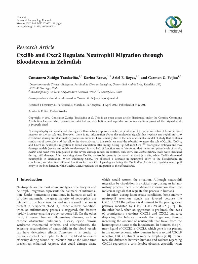
Figure 1: Severe and mild damage differentially regulate the transcription of cxcl8 paralogues in zebrafish. (a) Diagram showing the locationof severe (S) and mild (M) damage on the caudal region and caudal fin of the embryo, respectively. The red line corresponds to the caudalartery, and the blue line to the caudal vein. (b, c) Transcription levels of cxcl8a, cxcl8b, cxcr2, cxcr1, and gcsf-chr19 were quantified byqPCR after (b) severe or (c) mild damage. Data are presented as fold of change over each level at 0 hours post damage and normalized tob-actin1. ∗p value < 0.05; ∗∗p value < 0.01; ∗∗∗p value < 0.005.
3Journal of Immunology Research
the QImaging MicroPublisher 5.0 RVT camera. Images wereprocessed with Photoshop CS5 or ImageJ 1.44o [29]. All ofthe described experiments were performed at least threetimes, and the images shown are representative of the effectsobserved in at least 70% of the individuals.
3. Results
3.1. Severe Damage Upregulates cxcl8a, cxcl8b, gcsf-chr19, andcxcr2, While Mild Damage Only Upregulates cxcl8a and cxcr2.To determine the roles of Cxcl8a (previously named Cxcl8l1[14] and zCxcl8 [30]), Cxcl8b (previously named Cxcl8l2[14]), and Cxcr2 in neutrophil migration through the blood-stream during mechanical damage, the transcriptional levelsof these genes were determined in vivo using severe and milddamage models, taking into consideration the differences in
the inflammatory processes generated by each type of injury[16]. As a control of the type of damage generated, themRNA levels of gcsf-chr19, a critical cytokine for neutrophilblood vessel entry, were assessed [16]. Severe damageincreased the mRNA levels of cxcl8a, cxcl8b, gcsf-chr19, andcxcr2. Specifically, as early as 30 minutes after severe damage,all of these molecules were upregulated, with peak expressionoccurring 1 hpd before slowly declining, reaching normallevels at 3 hpd (Figure 1(b)). On the other hand, mild damageonly increased the transcription of cxcl8a and cxcr2(Figures 1(b) and 1(c)). Furthermore, cxcl8a and cxcr2 upreg-ulation was delayed in comparison with the severe damagemodel, starting at 1 hpd for cxl8a and at 2 hpd for cxcr2(Figure 1(c)). There was no increase in the mRNA levelsof cxcr1 during the entire time course for either severeor mild damage.
Damaged area
Circulation
hpd Control Cxcl8a MO Cxcl8b MO
1
2
3
(a) (b)
02
468
10
1 hpd 2 hpd 3 hpd
Damaged area
Num
ber o
f neu
trop
hils/
defin
ed ar
ea
Cont
rol
Cont
rol
Cont
rol
Cxcl
8a M
O
Cxcl
8a M
O
Cxcl
8a M
O
Cxcl
8b M
O
Cxcl
8b M
O
Cxcl
8b M
O
⁎
⁎⁎ ⁎⁎
⁎
⁎⁎
⁎⁎⁎
012345678
1 hpd 2 hpd 3 hpd
Circulation
Num
ber o
f neu
trop
hils/
defin
ed ar
ea
Cont
rol
Cont
rol
Cont
rol
Cxcl
8a M
O
Cxcl
8a M
O
Cxcl
8a M
O
Cxcl
8b M
O
Cxcl
8b M
O
Cxcl
8b M
O
Cont
rol d
amag
e
Cont
rol d
amag
e
Cont
rol d
amag
e
⁎⁎⁎ ⁎⁎⁎⁎
⁎⁎⁎⁎⁎⁎⁎⁎
(c) (d)
Figure 2: Neutrophil migration decreases when Cxcl8a or Cxcl8b are inhibited during severe damage. (a) Diagram showing the quantifiedneutrophils in two areas. (b) Lateral view of the caudal section of the embryo tail at 1, 2, and 3 hpd in control and morphant embryos. (c)Quantified neutrophils at the damaged area at 1, 2, and 3 hpd. (d) Quantified neutrophils in circulation during homeostasis, after severedamage and in the absence of Cxcl8a or Cxcl8b function. The neutrophils were quantified at 1, 2, and 3 hpd. ∗p value < 0.05; ∗∗p value < 0.01;∗∗∗p value < 0.001; ∗∗∗∗p value < 0.0001.
4 Journal of Immunology Research
3.2. Cxcl8a Knockdown Decreases Neutrophil Quantity at theInjury, While Cxcl8b Decreases Neutrophils in Circulation.Considering the transcriptional differences observed in theqPCR analysis for cxcl8a and cxcl8b between the severe andmild damage models, the functions of both genes were inhib-ited by MO and the effects of this on neutrophil migration tothe wound were determined in vivo. Since Cxcl8 plays animportant role in angiogenesis, particularly in intersegmentalvessel formation [31], Cxcl8a MO or Cxcl8b MO was micro-injected in double transgenic embryos, Tg(BACmpx:GFP)i114
X Tg(fli1a:EGFP)y1, to correctly identify morphant embryos(Supplementary Figure 1). Both neutrophils and blood ves-sels of double transgenic fish are fluorescently labeled.
In the severe damage model, the absence of either Cxcl8aor Cxcl8b significantly decreased the amount of neutrophilspresent at the wound in comparison with that of control-damage embryos, in which the amount of neutrophils pres-ent at the damaged area continuously increased over the timecourse trial (Figures 2(b) and 2(c)). In addition, neutrophilsin blood circulation were quantified (Figure 2(d)). Incontrol-damage embryos, neutrophils were still high in circu-lation at 3 hpd (Video 1), in contrast to the noninjured con-trol embryos that lack neutrophils in the bloodstream(Videos 2 and 3). No differences were observed betweenMO-injected Cxcl8a and control-damage fish. Remarkably,severely damaged morphant embryos for Cxcl8b showed noneutrophils in circulation, just as observed in the noninjured
control embryos. On the other hand, in the mild damagemodel, only the absence of Cxcl8a affected the quantity ofneutrophils at the wound. The amount of neutrophils thatreached the wound in Cxcl8b morphant embryos presentedno significant difference with control-damage embryos(Figure 3). Thus, the results obtained through in vivo analysisusing MOs to block the functioning of each Cxcl8 paraloguewere consistent with qPCR analyses and suggest differentfunctions for Cxcl8a and Cxcl8b.
3.3. Pharmacological Inhibition of Cxcr2 Decreases theAmount of Neutrophil in the Bloodstream. To analyze Cxcr2participation in neutrophil migration trough circulation, itsfunction was pharmacologically inhibited, using the specificinhibitor SB225002. White and collaborators [32] demon-strated that SB225002 is a potent and selective nonpeptideinhibitor of Cxcr2, both in vitro and in vivo. Thus, in thesevere damage model, we quantified neutrophil number incirculation and in damaged area and included a third area(dorsal area) as a nonspecific region (Figure 4(a)). The resultsobtained showed that the number of neutrophils present atthe dorsal area was not different from that observed incontrol-damage embryos (Figure 4(d)). In contrast, theamount of neutrophils detected in the bloodstream ofinhibitor-treated embryos was significantly lower than thatof control-damage embryos at least until 3 hpd (Figure 4(c),Videos 4 and 5). Likewise, the number of neutrophils that
Damaged area
(a)
02
468
10
1 hpd 2 hpd 3 hpd
ns ns nsDamaged area
Num
ber o
f neu
trop
hils/
defin
ed ar
ea
Cont
rol
Cont
rol
Cont
rol
Cxcl
8a M
O
Cxcl
8a M
O
Cxcl
8a M
O
Cxcl
8b M
O
Cxcl
8b M
O
Cxcl
8b M
O
⁎⁎⁎ ⁎⁎⁎ ⁎⁎⁎
hpd Control Cxcl8a MO Cxcl8b MO
1
2
3
(b) (c)
Figure 3: Neutrophil migration decreases when Cxcl8a, but not Cxcl8b, is inhibited during mild damage. (a) Diagram showing the quantifiedneutrophils at the wound area. (b) Quantified neutrophils at damaged area at 1, 2, and 3 hpd. (c) Lateral view of the caudal section of theembryo tail at 1, 2, and 3 hpd in the control and morphant embryos. ∗∗∗p value < 0.005.
5Journal of Immunology Research
reached the injury site was lower in inhibitor-treatedembryos than that in controls during the entire time coursetrial (Figures 4(b) and 4(e)). In the mild damage model(Figure 5), the number of neutrophils present at the damagedarea in inhibitor-treated embryos was drastically lower ateach of the analyzed time points (Figures 5(a) and 5(c)). Incontrast, the number of neutrophils detected in the dorsal
area of inhibitor-treated embryos showed no difference com-pared with that of controls (Figure 5(b)).
4. Discussion
Neutrophils are the first cells to be recruited to a site ofinfection or damage, and neutrophil migration is regulated
(a)
hpd Control
1
2
3
SB225002 Circulation
Con
trol
SB 2
2500
2
Con
trol
SB 2
2500
2
Con
trol
SB 2
2500
2
02468
10121416
Num
ber o
f neu
trop
hils/
defin
ed a
rea
1 hpd 2 hpd 3 hpd
ns ⁎⁎⁎⁎⁎⁎
(b) (c)
Dorsal area
0
5
10
15
20
25
30
35
Num
ber o
f neu
trop
hils/
defin
ed ar
ea
1 hpd 2 hpd 3 hpd
ns ns ns
Cont
rol
SB 2
2500
2
Cont
rol
SB 2
2500
2
Cont
rol
SB 2
2500
2
Damaged area
0
5
10
15
20
25
30
35
Num
ber o
f neu
trop
hils/
defin
ed ar
ea
1 hpd 2 hpd 3 hpd
Cont
rol
SB 2
2500
2
Cont
rol
SB 2
2500
2
Cont
rol
SB 2
2500
2
⁎⁎ ⁎⁎⁎ ⁎⁎⁎⁎
(d) (e)
Figure 4: Cxcr2 inhibition decreases neutrophil entrance to the bloodstream and tissue infiltration in severe damage. (a) Diagramshowing the quantified neutrophils, in circulation and at the dorsal and damaged area. (b) Neutrophils in circulation at 1, 2, and 3 hpd incontrol embryos and treated with the inhibitor SB225002. (c) Lateral view of the caudal section of the embryo tail at 1, 2, and 3 hpd in thecontrol and treated embryos. (d, e) Quantified neutrophils at the dorsal and damaged areas at 1, 2, and 3 hpd. ∗∗p value < 0.01; ∗∗∗p value< 0.005; ∗∗∗∗p value < 0.0001.
6 Journal of Immunology Research
by different chemokines. However, which chemokines andreceptors involved in the regulation of these leukocytes’migration into blood circulation is unknown in both humansand, prior to this study, zebrafish. By using a series of meth-odological approaches and two different models of damages(severe and mild), a role for Cxcl8b and Cxcr2 in neutrophilentry to bloodstream was identified. Similarly, it was deter-mined that Cxcl8a, but not Cxcl8b, attracts neutrophils to
the wound area. Taken together, these data provide the firstfunctional characterization of neutrophil migration bybloodstream after mechanical damage in zebrafish.
The transcriptional analysis showed that in a severedamage model, all the genes analyzed are increased, suggest-ing the participation of all of them in the inflammation pro-cess. On the other hand, during the mild damage, only cxcr2and cxcl8a were upregulated, implying that some events
hpd Control
1
2
3
SB225002
(a)
Dorsal area
0
5
10
15
20
25
30
Num
ber o
f neu
trop
hils/
defin
ed ar
ea
ns ns ns
1 hpd 2 hpd 3 hpd
Cont
rol
SB 2
2500
2
Cont
rol
SB 2
2500
2
Cont
rol
SB 2
2500
2
Damaged area
0
5
10
15
20
25
30
Num
ber o
f neu
trop
hils/
defin
ed ar
ea
1 hpd 2 hpd 3 hpd
Cont
rol
SB 2
2500
2
Cont
rol
SB 2
2500
2
Cont
rol
SB 2
2500
2
⁎⁎⁎⁎ ⁎⁎⁎⁎ ⁎⁎⁎
(b) (c)
Figure 5: Cxcr2 inhibition decreases neutrophil infiltration in mild damage. (a) Lateral view of the caudal section of the embryo tail at 1, 2,and 3 hpd in the control and treated embryos. Demarked with a white rectangle are the two quantified areas. (b, c) Quantified neutrophils atthe dorsal and damaged areas at 1, 2, and 3 hpd. ∗∗∗p value < 0.005; ∗∗∗∗p value < 0.0001.
7Journal of Immunology Research
occurring during the severe damage are not activated inthis situation. One of these events is neutrophil migrationby blood circulation, thus suggesting that Cxcl8a is onlyinvolved in the chemoattraction of neutrophils throughthe extracellular matrix. The lack of function assays devel-oped confirmed these results. In the absence of Cxcl8a, thenumber on neutrophils present in the bloodstream isindistinguishable from control-damage embryos. In a pre-liminary analysis, it seems that these results do not agreewith those obtained by the group of De Oliveira [26]. Intheir work, they indicate that the absence of either Cxcl8aor Cxcl8b decreases the number of neutrophils that reachthe wound and conclude that both chemokines regulateneutrophil migration to the injury site. We observe thesame in our severe damage model, the lack of each Cxcl8paralogue affects the number of neutrophils that arrive atthe wound area, but only the absence of Cxcl8b decreasesthe number of circulation neutrophils. Thus, we agree thatthe lack of each Cxcl8 paralogue affects the final numberof neutrophils that reach the damage, but we think thatthe process altered in each case is different suggesting thatCxcl8a and Cxcl8b regulate different steps of the neutro-phils’ journey to the inflamed site. Also, they should beexpressed in different tissues, Cxcl8a at the wound andCxcl8b at the endothelium near the CHT.
On the other hand and although the function of CXCL8in neutrophil entry to circulation is not clear in humans,the function of CXCL8 in a similar process, such as neutro-phil extravasation, is well documented [33–36]. Duringneutrophil transendothelial migration, glycosaminoglycan-immobilized CXCL8 at the luminal surface of endothelialcells allows neutrophil adhesion and posterior emigrationto surrounding tissue. This mechanism could shed light ontohow neutrophils enter the bloodstream. Considering this andthe present results regarding Cxcl8b, the CXCL8-endothelialcells-neutrophils interaction could also function in the oppo-site direction. In other words, zebrafish Cxlc8b could beimmobilized and exposed to the abluminal endothelial sur-face, thereby allowing neutrophil contact with and entranceto the vasculature. Indeed, the entry of neutrophils to bloodcirculation occurs not only at the begging of the inflamma-tory process but also during resolution by reverse migration,a process that has been observed in vitro and in vivo in zebra-fish and mice [37–41].
The function exerted by CXCL8 on neutrophils inhumans can be divided into roles related to the vasculatureand to the interstitial tissue. In zebrafish, CXCL8 orthologuescontribute to both functions, but each role is performed by aseparate paralogous gene, Cxcl8a or Cxcl8b. The existence oftwo orthologous CXCL8 genes in zebrafish is attributable tothe genome duplication event that occurred near the baseof the ray-finned fish evolutionary tree [42]. Indeed, the rep-ertoire of chemokines present in zebrafish is twice that ofhumans (89 and 44, resp.) [43, 44].
On the other hand, in the current study, Cxcr2 wasfound to participate not only in the final neutrophilmigration to the wound but also in neutrophil migrationthrough the bloodstream. It is interesting that in the Cxcr2lack of function assay, a low amount of neutrophils still
circulate, suggesting that another chemokine receptorcould also participate in the process but to a lesser extent.This is supported by the fact that in the Cxcl8b lack offunction assay, no neutrophil was detected in the blood-stream. A receptor that is a good candidate to be involvedin this process is Cxcr1, mainly because it interacts withCXCL8 in humans [8]. Finally, and not expected, wefound that there is a neutrophil subpopulation that afterinjury migrates through the interstitial tissue in a Cxcr2-independent form. Moreover, these neutrophils did notmigrate in wound direction but to the dorsal area andcould or not be found later at the injury site.
The participation of CXCR2 in bone marrow neutro-phil release is documented in mice, where neutrophilslacking CXCR2 are preferentially retained in the bonemarrow, causing chronic neutropenia [7, 9, 45–47]. Severalstudies support the hypothesis that neutrophil release isantagonistically regulated by the CXCR2 and CXCR4 che-mokine receptor system [7, 48]. Under homeostatic condi-tions, neutrophil retention signals are favored in the bonemarrow since the CXCL12/CXCR4 pathway is dominantto the promigratory pathway mediated by the CXCR2/CXCL1-2 axis. When neutrophil release from the hemato-poietic tissue is required, the levels of the promigrationcytokines CXCL1 and CXCL2, as well as G-CSF, increase,thereby displacing the balance towards migration [7]. In aprevious study, we demonstrated that zebrafish Gcsf-Chr19 regulates neutrophil migration by the bloodstreamafter mechanical damage [16]. In turn, the present studyprovided new details for how neutrophils are mobilizedfrom the caudal hematopoietic tissue to the circulationafter a sterile stimulus by addressing the role of Cxcr2 inthis process and by confirming the evolutionary conserva-tion of Cxcr2 function in lower vertebrates, such as fish.Furthermore, the present results suggest that Cxcr2 is thereceptor for both Cxcl8 paralogues Cxcl8a and Cxcl8b.
In conclusion, and by consolidating previous and ourpresent data, Cxcl8b and Cxcr2 are key regulators of neutro-phil entrance into blood circulation in zebrafish. In moredetail, we propose the following model regarding neutrophilmigration during an inflammatory process in zebrafish(Figure 6). During homeostasis, neutrophils are retained inthe caudal hematopoietic tissue by Cxcr4/Cxcl12 [49], mean-ing only a few neutrophils would be in the bloodstream(Figure 6(a)). After severe damage (Figure 6(b)), Gcsf-Chr19, Cxcl8b, and Cxcr2 expression would increase, andCxcl8b would bind to Cxcr2. Considering the overexpres-sion of Gcsf-Chr19, it is plausible to hypothesize that thismolecule would functionally interact with its receptor,Gcsfr. Therefore, both the Cxcl8b/Cxcr2 and Gcsf-Chr19/Gcsfr signaling pathways would allow neutrophils to leavethe caudal hematopoietic tissue and enter the bloodstream[16]. This would induce neutrophils to enter and remainin circulation until sensing an unknown signal (probablyCxcl8b) in the endothelium near the site of injury, whereneutrophils would then leave the blood vessels. Further-more, in the interstitial tissue, Cxcl8a would bind to Cxcr2present in neutrophils to enable neutrophils to reach thewound [26, 28, 50].
8 Journal of Immunology Research
Our results significantly contribute tofill the gap regardingthe molecular signals that regulate inflammation and neutro-phil recruitment from the hematopoietic tissue to the vascula-ture in zebrafish, a key step of the journey of this granulocyteduring an inflammatory process. Considering the similarityin molecules between zebrafish and humans—which madethisfish a suitablemodel for this study—our research providesnew avenues for understanding neutrophil biology duringhomeostasis and pathologic conditions.
Conflicts of Interest
The authors declare no conflicts of interest in relation to theresearch and authorship.
Acknowledgments
The transgenic zebrafish line Tg(BACmpx:GFP)i114 waskindly provided by Dr. Steve Renshaw. This work wassupported by the Fondo Nacional de Desarrollo Cientifico yTecnologico, 1171199 and 1150816, Direccion de Investiga-cion UNAB DI-483-14/R, and Fondo de Financiamiento deCentros de Investigación en Áreas Prioritarias, 15110027.
References
[1] C. L. Semerad, F. Liu, A. D. Gregory, K. Stumpf, and D. C.Link, “G-CSF is an essential regulator of neutrophil trafficking
from the bone marrow to the blood,” Immunity, vol. 17, no. 4,pp. 413–423, 2002.
[2] R. B. Day and D. C. Link, “Regulation of neutrophil traffickingfrom the bone marrow,” Cellular and Molecular Life Sciences,vol. 69, no. 9, pp. 1415–1423, 2012.
[3] Q. Ma, D. Jones, and T. A. Springer, “The chemokine receptorCXCR4 is required for the retention of B lineage and granulo-cytic precursors within the bone marrow microenvironment,”Immunity, vol. 10, no. 4, pp. 463–471, 1999.
[4] W. C. Liles, H. E. Broxmeyer, E. Rodger et al., “Mobilization ofhematopoietic progenitor cells in healthy volunteers byAMD3100, a CXCR4 antagonist,” Blood, vol. 102, no. 8,pp. 2728–2730, 2013.
[5] H. E. Broxmeyer, C. M. Orschell, D. W. Clapp et al.,“Rapid mobilization of murine and human hematopoieticstem and progenitor cells with AMD3100, a CXCR4 antago-nist,” The Journal of Experimental Medicine, vol. 201, no. 8,pp. 1307–1318, 2005.
[6] K. J. Eash, J. M. Means, D. W. White, and D. C. Link, “CXCR4is a key regulator of neutrophil release from the bone marrowunder basal and stress granulopoiesis conditions,” Blood,vol. 113, no. 19, pp. 4711–4719, 2009.
[7] K. J. Eash, A. M. Greenbaum, P. K. Gopalan, and D. C.Link, “CXCR2 and CXCR4 antagonistically regulateneutrophil trafficking from murine bone marrow,” TheJournal of Clinical Investigation, vol. 120, no. 7,pp. 2423–2243, 2010.
[8] J. Lee, R. Horuk, G. C. Rice, G. L. Bennett, T. Camerato, andW. I. Wood, “Characterization of two high affinity human
Caudal hematopoietic tissue
Cxcr1
GcsfrCxcr2
Sdf-1
Gcsf-Chr19
Neutrophil
Epithelial cellCaudal hematopoietic tissue
Cxcr1
Gcsfr
Cxcr2
Sdf-1
Gcsf-Chr19Cxcl8b
Cxcl8a
Neutrophil
Epithelial cell
(a) (b)
Figure 6: Model for the regulation of neutrophil migration to a wound. (a) During homeostasis, neutrophils are retained in the caudalhematopoietic tissue by Cxcr4/Cxcl12. (b) After damage, the expression of Gcsf-Chr19 would increase and interact with its receptor,Gcsfr. Likewise, mRNA levels of Cxcl8b and Cxcr2 would increase, and both proteins would interact. Therefore, both signaling pathwayswould allow neutrophils to leave the caudal hematopoietic tissue and enter the bloodstream. Neutrophils would stay in circulation untilsensing an unknown signal (probably Cxcl8b) in the endothelium, then leaving the blood vessel. Finally, in the interstitial tissue, Cxcl8a/Cxcr2 would guide neutrophils to the wound.
9Journal of Immunology Research
interleukin-8 receptors,” The Journal of Biological Chemistry,vol. 267, no. 23, pp. 16283–16287, 1992.
[9] A. Stadtmann and A. Zarbock, “CXCR2: from bench tobedside,” Frontiers in Immunology, vol. 3, p. 263, 2012.
[10] Y. Kobayashi, “The role of chemokines in neutrophil biology,”Frontiers in Bioscience, vol. 13, pp. 2400–2407, 2008.
[11] M. K. Mamik and A. Ghorpade, “CXCL8 as a potential thera-peutic target for HIV-associated neurocognitive disorders,”Current Drug Targets, vol. 17, no. 1, pp. 111–121, 2016.
[12] G. Caramori, I. M. Adcock, A. Di Stefano, and K. F. Chung,“Cytokine inhibition in the treatment of COPD,” InternationalJournal of Chronic Obstructive Pulmonary Disease, vol. 9,pp. 397–412, 2014.
[13] H. Ha, T. Bensman, H. Ho, P. M. Beringer, and N. Neamati, “Anovel phenylcyclohex-1-enecarbothioamide derivative inhibitsCXCL8-mediated chemotaxis through selective regulation ofCXCR2-mediated signalling,” British Journal of Pharmacology,vol. 171, no. 6, pp. 1551–1565, 2014.
[14] L. M. van der Aa, M. Chadzinska, E. Tijhaar, P. Boudinot,and B. M. Verburg-van Kemenade, “CXCL8 chemokinesin teleost fish: two lineages with distinct expression profilesduring early phases of inflammation,” PloS One, vol. 5,no. 8, article e12384, 2010.
[15] S. H. Oehlers et al.M. V. Flores, C. J. Hall, R. O'Toole et al.,“Expression of zebrafish cxcl8 (interleukin-8) and its receptorsduring development and in response to immune stimulation,”Developmental and Comparative Immunology, vol. 34, no. 3,pp. 352–359, 2010.
[16] J. A. Galdames, C. Zuñiga-Traslaviña, A. E. Reyes, and C. G.Feijoo, “Gcsf-Chr19 promotes neutrophil migration to dam-aged tissue through blood vessels in zebrafish,” Journal ofImmunology, vol. 193, no. 1, pp. 372–378, 2014.
[17] B. Bajoghli, “Evolution and function of chemokine receptors inthe immune system of lower vertebrates,” European Journal ofImmunology, vol. 43, no. 7, pp. 1686–1692, 2013.
[18] B. Novoa and A. Figueras, “Zebrafish: model for the study ofinflammation and the innate immune response to infectiousdiseases,” Advances in Experimental Medicine and Biology,vol. 946, pp. 253–275, 2012.
[19] N. D. Meeker and N. S. Trede, “Immunology and zebrafish:spawning new models of human disease,” Developmentaland Comparative Immunology, vol. 32, no. 7, pp. 745–757,2008.
[20] M. Westerfield, L. I. Zon, and H. W. Detrich, Essential Zebra-fish Methods. Cell & Developmental Biology, Academic Press,London, 2009.
[21] S. A. Renshaw, C. A. Loynes, D. M. I. Trushell, S. Elworthy, P.W. Ingham, and M. K. Whyte, “A transgenic zebrafish modelof neutrophilic inflammation,” Blood Journal of Hematology,vol. 108, no. 13, pp. 3976–3978, 2006.
[22] N. D. Lawson and B. M.Weinstein, “In vivo imaging of embry-onic vascular development using transgenic zebrafish,” Devel-opmental Biology, vol. 248, no. 2, pp. 307–318, 2002.
[23] C. B. Kimmel, W. W. Ballard, S. R. Kimmel, B. Ullmann,and T. F. Schilling, “Stages of embryonic development ofthe zebrafish,” Developmental Dynamics, vol. 203, no. 3,pp. 253–310, 1995.
[24] P. M. Elks, A. C. Loynes, and S. A. Renshaw, “Measuringinflammatory cell migration in zebrafish,” Cell Migration:Developmental Methods and Protocols, vol. 769, pp. 261–275,2011, Chapter 18.
[25] F. Ellet and G. Lieschke, “Computational quantification offluorescent leukocyte numbers in zebrafish embryos,”Methodsin Enzymology, vol. 506, pp. 425–435, 2011.
[26] S. de Oliveira, C. C. Reyes-Aldasoro, S. Candel, S. A. Renshaw,V. Mulero, and A. Calado, “Cxcl8 (IL-8) mediates neutrophilrecruitment and behavior in the zebrafish inflammatoryresponse,” Journal of Immunology, vol. 190, no. 8, pp. 4349–4359, 2013.
[27] M. W. Pfaffl, G. W. Horgan, and L. Dempfle, “Relative expres-sion software tool (REST) for group-wise comparison andstatistical analysis of relative expression results in real-timePCR,” Nucleic Acids Research, vol. 30, no. 9, article e36, 2002.
[28] Q. Deng, M. Sarris, D. Bennin, J. M. Green, P. Herbomel, andA. Huttenlocher, “Localized bacterial infection inducessystemic activation of neutrophils through CXCR2 signalingin zebrafish,” Journal of Leukocyte Biology, vol. 93, no. 5,pp. 761–769, 2013.
[29] M. D. Abramoff, P. J. Magalhaes, and S. J. Ram, “Image pro-cessing with Image J,” Biophotonics International, vol. 11,pp. 36–42, 2004.
[30] M. Sarris, J. B. Masson, D. Maurin et al., “Inflammatorychemokines direct and restrict leukocyte migration withinlive tissues as glycan-bound gradients,” Current Biology,vol. 24, no. 24, pp. 2375–2382, 2012.
[31] S. J. Stoll, S. Bartsch, H. G. Augustin, and J. Kroll, “The tran-scription factor HOXC9 regulates endothelial cell quiescenceand vascular morphogenesis in zebrafish via inhibition ofinterleukin 8,” Circulation Research, vol. 108, no. 11,pp. 1367–1377, 2011.
[32] J. R. White, J. M. Lee, P. R. Young et al., “Identification of apotent, selective non-peptide CXCR2 antagonist that inhibitsinterleukin-8-induced neutrophil migration,” The Journal ofBiological Chemistry, vol. 273, no. 17, pp. 10095–10098, 1998.
[33] W. B. Smith, I. Gamble, M. Clark-Lewis, and M. A. Vadas,“Interleukin-8 induces neutrophil transendothelial migra-tion,” Immunology, vol. 72, no. 1, pp. 65–72, 1991.
[34] A. Rot, “Binding of neutrophil attractant/activation protein-1(interleukin-8) to resident dermal cells,” Cytokine, vol. 4,no. 5, pp. 347–352, 1992.
[35] J. Middleton, S. Neil, J. Wintle et al., “Transcytosis and surfacepresentation of IL-8 by venular endothelial cells,” Cell, vol. 91,pp. 385–395, 1997.
[36] A. Woodfin, C. A. Reichel, A. Khandoga et al., “JAM-A medi-ates neutrophil transmigration in a stimulus-specific mannerin vivo: evidence for sequential roles for JAM-A andPECAM-1 in neutrophil transmigration,” Blood, vol. 110,no. 6, pp. 1848–1856, 2007.
[37] C. D. Buckley, E. A. Ross, H. M. McGettrick et al., “Identifica-tion of a phenotypically and functionally distinct populationof long-lived neutrophils in a model of reverse endothelialmigration,” Journal of Leukocyte Biology, vol. 79, no. 2,pp. 303–311, 2006.
[38] J. R. Mathias, B. J. Perrin, T. X. Liu, J. Kanki, A. T. Look,and A. Huttenlocher, “Resolution of inflammation by retro-grade chemotaxis of neutrophils in transgenic zebrafish,” Jour-nal of Leukocyte Biology, vol. 80, no. 6, pp. 1281–1288, 2006.
[39] P. M. Elks, F. J. van Eeden, G. Dixon et al., “Activation ofhypoxia-inducible factor-1α (Hif-1α) delays inflammationresolution by reducing neutrophil apoptosis and reversemigration in a zebrafish inflammation model,” Blood, vol. 118,no. 3, pp. 712–722, 2011.
10 Journal of Immunology Research
[40] A. Woodfin, M. B. Voisin, M. Beyrau et al., “The junctionaladhesion molecule JAM-C regulates polarized transendothe-lial migration of neutrophils in vivo,” Nature Immunology,vol. 12, no. 8, pp. 761–769, 2011.
[41] F. Ellett, P. M. Elks, A. L. Robertson, N. V. Ogryzko, and S. A.Renshaw, “Defining the phenotype of neutrophils followingreverse migration in zebrafish,” Journal of Leukocyte Biology,vol. 98, no. 6, pp. 975–981, 2015.
[42] J. Lu, E. Peatman, H. Tang, J. Lewis, and Z. Liu, “Profiling ofgene duplication patterns of sequenced teleost genomes:evidence for rapid lineage-specific genome expansion medi-ated by recent tandem duplications,” BMC Genomics, vol. 13,no. 1, p. 246, 2012.
[43] H. Nomiyama, N. Osada, and O. Yoshie, “The evolution ofmammalian chemokine genes,” Cytokine & Growth FactorReviews, vol. 21, no. 4, pp. 253–262, 2010.
[44] H. Nomiyama, N. Osada, and O. Yoshie, “Systematic classifi-cation of vertebrate chemokines based on conserved syntenyand evolutionary history,” Genes to Cells, vol. 18, no. 1,pp. 1–16, 2013.
[45] R. M. Devalaraja, L. B. Nanney, J. Du et al., “Delayed woundhealing in CXCR2 knockout mice,” The Journal of InvestigativeDermatology, vol. 115, no. 2, pp. 234–244, 2000.
[46] S. von Vietinghoff, M. Asagiri, D. Azar, A. Hoffmann, andK. Ley, “Defective regulation of CXCR2 facilitates neutro-phil release from bone marrow causing spontaneous inflam-mation in severely NF-kappa B-deficient mice,” Journal ofImmunology, vol. 185, no. 1, pp. 670–678, 2010.
[47] A. Köhler, K. De Filippo, M. Hasenberg et al., “G-CSF medi-ated thrombopoietin release triggers neutrophil motility andmobilization from bone marrow via induction of Cxcr2ligands,” Blood, vol. 117, no. 16, pp. 4349–4357, 2011.
[48] C. Martin, P. C. Burdon, G. Bridger, J. C. Gutierrez-Ramos,T. J. Williams, and S. M. Rankin, “Chemokines acting viaCXCR2 and CXCR4 control the release of neutrophils fromthe bone marrow and their return following senescence,”Immunity, vol. 19, no. 4, pp. 583–593, 2003.
[49] K. B. Walters, J. M. Green, J. C. Surfus, S. K. Yoo, andA. Huttenlocher, “Live imaging of neutrophil motility in azebrafish model of WHIM syndrome,” Blood, vol. 116, no. 15,pp. 2803–2811, 2010.
[50] M. Sarris, J. B. Masson, D. Maurin et al., “Inflammatorychemokines direct and restrict leukocyte migration withinlive tissues as glycan-bound gradients,” Current Biology,vol. 22, no. 24, pp. 2375–2382, 2012.