CyberKnife Radiosurgery for the Treatment of Orbital Metastases
www.tcrt.org
Purpose of this study is to evaluate radiographic therapy response, clinical outcome and adverse effects of CyberKnife radiosurgery in patients suffering from orbital metastases. Sixteen orbital metastases originating from different solid cancers in fourteen patients were treated by single fraction CyberKnife radiosurgery. Radiographic response and clinical out-come were evaluated. The treated tumor volume ranged from 0.2 to 35 cm3 (median 2.3 cm3, mean 7.0 cm3, SD 6 10.4 cm3, CI 0.9-9.4 cm3). The prescription dose ranged from 16.5-21 Gy (median 18 Gy, mean 18.2 Gy, SD 6 1.2 Gy, CI 17.0-18.4 Gy). A no change situation was observed in nine lesions, partial remission in four as well as complete remission in one metas-tasis. Tumor growth was stabilized or regressive following CyberKnife therapy in 87% of the cases. Recurrence was observed in two cases (13%). Before therapy, three patients suffered from visual disturbance and five patients reported diplopia. Six patients had no initial symp-toms. After therapy, one patient indicated improvement of the present visual deficit and two patients no change. Out of the two patients with persistent diplopia, two reported improve-ment after therapy and three no change. No progression of symptoms was noted in any of the cases. Fourteen out of sixteen treated lesions were stable or regressive following CyberKnife radiosurgery (87%). As no serious adverse effects were reported in this series, CyberKnife therapy was shown to be of great value for local management of orbital metastases.
Key words: CyberKnife; Radiosurgery; Orbital metastases.
Introduction
Approximately 0.1% of all tumors and 18% of all orbital diseases are orbital tumors (1). The actual incidence of metastases to the eye and orbit is difficult to quantify, as patients may remain asymptomatic and succumb to their primary cancer before clinical detection of the ocular metastasis. With improvements in systemic therapy and oncologic management, patients suffering from metastatic cancer may live longer and therefore, orbital metastases may manifest more frequently (2). Nearly all solid malignancies have been reported to metastasize to the orbit, but the most frequent primary tumors in adults to metastasize here are breast cancer (42%), lung (11%), unknown primary (11%), prostate (8.3%), melanoma (5.2%), gastro-intestinal tract (4.4%) and kidney cancer (3.2%) (1). Average patient survival after diagnosis of orbital metastasis has been reported to be approximately 9 months (1, 3). Onset of symptoms occurs more rapidly in orbital metastases than in other types of orbital neoplasia. The most frequent symptoms (if present) include: Prop-tosis or enophthalmos, disturbances of motility (infiltrative effect), pain, swelling
D-80336 Munich, Germany2European CyberKnife Center,
Max-Lebsche-Platz 31, D-81377 München
Munich, Germany
Abbreviations: SD: Standard Deviation; CI: Confidence Interval; LINAC: Linear Accelerator; CT: Computed Tomography; MRI: Magnetic Resonance Imaging; CTX: Chemotherapy; DV: Double Vision; VA: Visual Acuity; NC: No Change; PR: Partial Remission; Rec: Recurrence; CR: Complete Remission; RS: Radiosurgery.
2 Klingenstein et al.
Technology in Cancer Research & Treatment 2012 March 28 [Epub ahead of print]
(inflammatory effect), visible or palpable mass (mass effect) and decrease in visual acuity (functional effect including neu-rological deficits). The overall treatment goals for patients with orbital metastases are palliative, focusing on the improvement of quality of life (preservation of vision or prevention of pain) (2). Treatment options include external beam radiotherapy, surgery and stereotactic radiotherapy (4), observation and sys-temic therapy (5).
Palliative treatment of metastatic disease could be improved by technology that would allow treatment of specific orbital sub-sites (6, 7). Radiosurgery has gained wide acceptance and has become a routine procedure for the treatment of mul-tiple tumors in the optic system (8-10). Acute and late effects of radiotherapy to sensitive eye structures must be balanced against the overall treatment efficacy. Sequelae of radiation applied to the eye include cataract formation, radiation retin-opathy, optic neuropathy, dry eye syndrome as well as glau-coma due to neovascularization or narrow-angle (2).
The CyberKnife is a robotic linear accelerator capable of precise radiosurgery delivery to targets as small as 5 mm. It is an image-guided radiosurgical system with a linear accel-erator on a robotic arm, delivering precisely focused exter-nal beam radiation from a multidirectional perspective and thereby limiting damage to the surrounding healthy tissue. CyberKnife permits high patient comfort during frameless radiosurgery and the possibility of fractionating the treatment dose. By using this non-coplanar treatment approach, irreg-ular radiation fields can be generated, protecting surround-ing structures outside of the 3-dimensional target volume (11-13). Stereotactic fractionated radiotherapy is employed using dose distributions with consequent substantial sparing of surrounding tissues and has been proven more sophisti-cated and safer than conventional radiotherapy (9).
Previous publications on radiation treatment of orbital metas-tases have reported fractionated schemes (10, 14, 15), as these are assumed to minimize damage to the optic apparatus. We now evaluated radiographic response and clinical outcome of single-session CyberKnife radiosurgery for the treatment of orbital metastases which makes the results of our study impor-tant. Only documented continuing tumor growth was consid-ered as local recurrence and failure of therapy, respectively.
Material and Methods
Patients
All patients were archived in a digital database and evaluated retrospectively. CyberKnife radiation was performed as an out-patient procedure. Patients who presented with orbital metas-tases that were not accessible to other treatment options due to size or risky location were included. Further inclusion criteria
were (continuing) documented tumor growth or clinical symp-toms. Patients were consecutive and evaluated for radiosurgery treatment eligibility by a dedicated board of tumor specialists from the University Hospital of Munich consisting of ophthal-mologists, radiation oncologists, and physicians specialized in stereotactic radiosurgery. Informed consent was obtained from all patients before enrollment in this study.
We treated sixteen intraorbital metastases in fourteen patients with CyberKnife radiosurgery. Six patients were female (43%) and eight patients male (57%). Patients’ median age was 57 years (range 42-69 years). In twelve patients, one sole metastasis was treated (86%) and in two patients two lesions were treated (14%), respectively. The primary tumors were breast cancer in five cases, prostate cancer in three, mela-noma in three, as well as renal, pancreatic and pharyngeal cancer in one patient. Eight patients had already undergone chemotherapy. Three lesions had previously been treated by conventional fractionated radiotherapy without therapy response. Ongoing tumor growth had been observed before CyberKnife radiosurgery. No surgery had been performed in any of the included patients, either because of lacking accessi-bility due to the tumor location or due to tumor size. Patients’ mean follow-up was 6 months (maximum 24 months). Six patients died throughout follow-up.
Radiosurgery
The CyberKnife is a frameless 6 MV LINAC system, which performs image-guided robotic radiosurgery (9). The robotic arm offers high flexibility and submillimetric accuracy. Treatment procedure includes CT image acquisition based on skull-bone landmarks, treatment planning and of course treatment delivery (16). Metastases were treated and local-ized via real-time image guidance based on digitally recon-structed radiographs of the skull. As with other forms of stereotaxy, the CyberKnife system presumes a fixed rela-tionship between the lesion to be treated and the skull. The patient’s head is supported during treatment using a custom-fitted thermo-plastic face mask. Real-time digital radiographs obtained by two x-ray devices positioned on either side of the patient’s body were acquired at repeated intervals. Dur-ing intervention, the exposure to radiation remains modest through amorphous silicon sensors (10 mA, 75 kV; corre-sponding to a dose per image of approximately 25 mrad) (9). These acquired images are automatically registered to digi-tally reconstructed radiographs derived from the treatment planning CT scans. CT is particularly helpful in visualizing bony details of orbital metastases and can be employed to evaluate metastatic extension into the cranial cavity and to reveal calcifications. MRI is superior to CT in the visual-ization of soft tissue structures and non-calcific optic nerve abnormalities (17). According to clinical and radiographic findings, an individualized treatment plan was developed for
Technology in Cancer Research & Treatment 2012 March 28 [Epub ahead of print]
CyberKnife Radiosurgery for Orbital Metastases 3
every patient. Tumor volume was defined as visible tumor on imaging (CT simulation images fused with MRI made cal-culation of target volume possible and revealed risk of the optic nerve). In CyberKnife radiosurgery, the position of the skull and thereby also the intraorbital metastasis is translated to the coordinate frame of the LINAC. Patient movement is detected by a control loop between the imaging system and the robotic arm and the LINAC therapeutic beam is consecu-tively repositioned (9). Further details of the treatment proce-dure have already been published (12, 18). All patients were treated using single fraction radiosurgery. Patient number three was treated with single fraction at different time-points, as two lesions were treated.
Evaluation
Serial CT and MRI imaging were employed in the diagno-sis, treatment planning and follow-up of the treated orbital metastases. Changes in tumor size after therapy were consid-ered as main outcome measure and qualified as recurrence in case of documented tumor growth .20%, stable in case of tumor volume 620%, partial remission in case of decrease of tumor volume .20% and complete remission if no lesion could be visualized on follow-up imaging.
Follow-up visits were scheduled at 3-monthly intervals after therapy until the patients’ Karnofsky index had decreased to an extent making further follow-up visits impossible. At every visit, patients’ quality of life and secondary out-come measures (visual acuity, diplopia, pain, paraesthesia) were assessed and evaluated semiquantitatively via patient questionnaire.
Results
Radiographic Response
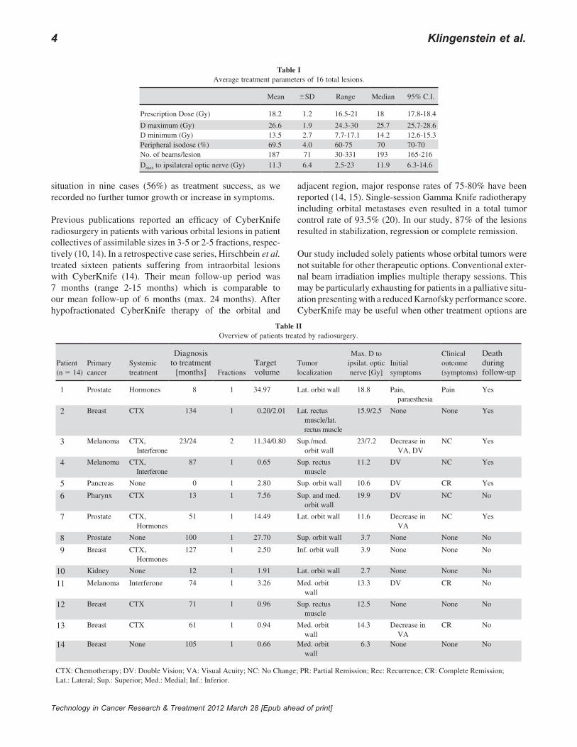
Maximum tumor dimensions were measured by MRI or CT. The total tumor volume prior to therapy ranged from 0.2 to 35 cm3 (median 2.3 cm3, mean 7.0 cm3, SD 6 10.4 cm3, 95%-CI 0.9-9.4 cm3). For details of treatment parameters of the sixteen treated lesions see Table I. After single-session radiosurgery, we observed no change in nine lesions (56%), partial resolu-tion in 4 (25%) as well as complete resolution in one metastasis (6%), respectively. Table II shows an overview of the indi-vidual outcome of each patient. Interstitial regression, tumor necrosis or reduced contrast-enhancement on follow-up imag-ing (CT or MRI) were considered as secondary criteria for ther-apy response of the orbital tumor after radiosurgical treatment. Consecutively, tumor growth was stabilized or regressive fol-lowing CyberKnife therapy in 87% of the cases. Recurrence was observed in two cases (13%) (Table III). Figures one and two exemplarily show the orbital metastasis in patient two (59 years, female) suffering from breast cancer. Primary
diagnosis was in 1997 in the right (pT3 N1 M0 G3) and in 1999 in the left breast (pT1 N0 M0 G2). Pathology showed an invasive lobular carcinoma. Primary treatment included abla-tion and radiation therapy of the chest wall and lymph nodes. Osseous metastases were initially diagnosed in 2001. Previ-ous therapies of metastases included chemotherapy as well as radiation therapy of the intraorbital metastasis with a total dose of 40 Gy in 2002. CyberKnife radiation of the orbital metastasis was performed in 2008 (Figure 1). The metastasis infiltrating the inferior muscles of the left orbit was treated with a prescription dose of 18 Gy to the 70% isodose and a maximum dose of 25.71 Gy using 216 different beam direc-tions. Figure 2 shows the subtotal decrease of the treated lesion at follow-up 6 months later. Visual acuity was unchanged.
Clinical Outcome
Three patients reported decrease in visual acuity and five patients suffered from double vision before therapy. Six patients had no initial symptoms. After therapy, one patient indicated improvement of the previous visual deficit and two patients reported no change. Out of the five patients having complained about double vision, two reported improvement after therapy and three reported no change. Patient one suf-fered from pain and paraesthesia before radiosurgery. The paraesthesia was completely resolved after therapy, whereas pain persisted. In summary, there was no worsening of symp-toms in any case observed in this study (Table IV). No seri-ous adverse effects related to radiosurgery were noted during the observation period.
Discussion
Metastatic disease has been reported to account for 2-12% of all orbital neoplasia (1). Orbital metastases spread hematogenously, as there are no significant lymphatics in the orbit. Early orbital metastasis seems to be associated with more aggres-sive cancers (19). This means clinical symptoms must be expected soon. Although patient survival is reportedly lim-ited after diagnosis of intraorbital metastases (1, 3), best possible palliative care with maximum quality of life is crucial. Up to now, surgical resection or fractionated radia-tion therapy are considered as the mainstays for extraocular treatment (5).
This study presents a promising treatment option of orbital metastases via single fraction CyberKnife radiosurgery. Tumor stabilization or decrease in size could be achieved in 87% of the lesions treated. This result is encouraging, espe-cially regarding that radiosurgery was the therapy of last resort, considering the high percentage of failed chemother-apy or conventional radiotherapy as well as the lacking access to surgical intervention. Partial and complete remission was observed in five lesions (31%). We consider the no-change
4 Klingenstein et al.
Technology in Cancer Research & Treatment 2012 March 28 [Epub ahead of print]
situation in nine cases (56%) as treatment success, as we recorded no further tumor growth or increase in symptoms.
Previous publications reported an efficacy of CyberKnife radiosurgery in patients with various orbital lesions in patient collectives of assimilable sizes in 3-5 or 2-5 fractions, respec-tively (10, 14). In a retrospective case series, Hirschbein et al. treated sixteen patients suffering from intraorbital lesions with CyberKnife (14). Their mean follow-up period was 7 months (range 2-15 months) which is comparable to our mean follow-up of 6 months (max. 24 months). After hypofractionated CyberKnife therapy of the orbital and
adjacent region, major response rates of 75-80% have been reported (14, 15). Single-session Gamma Knife radiotherapy including orbital metastases even resulted in a total tumor control rate of 93.5% (20). In our study, 87% of the lesions resulted in stabilization, regression or complete remission.
Our study included solely patients whose orbital tumors were not suitable for other therapeutic options. Conventional exter-nal beam irradiation implies multiple therapy sessions. This may be particularly exhausting for patients in a palliative situ-ation presenting with a reduced Karnofsky performance score. CyberKnife may be useful when other treatment options are
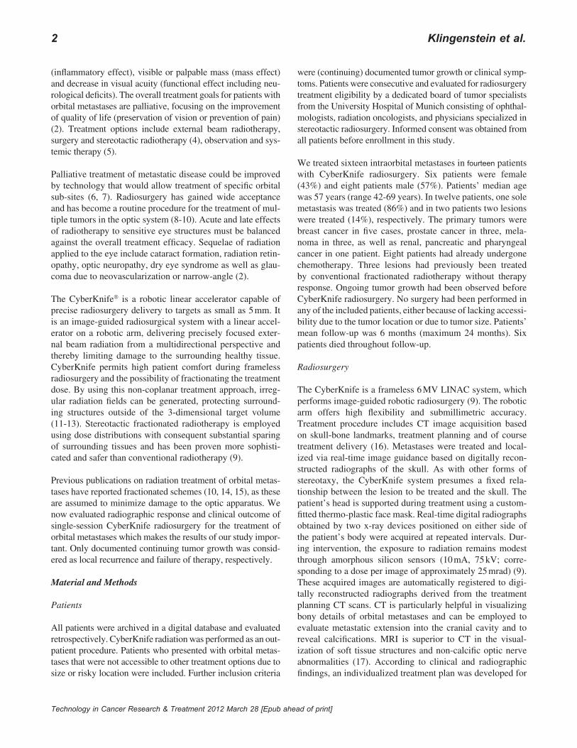
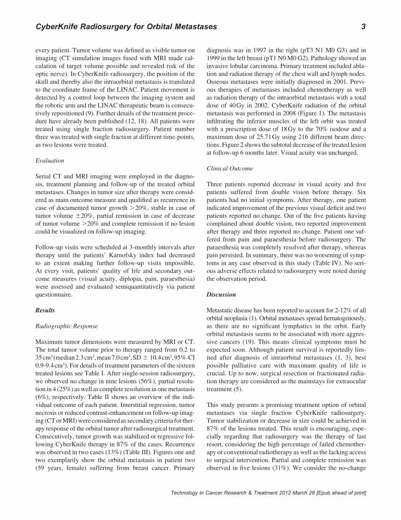
Table IAverage treatment parameters of 16 total lesions.
Technology in Cancer Research & Treatment 2012 March 28 [Epub ahead of print]
CyberKnife Radiosurgery for Orbital Metastases 5
not feasible due to poor general condition, advanced age and significant medical comorbidity (21). We therefore see great potential especially in hypo- or single fraction therapy via CyberKnife. Taking into account the results of the present study, we subscribe to Daftari’s opinion that CyberKnife is feasible and may even be ideal for large-diameter orbital tumors as it allows stereotactic irradiation and requires no additional surgical procedures (21).
With regard to the radiation sensitivity of the globe and the optic nerve, minimal radiation exposure of the adjacent structures of
the lesion is of great importance. A significantly increased inci-dence of dose-dependent complications concerning the optic apparatus has been reported (22). Based on the know-ledge of optic nerve tolerance to single-session radiosurgery, radiation optic neuropathy rates are low around a median dose of 10-12 Gy applied (23-25) to a functioning optic nerve. Due to the very limited awaited survival-time of patients with orbital metastases, we feel that the risk of potential remote damage to the optic apparatus within the range of doses applied here is low compared with the high rate of local tumor control after single-session treatment. Of course, radiation doses should always be applied with great care and be held to the necessary minimum. In the literature reporting the treatment of intraor-bital lesions via CyberKnife, radiosurgery appears to be a safe procedure with minimal adverse effects (10, 16). As the CyberKnife system is image-guided, it is possible to deliver high doses of conformal radiation therapy to tumors and keep the dose to the anterior visual pathways to a minimum (26). Hypofractionated CyberKnife radiosurgery has been reported to be tolerated quite well as salvage therapy in patients with
Table IIIRadiographic response.
Lesions (n 5 16) %
No change 9 56
Partial remission 4 25Complete remission 1 6
Recurrence 2 13
Figure 1: Treatment of CyberKnife therapy in 2008 (2.5 cm3, 18 Gy, 70% isodose). Multiple beam tranjectories (upper left), axial (upper right), sagittal (lower left) and coronal (lower right) scans.
6 Klingenstein et al.
Technology in Cancer Research & Treatment 2012 March 28 [Epub ahead of print]
large orbital metastases (21). In this study, a maximum radi-ation dose of 2.5-23 Gy to the ipsilateral optic nerve was applied (median dose 11.9 Gy, mean 11.3 Gy, SD 6 6.4 Gy, CI 6.3-14.6 Gy) (Tables I and II).
Most commonly, adverse effects occur within the first 9 months after radiation (14). In a recent series of 14 eyes with lymphoma treated with CyberKnife, the only adverse effects observed were mild dry eye syndrome in 2 eyes and cataract in 1 eye after a mean follow-up of 23 months, demonstrating relative safety (10). Hirschbein et al. observed stabiliza-tion, partial regression or even complete disappearance with reported visual field preservation and no reported loss of
visual acuity (14). They did report the development of diplo-pia in one patient 2 months after radiosurgery. However, in the opinion of the treating ophthalmologist, this adverse effect was not caused by the treatment itself (14). During our follow-up, we found no adverse reactions related to CyberKnife treatment. Of particular note, visual acuity could be preserved in all cases. Yet, there is still no overall answer, as long-terms results are still not available. However, our results indicate that the risk of CyberKnife-radiation-induced complications is low during the metastatic patients’ expected life span. Tumor regression reportedly occurs slowly after radiotherapy, beginning approximately after 6-12 months, continuing even years later (19). After Gamma Knife radia-tion with a prescription dose for orbital metastasis (16-20 Gy, median 18 Gy) similar to the dose employed in this study (16.5-21 Gy, median 18 Gy), excellent preservation of neu-rological function with few treatment-related complications was observed (19).
We do acknowledge the limited number of lesions treated in this series. Nevertheless, our preliminary results appear to be very promising, considering the lack of other therapeutic options in these challenging cases. In intraorbital metastases originating from different primary tumors, stereotactic radio-surgery was shown to be able to stabilize or diminish local tumors in fourteen out of sixteen lesions (87%). As our patient collective includes merely metatastatic lesions and is thereby
Figure 2: CT-scan before (left) and after radiosurgery (right). Diminuation of the orbital metastasis (see arrow).
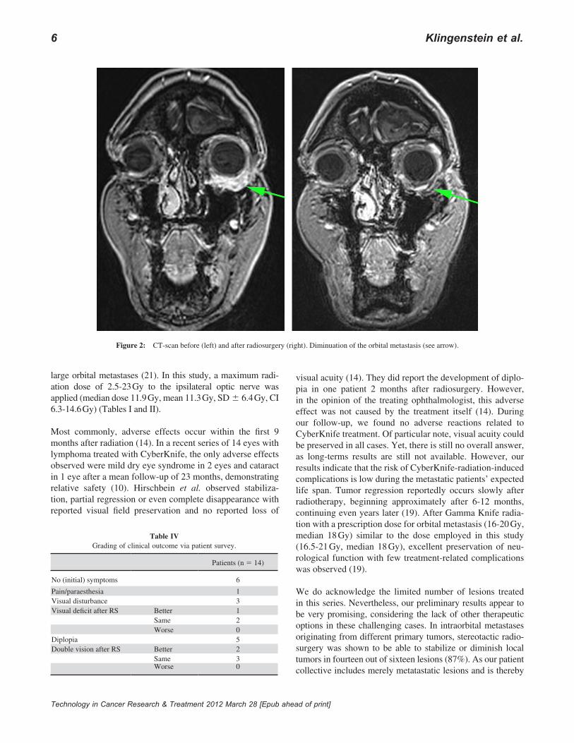
Table IVGrading of clinical outcome via patient survey.
Patients (n 5 14)
No (initial) symptoms 6
Pain/paraesthesia 1Visual disturbance 3Visual deficit after RS Better 1
Same 2Worse 0
Diplopia 5Double vision after RS Better 2
Same 3Worse 0
Technology in Cancer Research & Treatment 2012 March 28 [Epub ahead of print]
CyberKnife Radiosurgery for Orbital Metastases 7
consistent with the poor prognosis of the four patients who dropped out of Hirschbein’s study because of death prior to follow-up (14), this makes our results achieved via CyberKnife radiation more promising. In order to evaluate the response rate of the different sources of metastasis, studies on larger patient collectives are warranted. Other than in previous publications, merely orbital metastases of solid tumors were included.
As no serious adverse effects were reported in this series, CyberKnife therapy has proven to be of great value in the local management of orbital metastases. The CyberKnife sys-tem seems to be an effective and safe alternative to traditional radiotherapy in orbital metastases. Further studies should be initiated in order to compare the outcome of single fraction versus multisession radiosurgery.
Disclosure/Conflict of Interest
A. Muacevic occasionally receives speaker fees from Accuray Inc.
The other authors declare no conflict of interest.
References
Goldberg, R. A., Rootman. J., Cline, R. A. Tumors metastatic to the 1. orbit: a changing picture. Surv Ophthalmol 35, 1-24 (1990).Rudoler, S. B., Shields, C. L., Shields, J. A. Radiation Therapy 2. of Uveal and Orbital Metastases. In: Radiotherapy of Intraocular and Orbital Tumors. pp. 87-93. Eds., Sagerman, R. H., Alberti, W. E. Berlin Heidelberg, New York: Springer (2003).Freedman, M. I., Folk, J. C. Metastatic tumor to the eye and orbit. 3. Patient survival and clinical characteristics. Arch Ophthalmol 105, 1215-1219 (1987).Rosenberg, C., Finger, P. T. Cutaneous Malignant Melanoma Metastatic 4. to the Eye, Lids, and Orbit. Surv Ophthalmol 53, 187-202 (2008).Patel, B. C. K. Malignant tumors of the orbit. In: 5. Clinical Ophthalmic Oncology. pp. 571-580. Eds., Singh, A. D., Damato, B. E., Pe’er, J., Murphree, A. L., Perry, J. D. Philadelphia, PA: Elsevier (2007).Rudoler, S. B., Shields, C. L., Corn, B. W., De Potter, P., Hyslop, T., 6. Curran, W. J. Jr., Shields, J. A. Functional vision is improved in the majority of patients treated with external-beam radiotherapy for choroid metastases: a multivariate analysis of 188 patients. J Clin Oncol 15, 1244-1251 (1997).Shields, J. A., Shields, C. L., Brotman, H. K., Carvalho, C., Perez, N., 7. Eagle, R. C., Jr. Cancer metastatic to the orbit: the 2000 Robert M. Curts Lecture. Ophthal Plast Reconstr Surg 17, 346-354 (2001).Muacevic, A., Nentwich, M., Wowra, B., Staerk, S., Kampik, A., 8. Schaller, U. Development of a streamlined, non-invasive robotic radiosurgery method for treatment of uveal melanoma. Technol Cancer Res Treat 7, 369-374 (2008).Romanelli, P., Wowra, B., Muacevic, A. Multisession CyberKnife 9. radiosurgery for optic nerve sheath meningiomas. Neurosurg Focus 23, E11 (2007).
Bianciotto, C., Shields, C. L., Lally, S. E., Freire, J., Shields, J. A. 10. CyberKnife Radiosurgery for the Treatment of Intraocular and Perio-cular Lymphoma. Arch Ophthalmol 128, 1561-1567 (2010).Andrews, D. W., Bednarz, G., Evans, J. J., Downes, B. A review of 11. 3 current radiosurgery systems. Surg Neurol 66, 559-564 (2006).Adler, J. R. Jr., Chang, S. D., Murphy, M. J., Doty, J., Geis, P., 12. Hancock, S. L. The CyberKnife: a frameless robotic system for radio-surgery. Stereotact Funct Neurosurg 69 (1-4 Pt2), 124-128 (1997).Adler, J. R. Jr., Murphy, M. J., Chang, S. D., Hancock, S. L. Image-13. guided robotic radiosurgery. Neurosurgery 44(6), 1299-1307 (1999).Hirschbein, M. J., Collins, S., Jean, W. C., Chang, S. D., Adler, J. R. 14. Jr. Treatment of intraorbital lesions using the Accuracy CyberKnife system. Orbit 27, 97-105 (2008).Roh, K. W., Jang, J. S., Kim, M. S., Sun, D. I., Kim, B. S., Jung, S. L. 15. Fractionated Stereotactic Radiotherapy as Reirradiation for Locally Recurrent Head and Neck Cancer. Int J Radiat Oncol Biol Phys 74, 1348-1355 (2009).Kodani, N., Ymazaki, H., Tsubokura, T., Shiomi, H., Kobayashi, K., 16. Nishimura, T., Aibe, N., Ikeno, H., Nishimura, T. Stereotactic Body Radiation Therapy for Head and Neck Tumor: Disease Control and Morbidity Outcomes. J Radiat Res 52, 24-31 (2011).Conill, C., Morilla, I., Malvehy, J., Toscas, I., Puig, S., Pujol, T. Sec-17. ondary orbital metastases from cutaneous melanoma. Melanoma Res 14, 437-438 (2004).Chang, S. D., Main, W., Martin, D. P., Gibbs, I. C., Heilbrun, M. P. 18. An analysis of the accuracy of the CyberKnife: a robotic frame-less stereotactic radiosurgical system. Neurosurgery 52, 140-147 (2003).Taban, M., Perry, J. D. Classification of orbital tumors. In: 19. Clinical Ophthalmic Oncology, pp. 517-519. Eds., Singh, A. D., Damato, B. E., Pe’er, J., Murphree, A. L., Perry, J. D. Philadelphia, PA: Elsevier (2007).Xu, D., Liu, D., Zhang, Z., Zhang, Y., Li, Y., Liu, X., Jia, Q., Zheng, 20. L., Song, G. Gamma Knife surgery in the management of orbital tumors. J Neurosurg 113, 34-38 (2010).Daftari, I. K., Petti, P. L., Larson, D. A., O’Brien, J. M., Phillips, 21. T. L. A noninvasive eye fixation monitoring system for CyberKnife radiotherapy of choroidal and orbital tumors. Med Phys 36, 719-724 (2009).Tischler, R. B., Loeffler, J. S., Lunsford, L. D., Duma, C., Alexander, 22. E. III, Kooy, H. M., Flickinger, J. C. Tolerance of cranial nerves oft he cavernous sinus to radiosurgery. Int J Radiat Oncol Biol Phys 27, 215-221 (1993).Leber, K. A., Berlöff, J., Pendl, G. Dose-response tolerance of the 23. visual pathways and cranial nerves of the cavernous sinus to stereot-actic radiosurgery. J Neurosurg 88, 43-50 (1998).Morita, A., Coffey, R. J., Foote, R. L., Schiff, D., Gorman, D. Risk 24. of injury to cranial nerves after gamma knife radiosurgery for skull base meningiomas: experience in 88 patients. J Neurosurg 90, 42-49 (1999).Stafford, S. L., Pollock, B. E., Leavitt, J. A., Foote, R. L., Brown, 25. P. D., Link, M. J., Gorman, D. A., Schomberg, P. J. A study on the radiation tolerance of the optic nerves and chiasm after stereot-actic radiosurgery. Int J Radiat Oncol Biol Phys 55, 1177-1181 (2003).Pham, C. J., Chang, S. D., Gibbs, I. C., Jones, P., Heilbrun, M. P., Adler, 26. J. R., Jr. Preliminary visual field preservation after staged CyberKnife radiosurgery for perioptic lesions. Neurosurgery 5, 799-810 (2004).
Received: June 13, 2011; Revised: January 13, 2012; Accepted: January 23, 2012