101
Neotropical Ichthyology, 6(1):101-108, 2008Copyright © 2008 Sociedade Brasileira de Ictiologia
ISSN 1679-6225 (Print Edition)ISSN 1982-0224 (Online Edition)
Cytogenetic studies in three species of Lutjanus (Perciformes: Lutjanidae:
Lutjaninae) from the Isla Margarita, Venezuela
Mauro Nirchio1, Rodolfo Rondón1, Claudio Oliveira2, Irani A. Ferreira2, Cesar Martins2,
Julio Pérez3, Luciana Sola4 and Anna Rita Rossi4
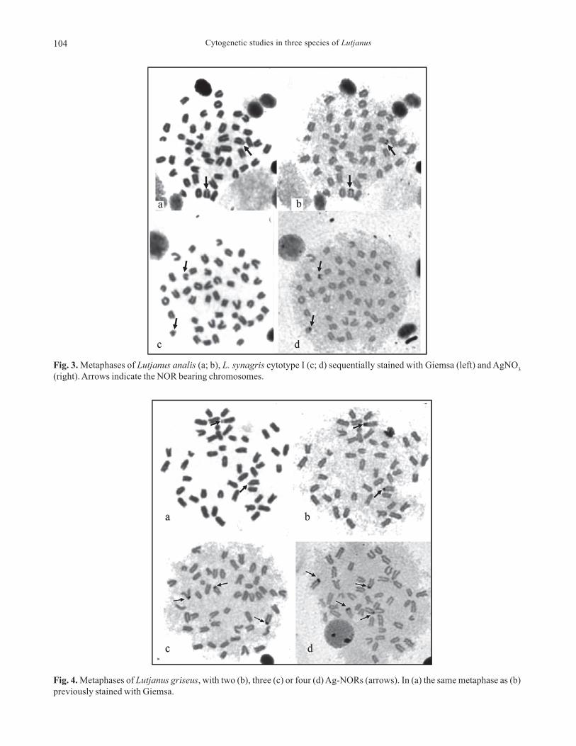
In the present study, three species of Lutjaninae, Lutjanus analis, L. griseus and L. synagris, were analyzed by conventionalGiemsa staining, C-banding and silver staining, to reveal active Nucleolus Organizer Regions (NORs). Fluorescent in situhybridization (FISH) was also applied to establish the number and location of the ribosomal gene clusters (18S and 5S rRNAgenes). Counts of diploid metaphasic cells revealed a diploid modal chromosome complement composed of 48 acrocentricchromosomes in both L. analis and L. griseus. Two cytotypes were observed in L. synagris: cytotype I, with 2n=48 acrocentricchromosomes, found in 19 specimens, and cytotype II, with 46 acrocentric chromosomes and one large metacentric, found intwo specimens. The large metacentric, which possibly originated from a Robertsonian rearrangement, was not found to be sex-related. In the three species, constitutive heterochromatin is located in the centromeres of all chromosomes. NORs weredetected on the short arms of a single chromosome pair, number 24 in L. analis and number 6 in both cytotypes of L. synagris.In L. griseus, a polymorphism of the NORs number was detected, by both Ag-staining and FISH, as females show a maximumof three NORs, and males a maximum of six NORs. In all species, minor ribosomal genes were found located on a singlechromosome pair. The obtained data, along with those previously reported for other five Lutjanidae species, show that ageneral chromosome homogeneity occurs within the family, but that derived karyotypes based on Robertsonian rearrange-ments as well as multiple and variable NORs sites can also be found.
No presente estudo três espécies de Lutjaninae, Lutjanus analis, L. griseus e L. synagris foram analisadas através da coloraçãoconvencional com Giemsa, banda C e coloração com nitrato de prata para identificar as Regiões Organizadoras de Nucléolo(NORs) ativas. Hibridação fluorescente in situ (FISH) foi também aplicada para estabelecimento do número e localização dosagrupamentos de genes ribossômicos (18S e 5S rRNA). A contagem de células metafásicas revelou um número diplóide modalde 48 cromossomos acrocêntricos em L. analis e L. griseus. Dois citótipos foram observados em L. synagris: citótipo I com2n=48 cromossomos acrocêntricos, encontrado em 19 espécimes, e citótipo II com 46 cromossomos acrocêntricos e um grandemetacêntrico, encontrado em dois espécimes. O grande metacêntrico, que possivelmente se originou por um rearranjoRobertsoniano, não está relacionado com o sexo. Nas três espécies a heterocromatina constitutiva está localizada nas regiõescentroméricas de todos os cromossomos. NORs foram detectadas no braço curto de um único par cromossômico, número 24em L. analis e número 6 em ambos os citótipos de L. synagris. Em L. griseus, um polimorfismo de número de NORs foiobservado, após coloração com prata e por FISH, as fêmeas apresentaram um máximo de três NORs e os machos um máximo deseis NORs. Em todas as espécies os genes ribossômicos 5S foram encontrados em um único par cromossômico. Os dadosobtidos, somados aos demais previamente publicados para cinco outras espécies de Lutjanidae, mostram que na família há umahomogeneidade cromossômica, porém também são encontrados cariótipos derivados, originados por rearranjos Robertsonianos,assim como pela ocorrência de sítios múltiplos e variados de NORs.
Key words: Karyotype, Ribosomal genes, NOR polymorphism, C-banding, Robertsonian rearrangement.
1Escuela de Ciencias Aplicadas del Mar, Universidad de Oriente, Apartado Postal 147, Porlamar, Venezuela. [email protected] de Morfologia, Instituto de Biociências Universidade Estadual Paulista, 18618-000 Botucatu, São Paulo, Brazil.3Instituto Oceanográfico de Venezuela, Universidad de Oriente, Cumaná, Venezuela.4Department of Human and Animal Biology, University of Rome “La Sapienza”, via Borelli 50, 00161 Rome, Italy.
Cytogenetic studies in three species of Lutjanus102
Introduction
The Lutjanidae (snappers) is a group composed of 17 gen-
era and 105 species of mostly reef-associated marine fishes,
which are distributed in all the tropical and subtropical seas
of the world (Nelson, 2006). The family is divided in four
subfamilies. Three smaller subfamilies include the
Paradichthyinae, with two monotypic genera (Symphorus and
Symphorichthys), the Etelinae, with five genera (Aphareus,
Aprion, Etelis, Pristipomoides and Rhandallichthys) and 19
species, and the Apsilinae, with four genera (Apsilus,
Lipocheilus, Paracesio and Parapristipomoides) and 12 spe-
cies (Nelson, 2006). The subfamily Lutjaninae is the largest,
with three monotypic genera (Hoplopagrus, Ocyurus and
Rhomboplites), the genera Macolor and Pinjalo with two
species each, and the genus Lutjanus, which is the most
speciose, with 64 species. In Venezuela, Cervigón (1993) rec-
ognizes six genera of Lutjanidae (Etelis, Pristipomoides,
Apsilus, Ocyurus, Rhomboplites and Lutjanus ) including 15
species, 10 of which belong to the genus Lutjanus (L. analis,
L. apodus, L. aya, L. bucanella, L. cyanopterus, L. griseus, L.
jocu, L. mahogoni, L. purpureus, L. synagris and L. vivanus).
In spite of their high number and their ecological and eco-
nomic importance, cytogenetic studies on Lutjanidae are
scarce. In fact, among the 105 recognized species of
Lutjanidae, barely five species have been karyotyped to date:
Lutjanus argentimaculatus (Raghunath & Prasad, 1980), L.
kasmira (Choudhury et al., 1979; Ueno & Takai, 2008), L.
sanguineus (Rishi, 1973), L. russelli (Ueno & Ojima 1992),
and L. quinquelineatus (Ueno & Takai, 2008). For most of
them, only the chromosome number and morphology have
been reported and there is no data regarding the chromo-
somal distribution and composition of the constitutive het-
erochromatin or numbers and locations of the major and mi-
nor ribosomal genes, which have proved to be useful markers
in the investigation of the phylogenetic relationships among
fish species within a family (Sola et al., 2007).
In the present study, three species of Lutjaninae, Lutjanus
analis, L. griseus and L. synagris were analyzed by conven-
tional Giemsa staining and C-banding, and by Fluorescent in
situ hybridization with 18S rDNA and 5S rDNA, in order to
obtain a fine karyotype characterization, and, thus, chromo-
some markers which can provide useful information concern-
ing relationships within the family.
Materials and Methods
Eight sexually immature (unsexed) specimens of L. analis,
seven specimens of L. griseus (3 males, 3 females, 1 unsexed)
and 21 specimens of L. synagris (9 males, 10 females, 2
unsexed) were captured with a fishing trap in the locality of
Guayacancito, on Margarita Island, Venezuela. Voucher speci-
mens (Table 1) were deposited at the Ichthyology Collection
of the Escuela de Ciencias Aplicadas del Mar (ECAM),
Universidad de Oriente.
Twenty four hours before chromosome preparations, the
fishes were injected intramuscularly with a yeast glucose
solution (Lee & Elder, 1980) for mitosis stimulation. Chromo-
somes were obtained from kidney cells according to Foresti
et al. (1993). C-bands were obtained according to the method
described by Sumner (1972), modified by testing different
time of exposition to barium hydroxide, from 1 to 180 sec-
onds, in order to enhance the contrast of constitutive hetero-
chromatin on chromosomes. For detection of the active
Nucleolus Organizer Regions (NORs), slides were stained with
silver nitrate using the method of Howell & Black (1980).
The 5S and 18S rDNA sites were identified by FISH ac-
cording to the method of Pinkel et al. (1986). A sequence of
1800 base pairs of the 18S rRNA gene of Oreochromis
niloticus (Nile tilapia), cloned in pGEM-T plasmid, was used
as a probe to localize sites for 45S rDNA. PCR products con-
taining 5S rDNA repeats from each species were used as
probes for the chromosome mapping of 5S rDNA. DNA was
extracted from muscle (Sambrook & Russel, 2001) and the 5S
rDNA repeats were generated by Polymerase Chain Reaction
(PCR) with the primers 5SA (5’TAC GCC CGA TCT CGT CCG
ATC3’) and 5SB (5’CAG GCT GGT ATG GCC GTA AGC3’)
according to Martins & Galetti (1999).
The 18S rDNA and 5S rDNA probes were labeled by nick
translation with biotin-14-dATP, following the manufacturer’s
(BionickTM Labelling System-Gibco.BRL) instructions. Signals
were detected and amplified by a three-round application of
Avidin-FITC/biotinilated Anti-avidin. Chromosomes were
counter-stained with Propidium Iodide (50µg/ml) diluted in
Antifade.
The mitotic figures were photographed using a Motic B400
microscope equipped with a Moticam 5000C digital camera.
The fundamental number (NF) of arms was determined con-
sidering acrocentrics (A) as having one chromosome arm and
metacentrics (M) as having two chromosome arms. FISH
metaphases were photographed with a Olympus BX61 pho-
tomicroscope equipped with a DP70 digital camera.
Results
The counts of diploid metaphasic cells (Table 1) revealed
a modal chromosome complement composed of 2n=48 acro-
centric chromosomes (NF=48) in both L. analis and L. griseus
and in 19 out of the 21 examined specimens of L. synagris
(cytotype I). The two remaining specimens, one male and one
unsexed, of L. synagris show a modal count of 2n=47 (NF=48),
made up of one large metacentric and 46 acrocentric chromo-
somes. This karyomorph was named cytotype II. The karyo-
types obtained by arranging the chromosomes in order of
decreasing size are shown in Fig. 1. The negligible differ-
ences in chromosome sizes make it impossible to identify
homologous pairs with any certainty, with the exception of a
chromosome pair, classified as number 6, in L. analis (Fig.
1a), which shows a secondary constriction when chromo-
somes are elongated, and chromosome pair number 24 in all
the three species, clearly the smallest of the chromosome
complements.
M. Nirchio, R. Rondón, C. Oliveira, I. A. Ferreira, C. Martins, J. Pérez, L. Sola & A. R. Rossi 103
In all the three Lutjanus species examined, C-banding (Fig.
2) revealed that the heterochromatin is restricted to the cen-
tromeres of all chromosomes, including the large metacentric
in cytotype II of L. synagris, though some of them show
weaker signals.
The analysis of the nucleolus organizer regions with the
Ag-NOR staining technique detected a maximum of two Ag-
positive paracentromeric signals in L. analis (Fig. 3b) and in
both cytotypes of L. synagris (Fig. 3d for cytotype I, data not
shown for cytotype II). In L. analis the Ag-signals (Fig. 3b)
are located on the secondary constriction, often evident in
Giemsa-stained metaphases (Fig. 3a), of chromosome pair
number 6. In L. synagris the Ag-signals (Fig. 3d) are located
on the smallest chromosome pair number 24 of the comple-
ment. In L. griseus, a variable number of paracentromeric Ag-
positive signals per metaphase was observed, up to two in
females (Fig. 4a, b) and up to four in males (Fig. 4c, d). By its
morphology and size and for the presence of a secondary
constriction (Fig. 4a, b), one of the NOR-bearing chromo-
some pair in this is likely to be homoeologous to the chromo-
Fig. 1. Giemsa-stained karyotypes of Lutjanus analis (a), L. griseus (b), L. synagris cytotype I (c) and L. synagris cytotype II (d).
Fig. 2. C-banded metaphases of Lutjanus analis (a), L. griseus (b), L. synagris cytotype I (c) and L. synagris cytotype II (d).
Cytogenetic studies in three species of Lutjanus104
Fig. 3. Metaphases of Lutjanus analis (a; b), L. synagris cytotype I (c; d) sequentially stained with Giemsa (left) and AgNO3
(right). Arrows indicate the NOR bearing chromosomes.
Fig. 4. Metaphases of Lutjanus griseus, with two (b), three (c) or four (d) Ag-NORs (arrows). In (a) the same metaphase as (b)
previously stained with Giemsa.
M. Nirchio, R. Rondón, C. Oliveira, I. A. Ferreira, C. Martins, J. Pérez, L. Sola & A. R. Rossi 105
some pair number 6 identified in L. analis (Fig. 3a, b). The
remaining signals are located on medium-sized chromosomes.
After FISH with 18S rDNA, in L. analis and in L. synagris
two fluorescence hybridization signals were seen at the same
chromosome locations detected with Ag-staining, i.e., in
paracentromeric positions of chromosome pair number 6 in L.
analis (Fig. 5a) and of chromosome pair number 24 in both
cytotypes of L. synagris (Fig. 5c, e), confirming that in these
species a single pair of NOR-bearing chromosomes is present.
In L. griseus (Fig. 6) hybridization with 18S rDNA confirms
the NORs sites detected by Ag-staining, but also reveals
further positive sites, so that up to three NORs were ob-
served in females (Fig. 6a) and up to six in males (Fig. 6b). All
FISH positive chromosomes are medium-sized, thus major ri-
bosomal gene clusters location on chromosome pair 24 can
be excluded.
After FISH with 5S rDNA, in all species - L. analis (Fig.
5b), both cytotypes of L. synagris (Fig. 5d, f) and L. griseus
(Fig. 6c) - one cluster of 5S rRNA genes was found close to
the centromere of a medium-sized acrocentric chromosome
pair, which is apparently the same in the three species.
Discussion
The cytogenetic features here reported for the examined
specimens of L. analis, L. griseus and L. synagris revealed
that the three species have the 48-acrocentric karyotype which
is shared by most of the Lutjaninae species previously ana-
lyzed, such as Lutjanus argentimaculatus (Raghunath &
Prasad, 1980), L. kasmira (Choudhury et al., 1979; Ueno &
Takai, 2008), L. sanguineus (Rishi, 1973) and L. russelli (Ueno
& Ojima, 1992). The only exception is L. quinquelineatus
which has been reported to possess 2n=48 A in females and
2n = 47 (1M+46A) in males (Ueno & Takai, 2008).
In fish, Robertsonian rearrangements, which, by fusions
(or fissions) of two uniarmed chromosomes into one biarmed
chromosome (or vice-versa), cause changes in chromosome
number, but leave the fundamental number of arms unchanged,
are infrequent. However, examples of Robertsonian polymor-
phism in marine fishes have been reported for species of
Gobiidae (Gobius paganellus, Giles et al., 1985; Neogobius
eurycephalus, Ene, 2003), Sparidae (Diplodus annularis,
Vitturi et al., 1996), Pomacentridae (Chromis insolata and C.
flavicauda, Molina & Galetti, 2002), Cottidae (Myoxocephalus
scorpius, Yershov, 2005), Gadidae and Pleuronectidae (Ga-
dus morhua and Pleuronectes platessa, Fana & Fox, 1991). In
this context, the presence of cytotype II, with a large un-
paired metacentric chromosome, found in one male and one
unsexed specimen of L. synagris, is quite interesting. In fact,
when chromosome differences are restricted to one sex, the
presence of sex chromosomes is strongly suggested (Devlin
& Nagahama, 2002). As a matter of fact, the presence of 2n=48
acrocentric chromosomes in females and 2n=47 (1 M+46A) in
males of L. quinquelineatus was interpreted by Ueno & Takai
(2008) as the result of a single Robertsonian fusion which
formed a neo-Y chromosome, establishing a multiple sex chro-
mosome system of X1X
1X
2X
2/X
1X
2Y type. In the case reported
here for L. synagris, the reduced 2n=47 cytotype II character-
izes only two (one male, one immature) of the 21 specimens
analyzed, but, among these latter, the remaining eight males
present the same 48 acrocentric chromosome cytotype I
shown by all ten females and one immature specimens. Thus,
the two cytotypes co-exist in different males and the pres-
ence of the large unpaired metacentric chromosome does not
seem to be associated to sex determination. Due to the fre-
quency of cytotype II, approximately 9.5%, this rearrange-
ment suggests the existence of a chromosomal polymorphism
in the studied population of L. synagris. Nevertheless, the
possibility that the observed fusion in L. synagris might origi-
nate a neo-Y chromosome and that an incipient stage of mul-
tiple sex chromosome differentiation, as the one reported in
L. quinquelineatus (Ueno & Takai, 2008), has been identi-
fied, can not be discarded.
In all the three Lutjanus species examined, the C-positive
heterochromatin distribution is restricted to the centromeres
Frequency Species (n)
Voucher number
Sex Number of scored cells 46 47 48 49
ECAM-365 ? 45 8.9 11.1 73.3 6.7
ECAM-366 ? 26 3.8 11.5 76.9 7.7
ECAM-422 ? 24 0.0 8.3 87.5 4.2
ECAM-425 ? 34 0.0 5.9 85.3 8.8 ECAM-426 ? 28 7.1 3.6 85.7 3.6
ECAM-427 ? 40 2.5 7.5 85.0 5.0
ECAM-405 ? 38 0.0 7.9 81.6 10.5
L. analis
(8)
ECAM-428 ? 15 0.0 13.3 80.0 6.7
Subtotal 250 2.8 8.6 81.9 6.7
ECAM-399 F 67 4.5 7.5 82.1 6.0
ECAM-408 ? 23 4.3 8.7 82.6 4.3 ECAM-410 M 11 0.0 18.2 81.8 0.0
ECAM-409 F 30 0.0 13.3 76.7 10.0
ECAM-002 F 36 5.6 11.1 80.6 2.8 ECAM-413 M 26 3.8 15.4 80.8 0.0
L. griseus (7)
ECAM-429 M 76 3.9 10.5 78.9 6.6
Subtotal 269 3.2 12.1 80.5 4.2
ECAM-364 ? 70 4.3 4.3 85.7 5.7
ECAM-367 ? 56 5.4 83.9 7.1 3.6
ECAM-423 M 16 6.3 12.5 75.0 6.3
ECAM-424 F 59 3.4 10.2 81.4 5.1 ECAM-430 F 49 6.1 10.2 77.6 6.1
ECAM-438 F 83 3.6 4.8 83.1 8.4
ECAM-439 F 26 0.0 3.8 88.5 7.7 ECAM-451 M 47 8.5 76.6 10.6 4.3
ECAM-452 F 64 3.1 6.3 84.4 6.3 ECAM-453 F 33 3.0 9.1 84.8 3.0
ECAM-454 F 25 16.0 8.0 72.0 4.0
ECAM-455 M 50 6.0 14.0 74.0 6.0 ECAM-456 F 38 5.3 7.9 81.6 5.3
ECAM-457 F 51 5.9 7.8 78.4 7.8
ECAM-458 M 18 5.6 5.6 77.8 11.1 ECAM-459 M 21 0.0 9.5 85.7 4.8
ECAM-460 M 13 0.0 7.7 92.3 0.0
ECAM-461 M 40 0.0 10.0 87.5 2.5 ECAM-462 F 37 5.4 8.1 86.5 0.0
ECAM-463 M 43 4.7 4.7 83.7 7.0
L.
synagris
(21)
ECAM-464 M 62 1.6 8.1 87.1 3.2
Subtotal 901 4.5 14.9 75.5 5.1
TOTAL 1440 3.5 11.9 79.2 5.45
Table 1. Number of scored cell and percentages of different
diploid counts in specimens of the three species of Lutjanus
examined. Undetermined sex (?), male (M), and female (F).
Cytogenetic studies in three species of Lutjanus106
Fig. 5. Metaphases of Lutjanus analis (a, b), L. synagris cytotype I (c, d) and L. synagris cytotype II (e, f) after FISH with 18S
rDNA (left) and with 5S rDNA (right). Arrows indicate the NOR bearing chromosomes. Asterisks indicate the 5S rDNA bearing
chromosomes.
M. Nirchio, R. Rondón, C. Oliveira, I. A. Ferreira, C. Martins, J. Pérez, L. Sola & A. R. Rossi 107
of all chromosomes, a pattern similar to the one reported for
the Pacific L. kasmira and L. quinquelineatus (Ueno & Takai,
2008), the only two, among the five karyologically investi-
gated Lutjaninae species, for which data are available. Al-
though the heterochromatin distribution does not identify
species-specific chromosome markers, it is worth noting that
considerably different exposition times to BaOH, during the
C-banding procedure, were applied, from 4 seconds in L.
analis, to 30 seconds in L. synagris and up to 180 seconds in
L. griseus, a fact which might reflect differences in chromatin
condensation levels and/or composition among the species
of Lutjaninae here studied.
As far as NORs are concerned, different aspects can be
considered. The first one is related to the methodology. In fish,
NORs are usually indirectly visualized by silver staining (Nirchio
& Oliveira, 2006), which localizes the transcribing major ribo-
somal genes because silver precipitates in correspondence to
a rRNA protein complex synthesized only when NORs are ac-
tive in the preceding interphase (Hubbel 1985, Sánchez-Pina et
al. 1984, Jimenez et al. 1988). However, recent evidence in some
fish species (Gromicho et al., 2005) have cast doubt on the
accuracy and reliability of Ag in NORs detection, both be-
cause other regions, besides NORs, can be stained with Ag or
because most of the 28S rDNA sites, as revealed by FISH, were
not detected. In the studied species of Lutjaninae, all the Ag-
positive sites were also FISH-positive, so the technique is NOR-
specific, but FISH revealed the presence of additional and in-
active NORs in only one of them, L. griseus, confirming that
this species has a higher NOR variability, both in their number
and location, compared to the other congeneric species. As far
as this variability is concerned, though a sex-associated trend
is identifiable, as males show a higher number of structural
NORs, compared to females, a larger sample is needed to verify
the extent and the basis of this variability.
A further aspect is related to the cytotaxonomic consider-
ations. A single pair of NOR-bearing chromosomes has been
suggested to represent the primitive karyotypical condition in
most vertebrate species (Hsu et al., 1975; Schmidt, 1978). In
most of the teleost fish displaying the primitive karyotype with
48 acrocentric chromosomes, two terminal NORs near the cen-
tromere can be found (Vitturi et al., 1995). Therefore, on one
hand, a single NOR bearing chromosome pair, with ribosomal
sites interstitially located, as observed in L. analis and L.
synagris (present study) and in L. kasmira and L.
quinquelineatus (Ueno & Takai, 2008), could be considered a
plesiomorphic condition for Lutjanus, whereas the presence of
multiple NOR bearing chromosomes, found in L. griseus, would
be an apomorphic feature. These data would indicate that L.
analis and L. synagris are more closely related, and that the
occurrence of duplication and translocation of ribosomal genes
in L. griseus might reflect an important mechanism involved in
the diversification of this taxon. On the other, when consider-
ing NORs location, a different perspective highlights. Indeed,
the single NOR-bearing chromosome pair observed in L. analis
(chromosome pair 6), as well as one of the NOR-bearing chro-
mosome pairs observed in L. griseus, when pursuing a parsi-
monious criterion, appears to be homeologous to the one iden-
tified both in L. kasmira and L. quinquelineatus (Ueno & Takai,
2008), and, thus, it could be regarded as the primitive condition
in the genus. As a consequence, no homeology could be iden-
tified with the NOR-bearing chromosome pair of L. synagris.
Similar ambiguous results were obtained from molecular and
morphological data. Indeed, in a phylogenetic study of
Lutjanidae, based on isozyme and morphological data, Chow
& Walsh (1992) suggested that L. analis and L. synagris form
a clade, which is a sister group of L. griseus. However, subse-
quent molecular phylogenetic studies, based on mitochondrial
DNA (Sarver et al., 1996) have not provided a similar clear-cut
subdivision.
FISH experiment with 5S rDNA, revealed their location on
a chromosome which is clearly distinct from the one (number
24) bearing major rDNA genes in L. synagris, but, though the
difference in size is smaller, also from the NOR bearing chro-
mosome pair number 6 of L. analis and L. griseus. The 5S
rRNA gene sites are usually located in separate areas from
45S rRNA genes of the genome in higher eukaryotes (Mar-
tins & Galetti, 1999). This is probably because a separate
location may permit them to independently evolve, since the
divergent functional dynamics of these sequences require
physical distancing (Martins & Galetti, 2000).
Fig. 6. Metaphases of Lutjanus griseus after FISH with 18S rDNA (arrows) in a female (a) and in a male (b) and (c) with 5S rDNA
(asterisks).
Cytogenetic studies in three species of Lutjanus108
In conclusion, data here obtained for Lutjanus analis, L.
griseus and L. synagris, along with those previously reported
for L. kasmira and L. quinquelineatus (Ueno & Takai, 2008),
show that a general chromosome homogeneity occurs within
the family, but that derived karyotypes based on Robertsonian
rearrangements as well as multiple and variable NORs sites
can also be found. Phylogenetic relationships among spe-
cies remained unresolved and further data on the other spe-
cies of Lutjanus are needed to obtain a more general picture
of the karyoevolutive trends in the family.
Acknowledgements
Financial support was provided by Consejo de
Investigación, Universidad de Oriente, Venezuela, by the Ital-
ian Ministry of Instruction, University and Research (MIUR),
and by Fundação de Amparo a Pesquisa do Estado de São
Paulo (FAPESP) Brazil and Conselho Nacional de Desenvolvi-
mento Científico e Tecnológico (CNPq) Brazil.
Literature Cited
Cervigón, F. 1993. Los Peces Marinos de Venezuela. Volumen II.
Fundación Científica Los Roques. Caracas - Venezuela. 499 p.
Choudhury, R. C., R. Prasad & C. C. Das. 1979. Chromosomes of
six species of marine fishes. Caryologia, 32: 15-21.
Chow, S. & P. J. Walsh. 1992. Biochemical and morphometric
analyses for phylogenetic relationships between seven snapper
species (Subfamily: Lutjaninae) of the western Atlantic. Bulletin
of Marine Science, 50: 508-519.
Devlin, R. H. & Y. Nagahama. 2002. Sex determination and sex
differentiation in fish: an overview of genetic, physiological,
and environmental influences. Aquaculture, 208: 191-364.
Ene, A. C. 2003. Chromosomal polymorphism in the goby Neogobius
eurycephalus (Perciformes: Gobiidae). Marine Biology, 142:
583-588.
Fana, Z. & D. P. Fox. 1991. Robertsonian polymorphism in plaice,
Pleuronectes platessa L., and cod, Gadus morhua L., (Pisces
Pleuronectiformes and Gadiformes). Journal of Fish Biology,
38: 635-640.
Foresti F, C. Oliveira & L. F. Almeida-Toledo. 1993. A method for
chromosome preparations from large specimens of fishes using in
vitro short treatment with colchicine. Experientia, 49: 810-813.
Giles, V., G. Thode & C. Alvarez. 1985. A new Robertsonian fusion
in the multiple chromosome polymorphism of a mediterranean
population of Gobius paganellus (Gobiidae, Perciformes).
Heredity, 55: 255-260.
Gromicho, M., C. Ozouf-Costaz & M. J. Collares-Pereira. 2005.
Lack of correspondence between CMA3-, Ag-positive signals
and 28S rDNA loci in two Iberian minnows (Teleostei,
Cyprinidae) evidenced by sequential banding. Cytogenetic and
Genome Research, 109: 507-511.
Howell, W. M. & D. A. Black. 1980. Controlled silver staining of
nucleolus organizer regions with a protective colloidal developer:
a 1-step method. Experientia, 3: 1014-1015.
Hubbel, H. R. 1985. Silver staining as an indicator of active ribosomal
genes. Stain Technology, 60: 285-294.
Hsu, T. C., S, Patak & T. R. Chen. 1975. The possibility of latent
centromeres and a proposed nomenclature system for total
chromosome and whole arm translocations. Cytogenetics and
Cell Genetics, 15: 41-49.
Jiménez, R., M. Burgos & R. Diaz de La Guardia. 1988. A study of
the silver staining significance in mitotic NORs. Heredity, 60:
125-127.
Lee, M. R. & F. F. B. Elder. 1980. Yeast stimulation of bone marrow
mitosis for cytogenetic investigations. Cytogenetics and Cell
Genetics, 26: 36-40.
Martins, C. & P. M. Galetti Jr. 1999. Chromosomal localization of
5S rDNA genes in Leporinus fish (Anostomidae, Characifor-
mes). Chromosome Research, 7: 363-367.
Martins, C. & P.M. Galetti Jr. 2000. Conservative distribution of
5S rDNA loci in Schizodon (Pisces, Anostomidae)
chromosomes. Chromosome Research, 8: 353-355.
Molina, W. F. & P. M. Galetti Jr. 2002. Robertsonian rearrangements
in the reef fish Chromis (Perciformes, Pomacentridae) involving
chromosomes bearing 5S rRNA genes. Genetics and Molecular
Biology, 25: 373-377.
Nelson, J. S. 2006. Fishes of the World. 4th eds. New York: John
Wiley and Sons, Inc. 601 p.
Nirchio, M. & C. Oliveira. 2006. Citogenética de Peces/Editado por
Universidad de Oriente, Cumaná, Venezuela, 216 p.
Pinkel D., T. Straume & J. W. Gray. 1986. Cytogenetic analysis using
quantitative, high-sensitivity, fluorescence hybridization.
Proceedings of the National Academy of Sciences, 83: 2934-2938.
Raghunath, P. & R. Prasad. 1980. Chromosomes of six marine
percoids from the Indian Sea. Indian Biologist, 11: 9-12.
Rishi, K. K. 1973. A preliminary report on the karyotypes of
eighteen marine fishes. Research Bulletin of the Panjab
University, 24:161-162
Sambrook, J. & D. W. Russell. 2001. Molecular cloning: a laboratory
manual. Cold Spring Harbor Laboratory Press. Cold Spring
Harbor, New York. 2344 pp.
Sánchez-Pina, M. A., F. J. Medina, M. M. Fernández-Gómez &
M. C. Risueno. 1984. Ag-NOR proteins are present when
transcription is impaired. Biology of the Cell, 5: 199-202.
Schmidt, M. 1978. Chromosome banding in Amphibia. II. Constitutive
heterochromatin and nucleolus organizer regions in Ranidae,
Microhylidae and Racophoridae. Chromosoma, 68: 131-148.
Sola, L., E. Gornung, M. E. Mannarelli & A. R Rossi. 2007.
Chromosomal evolution in Mugilidae, Mugilomorpha: an
overview, Pp. 165-194. In: Pisano, E., C. Ozouf-Costaz, F.
Foresti & B. G. Kapoor (Eds.). Fish Cytogenetics. Science
Publishers, Enfield, NH, USA. 502 p.
Sumner, A. T. 1972. A simple technique for demonstrating centromeric
heterocromatin. Experimental Cell Research, 75: 304-306.
Ueno, K. & Y. Ojima. 1992. Notes on the chromosomes of Girella
melanichthys and Lutjanus russelli (Pisces, Perciformes).
Chromosome Information Service, 52: 3–5.
Ueno, K & A. Takai. 2008. Multiple sex chromosome system of
X1X
1X
2X
2/X
1X
2Y type in lutjanid fish, Lutjanus
quinquelineatus (Perciformes). Genetica, 132: 35-41.
Vitturi, R., A. Libertini, A. Mazzola, M. S. Colomba & G. Sara. 1996.
Characterization of mitotic chromosomes of four species of the
genus Diplodus: karyotypes and chromosomal nucleolar organizer
region phenotypes. Journal of Fish Biology, 49: 1128-1137.
Yershov, P. N. 2005. Chromosomal studies of Zoarces viviparus L.
(Zoarcidae) and Myoxocephalus scorpius L. (Cottidae) from
different parts of distribution area. Ecohydrology and
Hydrobiology, 5: 237-243.
Accepted December 2007
Published 31 March 2008