Cytokine Factors Present in Dengue Patient Sera Induces Alterations of Junctional
Proteins in Human Endothelial Cells
Ramapraba Appanna, Seok Mui Wang, Sasheela A. Ponnampalavanar, Lucy Chai See Lum, and Shamala Devi Sekaran*Department of Medical Microbiology, Department of Medicine, and Department of Pediatrics, Faculty of Medicine,University of Malaya, Kuala Lumpur, Malaysia; Institute of Medical Molecular Biotechnology, Faculty of Medicine,
Universiti Teknologi MARA, Selangor, Malaysia
Abstract. Plasma leakage in severe dengue has been postulated to be associated with skewed cytokine immuneresponses. In this study, the association of cytokines with vascular permeability in dengue patients was investigated.Human serum samples collected from 48 persons (13 with dengue fever, 29 with dengue hemorrhagic fever, and6 healthy) were subjected to cytokines analysis by using Luminex Multiplex Technology. Selected serum samples frompatients with dengue hemorrhagic fever sera and recombinant human cytokines were then tested for roles on inducingvascular permeability by treatment of human umbilical vein endothelial cells. Confocal immunofluorescence stainingindicated morphologic alteration of human umbilical vein endothelial cells treated with serum samples from patientswith dengue hemorrhagic fever compared with serum samples from healthy persons. The findings suggest that cytokinesproduced during dengue hemorrhagic infections could induce alterations in the vascular endothelium, which may play afundamental role in the pathophysiology of dengue.
INTRODUCTION
Dengue is the most extensive vector borne infectious dis-ease and a major source of public health concern in tropicaland sub-tropical regions of the world. The Pediatric DengueVaccine Initiative has estimated that globally there are some3.61 billion persons (55% of the world’s population) are atrisk for dengue, and there are approximately 36 million casesof dengue fever (DF) and 2.1 million severe dengue infectionsevery year.1 In Southeast Asia, by the late 1990s, dengue hadbecome the most important mosquito-borne disease affectinghumans aftermalaria, and therewere approximately 40millioncases of DF and several hundred thousand cases of denguehemorrhagic fever (DHF) each year.2,3 Because this diseaseaffects persons of all ages, it has inevitably contributed to asubstantial economic burden to society.4
Dengue virus comprises four genetically distinct serotypes:dengue virus serotype 1, 2, 3, and 4 (DENV-1–DENV-4).Infection by any one of the dengue serotypes can cause a spec-trum of illnesses ranging from clinically silent infection, mildfebrile illness, and DF, to life-threatening DHF (grades I andII) or dengue shock syndrome (DSS) (grades III and IV).5,6
Increased vascular permeability, which leads to plasmaleakage, has been demonstrated as the fundamental featurein DHF that implies damage to the vascular endothelium andinduces major clinical complications; it is the critical stage ofthe disease that can cause hypovolemic shock (DSS).5 Plasmaleakage in DHF generally lasts no more than 48 hours andis usually follow by rapid complete recovery.7 Consistent withthe clinical course, plasma leakage inDHFoccurswith a relativelack of tissue inflammation, suggesting that a transient change infactors that regulate vascular permeability in the physiologicstate may be themechanism of plasma leakage in the disease.8
The pathogenesis of dengue is not fully understood, eventhough many studies have been conducted on its pathogenesisin the past few decades. This lack of understanding is causedmainly by the lack of an appropriate animal model that can
precisely simulate dengue virus infections in humans. Recently,human umbilical vein endothelial cells (HUVEC) were identi-fied as the central model used to understand infection and path-ologic events during severe dengue.8 Although increased levelsof cytokines, such as interferons (IFNs), interleukin-2 (IL-2),IL-8, tumor necrosis factor-a (TNFa), and vascular endothelialgrowth factor A (VEGF-A), have been reported to be associ-ated with enhancement of vascular permeability, the relativerole of these cytokines in plasma leakage is not known.9,10
In this study, we investigated the effect of cytokines in den-gue patient serum by using an in vitromodel withHUVEC.Wealso performed confocal immunofluorescence analysis on themorphologic changes of HUVEC treated with serum samplesfromdengue patients and compared themwith those of healthydonors. Our data suggest that cytokines produced during den-gue hemorrhagic infections participate in the regulation ofvascular permeability.
MATERIALS AND METHODS
Study participants. Forty-two persons (13 with DF and29 with DHF) who were admitted to University Malaya Med-ical Center, Kuala Lumpur, Malaysia, for acute dengue infec-tion, were recruited in this preliminary study of cytokineprofiling in dengue patients. Blood samples were collectedduring acute, defervescence, and convalescence phases. Lab-oratory tests, including a dengue polymerase chain reaction,virus isolation, hemagglutination, IgM-capture enzyme-linkedimmunosorbent assay, and test for non-structural protein 1,were conducted for confirmation of dengue virus infection.11
Persons were subsequently clinically diagnosed as either havingDF or DHF by clinicians on the basis of World Health Organi-zation criteria.2 Ethical clearance (Ref. no.: 321.4) for the studywas approved by the Scientific and Ethical Committee of Uni-versityMalayaMedical Center, and procedureswere conductedin accordance with the Helsinki Declaration of 1975, as revisedin 2000.12
Detection of cytokines by bead-based enzyme-linkedimmunosorbent assay. Multiple cytokines in patient serumsamples were identified and quantified by using the humancytokine 27-plex panel kit (catalog no. 171-A11127; Bio-Rad,
*Address correspondence to Shamala Devi Sekaran, Department ofMedical Microbiology, Faculty of Medicine, University of Malaya,50603 Kuala Lumpur, Malaysia. E-mail: [email protected]
936
Hercules, CA) and the 2-plex panel kit (catalog no.XF0000ZG2Y) with the Bio-Plex suspension array system(Bio-Rad) in accordance to the manufacturer’s protocol.Briefly, diluted serum samples (1:3 dilutions) were added intoeach of the 96-well filter plates containing multiplex beads.Plates were then placed on a micro-plate shaker and gentlyshaken in the dark for 30 minutes at room temperature.Plates were then washed three times with 100 mL of Bio-Plexwash buffer by vacuum filtration. Twenty-five microliters ofantibodies to various cytokines was added and the plateswere incubated in the dark for 30 minutes with gently shaking.After three washes, 50 mL of phycoerythrin-conjugatedstreptavidin was added and the plates were incubated for10 minutes. Fluorescent signals were read by using a LuminexMachine (Bio-Rad). The analyte concentration was calcu-lated by using software provided by the manufacturer.Raw data was initially measured as the relative fluores-
cence intensity and then converted to cytokine concentrationon the basis of a standard curve generated from referenceconcentrations supplied in the kit. The following cytokineswere measured: IL-1b, IL-1 receptor antagonist (IL-1Ra),IL-2, IL-4, IL-5, IL-6, IL-7, IL-8, IL-9, IL-10, IL-12 (p70),IL-13, IL-15, IL-17, eotaxin, basic fibroblast growth factor(FGF-basic), granulocyte colony-stimulating factor (G-CSF),granulocyte–macrophage colony-stimulating factor (GM-CSF), IFN-g, IFN-g–induced protein 10 (IP-10), monocytechemoattractant protein 1 (MCP-1), macrophage inflamma-tory protein-1a (MIP-1a), MIP-1b, platelet-derived growthfactor-bb (PDGF-bb), regulated-on-activation normal T-cellexpressed and secreted (RANTES), TNF-a, and VEGF in the27-plex assay; and IL-18 and intercellular adhesion molecule 1(ICAM-1) in the 2-plex assay.Cell culture. Human umbilical vein endothelial cells
(American Type Culture Collection, Manassas, VA) were main-tained in endothelial cell–based medium 2 (Lonza, Basel,Switzerland) supplemented with 10% fetal calf serum, andcultured at 37 °C in an atmosphere of 5% CO2. For sub-culturing or experiments, cells were seeded into cultureflasks or chamber slides that were pre-coated with 50 mg/mLof fibronectin (Roche, Mannheim, Germany). Pre-coating wasperformed by incubating flasks with 100 mL/cm2 of 50 mg/mL offibronectin and incubated at room temperature for 45 minutes.The solution was then removed and flasks were used immedi-ately for cell culture.Recombinant human cytokines. Recombinant human (rh)
cytokines (rhIL-1ra, rhIL-9, rhMCP-1, rhRANTES, rhEotaxin,and rhIP-10) (R&D Systems, Minneapolis, MN) were used totest their effects on inducing endothelial cells permeabilitychanges. All of these cytokines except rhIL-9 were selectedon the basis of our findings that their levels were significantlyincreased (P < 0.05) in DHF patients compared with healthycontrols, and on their availability in the laboratory when theassay was performed. Although the level of IL-9 was increased ina DHF patient, there was no significant association (Appanna Rand others, unpublished data).Assay for endothelial cell permeability changes. The assay
was performed to determine alterations in the distributionof two junctional complexes: adherens junction protein vascu-lar endothelial cadherin (VE-cadherin) and endothelial tightjunction protein zonula occludens-1 (ZO-1) after treatmentof HUVEC with dengue patient serum samples and recombi-nant human cytokines. Briefly, endothelial cells were seeded
onto eight-well chamber slides coated with fibronectin. Thenext day, serum samples from dengue patients (diluted inendothelial cell–based medium 2 at a ratio of 1:3) and rh cyto-kines (rhIL-1ra, rhIL-9, rhMCP-1, rhRANTES, rhEotaxin,and rhIP-10) at appropriate working concentrations wereadded and incubated for 3 hours at 37°C in an atmosphereof 5%CO2.Cells were then subjected to immunofluorescence staining
of VE-cadherin and ZO-1. For testing the effects of cytokine-neutralizing antibodies onVE-cadherin andZO-1, anti-humancytokine monoclonal antibodies were pre-incubated withculture medium in the chamber for 3 hours at 37°C beforeaddition of rh cytokines. Working concentrations of rh cyto-kines and their respective cytokine neutralizing monoclonalantibodies (ah) (R&D Systems) were rhIP-10 (200,000 pg/mL),rhRANTES (800,000 pg/mL), rhMCP-1 (3,000 pg/mL), rhIL-1ra(20,000 pg/mL), rhEotaxin (1,500 pg/mL), rhIL-9 (6,000 pg/mL),ahIP-10 (20 mg/mL), ahRANTES (32 mg/mL), ahMCP-1(0.9 mg/mL), ahIL-1ra (12 mg/mL), ahEotaxin (0.75 mg/mL),and ahIL-9 (12 mg/mL).Immunofluorescence staining of VE-cadherin and ZO-1.
The HUVEC were placed in cell chambers, washed threetimes with phosphate-buffered saline (PBS), and fixed with2% paraformaldehyde (Sigma, St. Louis, MO) for 10 minutes.After three washes with PBS, cells were permeabilized with0.1% Triton X-100 (BDH Laboratory Supplies, London,United Kingdom) for five minutes at room temperature. Thecells were then blocked with 2% BSA for 30 minutes atroom temperature before rinsing with PBS. Cells were thenimmunolabeled with purified mouse monoclonal anti-humanZO-1 antibody (BD Biosciences, San Jose, CA) (1:200 dilu-tion of a 250 mg/mL stock) and rabbit monoclonal anti-humanVE-cadherin antibody (Sigma) (1:100 dilution of a 1.0 mg/mLstock) and incubated for one hour at room temperature. Afterthree washes with PBS, Alexa Fluor 488 goat anti-mouse IgG(heavy and light chains) (Invitrogen, Carlsbad, CA) and AlexaFluora 568 goat anti-rabbit IgG (heavy and light chains) withworking dilution of 5 mg/mL were added to the cells and incu-bated for one hour. 4¢,6-diamidino-2-phenylindole (0.05 mg/mL)(Sigma) was added to the chambers for 5 minutes to stainnuclei, and the chambers were washed three times with PBS.Cells were then mounted with fluorescent mounting medium(Dako, Carpinteria, CA), and viewed under a laser confocalmicroscope (TCS SP5; Leica,Wetzlar, Germany).
RESULTS
Forty-two adults with confirmed dengue (13 with DF and29 with DHF) and 6 healthy controls were selected for pre-liminary study of their cytokine profiles at different daysof fever onset. All patients were given a clinical diagnosisof DF or DHF by a clinician on the basis of World HealthOrganization criteria.2 The mean age of the patients was29.3 years (range = 14–67 years), and there were 22 malesand 20 females. Detailed clinical and laboratory data of alldengue patients were summarized as supplementary data.Blood collection was performed at 2–14 days of illness (meanduration of illness = 6 days). The mean of the defervescencephase was day 5 of illness (range = 3–7 days). The highest(70.45%) number of patients was in defervescence on days4–6 of illness. IgM was detected in 39 patients. Dengue viruswas detected in 32 patients (13 with DENV-1, 4 with DENV-2,
CYTOKINES AND ALTERATION OF HUVEC 937
7 with DENV-3, and 8 with DENV-4), and dengue virus non-structural protein 1 was detected in 27 patients.The profile of 29 different type of cytokine levels was deter-
mined during the acute (2–3 days of illness), defervescence(4–6 days of illness) or convalescent (> 7 days of illness)phases in DF and DHF patients, as shown in Table 1. Pro-inflammatory cytokines IL-18, anti-inflammatory cytokinesIL-1ra, adhesion molecule ICAM-1, and chemokines Eotaxin,IP-10, MCP-1, MIP-1b, and RANTES were significantlyincreased in serum samples from DHF patients (P < 0.05).
However, only IP-10, MCP-1, and MIP-1b were significantlyincreased in serum samples from DF patients. Cytokines thatwere significantly reduced in serum samples from DHFpatients were pro-inflammatory cytokines IFN-g, IL-5, IL-12,anti-inflammatory cytokines IL-4, and growth factors FGF-basic, G-CSF, PDGF, and VEGF. Pro-inflammatory cytokineIL-2, anti-inflammatory cytokine IL-13, and growth factorG-CSF were significantly decreased in DF patients.Effects of cytokines on permeability of monolayers of
HUVECwere accessed by treatment of HUVEC directly with
Table 1
Cytokine levels in serum samples of dengue patients compared with those of healthy donors*
*DF = dengue fever; DHF = dengue hemorrhagic fever; IFN-g = interferon-g; IL = interleukin; ICAM-1 = intercellular adhesion molecule 1; FGF-basic; = basic fibroblast growth factor; G-CSF =granulocyte colony-stimulating factor; PDGF = platelet-derived growth factor; VEGF = vascular endothelial growth factor; IP-10 = interferon-g–induced protein 10; MCP-1 = monocytechemoattractant protein 1; MIP-1b = macrophage inflammatory protein-b; RANTES = regulated-on-activation normal T-cell expressed and secreted.†P < 0.05 (cytokines that showed significant difference between healthy persons and dengue patients).
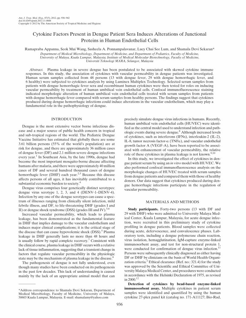
Figure 1. Immunofluorescence analysis of perturbation of tight junction protein ZO-1 (Alexa 488; Invitrogen, Carlsbad, CA) and adherensjunction protein VE-cadherin (Alexa 568) on the outer membrane of human umbilical vein endothelial cells. Cells were treated with A, acute-phase serum sample from patient A with dengue hemorrhagic fever (DHF); B, defervescence serum sample from DHF patient A; C, acute-phaseserum from patient B with dengue fever; D, healthy donor serum; and E, culture medium only. Arrows indicate ZO-1 and VE-cadherindistributions. Assay was done in duplicate and repeated twice.
938 APPANNA AND OTHERS
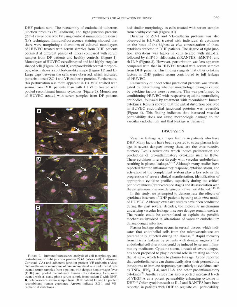
DHF patient sera. The reassembly of endothelial adherensjunction proteins (VE-cadherin) and tight junction proteins(ZO-1) were observed by using confocal immunofluorescence(IF) techniques. Immunofluorescence staining showed thatthere were morphologic alterations of cultured monolayersof HUVEC treated with serum samples from DHF patientsobtained at different phases of illness compared with serumsamples from DF patients and healthy controls. (Figure 1).Monolayers ofHUVECwere disrupted andhad highly irregularshaped cells (Figure 1AandB) comparedwith normalmorphol-ogy, which shows a cobblestone-like shape (Figure 1D and E).Large gaps between the cells were observed, which indicatedperturbations of ZO-1 andVE-cadherin proteins. Furthermore,this perturbation was more apparent in HUVEC treated withserum from DHF patients than with HUVEC treated withpooled recombinant human cytokines (Figure 2). Monolayersof HUVEC treated with serum samples from DF patients
had similar morphology as cells treated with serum samplesfrom healthy controls (Figure 1C).Disarray of ZO-1 and VE-cadherin proteins was also
observed in HUVEC treated with individual rh cytokineson the basis of the highest in vivo concentration of thesecytokines detected in DHF patients. The degree of tight junc-tion alterations was higher in cells treated with rhIL-1ra,followed by rhIP-10, rhEotaxin, rhRANTES, rhMCP-1, andrh-IL-9 (Figure 3). However, perturbation was less apparentcompared with that in HUVEC treated with serum samplesfrom DHF patients. This finding suggests that other cytokinefactors in DHF patient serum contributed to full leakageof HUVEC.Reassembly of endothelial junctional proteins was investi-
gated by determining whether morphologic changes causedby cytokine factors were reversible. This was performed byconditioning HUVEC with respective cytokine-neutralizingantibodies, followed by treatment with recombinant humancytokines. Results showed that the initial distortion observedin HUVEC endothelial junctional proteins was reversible(Figure 4). This finding indicates that increased vascularpermeability does not cause morphologic damage to thevascular endothelium and that leakage is transient.
DISCUSSION
Vascular leakage is a major feature in patients who haveDHF. Many factors have been reported to cause plasma leak-age in severe dengue; among these are the cross-reactivememory T-cells activations, which induce proliferation andproduction of pro-inflammatory cytokines such as IFN-g.These cytokines interact directly with vascular endothelium,resulting in plasma leakage.13,14 Although many studies havereported that the inflammatory response, cytokine storm, andactivation of the complement system play a key role in theprogression of severe clinical manifestation, identification ofappropriate cytokine profiles, especially during the criticalperiod of illness (defervescence stage) and its association withthe progression of severe dengue, is not well established.8,15–18
In this study, we attempted to demonstrate the effects ofcytokines in serum of DHF patients by using an in vitromodelof HUVEC. Although extensive studies have been conductedduring the past several decades, the molecular mechanismsunderlying vascular leakage in severe dengue remain unclear.The results could be extrapolated to explain the possiblemechanism involved in alterations of vascular endotheliumduring dengue infection.Plasma leakage often occurs in serosal tissues, which indi-
cates that endothelial cells from the microvasculature arepreferentially affected during the disease.19 Rapid recoveryfrom plasma leakage by patients with dengue suggests thatendothelial cell alterations could be induced by serum inflam-matory mediators. Cytokine storm, a result of severe dengue,has been proposed to play a central role in creating an endo-thelial sieve, which leads to plasma leakage. Crone reportedthat endothelial cells can dramatically alter their permeabilityin response to immune responses, particularly to cytokines suchas TNFa, IFNg, IL-6, and IL-8, and other pro-inflammatorycytokines.20 Another study has also reported increased levelsof VEGF-A as a potent permeability-enhancing cytokine inDHF.21 Other cytokines such as IL-2 and RANTES have beenreported in patients with DHF to regulate cell permeability,
Figure 2. Immunofluorescence analysis of cell morphology andperturbation of tight junction protein ZO-1 (Alexa 488; Invitrogen,Carlsbad, CA) and adherens junction protein VE-cadherin (Alexa568) on the outer membrane of human umbilical vein endothelial cellstreated serum samples from a patient with dengue hemorrhagic fever(DHF) and pooled recombinant human (rh) cytokines. Cells weretreated with A, acute-phase serum sample from patient C with DHF;B, defervescence serum sample from DHF patient D; and C, pooledrecombinant human cytokines. Arrows indicate ZO-1 and VE-cadherin distributions.
CYTOKINES AND ALTERATION OF HUVEC 939
Figure 3. Immunofluorescent analysis of perturbation of the tight junction protein ZO-1 (Alexa 488; Invitrogen, Carlsbad, CA) and adherensjunction protein VE-cadherin (Alexa 568) on the outer membrane of human umbilical vein endothelial cells treated with individual recombinanthuman (rh) cytokines. A, rhIL-1ra; B, rhIP-10; C, rhEotaxin; D, rhRANTES; E, rhMCP-1; F, rhIL-9; and G, control treated with culture media.IL = interleukin; IP-10 = interferon-g–induced protein 10; RANTES = regulated-on-activation normal T-cell expressed and secreted; MCP-1 =monocyte chemoattractant protein 1. Arrows indicate ZO-1 and VE-cadherin distributions.
940 APPANNA AND OTHERS
although the relative role of these cytokines in plasma leakageis not known.22,23
In this study, analysis of multiple cytokines in serumobtained from DHF patients showed an association betweencytokine profiles and disease severity, and the major cyto-kines implicated are the inflammatory group and chemokines.Our investigations show a probable role of pro-inflammatorycytokines (IL-18, IFN-g, IL-5, and IL-12), anti-inflammatorycytokines (IL-1ra and IL-4), adhesion molecule (ICAM-1),chemokines (Eotaxin, IP-10, MCP-1, MIP-1b, and RANTES),and growth factors (FGF-basic, G-CSF, PDGF, and VEGF)
in DHF patients. The effect of serum from DHF patientson monolayers of HUVEC has further suggested that thesecytokines might play an important role in the pathophysiologyof the vascular disorder. Using selected individual humanrecombinant cytokines, we showed that markedly increaselevels of IL-1ra, IL-8, MCP-1, IP-10, and RANTES wouldhave perturbed organized cells in HUVEC and cause mor-phologic alterations in endothelial cells.One limitation of this study was that only six rh cytokines
were analyzed. Although the degree of permeability changesin monolayers of HUVEC and reversion of permeability were
Figure 4. Immunofluorescent analysis of perturbation of tight junction protein ZO-1 (Alexa 488; Invitrogen, Carlsbad, CA) and adherensjunction protein VE-cadherin (Alexa 568) on the outer membrane of human umbilical vein endothelial cells conditioned medium containingrecombinant human (rh) cytokine treated with respective neutralizing antibody (ab). A, abIL-1ra; B, abIP-10, C, abEotaxin; D, abRANTES;E, abMCP-1; and F, abIL-9. IL = interleukin; IP-10 = interferon-g–induced protein 10; RANTES = regulated-on-activation normal T-cellexpressed and secreted; MCP-1 = monocyte chemoattractant protein 1. Arrows indicate ZO-1 and VE-cadherin distributions.
CYTOKINES AND ALTERATION OF HUVEC 941
not quantified, preliminary findings in this study have demon-strated the effect of cytokines in increasing vascular perme-ability. Observations of highly irregular-shaped cells and largegaps between cells in monolayers of HUVEC treated withserum from DHF patients indicated perturbations of ZO-1and VE-cadherin. Endothelial integrity is maintained mainlyby adherens junction protein, in which the adhesive proteincadherin form predominates, whereas the connector proteinZO-1 is a marker of a tight junction and serves as a linkerbetween the adherens junction and the actin cytoskeleton.19
Observations of altered cells after treatment with serum fromDHF serum treated HUVEC cells comparing to the untreatedcells suggest that remodeling of the actin cytoskeleton,cadherin, and ZO-1 may be the molecular basis that underliesincreased vascular permeability during DHF/DSS. In addi-tion, our results also showed that the effect on vascular alter-ation was transient and reversible. These findings have furthersupported the characteristic feature of DHF, whereby capil-lary permeability changes occur without morphologic damageto capillary endothelium.In summary, we showed that serum samples from DHF
dengue patients induce alterations in the vascular endothe-lium, which may play a fundamental role in the pathophysiol-ogy of the disease. Our results also indicated that alterationswere only transient. These findings suggest that productionof cytokines during dengue significantly influence vascularpermeability. We postulate that analysis of a broad range ofcytokines and their correlation with endothelial damage wouldhelp in understanding dengue immunopathogenesis.
Received September 30, 2011. Accepted for publication May 15, 2012.
Published online September 17, 2012.
Acknowledgments: We thank the clinicians and the nurses at theUniversity Malaya Medical Centre, Kuala Lumpur, Malaysia, forassistance in collecting samples, and the patients for participating inthe study.
Financial support: This study was supported by University of Malayaresearch grants UMRG RG081-09HTM and HIRG J-00000-73560.
Disclosure: None of the authors have any conflicts of interest.
Authors’ addresses: RamaprabaAppanna and ShamalaDevi Sekaran,Department ofMedicalMicrobiology, Faculty ofMedicine,Universityof Malaya, Kuala Lumpur 50603, Malaysia, E-mails: [email protected] and [email protected]. Seok Mui Wang, Institute of MedicalMolecular Biotechnology, Faculty of Medicine, Universiti TeknologiMARA, Selangor, Malaysia, E-mail: [email protected] A. Ponnampalavanar, Department of Medicine, Faculty ofMedicine, University ofMalaya, Kuala Lumpur 50603,Malaysia, E-mail:[email protected]. Lucy Chai See Lum, Department of Pediatrics,Faculty of Medicine, University of Malaya, Kuala Lumpur 50603,Malaysia, E-mail: [email protected].
Reprint requests: Shamala Devi Sekaran, Department of MedicalMicrobiology, Faculty ofMedicine, University ofMalaya, Kuala Lumpur50603, Malaysia, E-mail: [email protected].
REFERENCES
1. International Vaccine Institute, 2008. The Pediatric DengueVaccine Initiative. Vaccines, Children and a Better WorldAnnual Report. Seoul: International Vaccine Institute, 28–35.
2. World Health Organization, 2009.Dengue Guidelines for Diagno-sis, Treatment, Prevention and Control. Geneva: World HealthOrganization, 3–54.
3. Nielsen DG, 2009. The relationship of interacting immunologicalcomponents in dengue pathogenesis. J Virol 6: 211.
4. Singhasivanon P, Jacobson J, 2009. Dengue is a major globalhealth problem. J Clin Virol 46: S1–S2.
5. Halstead SB, 2007. Dengue. Lancet 370: 1644–1652.6. Martina BE, Koraka P, Osterhaus AD, 2009. Dengue virus
pathogenesis: an integrated view. Clin Microbiol Rev 22:564–581.
7. Srikiatkhachorn A, Ajariyakhajorn C, Endy TP, Kalayanarooj S,Libraty DH, Green S, Ennis FA, Rothman AL, 2007. Virus-induced decline in soluble vascular endothelial growth recep-tor 2 is associated with plasma leakage in dengue hemorrhagicfever. J Virol 1: 1592–1600.
8. Basu A, Chaturvedi UC, 2008. Vascular endothelium: the battle-field of dengue viruses. FEMS Immunol Med Microbiol 53:287–299.
9. Srikiatkhachorn A, Ajariyakhajorn C, Endy TP, Kalayanarooj S,Libraty DH, Green S, Ennis FA, Rothman AL, 2007. Virus-induced decline in soluble vascular endothelial growth recep-tor 2 is associated with plasma leakage in dengue hemorrhagicfever. J Virol 1: 1592–1600.
10. Chaturvedi UC, Agarwal R, Elbishbishi EA, Mustafa AS,2000. Cytokine cascade in dengue hemorrhagic fever: impli-cations for pathogenesis. FEMS Immunol Med Microbiol 28:183–188.
11. Wang SM, Sekaran SD, 2010. Early diagnosis of dengue infectionusing a commercial dengue duo rapid test kit for the detectionof NS1, IgM, IgG. Am J Trop Med Hyg 83: 690–695.
12. World Medical Association Declaration of Helsinki, 2000. Ethi-cal principles for medical research involving human subjects.JAMA 24: 3043–3045.
14. Mongkolsapaya J, Duangchinda T, Dejnirattisai W, VasanawathanaS, Avirutnan P, Jairungsri A, KhemnuN, Tangthawornchaikul N,Chotiyarnwong P, Sae-Jang K, Koch M, Jones Y, McMichael A,Xu X, Malasit P, Screaton G, 2006. T cell responses in denguehemorrhagic fever: are cross-reactive T cells suboptimal?J Immunol 176: 3821–3829.
15. Chaturvedi UC, Agarwal R, Elbishbishi EA, Mustafa AS,2000. Cytokine cascade in dengue hemorrhagic fever: impli-cations for pathogenesis. FEMS Immunol Med Microbiol 28:183–188.
16. Mustafa AS, Elbishbishi EA, Agarwal R, Chaturvedi UC, 2001.Elevated levels of interleukin-13 and IL-18 in patients withdengue hemorrhagic fever. FEMS Immunol Med Microbiol30: 229–233.
17. Priyadarshini D, Gadia RR, Tripathy A, Gurukumar KR, BhagatA, Patwardhan S, Mokashi N, Vaidva D, Shah PS, Cecelia D,2010. Clinical findings and pro-inflammatory cytokines in den-gue patients in western India: a facility-based study. PLoSONE 5: e8709.
18. Shresta S, 2012. Role of complement in dengue virus infection:protection or pathogenesis. mBio 3: e00003–12.
19. Kanlaya R, Pattanakitsakul S, Sinchaikul S, Chen ST,ThongboonkerdV, 2009. Alterations in actin cytoskeletal assem-bly and junctional protein complexes in human endothelial cellsinduced by dengue virus infection and mimicry of leukocytetransendothelial migration. J ProteomeRes 8: 2551–2562.
20. Crone C, 1986. Modulation of solute permeability in microvascu-lar endothelium. Fed Proc 45: 77–83.
21. Tseng CS, Lo HW, Teng HC, Lo WC, Ker CG, 2005. Elevatedlevels of plasma VEGF in patients with dengue hemorrhagicfever. FEMS Immunol Med Microbiol 43: 99–102.
22. Huang YH, Lei HY, Liu HS, Lin YS, Liu CC, Yeh TM, 2000.Dengue virus infects human endothelial cells and induces IL-6and IL-8 production. Am J Trop Med Hyg 63: 71–75.
23. Avirutnan P, Malasit P, Seliger B, Bhakdi S, Husmann M, 1998.Dengue virus infection of human endothelial cells leads tochemokine production, complement activation, and apoptosis.J Immunol 161: 6338–6346.


![Dengue Fever/Severe Dengue Fever/Chikungunya Fever · Dengue fever and severe dengue (dengue hemorrhagic fever [DHF] and dengue shock syndrome [DSS]) are caused by any of four closely](https://static.documents.pub/doc/80x56/5e87bf3e7a86e85d3b149cd7/dengue-feversevere-dengue-feverchikungunya-dengue-fever-and-severe-dengue-dengue.jpg)


