_______________________________________________________________________ Concord Hospital Laboratory Services Handbook 1
May 13, 2016
HISTOLOGY AND CYTOLOGY
TABLE OF CONTENTS
LABELING AND REQUISITION REQUIREMENTS 2
HISTOLOGY- Specimen Collection; Ordering and Work Instruction 3
Lymph Node OR Frozen Section specimen collection
Kidney Biopsy specimen collection
Liver Biopsy specimen collection
Bone Marrow specimen collection
Products of Conception/Fetus 4
CYTOLOGY- Specimen Collection; Ordering and Work instruction 5
Bladder Irrigation (Washings) 6
Breast Smears and Fine Needle Aspiration for Cytology 6
Cerebrospinal Fluid 6
Gastro- Intestinal Specimens 7
Colonic Brushings 7
Colonic Washings 7
Duodenal and Gastric Washings 7
Esophagus 7
Rectal Washings 7
Gynecological Smears 8
Thin Prep Liquid-Based Pap Smear Collection Technique 8
Broom like collection device Use
Spatula like collection device Use
PAP Urine 8
Pulmonary Specimens 9
EUS Procedure 9
Lung Aspirate and Needle Biopsy 9
Bronchial Aspirates or Saline Washings 10
EBUS Procedure 10
Pleural, Peritoneal, and Pericardial Fluids 11
Sputum 11
PCP Stain 11
Renal Aspiration Biopsies 11
Renal Pelvis and Ureters 12
Tzanck Preparations 12
_______________________________________________________________________ Concord Hospital Laboratory Services Handbook 2
May 13, 2016
HISTOLOGY AND CYTOLOGY
Specimen Labeling and Requisitions Requirements
Specimens will not be processed unless:
1. All specimens labeled with: Patient Name, DOB or MR#, Date and Time of
collection, Collector’s initials and Specimen Source
2. All requisitions adequately completed with pertinent information regarding
clinical history
3. Specimen label and requisition information match.
4. Requisition is signed by the ordering physician
WORK INSTRUCTIONS:
_______________________________________________________________________ Concord Hospital Laboratory Services Handbook 3
May 13, 2016
HISTOLOGY SPECIMEN COLLECTION AND ORDERING
Tissue specimens and biopsies (e.g. placentas, renal, liver, pleural biopsies, etc.)
Collect in clean container with at least 10x the amount of 10% Buffered Formalin
per specimen size. Specimens must be completely covered and floating, for
adequate fixation.
Complete label on specimen and send to Histology lab with the following form:
Patient Care Units accompanied by a Pathology requisition.
Operating Room accompanied by the Operating Room Data Sheet.
Lymph Node Studies and Frozen Sections ****SENT FRESH****
Do Not Add Formalin to these Specimens
Notify Histology at ext. 4660 before the specimen arrives.
Label the specimen “FRESH” and send directly to the Histology Lab accompanied
by the O.R. Data Sheet and a Frozen Section form Provide O.R. room extension
number to contact the operating surgeon
Notify Histology upon arrival in the lab.
KIDNEY BIOPSY
ALL Kidney biopsies MUST be received “FRESH” on saline dampened gauze, in a
specimen container. The core sample should be a minimum of 1mm in size with 3 mm
being ideal for diagnostic interpretation. The Specimen MUST be received with a
Pathology requisition AND a completed Brigham and Women’s Hospital Pathology
Requisition signed by the ordering doctor.
LIVER BIOPSY
ALL Liver biopsies sent for special studies (i.e. Quantitative Iron Study; Copper study)
MUST be received fresh on dampened saline gauze OR within a Metal Free container.
The Liver Biopsy should measure at least 0.5mm x 1cm in size, for optimal testing
results. The ordering physician MUST fill out the Mayo Clinic Request form as well as
the Concord Hospital Pathology Requisition, and deliver directly to Pathology with the
specimen.
Bone Marrow Aspirations
Call Hematology at ext. 4650 to notify of appointment
Complete CH Bone Marrow Biopsy and Aspirate Order.
_______________________________________________________________________ Concord Hospital Laboratory Services Handbook 4
May 13, 2016
PRODUCTS OF CONCEPTION
If Genetic Studies are to be performed on the tissue, the specimen MUST be sent to Pathology
In the FRESH state OR in SALINE in an appropriate labeled plastic surgical
container. (Chromosomal analysis for example)
If NO GENETIC STUDIES are to be performed the specimen may be sent to
Pathology in 10% formalin in an appropriate labeled plastic surgical container.
All products of conception sent to the Pathology department must be labeled with
patient name, date of birth, date and time of collection.
Completed pathology requisitions must accompany specimen.
FETUS/FETAL TISSUE
If the Fetus/Baby is less than 20 weeks old:
The tissue is sent to the Pathology Dept. in saline in an appropriate plastic surgical
container. NOTE: Saline will allow for Genetic Studies if needed.
Label container with patient name, date of birth, date and time of collection
Completed Pathology requisitions must accompany specimen- Order as a surgical
specimen.
.
If the Fetus/Baby is over 20 weeks old:
Over 20 weeks the Fetus/Baby should be wrapped in a blanket and sent to the Morgue
for either autopsy or disposition.
PLEASE CALL PATHOLOGY TO LET THEM KNOW A BABY IS BEING
SENT.
NOTE: Any Fetus/Baby born alive or shows signs of life (regardless of how briefly)
MUST be buried or cremated by a funeral director. Internal disposition is an option
only for Fetus/Baby death under twenty week’s gestation.
_______________________________________________________________________ Concord Hospital Laboratory Services Handbook 5
May 13, 2016
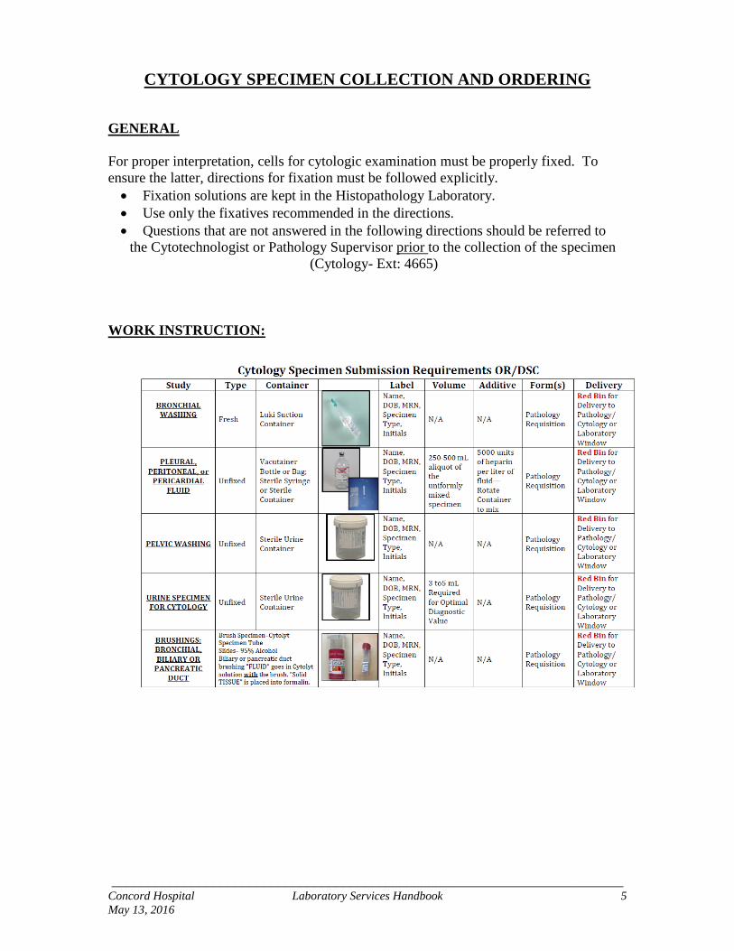
CYTOLOGY SPECIMEN COLLECTION AND ORDERING
GENERAL
For proper interpretation, cells for cytologic examination must be properly fixed. To
ensure the latter, directions for fixation must be followed explicitly.
Fixation solutions are kept in the Histopathology Laboratory.
Use only the fixatives recommended in the directions.
Questions that are not answered in the following directions should be referred to
the Cytotechnologist or Pathology Supervisor prior to the collection of the specimen
(Cytology- Ext: 4665)
WORK INSTRUCTION:
_______________________________________________________________________ Concord Hospital Laboratory Services Handbook 6
May 13, 2016
BLADDER IRRIGATION (WASHINGS)
Obtain washings of the bladder with hormonal saline or Ringer’s solution at the time of
cystoscopy. Label specimen and send to the Cytology Laboratory immediately,
accompanied by a Pathology Test Requisition Form.
BREAST SMEARS and FINE NEEDLE ASPIRATION FOR CYTOLOGY
Label two or more glass slides with patient’s name and other information. After
aspiration has been completed, place a drop or two of the material on the centers of as
many glass slides as possible, immediately covering each slide with a second, clean glass
slide. After all slides have been covered, gently pull the slides apart, smearing the fluid
along the length of each slide. Fix both slides IMMEDIATELY in 95% ethanol. The
slightest air drying of the smear prior to fixation will lead to some distortion of the cell
sample; therefore the smears that are fixed immediately (wet fixed) yield the best
preservation of the nuclear detail which is mandatory for accurate interpretation.
If there is any remaining material, draw saline solution into the barrel of syringe, using
the same needle. Expel entire contents into specimen container of CytoLyt Fixative
(about 50cc) and send it ASAP to the Cytology Laboratory for preparation, accompanied
by a Pathology Requisition form.
Reference: Koss, LG, Diagnostic Cytology and Its Histopathologic Bases, 3rd Ed., Vol. 2 p1005;
1979.
CEREBROSPINAL FLUID
The majority of specimens from the central nervous system fluid compartment are drawn
via lumbar puncture. Cisternal and ventricular taps, as well as direct aspirations of cystic
or solid masses also provide very useful cellular samples. Knowledge of the collection
method and site is essential to the interpretation of the cellular collection. Cellular
deterioration is the main problem encountered when delay occurs. Adequacy of
specimen volume is of equal significance to the diagnostic value; 3-5cc is preferred. Use
second or third sample drawn for cytology. Label and send to the Cytology Laboratory
immediately, accompanied by a Pathology Test Requisition form.
_______________________________________________________________________ Concord Hospital Laboratory Services Handbook 7
May 13, 2016
GASTRO-INTESTINAL SPECIMENS
COLONIC BRUSHINGS
Brushings of mucosal abnormalities at the time of fiber optic examination of the colon
are the generally accepted procedure in patients suspected of harboring a colonic lesion.
Specimen is smeared on clean glass slides and fixed immediately by placing in 95%
ethyl alcohol. Label and send to the Cytology Laboratory accompanied by a Pathology
requisition form.
COLONIC WASHINGS
Preparation of the patient is important since the colon must be free of fecal material. The
patient is instructed to take 2 oz. of castor oil 12 hours prior to the collection period and
to have only a very light meal on the morning of collection. Warm soap suds enemas are
given until the returns are clear. Two to three hours later the specimen is collected by
colonic irrigation. Five hundred (500) ml. of warm (37º C.) saline or Ringers solution is
instilled with the patient on the left decubitus position. Three to five (3-5) minutes later
the fluid is collected. Label and send specimen to the Cytology Laboratory accompanied
by a Pathology requisition form.
DUODENAL and GASTRIC WASHINGS
The patient should not have eaten food in the previous eight hours. The gastric tube
should be passed to the 70 cm mark without the use of any lubricants except glycerin.
Small sips of saline (not water) may be used to aid in passing the tube. Two hundred
fifty (250) ml of Ringers solution or physiologic saline are then introduced into the
stomach in small proportions, aspired and discarded. If the patient has pyloric
obstruction, several such lavages may be necessary. Introduce 250 ml of Ringers
solution or saline and re-aspirate and reintroduce the fluid several times using a 50 or 100
ml. syringe and with the patient in different positions ( prone, supine, left side, right side).
The entire gastric contents are then aspirated. Send specimen to the Cytology Laboratory
without delay, accompanied by a Pathology requisition form.
ESOPHAGEAL WASHINGS
Material is best obtained by direct esophagoscopy. Small amounts of physiologic saline
are injected through the esophagoscope and aspirated. Place the aspirate immediately in
CytoLyt fixative. Label and send to the Cytology Laboratory immediately, accompanied
by a Pathology Test Requisition form.
RECTAL WASHINGS
Preparation is the same as for the Colonic Washings. Two to three hours after the
cleansing enema, 10 ml of normal saline is introduced through the sigmoidoscope and
after 30 seconds is aspirated by strong suction. Label and send to the Cytology
Laboratory accompanied by a Pathology requisition form.
_______________________________________________________________________ Concord Hospital Laboratory Services Handbook 8
May 13, 2016
GYNECOLOGICAL SMEARS
Patient should not be douched for 24 hours before genital smears are obtained.
Preferably, smears should not be taken during menstrual bleeding.
No lubricant should be used.
Requests for special testing ( HPV; GC Chlamydia; Trichomonas) must be
requested at time of Pap testing.
NOTE: HPV, GC Chlamydia and Trichomonas are now performed in Concord
Hospital Laboratory directly off of the Thin Prep Pap Vial, on the Hologic Panther
System.
THIN PREP LIQUID BASED PAP PREPARATION COLLECTION
TECHNIQUE
http://www.thinprep.com/pdfs/man-01704-001.pdf
Sample Collection Technique using Broom-Like Collection Device:
1. Obtain a sample from the cervix using a broom-like device. Insert the Broom-like
collection device into the endocervical canal, push gently and rotate in a clockwise
direction five (5) times
2. Rinse the broom collection device immediately into the Thin Prep Vial by pushing
the broom into the bottom of the vial ten (10) times forcing the bristles apart. As a
final step, swirl the broom vigorously to further release material into the vial. Discard
the collection device.
3. Cap the vial tightly.
4. Label with patient’s name and date of birth. Send specimen to the Laboratory for
processing, accompanied by a PAP Test Requisition form.
Sample Collection Technique using Brush/Spatula Collection Device:
1. Obtain a sample from the cervix using the brush/spatula collection device. Insert the
contoured end of the plastic spatula and rotate 360 degrees around the entire
exocervix while maintaining tight contact with the exocervical surface.
2. Rinse the spatula immediately into the Thin Prep vial by swirling vigorously in the
vial ten (10) times. Discard the spatula.
3. Obtain a sample from the endocervix using the endocervical brush device. Insert the
Brush device into the cervix until only the bottom most fibers are exposed. Slowly
Rotate ¼ to ½ turn in ONE direction. DO NOT OVER ROTATE.
4. Cap the vial tightly
5. Label with patient’s name and date of birth. Send specimen to the Laboratory for
processing, accompanied by a PAP Test Requisition form.
PAP URINE
NOTE: Obtain separate specimens for routine and bacteriological examination. Voided
urine is preferable; however, catheterized urine is acceptable. Three morning non-initial
samples of urine, each of about 50 to 100 ml. obtained on consecutive days are
recommended. Hydration of patients by forced intake of fluids (1 glass of water every 30
minutes for a 3 hour period) is advocated. Label specimen and send to the Cytology
Laboratory immediately, accompanied by a Pathology Test Requisition Form.
_______________________________________________________________________ Concord Hospital Laboratory Services Handbook 9
May 13, 2016
EUS PROCEDURE
Endoscopic ultrasound (EUS) is a procedure that uses sound waves to create visual
images of the internal parts of the body. The Endoscopic ultrasound (EUS) allows the
surgeon to perform a biopsy to diagnose a tumor, plan treatment, and to check for
recurrence after treatment. This is done using fine needle aspiration (FNA). During this
process, a thin needle is placed through the endoscope and directed into the mass or
surrounding lymph nodes to obtain a biopsy specimen.
The needle is withdrawn from the area being biopsied and the aspirated matter is
expressed onto glass slides, the number of slides depending on the amount of aspirate.
Fix half of the total smears collected in 95% alcohol. Air dry the remaining number of
smears. The needle and syringe may be washed with sterile normal saline and expelled
into a specimen container containing CytoLyt fixative and the contents sent for
examination. Label and send specimen immediately to the Cytology Laboratory
accompanied by a Pathology requisition form. NOTE: If a tissue core is taken at time of
biopsy the tissue core should be placed into 10% Formalin and sent to Pathology
accompanied by a Pathology requisition form.
LUNG ASPIRATE and NEEDLE BIOPSY
Histologic and Cytologic study of pulmonary tissue are often made on material collected
with the aid of a wide-bore needle or a trocar and cannula. Needle biopsy of the lung is
most commonly performed to investigate lesions that are inaccessible to the
bronchoscope and do not desquamate into the bronchial tree. Image-intensifier television
fluoroscopy is used to monitor pulmonary aspiration. The patient is positioned
horizontally on an adjustable table. The point on the skin from which the underlying
lesion can be penetrated is marked. The skin, chest wall and pleura are anesthetized.
With television fluoroscope guidance, the needle is inserted close to the upper margin of
a rib (to avoid the intercostal artery) and introduced into the lesion.
Care must be taken to avoid large blood vessels and bronchi. The patient is instructed to
breathe normally. When the tip of the needle is in the desired position, the operator
rotates it so as to loosen small tissue fragments. A 10-20 cc. syringe is attached to the
needle, and the loosened tissue is aspirated while the patient holds his/her breath. The
needle is withdrawn from the chest, and the aspirated matter is expressed onto glass
slides, the number of slides depending on the amount of aspirate. Fix half of the total
smears collected in 95% alcohol. Air dry the remaining number of smears. The needle
and syringe may be washed with sterile normal saline and expelled into a specimen
container containing CytoLyt Fixative and the contents sent for examination. Label and
send specimen immediately to the Cytology Laboratory accompanied by a Pathology
requisition form.
_______________________________________________________________________ Concord Hospital Laboratory Services Handbook 10
May 13, 2016
EBUS PROCEDURE
The Endobronchial ultrasound (EBUS) is used to diagnose and stage lung cancer, and
to determine if the disease has spread to other parts of the body, such as the lymph nodes.
This technique allows the surgeon to obtain real-time images in and around the lungs and
to identify difficult-to-reach tumors. The Endobronchial ultrasound (EBUS) is also used
to obtain a biopsy of tissue or fluid sample from the lungs and surrounding lymph nodes
of the chest.
During an EBUS procedure, a thin, flexible instrument called a bronchoscope is fitted
with an ultrasound device and guided through the patient’s mount and trachea, a thin
needle is placed through the bronchoscope and directed into the mass or surrounding
lymph nodes to obtain a biopsy specimen.
The needle is withdrawn from the area being biopsied and the aspirated matter is
expressed onto glass slides, the number of slides depending on the amount of aspirate.
Fix half of the total smears collected in 95% alcohol. Air dry the remaining number of
smears. The needle and syringe may be washed with sterile normal saline and expelled
into a specimen container containing CytoLyt fixative and the contents sent for
examination. Label and send specimen immediately to the Cytology Laboratory
accompanied by a Pathology requisition form. NOTE: If a tissue core is taken at time of
biopsy the tissue core should be placed into 10% Formalin and sent to Pathology
accompanied by a Pathology requisition form.
PULMONARY SPECIMENS
BRONCHIAL ASPIRATES OR SALINE WASHINGS
Bronchial aspirates are obtained by suction during bronchoscopic procedures.
Bronchial Washings: With the bronchoscope in position, the patient is placed on the
table in such manner that the suspicious lung is dependent. The tip of the bronchoscope
is placed as close as possible to the area to be investigated. About 10 ml. of normal
saline is instilled in small portions of 2-3 ml. at a time and re-aspirated while the patient
is made to cough. The flexible tip of the aspirator may be placed also in the opening of
some of the smaller bronchi and the procedure repeated. All the cellular material is
collected in a clef collection. The collection tube should be rinsed thoroughly with saline
and the rinsing’s added to the specimen. The specimen must be labeled and send to the
Cytology Laboratory immediately, accompanied by a Pathology Test Requisition form.
_______________________________________________________________________ Concord Hospital Laboratory Services Handbook 11
May 13, 2016
PLEURAL, PERITONEAL and PERICARDIAL FLUIDS
Fluids are drawn by needle aspiration from pleural, pericardial and peritoneal spaces.
Specimens may also be obtained by needle insertion and irrigation of cavities or by
simple suction of free fluid encountered at surgical exploration of cavities. Do not add
fixative to the unprocessed specimen. An anticoagulant should be added to prevent fluid
clotting - 5000 units of heparin per liter of fluid is recommended. Rotate container to
properly mix specimen and anticoagulant. A 250 - 500 ml. aliquot of the uniformly mixed
specimen is sufficient for evaluation. Label the container. Complete a Pathology Test
Requisition form with patient information and clinical history.
It is essential to include the site, method of collection and clinical history.
Send specimen to the Cytology laboratory without delay.
Refrigerate specimen if delay is unavoidable.
SPUTUM
Instruct the patient to expectorate directly into a clean container. Morning specimens
resulting from overnight accumulation of secreta yield the best diagnostic results. Three
specimens on three successive days should be collected to ensure a maximum of
diagnostic accuracy. The patient must be carefully instructed not to spit into the
container without a deep cough, since saliva is of no diagnostic value. Specimen must be
labeled and send to the Cytology Laboratory immediately, accompanied by a Pathology
Test Requisition Form.
PCP STAIN
PCP Stain is a Special Stain used to rule out Pneumocystis Carinii in Bronchial
Specimens and Sputum Specimens. The Bronchial Specimen or the Sputum Specimen
should be sent to the Cytology Dept. in the Fresh State. The Physician should write the
orders for PCP Stain to be done on the Cytology Requisition which should accompany
the specimen to the lab.
RENAL ASPIRATION BIOPSIES
Renal aspiration biopsies are used almost exclusively in the diagnosis of lesions detected
by radiologic methods. Most are performed with the aid of television fluoroscopy or
ultrasound.
When the target has been exactly located by fluoroscopy or ultrasound, the patient is
positioned prone for television fluoroscopy with one or two pillows under the abdomen.
Approximately ten minutes before the biopsy is begun, intravenous pyelography is
performed. When the renal pelvis has been visualized, the site for aspiration biopsy is
selected. The skin is cleansed and the underlying tissues are anesthetized. A fairly thick
needle (external diameter 1.2mm. length 10cm.) containing a mandrin is inserted 5 to 8
cm. A series of aspirations takes place and an abundance of blood or fluid is obtained
and deposited on clean glass slides. Slides are fixed immediately by placing in coplin jar
containing 95% ethyl alcohol. Label and send specimen immediately to the Cytology
Laboratory accompanied by a Pathology requisition form.
_______________________________________________________________________ Concord Hospital Laboratory Services Handbook 12
May 13, 2016
RENAL PELVIS and URETERS
Voided urine, retrograde catheterization, and direct brushings are satisfactory procedures
for suspected lesions of the renal pelvis or the ureter and may assist in the localization of
the lesion of the upper urinary tract.
Specimen must be labeled as to type (voided or catheterized), and for ureteral specimens
and brushings, as to origin (right or left). Label and send specimen immediately to the
Cytology Laboratory accompanied by a Pathology requisition form.
TZANCK PREPARATIONS
Multiple scrapings are taken from patients with localized herpes zoster, disseminated
herpes zoster or herpes simplex infections.
The scrapings are prepared by selecting an early intact vesicle and swabbing the lesion
with a swab saturated with 70% isopropyl alcohol. A blade or needle is used to open the
vesicle and the lesion edge is gently scraped to avoid hemorrhage. The material is placed
on clean, previously albumenized glass slides and gently spread.
Fix slides IMMEDIATELY by placing in 95% ethyl alcohol. Label and send specimen to
the Cytology Laboratory accompanied by a Pathology requisition form.
Reference: Koss, LG, Diagnostic Cytology and Its Histopathologic Bases, 3rd Ed., Vol.
2, pp 1165-1187; 1979.