+ All Categories
Home > Documents > Cytoskeleton-Structure and Function

Cytoskeleton-Structure and Function

Date post: 13-Jan-2022
Category:
Upload: others
View: 4 times
Download: 0 times
Share this document with a friend
12
SUBJECT ZOOLOGY PaperNo. And Title V Cell Biology Module No. and Title Cytoskeleton Module tag DAYA-ZOO-CS Cytoskeleton-Structure and Function Dr.L.C.Mushan Assistant Professor D. B. F. Dayanand College of Arts and Science, Solapur.
Transcript

SUBJECT ZOOLOGY

PaperNo. And Title V Cell Biology

Module No. and Title Cytoskeleton

Module tag DAYA-ZOO-CS

Cytoskeleton-Structure and Function

Dr.L.C.Mushan

Assistant Professor

D. B. F. Dayanand College of Arts and Science, Solapur.

1

Learning Outcomes

The course provides a detailed insight into basic concepts of cytoskeleton structure and function.

Understand the structure and function of cytoskeleton –microtubules,microfilaments and

intermediate filaments.

Develop an understanding how cells move and spindlefibres help in chromosome movement

during cell division.How cell shape is maintained. Movement of cell is understood.

Table of contents

S.No. Cytoskeleton

1 Introduction

2 Structure of Microtubules

3 Functions of Microtubules

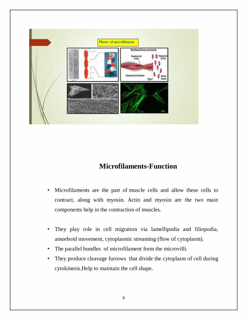
4 Microfilaments -structure and Function

5 Intermediate filaments-Structure and Function

Cytoskeleton

Introduction:

• In 1903, Nikolai Koltsov proposed that the shape of cells is determined by a

network of tubules that he termed the ‘cytoskeleton’.

• The cytoskeleton is a complex, dynamic network of interlinking protein

filaments present in the cytoplasm of all cells including bacteria and

archaea.

• It extends from the cell nucleus to the cell membrane and is composed of

similar proteins in various organisms.

2

• In eukaryotes, it is composed of three main components, microfilaments,

intermediate filaments and microtubules.

• All are capable of rapid growth or disassembly dependent on the cell's

requirements.

• Cytoskeleton’s primary function is to give the cell its shape and

mechanical resistance to deformation.

• The cytoskeleton can also contract, thereby deforming the cell and the

cell's environment, which allows the cells to migrate.

• Cytoskeleton is involved in many cell signalling pathways and in the

uptake of extracellular material. During cell division it helps in the

segregation of chromosomes.

• Helps in intracellular transport of vesicles and organelles within the

cell.It can be a template for the construction of a cell wall. It can form

specialized structures, such as cilia, flagella, lamelliopodia and

podosomes

• The structure, function and dynamic behaviour of the cytoskeleton can

be very different, depending on organism and cell types.

• The cytoskeleton consists of three components:

Microtubules.

Microfilaments (Actin filament).

Intermediate filament

3

Microtubules-Structure

• Microtubules are long, hollow cylindrical and filamentous or fibrilar

structures found the cytoplasm of all eukaryotic cells. Absent in prokaryotes.

• Microtubules are found in the thrombocytes (blood platelets) of human and

rat.

• They are about 25 nm in diameter and 200 nm to 25 micrometre in length.

• Microtubules are composed of many subunits called protofilaments. The

number of protofilaments in microtubules is variable.

• The protofilaments is composed of a series of globular protein (tubulin) units.

• The tubulin is a dimer, made up of two similar polypeptides.

• The two tubulin dimers are α- tubulin and β- tubulin.

• These two units are arranged alternately in the protofilament.

4

• The tubulin shows helical structure with 13 tubulin molecules per turn of the

helix.They grow in length by adding tubulin dimer or they can be

disassembled.

https://drive.google.com/file/d/1qmx7w9zWMrHEVM3WblvPV9mx8eEUp04t/view

Microtubules-Function

• Microtubule are rigid structures which work as a supporting framework and

give shape to the cell.

• They maintain shape of long processes such as cilia, flagella, axons of nerve

cells, axopodia of protozoa.

• The motion of the cilia and flagella is created by the microtubules. The

centrioles are morphologically identical to the basal body of cilia or flagella

• Microtubules changes the cell shape during cell differentiation. Help in the

elongation of the cells in the lens of eye.

5

• The microtubules of many sensory cells act as a transducers which convert

stimuli into the nerve impulses.

• Microtubules help in the transport of cellular materials. During cell division,

the spindle fibre appear, they are the bundles of microtubules. Help in of the

formation of cell wall in plants.They also help in separation of chromosome.

Microfilaments: structure

• Microfilaments also known as actin filaments are solid rods of protein.

• The diameter of filament is about 7 nm and they are smallest of the

cytoskeletal filaments.

• They occur in almost all eukaryotic cells.

• They are called as actin filaments because they are mostly composed of the

protein actin.

• Actin is the protein building blocks of microfilament. Actin is found

abundantly in all eukaryotic cells.

• Their structure is two strands of actin wound in a spiral.

• It is synthesized as a single polypeptide consisting of 375 amino acids.

• Individual actin molecules are referred as G- actin. The G- actin molecule

polymerize to form microfilament which is called as F- actin (Filamentous

actin).

6

Microfilaments-Function

• Microfilaments are the part of muscle cells and allow these cells to

contract, along with myosin. Actin and myosin are the two main

components help in the contraction of muscles.

• They play role in cell migration via lamellipodia and filiopodia,

amoeboid movement, cytoplasmic streaming (flow of cytoplasm).

• The parallel bundles of microfilament form the microvilli.

• They produce cleavage furrows that divide the cytoplasm of cell during

cytokinesis.Help to maintain the cell shape.

7

Intermediate Filaments-Structure

• Intermediate filaments are a part of the cytoskeleton of many eukaryotic

cells.

• They are more stable (strongly bound) than microfilaments and are

heterogeneous constituents of the cytoskeleton

• Intermediate filaments are about 8-12 nm wide. They are called

intermediate because they are in-between the size of microfilaments and

microtubules.

• Intermediate filaments are made of different proteins such as keratin (found

in hair and nails, and also in animals with scales, horns, or hooves), vimentin

(mesenchymal cell) , desmin (muscle cell), and lamin (nuclear envelop).

• All intermediate filaments are found in the cytoplasm except for lamins,

which are found in the nucleus and help support the nuclear envelope that

surrounds the nucleus.

8

Intermediate Filaments-Function

• The intermediate filaments in the cytoplasm maintain the cell’s shape, bear

tension, and provide structural support to the cell.

• Fix the organization of certain cell organelles.

• Intermediate filaments organize the internal tridimensional structure of the

cell, anchoring organelles and serving as structural components of

the nuclear lamina.

• Keratin intermediate filaments in epithelial cells provide protection for

different mechanical stresses that skin may endure.

• They also provide protection for organs against metabolic, oxidative, and

chemical stresses.

• Strengthening of epithelial cells with these intermediate filaments may

prevent onset of apoptosis, or cell death, by reducing the probability of

stress.

• In combination with proteins and desmosomes, the intermediate filaments

form cell-cell connections and anchor the cell-matrix junctions that are used

in messaging between cells as well as vital functions of the cell.

Summary

The cytoskeleton is the structure consisting of fibrous proteins that occur in the

cytoplasm and maintain the shape of the cell.

It composed of three different types of components, namely microtubule,

microfilament and intermediate filaments,

9

The microtubules are made up of α- tubulin and β- tubulin arranged alternately

to form a protofilament.

The microfilament also called as actin filament is solid rod of protein formed

by two strands of actin filament and it shows spiral arrangement.

The intermediate filaments are made up of different types of proteins like

keratin, desmin, lamin and vimentin.

All these cytoskeletal elements perform many functions like maintain the cell

structure, acts as supporting framework, help in cell movement, intracellular

transport of material , locomotion and fix the organization of cell organelles.

Links:

https://docs.google.com/presentation/d/1473WiJB9-lZICCr_MFgAcUbnnXBvLwFk/edit#slide=id.p1

https://docs.google.com/document/d/1QnXIlcm1m4PFcSEWeDN7qyA_F3lpJy3FeVjCvlKUIrg/edi

t?usp=sharing

https://drive.google.com/file/d/1DiEw1LvdEWwf0NVGCMkunIUPhbx3j7mR/view

Vedio

https://youtu.be/VC1zbUEEw9k

https://drive.google.com/drive/folders/1Rbsb059X6OU171vR6BuSS06O-hR0SUWR

https://drive.google.com/file/d/1qmx7w9zWMrHEVM3WblvPV9mx8eEUp04t/view?usp=sharin

g

https://docs.google.com/forms/d/1IFDtxTGUIYVfl7eu8-SiUxJz72hMrSgcA0U0FqJNLrc/edit

https://drive.google.com/file/d/14Gwzy4g8jkQyRyfdpiCKsp6l8w5YwlOP/view?usp=sharing

Explore more

1. Karp, G. (2010) Cell and Molecular Biology: Concepts and Experiments (6th edition) John Wiley & Sons. Inc.

2. De Robertis, E.D.P. and De Robertis, E.M.F. (2006) Cell and Molecular Biology (8th edition) Lippincott

Williams and Wilkins, Philadelphia.

10

3. Cooper, G.M. and Hausman, R.E. (2009) The Cell: A Molecular Approach. (5th edition) ASM Press &

Sunderland, Washington, D.C.; Sinauer Associates, MA

. 4. Becker, W.M.; Kleinsmith, L.J.; Hardin. J. and Bertoni, G. P. (2009) The World of the Cell. (7th edition)

Pearson Benjamin Cummings Publishing, San Francisco.

ASSESSMENT

Module / Topic: CellBiology Year: SYBSc-Sem-III

ILO Teaching Activity Assessment Type Assessment Mode & Tool

1.Students will be able to draw neat labelled diagrams of different types of cytoskeleton

Strategies Used Explanation & drawing the diagram. Showing models and discussing. Ask students to prepare animation PPT’s

Assessment type: Draw a chart of the different parts of Cell Give a diagrammatic sketch with arrows and ask them to label the different parts. Sketch & label types of cytofilaments

Google class room code:un5po2c

2. Students will be able to differentiate between types of cytoskeleton

Strategies Used Quiz, MCQhttps://docs.google.com/document/d/1o_8fa3y7FH2f10XdWp0T1esK4-zRnlnj26_PPbuQ5Dk/edit

Assessment type: Grading till they are able to get minimum score of 60%

https://docs.google.com/forms/d/1IFDtxTGUIYVfl7eu8-SiUxJz72hMrSgcA0U0FqJNLrc/edit

3.To explain the mechanism & functions of each part of the cytoskeleton.

Strategies Used Explanation- Concept map

Give a concept map with blanks in different columns to be filled by the students, Quiz, https://forms.gle/dJ2EJBG5adFUYsPd7

MCQ’s Class test .https://forms.gle/dJ2EJBG5adFUYsPd7 MOODLE LMS has these


Recommended