THE JOURNAL OF BIOLOGICALCHEMISTHY Vol. 250, No. 17, Issue of September 10, pp. 6983-6989, 1975 Printed in U.S.A. D-Amino Acid Aminotransferase of BaciLZus sphaericus ENZYMOLOGIC AND SPECTROMETRIC PROPERTIES (Received for publication, March 25, 1975) KAZUO YONAHA, HARUO MISONO, TATSUO YAMAMOTO, AND KENJI SODA* From the Laboratory of Microbial Biochemistry, Institute for Chemical Research, Kyoto University, Uji, Kyoto-Fu 611, Japan D-Amino acid aminotransf’erase, purif’ied to homogeneity and crystallized from Bacillus sphaericus, has a molecular weight of about 60,000 and consists of two subunits identical in molecular weight (X0,000). The enzyme exhibits absorption maxima at 280,330, and 415 nm, which are independent of the pH (5.5 to lO.O), and contains 2 mol of pyridoxal 5’-phosphate per mol of enzyme. One of the pyridoxal-5’.P, absorbing at 415 nm, is bound in an aldimine linkage to the t-amino group of a lysine residue of the protein, and is released by incubation with phenylhydrazine to yield the catalytically inactive form. The inactive form, which is reactivated by addition of pyridoxal 5’-phosphate, still has a 330 nm peak and contains 1 mol of pyridoxal5’-phosphate. Therefore, this form is regarded as a semiapoenzyme. The holo- enzyme shows negative circular dichroic bands at 330 and 415 nm. D-Amino acid aminotransferase catalyzes CYtransamination of various D-amino acids and a-keto acids. n-Alanine, D-a-aminobutyrate and n-glutamate, and a-ketoglutarate, pyruvate, and Lu-ketobutyrate are the preferred amino donors and acceptors, respectively. The enzyme activity is significantly affected by both the carbonyl and sulf- hydryl reagents. The Michaelis constants are as follows: D-alanine (1.3 and 4.2 mM with a-ketobutyrate and a-ketoglutarate, respectively), a-ketobutyrate (14 ITIM with D-alanine), cu-ketoglutarate (3.4 mu with n-alanine), pyridoxal5’-phosphate (2.3 MM) and pyridoxamine 5’-phosphate (25 fiM). The first evidence for the occurrence of D-amino acid specific aminotransf’erase was found by Thorne et al. (1, 2) in the cell-free extract of Bacillus subtilis and Bacillus anthracis. They showed that the: enzyme of B. subtilis was activated a few-fold by pyridoxal-5’-P’ and suggested that this compound was a cofactor. D-Amino acid aminotransferase was subse- quently demonstrated in extracts of Rhodospirillum rubrum (3), Bacillus sphuericus (4), and Bacillus licheniformis (5), and recently has also been f’ound in higher plants (6, 7) and mammalian liver (8). n-Alanine aminotransferase was purified to near homogeneity from B. subtilis to elucidate the en- zymologic and kinetic properties (9-11). Exact knowledge of the enzymatic transamination of D-amino acids is of value not only for a study of the enzyme chemistry of D-amino acid metabolism, but also for understanding the reaction mech- anisms of L-amino acid aminotransf’erases and amino acid racemases. We found a high activity of n-amino acid amino- transferase in the cell-free extract of B. sphuericus IF0 3525 (12), and recently have purified the enzyme to homogeneity and crystallized it (13). In the present paper, more detailed studies on enzymological and physicochemical characteristics * To whom correspondence regarding this work should be addressed. 1The abbreviations used are: pyridoxal-5,-P, pyridoxal 5’.phos- phate; pyridoxamine-5,-P, pyridoxamine 5’-phosphate; pyridox- ine-5,-P, pyridoxine 5’-phosphate. of the crystalline n-amino acid aminotransferase are described with particular emphasis on the bound pyridoxal-j’-P. MATERIALS AND METHODS Chemicals-D-Cycloserine was obtained from Shionogi Seiyaku Co., Osaka; D-, L-, and oL-penicillamine from Calbiochem; Sephadex G-25 from Pharmacia, Upsala, Sweden; Bio-Gel P-150 from Bio-Rad Labora- tories, Richmond, California; pyridoxal-5’.P and L-amino acids from Kyowa Hakko Kogyo, Tokyo; and aminoxyacetate, ol-keto acids, pyridoxal, pyridoxamine, pyridoxamine-5,-P, and pyridoxine-5’-P from Nakarai Chemicals, Kyoto, Japan. D-Amino acids were products of Ajinomoto Co., Tokyo. Pyridoxamine-5’-P and pyridoxal-5’-P were chromatographically purified by the method of Peterson and Sober (14). 3-Methyl-2-benzothiazolone hydrazone hydrochloride was pur- chased from Aldrich Chemical Company, Inc., Wis. A’-Piperideine-2- carboxylic acid was synthesized from e-N-carbobenzoxy-L-lysine (15). A’-Piperideine-6-carboxylic acid was prepared from L-lysine with the crystalline L-lysine-cu-ketoglutarate e-aminotransferase (16). c-N- Pyridoxyllysine and @-chloro-D-alanine were kindly provided by Dr. Morino, Kumamoto University, Kumamoto, Japan. Sodium lauryl sulfate was a specially prepared reagent for protein research (Nakarai Chemicals) and urea was purified by recrystallization from ethanol before use. The other chemicals were analytical grade reagents. Enzyme Preparation-The enzyme was purified from a cell-free extract of Bacillus sphaericus IF0 3525 and crystallized as described previously (13), but with higher yield (about 30%). The specific activity of the crystalline preparation was approximately 110. Enzyme Assay-The standard assay mixture consisted of 25 Fmol of D-alanine or other n-amino acid, 80 pmol of potassium phosphate buffer (pH 8.0), 1 pmol of pyridoxal-5,-P, 25 rmol of cu-keto acid, and 6983 by guest on July 13, 2018 http://www.jbc.org/ Downloaded from
Transcript
THE JOURNAL OF BIOLOGICAL CHEMISTHY Vol. 250, No. 17, Issue of September 10, pp. 6983-6989, 1975
Printed in U.S.A.
D-Amino Acid Aminotransferase of BaciLZus sphaericus ENZYMOLOGIC AND SPECTROMETRIC PROPERTIES
(Received for publication, March 25, 1975)
KAZUO YONAHA, HARUO MISONO, TATSUO YAMAMOTO, AND KENJI SODA*
From the Laboratory of Microbial Biochemistry, Institute for Chemical Research, Kyoto University, Uji, Kyoto-Fu 611, Japan
D-Amino acid aminotransf’erase, purif’ied to homogeneity and crystallized from Bacillus sphaericus, has a molecular weight of about 60,000 and consists of two subunits identical in molecular weight (X0,000).
The enzyme exhibits absorption maxima at 280,330, and 415 nm, which are independent of the pH (5.5 to
lO.O), and contains 2 mol of pyridoxal 5’-phosphate per mol of enzyme. One of the pyridoxal-5’.P, absorbing at 415 nm, is bound in an aldimine linkage to the t-amino group of a lysine residue of the protein, and is released by incubation with phenylhydrazine to yield the catalytically inactive form. The inactive form, which is reactivated by addition of pyridoxal 5’-phosphate, still has a 330 nm peak and contains 1 mol of pyridoxal5’-phosphate. Therefore, this form is regarded as a semiapoenzyme. The holo- enzyme shows negative circular dichroic bands at 330 and 415 nm. D-Amino acid aminotransferase catalyzes CY transamination of various D-amino acids and a-keto acids. n-Alanine, D-a-aminobutyrate and n-glutamate, and a-ketoglutarate, pyruvate, and Lu-ketobutyrate are the preferred amino donors and acceptors, respectively. The enzyme activity is significantly affected by both the carbonyl and sulf- hydryl reagents. The Michaelis constants are as follows: D-alanine (1.3 and 4.2 mM with a-ketobutyrate
and a-ketoglutarate, respectively), a-ketobutyrate (14 ITIM with D-alanine), cu-ketoglutarate (3.4 mu with
n-alanine), pyridoxal5’-phosphate (2.3 MM) and pyridoxamine 5’-phosphate (25 fiM).
The first evidence for the occurrence of D-amino acid specific aminotransf’erase was found by Thorne et al. (1, 2) in the
cell-free extract of Bacillus subtilis and Bacillus anthracis. They showed that the: enzyme of B. subtilis was activated a few-fold by pyridoxal-5’-P’ and suggested that this compound
was a cofactor. D-Amino acid aminotransferase was subse- quently demonstrated in extracts of Rhodospirillum rubrum (3), Bacillus sphuericus (4), and Bacillus licheniformis (5), and recently has also been f’ound in higher plants (6, 7) and mammalian liver (8). n-Alanine aminotransferase was purified to near homogeneity from B. subtilis to elucidate the en-
zymologic and kinetic properties (9-11). Exact knowledge of the enzymatic transamination of D-amino acids is of value not only for a study of the enzyme chemistry of D-amino acid
metabolism, but also for understanding the reaction mech- anisms of L-amino acid aminotransf’erases and amino acid
racemases. We found a high activity of n-amino acid amino- transferase in the cell-free extract of B. sphuericus IF0 3525 (12), and recently have purified the enzyme to homogeneity and crystallized it (13). In the present paper, more detailed studies on enzymological and physicochemical characteristics
* To whom correspondence regarding this work should be addressed. 1 The abbreviations used are: pyridoxal-5,-P, pyridoxal 5’.phos-
of the crystalline n-amino acid aminotransferase are described with particular emphasis on the bound pyridoxal-j’-P.
MATERIALS AND METHODS
Chemicals-D-Cycloserine was obtained from Shionogi Seiyaku Co., Osaka; D-, L-, and oL-penicillamine from Calbiochem; Sephadex G-25 from Pharmacia, Upsala, Sweden; Bio-Gel P-150 from Bio-Rad Labora- tories, Richmond, California; pyridoxal-5’.P and L-amino acids from Kyowa Hakko Kogyo, Tokyo; and aminoxyacetate, ol-keto acids, pyridoxal, pyridoxamine, pyridoxamine-5,-P, and pyridoxine-5’-P from Nakarai Chemicals, Kyoto, Japan. D-Amino acids were products of Ajinomoto Co., Tokyo. Pyridoxamine-5’-P and pyridoxal-5’-P were chromatographically purified by the method of Peterson and Sober (14). 3-Methyl-2-benzothiazolone hydrazone hydrochloride was pur- chased from Aldrich Chemical Company, Inc., Wis. A’-Piperideine-2- carboxylic acid was synthesized from e-N-carbobenzoxy-L-lysine (15). A’-Piperideine-6-carboxylic acid was prepared from L-lysine with the crystalline L-lysine-cu-ketoglutarate e-aminotransferase (16). c-N- Pyridoxyllysine and @-chloro-D-alanine were kindly provided by Dr. Morino, Kumamoto University, Kumamoto, Japan. Sodium lauryl sulfate was a specially prepared reagent for protein research (Nakarai Chemicals) and urea was purified by recrystallization from ethanol before use. The other chemicals were analytical grade reagents.
Enzyme Preparation-The enzyme was purified from a cell-free extract of Bacillus sphaericus IF0 3525 and crystallized as described previously (13), but with higher yield (about 30%). The specific activity of the crystalline preparation was approximately 110.
Enzyme Assay-The standard assay mixture consisted of 25 Fmol of D-alanine or other n-amino acid, 80 pmol of potassium phosphate buffer (pH 8.0), 1 pmol of pyridoxal-5,-P, 25 rmol of cu-keto acid, and
enzyme in a final volume of 1.0 ml. Enzyme was replaced by water in a blank. The reaction was initiated by addition of n-amino acid, and incubation was carried out at 37” for 20 min. The enzyme was assayed by determining pyruvate or amino acid formed with salicylaldehyde as follows, or with ninhydrin, after addition of 0.1 ml of 50% trichloroace- tic acid and separation by paper chromatography (17), respectively.
Determination of Pyruuate-Pyruvate was determined by a modifi- cation of Berntsson’s method (18). The reaction was stopped by addition of 1.0 ml of 60% KOH. A 0.5.ml portion of 2% salicylaldehyde in 99% ethanol was added and incubation was performed at 37” for 30 min to develop an orange color. After addition of 1.5 ml of cold water, the absorbance was measured at 480 nm.
Protein Determination-Protein concentration of the enzyme was derived from the absorbance at 280 nm. The absorbance coefficient (A,,, I% = 12.64) was used throughout, obtained by absorbance and dry weight determinations.
Definction of Units and Specific Activity-One unit of enzyme is defined as the amount of enzyme that catalyzes the formation of 1 pmol of pyruvate or amino acid per min. Specific activity is expressed as units per mg of protein.
Spectrophotometry-Absorption and circular dichroism spectra were taken with a Shimadzu MPS-50 L recording spectrophotometer and a Jasco ORD/UV-5 recording spectropolarimeter equipped with a circular dichroism attachment with a 1.0.cm light path, respectively. Fluorescence measurements were performed with a Shimadzu spectro- fluorophotometer type RF 502 with a l.O-cm light path, and intensities of emission and excitation spectra were given arbitrarily.
RESULTS
Molecular Weight-The molecular weight of D-amino acid aminotransferase was determined to be 58,000 =t 2,000 by sedimentation equilibrium method as reported in a previous paper (13). A molecular weight of about 60,000 was also obtained by the Bio-gel P-150 gel filtration method of Andrews (19), with bovine serum albumin (M, 68,000), ovalbumin (M, 45,000), and cu-chymotrypsinogen A (M, 25,000) as standard proteins.
Structure of Subunit-n-Amino acid aminotransferase was found to be completely inhibited by 1% sodium lauryl sulfate in 0.02 M sodium phosphate buffer (pH 7.2), or 8 M urea in 0.02 M
potassium phosphate buffer (pH 7.4). No activity was re- covered after dialysis against two changes of 1000 volumes of the respective buffers at 4’. These results suggest that the denaturants cause irreversible conformation changes in the enzyme, possibly with dissociation into subunits.
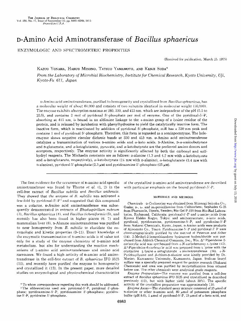
The subunit structure of the enzyme was examined by disc gel electrophoresis. The enzyme was incubated with 1.0% sodium lauryl sulfate in 0.01 M sodium phosphate buffer (pH 7.2) containing 1.0% 2-mercaptoethanol and 25% glycerol, or with 8 M urea in 0.02 M potassium phosphate buffer (pH 7.4) containing 1.0% 2-mercaptoethanol at 37” for about 12 hours. The treated enzyme preparations were subjected to electropho- resis in the presence of 0.1% sodium lauryl sulfate (20), or of 8 M
urea (Fig. 1). There was a single band of stained protein. To determine the molecular weight of the polypeptide in this band, we ran a series of marker proteins treated in the same manner; ovalbumin (M, 45,000), bovine serum albumin (M, 68.000), glutamate dehydrogenase (M, 50,000), cytochrome c (M, 12,000) and chymotrypsinogen A (M, 25,000). The molecu- lar weight was calculated to be approximately 30,000 from a semilogarithmic plot of molecular weight against mobility, suggesting the enzyme consists of two subunits identical in molecular weight.
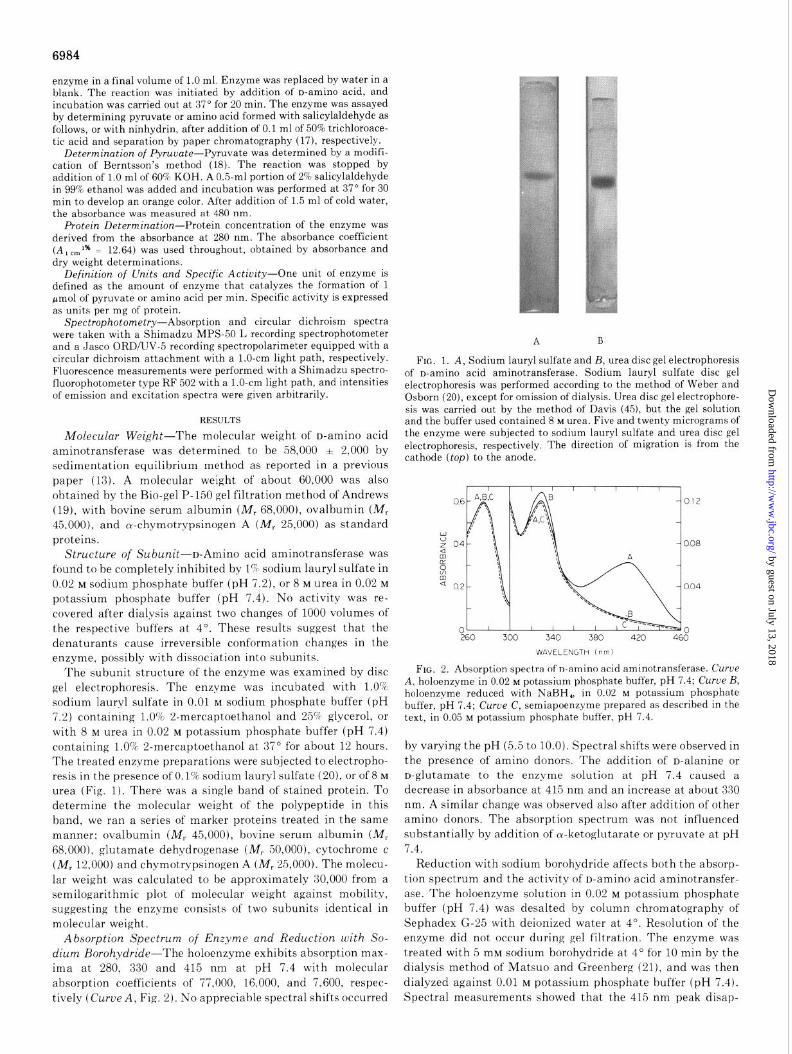
Absorption Spectrum of Enzyme and Reduction with So- dium Borohydride-The holoenzyme exhibits absorption max- ima at 280, 330 and 415 nm at pH 7.4 with molecular absorption coefficients of 77,000, 16,000, and 7,600, respec- tively (Curue A, Fig. 2). No appreciable spectral shifts occurred
A B
FIG. 1. A, Sodium lauryl sulfate and B, urea disc gel electrophoresis of n-amino acid aminotransferase. Sodium lauryl sulfate disc gel electrophoresis was performed according to the method of Weber and Osborn (20), except for omission of dialysis. Urea disc gel electrophore- sis was carried out by the method of Davis (45), but the gel solution and the buffer used contained 8 M urea. Five and twenty micrograms of the enzyme were subjected to sodium lauryl sulfate and urea disc gel electrophoresis, respectively. The direction of migration is from the cathode (top) to the anode.
WAVELENGTH 1 nm 1
FIG. 2. Absorption spectra of n-amino acid aminotransferase. Curve A, holoenzyme in 0.02 M potassium phosphate buffer, pH 7.4; Curve B, holoenzyme reduced with NaBH,, in 0.02 M potassium phosphate buffer, pH 7.4; Curve C, semiapoenzyme prepared as described in the text, in 0.05 M potassium phosphate buffer, pH 7.4.
by varying the pH (5.5 to 10.0). Spectral shifts were observed in the presence of amino donors. The addition of D-alanine or n-glutamate to the enzyme solution at pH 7.4 caused a decrease in absorbance at 415 nm and an increase at about 330 nm. A similar change was observed also after addition of other amino donors. The absorption spectrum was not influenced substantially by addition of oc-ketoglutarate or pyruvate at pH 7.4.
Reduction with sodium borohydride affects both the absorp- tion spectrum and the activity of o-amino acid aminotransfer- ase. The holoenzyme solution in 0.02 M potassium phosphate buffer (pH 7.4) was desalted by column chromatography of Sephadex G-25 with deionized water at 4”. Resolution of the enzyme did not occur during gel filtration. The enzyme was treated with 5 mM sodium borohydride at 4” for 10 min by the dialysis method of Matsuo and Greenberg (21), and was then dialyzed against 0.01 M potassium phosphate buffer (pH 7.4). Spectral measurements showed that the 415 nm peak disap-
for L-lysine-cY-ketoglutarate t-aminotransferase of Achromo- batter liquidum (24).
The 330 nm peak of semiapoenzyme was never shifted by addition of amino donors or amino acceptors, e.g. o-alanine or a-ketoglutarate, nor by varying the pH of enzyme solution in the range of 5.5 to 10.0, although it disappeared when the enzyme was incubated with 1% sodium lauryl sulfate, 6 M
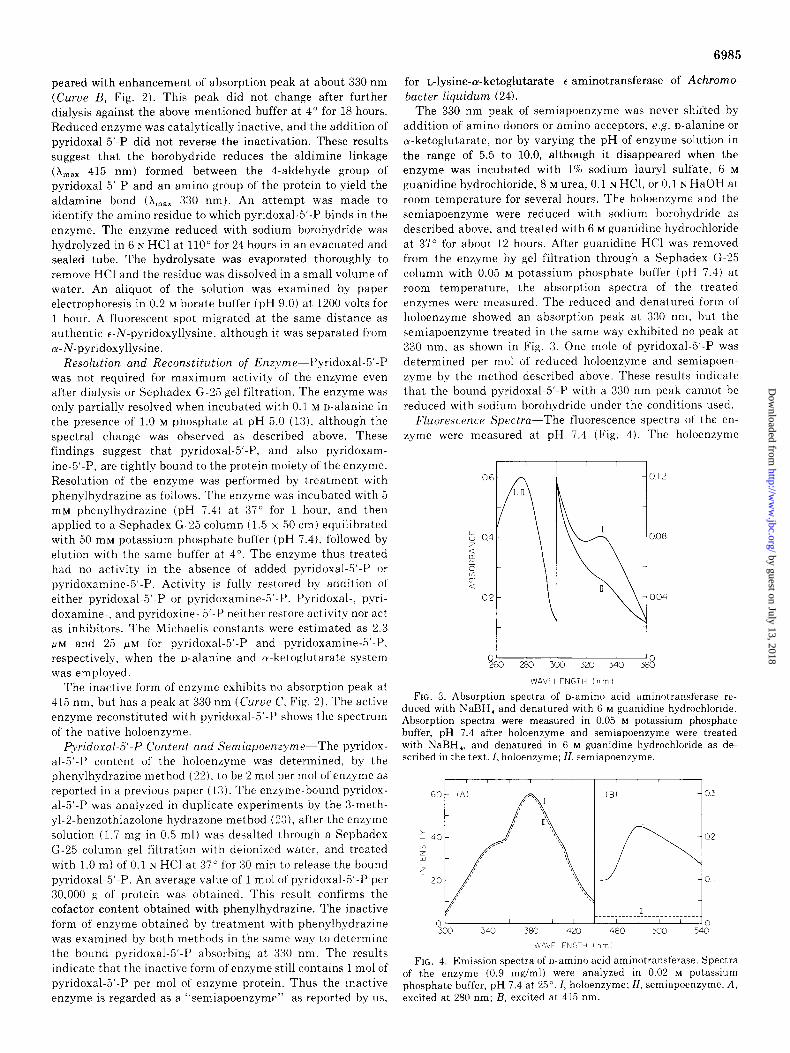
guanidine hydrochloride, 8 M urea, 0.1 N HCl, or 0.1 N HaOH at room temperature for several hours. The holoenzyme and the semiapoenzyme were reduced with sodium borohydride as described above, and treated with 6 M guanidine hydrochloride at 37” for about 12 hours. After guanidine HCl was removed from the enzyme by gel filtration through a Sephadex G-25 column with 0.05 M potassium phosphate buffer (pH 7.4) at room temperature, the absorption spectra of the treated enzymes were measured. The reduced and denatured form of holoenzyme showed an absorption peak at 330 nm, but the semiapoenzyme treated in the same way exhibited no peak at 330 nm, as shown in Fig. 3. One mole of pyridoxal-5,-P was determined per mol of reduced holoenzyme and semiapoen- zyme by the method described above. These results indicate that the bound pyridoxal-5’-P with a 330 nm peak cannot be reduced with sodium borohydride under the conditions used.
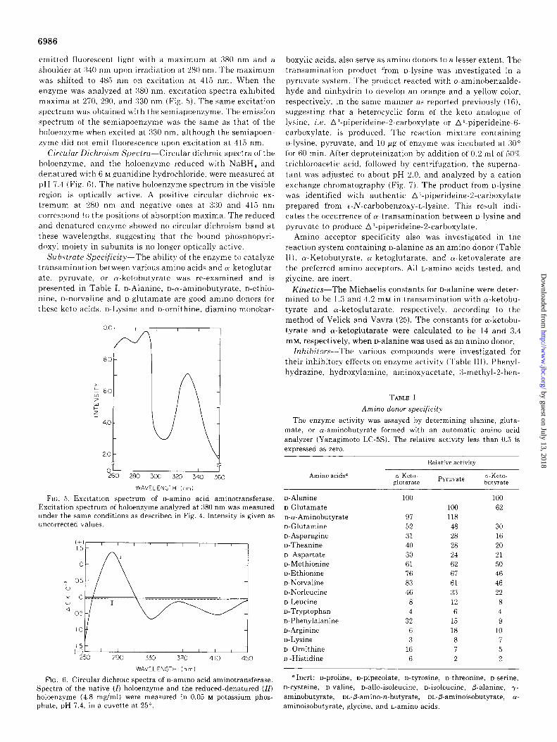
Fluorescence Spectra-The fluorescence spectra of the en- zyme were measured at pH 7.4 (Fig. 4). The holoenzyme
peared with enhancement of absorption peak at about 330 nm (Curue B, Fig. 2). This peak did not change after further dialysis against the above mentioned buffer at 4” for 18 hours. Reduced enzyme was catalytically inactive, and the addition of pyridoxal-5’.P did not reverse the inactivation. These results suggest that the borohydride reduces the aldimine linkage
(L,, 415 nm) formed between the 4-aldehyde group of pyridoxal-5’.P and an amino group of the protein to yield the aldamine bond (X,,, 330 nm). An attempt was made to identify the amino residue to which pyridoxal-5’-P binds in the enzyme. The enzyme reduced with sodium borohydride was hydrolyzed in 6 N HCl at 110” for 24 hours in an evacuated and sealed tube. The hydrolysate was evaporated thoroughly to remove HCl and the residue was dissolved in a small volume of water. An aliquot of the solution was examined by paper electrophoresis in 0.2 M borate buffer (pH 9.0) at 1200 volts for 1 hour. A fluorescent spot migrated at the same distance as authentic t-A-pyridoxyllysine, although it was separated from n-N-pyridoxyllysine.
Resolution and Reconstitution of Enzyme-Pyridoxal-5’.P was not required for maximum activity of the enzyme even after dialysis or Sephadex G-25 gel filtration. The enzyme was only partially resolved when incubated with 0.1 M n-alanine in the presence of 1.0 M phosphate at pH 5.0 (13). although the spectral change was observed as described above. These findings suggest that pyridoxal-5,-P, and also pyridoxam- ine-5’.P, are tightly bound to the protein moiety of the enzyme. Resolution of the enzyme was performed by treatment with phenylhydrazine as follows. The enzyme was incubated with 5 mM phenylhydrazine (pH 7.4) at 37” for 1 hour, and then applied to a Sephadex G-25 column (1.5 x 50 cm) equilibrated with 50 mM potassium phosphate buffer (pH 7.4). followed by elution with the same buffer at 4”. The enzyme thus treated had no activity in the absence of added pyridoxal-5’.P or pyridoxamine-5’-P. Activity is fully restored by addition of either pyridoxal-5-P or pyridoxamine-5-P. Pyridoxal-, pyri- doxamine-, and pyridoxine- 5’.P neither restore activity nor act as inhibitors. The Michaelis constants were estimated as 2.3 PM and 25 PM for pyridoxal-5’-P and pyridoxamine-5,-P, respectively, when the n-alanine and cu-ketoglutarate system was employed.
The inactive form of enzyme exhibits no absorption peak at 415 nm, but has a peak at 330 nm (&rue C, Fig. 2). The active enzyme reconstituted with pyridoxal-5’-P shows the spectrum of the native holoenzyme.
PyridoxalM-P Content and Semiapoenzyme-The pyridox- al-5’-P content of the holoenzyme was determined, by the phenylhydrazine method (22), to be 2 mol per mol of enzyme as reported in a previous paper (13). The enzyme-bound pyridox- al-5’.P was analyzed in duplicate experiments by the Smeth- yl-2-benzothiazolone hydrazone method (23), after the enzyme solution (1.7 mg in 0.5 ml) was desalted through a Sephadex G-25 column gel filtration with deionized water, and treated with 1.0 ml of 0.1 N HCl at 37” for 30 min to release the bound pyridoxal-5’.P. An average value of 1 mol of pyridoxal-5’.P per 30.000 g of protein was obtained. This result confirms the cofactor content obtained with phenylhydrazine. The inactive form of enzyme obtained by treatment with phenylhydrazine was examined by both methods in the same way to determine the bound pyridoxal-5’.P absorbing at 330 nm. The results indicate that the inactive form of enzyme still contains 1 mol of pyridoxal-5’.P per mol of enzyme protein. Thus the inactive enzyme is regarded as a “semiapoenzymr” as reported by us.
Y-----& 60 280 3co 3x) 340
WAVEI.ENGTH In m 1
FIG. 3. Absorption spectra of n-amino acid aminotransferase re- duced with NaBH, and denatured with 6 M guanidine hydrochloride. Absorption spectra were measured in 0.05 M potassium phosphate buffer, pH 7.4 after holoenzyme and semiapoenzyme were treated with NaBH,, and denatured in 6 M guanidine hydrochloride as de- scribed in the text. I, holoenzyme; II, semiapoenzyme.
\~AVELENGTH I nrn 1
FIG. 4. Emission spectra of o-amino acid aminotransferase. Spectra of the enzyme (0.9 mg/ml) were analyzed in 0.02 M potassium phosphate buf’fer, pH 7.4 at 25”. I, holoenzyme; II, semiapoenzyme. A, excited at 280 nm; B, excited at 415 nm.
emitted fluorescent light with a maximum at 3SO nm and a shoulder at :%40 nm upon irradiation at 280 nm. The maximum was shifted to 485 nm on excitation at 415 nm. When the enzyme was analyzed at 380 nm, excitation spectra exhibited maxima at 270, 290, and 330 nm (Fig. 5). The same excitation spectrum was obtained with the semiapoenzyme. The emission spectrum of the semiapoenzyme was the same as that of the holoenzvme when excited at 330 nm. although the semiapoen- zyme did not emit fluorescence upon excitation at 415 nm.
Circular Dichroism Spectra-Circular dichroic spectra of the holoenzyme. and the holoenzyme reduced with NaBH, and denatured with 6 M guanidine hydrochloride. were measured at pH 7.4 (Fig. 6). The native holoenzyme spectrum in the visible region is optically active. A positive circular dichroic ex- tremum at 280 nm and negative ones at 330 and 415 nm correspond to the positions of absorption maxima. The reduced and denatured enzyme showed no circular dichroism hand at these wavelengths, suggesting that the hound phosphopyri- doxyl moiety in suhunits is no longer optically active.
Substrate Specific&-The ability of the enzyme to catalyze transamination between various amino acids and cu-ketoglutar- ate. pyruvate, or cu-ketohutyrate was re-examined and is presented in Table I. D-Alanine, D-ru-aminohutyrate, D-ethio- nine, D-norvaline and o-glutamate are good amino donors for
these keto acids. n-Lysine and n-ornithine. diamino monocar-
OT 1 I 1 1
260 280 300 320 340 360
WAVELENGTH inm)
FIG. 5. Excitation spectrum of n-amino acid aminotransferase. Excitation spectrum of holoenzyme analyzed at 380 nm was measured under the same conditions as described in Fig. 4. Intensity is given as uncorrected values.
Spectra of the native (0 hoioenzyme and the reduced-denatured (10 o-cysteine, n-valine, n-allo-isoleucine, n-isoleucine, p-alanine, y- holoenzyme (4.8 mg/ml) were measured in 0.05 M potassium phos- aminobutyrate, DL-@-amino-n-butyrate, oL-P-aminoisobutyrate, (Y- phate, pH 7.4, in a cuvette at 25”. aminoisobutyrate, glycine, and L-amino acids.
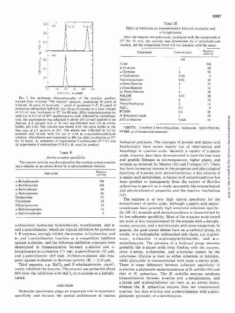
hoxylic acids, also serve as amino donors to a lesser extent. The transamination product from n-lysine was investigated in a pyruvate system. The product reacted with o-aminohenzalde- hyde and ninhydrin to develop an orange and a yellow color, respectively, in the same manner as reported previously (16), suggesting that a heterocyclic form of the keto analogue of Iysine, i.e. A’-piperideine-2carhoxylate or A’-piperideine-6- carhoxylate. is produced. The reaction mixture containing n-lysine. pyruvate, and 10 wg of enzyme was incubated at 30” for 60 min. After deproteinization by addition of 0.2 ml of 50% trichloroacetic acid, followed by centrifugation. the superna- tant was adjusted to about pH 2.0, and analyzed by a cation exchange chromatography (Fig. 7). The product from n-lysine was identified with authentic Al-piperideine-2carhoxylate prepared from c-N-carhohenzoxy-L-lysine. This result indi- cates the occurrence of oc-transamination between n-lysine and pyruvate to produce Al-piperideine-2carhoxylate.
Amino acceptor specificity also was investigated in the reaction system containing o-alanine as an amino donor (Table II). a-Ketohutyrate. cu-ketoglutarate. and cu-ketovalerate are the preferred amino acceptors. All L-amino acids tested, and glycine, are inert.
Kinetics-The Michaelis constants for n-alanine were deter- mined to he 1.3 and 4.2 mM in transamination with a-ketohu- tyrate and a-ketoglutarate. respectively, according to the method of Velick and Vavra (25). The constants for cu-ketohu- tyrate and Lu-ketoglutarate were calculated to he 14 and 3.4 mM, respectively, when n-alanine was used as an amino donor.
Inhibitors-The various compounds were investigated for their inhibitory effects on enzyme activity (Table III). Phenyl- hydrazine, hydroxylamine, aminoxyacetate, 3-methyl-2-hen-
TABLE I
Amino donor specificity
The enzyme activity was assayed by determining alanine, gluta- mate, or waminobutyrate formed with an automatic amino acid analyzer (Yanagimoto LC-5s). The relative activity less than 0.5 is expressed as zero.
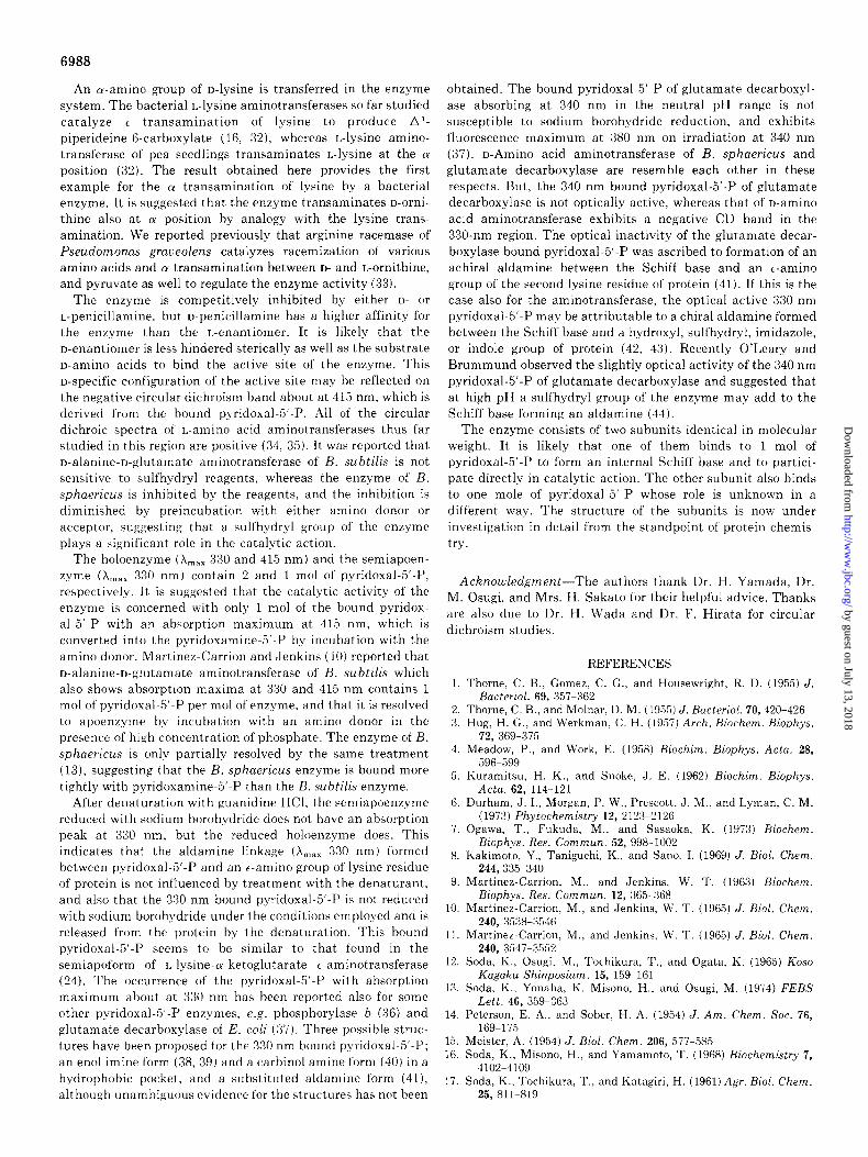
Effect of inhibitOrS on tranSUminatiOn between D-ahine and
a-ketoglutarate
After the enzyme was previously incubated with the compounds at 37” for 15 min, the activity was determined by a salicylaldehyde method. All the compounds listed did not interfere with the assay.
Compounds Concentration Relative activitv
FRACTION NUMBER
FIG. 7. Ion exchange chromatography of the reaction product formed from D-lysine. The reaction mixture, containing 50 Fmol of D-lysine, 50 Frnol of pyruvate, 1 pmol of pyridoxal 5,-P, 80 pmol of potassium phosphate (pH 8.0), and 10 lg of enzyme in a final volume of 2.0 ml, was incubated at 37” for 60 min. After deproteinization by addition of 0.2 ml of 50% trichloroacetic acid, followed by centrifuga- tion, the supernatant was adjusted to about pH 2.0 and applied to an Aminex A-4 column (0.8 x 55 cm) equilibrated with 0.2 M citrate buffer, pH 3.25. The column was eluted with the same buffer at the flow rate of 1.7 ml/min at 55”. The eluate was collected in 2.5-ml portions and mixed with 0.5 ml of 0.04 M o-aminobenzaldehyde solution. Absorbance was measured at 465 nm after incubation at 37” for 12 hours. A, authentic A’-piperideine-2.carboxylate (P-2-C) and A’-piperideine-6.carboxylate (P-6-C); B, reaction product.
TABLE II
Amino acceptor specificity
The enzyme activity was determined in the reaction system contain- ing o-alanine as an amino donor by a salicylaldehyde method.
zothiazolone hydrazone hydrochloride, D-cycloserine, and D- and L-penicillamine, which are typical inhibitors for pyridoxal 5’.P enzymes, strongly inhibit the enzyme. D-Cycloserine, and D- and L-penicillamine function as a competitive inhibitor against D-alanine, and the following inhibition constants were determined in transamination between D-alanine and 01. ketoglutarate:D-cycloserine (11 nM), D-penicillamine (57 PM),
and L-penicillamine (0.8 mM). P-Chloro-n-alanine also com- petes against n-alanine to decrease activity (K, = 2.25 fiLMI.
Thiol reagents, e.g. HgCl, and N-ethylmaleimide, signifi- cantly inhibited the enzyme. The enzyme was protected about 68% from the inhibition with H&l, by n-alanine or a-ketoglu- tarate.
DISCUSSION
Molecular asymmetry plays an important role in enzymatic specificity and dictates the spatial architecture of various
a MBTH, 3-methyl-2-benzothiazolone hydrazone hydrochloride; PCMB, p-chloromercuribenzoate.
biological polymers. The concepts of protein and amino acid biochemistry have grown mainly out of observations and knowledge of L-amino acids. Recently a variety of D-amino acids, however, have been demonstrated in both the free state and peptide linkages in microorganisms, higher plants, and animals as reviewed by Meister (26) and Corrigan (27). There has been increasing interest in the properties and physiological functions of D-amino acid aminotransferase, a key enzyme of n-amino acid metabolism. D-Amino acid aminotransferase has been purified to homogeneity from the extract of Bacillus sphaericus to permit us to study accurately the enzymological and physicochemical properties and the reaction mechanism (13).
The enzyme is of very high optical specificity for the n-enantiomer of amino acids. Although L-amino acid amino- transferases have generally high structural substrate specific- ity (2%31), D-amino acid aminotransferase is characterized by its low substrate specificity. Most of the n-amino acids tested are more or less transaminated by the enzyme with oc-ketoglu- tarate, pyruvate, and a-ketobutyrate with some exceptions. In general, the good amino donors have an w-carboxyl group, its amide, or a hydrophobic unbranched side chain, e.g. n-gluta- mate, D-theanine (y-n-glutamylethylamide), and D-LY-
aminobutyrate. The presence of a hydroxyl group prevents probably the n-amino acids from binding with the enzyme, since n-serine, D-threonine, and n-tyrosine cannot be the substrates. Glycine is inert as either substrate or inhibitor, while glyoxylate is transaminated with some D-amino acids. There is some difference between substrate specificity of D-alanine-n-glutamate aminotransferase of B. subtilis (10) and that of B. sphaericus. The B. subtillis enzyme catalyzes transamination between D-serine and cu-ketoglutarate, and n-lysine and D-phenylalanine are inert as an amino donor, whereas the B. sphaericus enzyme does not transaminate D-serine, but does n-lysine and n-phenylalanine with cy-keto- glutarate, pyruvate, or a-ketobutyrate.
An a-amino group of n-lysine is transferred in the enzyme system. The bacterial L-lysine aminotransferases so far studied catalyze 6 transamination of lysine to produce Al- piperideine-6-carboxylate (16, 32), whereas L-lysine amino- transferase of pea seedlings transaminates L-lysine at the cy position (32). The result obtained here provides the first example for the 01 transamination of lysine by a bacterial enzyme. It is suggested that the enzyme transaminates n-orni- thine also at N position by analogy with the lysine trans- amination. We reported previously that arginine racemase of Pseudomonas graveolens catalyzes racemization of various amino acids and cy transamination between D- and L-ornithine, and pyruvate as well to regulate the enzyme activity (33).
The enzyme is competitively inhibited by either D- or L-penicillamine, but o-penicillamine has a higher affinity for the enzyme than the L-enantiomer. It is likely that the o-enantiomer is less hindered sterically as well as the substrate n-amino acids to bind the active site of the enzyme. This n-specific configuration of the active site may be reflected on the negative circular dichroism band about at 415 nm, which is derived from the bound pyridoxal-5’.P. All of the circular dichroic spectra of L-amino acid aminotransferases thus far studied in this region are positive (34, 35). It was reported that n-alanine-n-glutamate aminotransferase of B. subtilis is not sensitive to sulfhydryl reagents, whereas the enzyme of B. sphaericus is inhibited by the reagents, and the inhibition is diminished by preincubation with either amino donor or acceptor, suggesting that a sulfhydryl group of the enzyme plays a significant role in the catalytic action.
The holoenzyme (A,,,,, 330 and 415 nm) and the semiapoen-
zyme (Lax 330 nm) contain 2 and 1 mol of pyridoxal-5,-P, respectively. It is suggested that the catalytic activity of the enzyme is concerned with only 1 mol of the bound pyridox- al-5’.P with an absorption maximum at 415 nm, which is converted into the pyridoxamine-5-P by incubation with the amino donor. Martinez-Carrion and cJenkins (10) reported that n-alanine-n-glutamate aminotransferase of B. subtilis which also shows absorption maxima at 330 and 415 nm contains 1 mol of pyridoxal-5’.P per mol of enzyme, and that it is resolved to apoenzyme by incubation with an amino donor in the presence of high concentration of phosphate. The enzyme of B. sphaericus is only partially resolved by the same treatment (13), suggesting that the B. sphaericus enzyme is bound more tightly with pyridoxamine-5-P than the B. subtilis enzyme.
After denaturation with guanidine HCl, the semiapoenzyme reduced with sodium borohydride does not have an absorption peak at 330 nm, but the reduced holoenzyme does. This indicates that the aldamine linkage (A,,,,, 330 nm) formed between pyridoxal-5’-P and an t-amino group of lysine residue of protein is not influenced by treatment with the denaturant, and also that the 330 nm bound pyridoxal-5’.P is not reduced with sodium borohydride under the conditions employed and is released from the protein by the denaturation. This bound pyridoxal-5-P seems to be similar to that found in the semiapoform of I.-lysine-uc-ketoglutarate t-aminotransferase (24). The occurrence of the pyridoxal-5’.P with absorption maximum about at 330 nm has been reported also for some other pyridoxal-5’-P enzymes, e.g. phosphorylase b (36) and glutamate decarboxylase of E. coli (37). Three possible struc- tures have been proposed for the 330 nm bound pyridoxal-5,-P; an enol imine form (38, 39) and a carbinol amine form (40) in a hydrophobic pocket, and a substituted aldamine form (41), although unambiguous evidence for the structures has not been
obtained. The bound pyridoxal-5’.P of glutamate decarboxyl- ase absorbing at 340 nm in the neutral pH range is not susceptible to sodium borohydride reduction, and exhibits fluorescence maximum at 380 nm on irradiation at 340 nm (37). n-Amino acid aminotransferase of B. sphaericus and glutamate decarboxylase are resemble each other in these respects. But, the 340 nm bound pyridoxal-5’-P of glutamate decarboxylase is not optically active, whereas that of n-amino acid aminotransferase exhibits a negative CD band in the 330-nm region. The optical inactivity of the glutamate decar- boxylase bound pyridoxal-5’.P was ascribed to formation of an achiral aldamine between the Schiff base and an t-amino group of the second lysine residue of protein (41). If this is the case also for the aminotransferase, the optical active 330 nm pyridoxal-5’.P may be attributable to a chiral aldamine formed between the Schiff base and a hydroxyl, sulfhydryl, imidazole, or indole group of protein (42, 43). Recently O’Leary and Brummund observed the slightly optical activity of the 340 nm pyridoxal-5’-P of glutamate decarboxylase and suggested that at high pH a sulfhydryl group of the enzyme may add to the Schiff base forming an aldamine (44).
The enzyme consists of two subunits identical in molecular weight. It is likely that one of them binds to 1 mol of pyridoxal-5’.P to form an internal Schiff base and to partici- pate directly in catalytic action. The other subunit also binds to one mole of pyridoxal-5’.P whose role is unknown in a different way. The structure of the subunits is now under investigation in detail from the standpoint of protein chemis- try.
Acknowledgment-The authors thank Dr. H. Yamada, Dr. M. Osugi, and Mrs. H. Sakato for their helpful advice. Thanks are also due to Dr. H. Wada and Dr. F. Hirata for circular dichroism studies.
1.
2. 3.
4.
5.
6.
7.
8.
9.
10.
11.
12.
13.
14.
15. 16.
17.
REFERENCES
Thorne, C. B., Gomez, C. G., and Housewright, R. D. (1955) J. Bacterial. 69, 357-362
Thorne, C. B., and Molnar, D. M. (1955) J. Bacterial. 70,420-426 Hug, H. G., and Werkman, C. H. (1957) Arch. Biochem. Biophys.
72, 369-375 Meadow, P., and Work, E. (1958) Biochim. Biophys. Acta. 28,
596-599 Kuramitsu, H. K., and Snoke, J. E. (1962) Biochim. Biophys.
Acta. 62, 114-121 Durham, J. I., Morgan, P. W., Prescott, J. M., and Lyman, C. M.
(1973) Phytochemistry 12, 2123S2126 Ogawa, T., Fukuda, M., and Sasaoka, K. (1973) Biochem.
Biophys. Res. Commun. 52, 998-1002 Kakimoto, Y., Taniguchi, K., and Sano, I. (1969) J. Biol. Chem.
244,335-340 Martinez-Carrion, M., and Jenkins, W. T. (1963) Biochem.
Biophys. Res. Commun. 12, 365-368 Martinez-Carrion, M., and Jenkins, W. T. (1965) J. Biol. Chem.
240, 3538-3546 Martinez-Carrion, M., and Jenkins, W. T. (1965) J. Biol. Chem.
240, 3547-3552 Soda, K., Osugi, M., Tochikura, T., and Ogata, K. (1965) Koso
Kugaku Shinposium. 15, 159-161 Soda, K., Yonaha, K. Misono, H., and Osugi, M. (1974) FEBS
Lett. 46, 359-363 Peterson, E. A., and Sober, H. A. (1954) J. Am. Chem. Sot. 76,
169-175 Meister, A. (1954) J. Biol. Chem. 206, 577-585 Soda, K., Misono, H., and Yamamoto, T. (1968) Biochemistry 7,
4102-4109 Soda, K., Tochikura, T., and Katagiri, H. (1961) Agr. Biol. Chem.
18. Berntsson, S. (1955) Anal. Chem. 27, 1659-1660 19. Andrews, P. (1964) Biochem. J. 91, 222-233 20. Weber, K., and Osborn, M. (1969) J. Biol. Chem. 244, 4406-4412 21. Matsuo, Y., and Greenberg, D. M. (1959) J. Biol. Chem. 234,
507-515 22. Wada, H., and Snell, E. E. (1961) J. Biol. Chem. 236, 2089-2095 23. Soda, K., Yorifuji, T., Misono, H., and Moriguchi, M. (1969)
Biochem. J. 114, 629-633 24. Soda, K., and Misono, H. (1968) Biochemistry 7, 4110-4119 25. Velick, S. F., and Vavra, J. (1962) J. Biol. Chem. 237, 2109-2122 26. Meister, A. (1965) Biochemistry of The Amino Acids, 2nd Ed., pp.
113-118, Academic Press, New York 27. Corrigan, J. J. (1969) Science 164, 142-149 28. Saier, M. H., Jr., and Jenkins, W. T. (1967) J. Biol. Chem. 242,
101-108 29. Taylor, R. T., and Jenkins, W. T. (1966) J. Biol. Chem. 241,
4396-4405 30. Jacoby, G. A., and La Du, B. N. (1964) J. Biol. Chem. 239,419-424 31. Novogrodsky, A., and Meister, A. (1964) Biochim. Biophys. Acta
81,605-608 32. Hasse, K., Ratych, 0. T., and Salinikow, J. (1967) 2. Physiol.
Chem. 348,843-851 33. Yorifuji, T., Misono, H., and Soda, K. (1971) J. Biol. Chem. 246,
5093-5101 34. Dunathan, H. C. (1971) Adu. Enzymol. 35,79-134 35. Torchinsky, Yu. M., Malakhova, E. A., Livanova, N. B., and
Pikhelgas, V. Ya. (1968) Pyridoral Catalysis, Enzymes and Model Systems. 2nd IUB Symposium. Moscow, 1966, pp. 269-290, Interscience, New York
36. Kent, A. B., Krebs, E. G., and Fischer, E. H. (1958) J. Biol. Chem. 232,549-558
37. Shukuya, R., and Schwert, G. W. (1960) J. Biol. Chem. 235, 1653-1657
38. Johnson, G. F., Jan-ITu, Bartlett, M. L. S., and Graves, D. J. (1970) J. Biol. Chem. 245,5560-5568
39. Shaltiel, S., and Cortijo, M. (1970) Biochem. Biophys. Res. Commun. 41,594-600
40. Honikel, K. O., and Madsen, N. B. (1972) J. Biol. Chem. 247, 1057-1064
41. O’Leary, M. H. (1971) Biochim. Biophys. Acta 242,484&492 42. Heyl, D., Harris, S. A., and Folkers, K. (1948) J. Am. Chem. Sot.
70,3429-3431 43. Mackay, D. (1962) Arch. Biochem. Biophys. 99,93-100 44. O’Leary, M. H., and Brummund, W. (1974) J. Biol. Chem. 249,
3737-3745 45. Davis, B. J. (1964) Ann. N. Y. Acad. Sci. 121,404-427


![Research Paper The Prognostic Value of aspartate aminotransferase … · 2019-05-22 · Aspartate aminotransferase (AST) to lymphocyte ratio (ALRI) [17], systemic immune-inflammation](https://static.documents.pub/doc/80x56/5f0222077e708231d402bbfe/research-paper-the-prognostic-value-of-aspartate-aminotransferase-2019-05-22-aspartate.jpg)