The Georgia Veterinary Diagnostic Laboratories D-Labs User Guide Using Laboratory Services and Specimen Submission Requirements The Georgia Veterinary Diagnostic Laboratories www.vet.uga.edu/dlab 6/16/2010
Transcript
The Georgia Veterinary Diagnostic Laboratories
D-Labs User Guide Using Laboratory Services and Specimen Submission Requirements
The Georgia Veterinary Diagnostic Laboratories www.vet.uga.edu/dlab
6/16/2010
The Georgia Veterinary Diagnostic Laboratories – www.vet.uga.edu/dlab The University of Georgia College of Veterinary Medicine September 2, 2010
Table of Contents INTRODUCTION ............................................................................................................................................. 4
Turnaround times ................................................................................................................................... 10
What samples are to be submitted for testing? ..................................................................................... 20
The Georgia Veterinary Diagnostic Laboratories – www.vet.uga.edu/dlab The University of Georgia College of Veterinary Medicine September 2, 2010
INTRODUCTION The two Georgia veterinary diagnostic laboratories, located in Athens and Tifton, are units of The University of Georgia College of Veterinary Medicine and are partially funded by the Georgia Department of Agriculture through a contract with the UGA Board of Regents.
This guide is intended to provide information about the laboratories, the scope of tests performed, recommendations for selection of tests, and instructions on sample submission including specimen collection, preservation, packaging, and appropriate shipment.
Mission The core mission of the two laboratories is to provide accurate, expeditious and comprehensive diagnostic service for veterinarians and animal owners. The laboratories provide diagnostic consultation and assistance to veterinarians. The laboratories also conduct surveillance testing for non-endemic diseases, as well as testing for diseases in non-traditional species such as wildlife, aquatic, and laboratory animals.
Organization The laboratories are divided into the following sections, which are staffed by specialists:
I. LABORATORY OPERATION II. BACTERIOLOGY and MYCOLOGY
III. CLINICAL PATHOLOGY & CYTOLOGY IV. GEORGIA LABORATORY ANIMAL DIAGNOSTIC SERVICE (GLADS) V. MOLECULAR DIAGNOSTICS
VI. PATHOLOGY/HISTOPATHOLOGY VII. VIROLOGY/SEROLOGY & ELECTRON MICROSCOPY
VIII. TOXICOLOGY
The multidisciplinary organization of the laboratories provides the capabilities necessary for the detection and characterization of most animal diseases. Both laboratories are accredited as full service laboratories by the AMERICAN ASSOCIATION OF VETERINARY LABORATORY DIAGNOSTICIANS.
Services Laboratory services are funded in part by the Georgia Department of Agriculture. Additional funding is derived from fee income and Federal service contracts. See our CURRENT FEE SCHEDULE for pricing information. Note that as our services are subsidized by Georgia tax payers, out-of-state specimens will be charged at a higher rate for those assays for which we do not recoup costs.
Cases are accepted only when referred by a licensed veterinarian with a completed SUBMISSION FORM.
Receiving A case number is assigned to each submitted sample for identification throughout processing, testing, reporting, and data storage.
Specimens are distributed to appropriate laboratory sections as indicated by either the submitting veterinary practitioner or the pathologist on duty.
Reporting Results will only be reported to and discussed with the referring veterinarian. Results may be disclosed to a non-governmental third party only with the veterinarian’s permission.
Regulatory agencies will be notified when positive results are obtained on reportable animal diseases.
Preliminary reports on finalized tests will be sent to the submitting veterinarian via e-mail or fax at no cost. A final report will be sent by regular mail, fax, or e-mail, as indicated by the submitting veterinarian on the submission form. Reports are also available ON OUR WEBSITE. To set up your on-line account, please call 706-542-5568.
Results and reports are archived electronically. Original submission forms and associated documents will be archived for at least 7 years.
Collection and Preservation of Specimens Properly collected and submitted specimens are necessary for accurate and meaningful laboratory test results. The accuracy of any laboratory result is dependent on submission of proper specimens with a
The Georgia Veterinary Diagnostic Laboratories – www.vet.uga.edu/dlab The University of Georgia College of Veterinary Medicine September 2, 2010
complete case history. If you are unsure of what samples to collect or how they should be preserved or shipped, please CALL THE LABORATORY for assistance. Some general guidelines are:
Animals If multiple animals are affected, more than one animal carcass should be submitted. With the exception of small laboratory animals, live animals are not accepted for necropsy. Please, make arrangements for euthanasia through the referring veterinarian or the Veterinary Teaching Hospital prior to submission for necropsy.
Organs and Tissues A complete set of both fresh and formalin fixed tissues are required to adequately provide a diagnosis for mail-in necropsies. Each fresh tissue should be placed in an individual leak-proof plastic container (preferably a capped tube, jar, or sterilized sampling bag such as Whirl-Pak® bags) and maintained at refrigerator temperatures from collection until receipt at the laboratory. Tissues for histopathologic examination must be preserved in 10% buffered neutral formalin solution (see the PATHOLOGY/HISTOPATHOLOGY for more information) and submitted in a wide-mouth, leak-proof plastic container with sufficient room for formalin. Do not submit formalin-fixed tissues in glass jars, jars with a neck, in Whirl-Pak bags, or in jars whose lid does not seal (e.g., baby food jars).
Blood, urine, plants, and other specimens Follow the laboratory section’s instructions for specimen submission. Our INTERACTIVE LAB TESTS & FEE
SCHEDULE also provides some submission information. When requesting multiple tests that require more than one laboratory section to perform, submit separate samples for each test. This is particularly important when submitting small samples such as fecal swabs or when submitting a sterile sample for more than one test (i.e., fluid for culture as well as cytology).
Viruses When diagnosing enteric and skin infections, it is advantageous to submit specimens to both Virology and Electron Microscopy sections.
Federal Regulations Federal regulations govern the packaging and labeling of diagnostic specimens. If, during shipment, spillage occurs that damages mail, equipment, or personnel, the shipper may face prosecution even though the material involved was not infectious or hazardous. Careful packaging is essential to prevent any leaking fluid or breakage. The outside of the container should be appropriately labeled (e.g. "Perishable, Biologic Specimen", etc.). Specimens submitted in an unsafe manner may incur additional expenses to cover the costs of special handling. For additional details, please consult AVMA
GUIDELINES.
Test Results and Interpretation We make every effort to provide reports with useful interpretations relevant to individual case histories. However, some of the laboratory data we provide does not constitute a diagnosis and must be carefully interpreted to arrive at a final diagnosis. For case consultation, faculty and technical staff members are available by telephone.
The Georgia Veterinary Diagnostic Laboratories – www.vet.uga.edu/dlab The University of Georgia College of Veterinary Medicine September 2, 2010
BACTERIOLOGY and MYCOLOGY The primary responsibility of the Bacteriology section of both diagnostic laboratories is to isolate and identify bacterial and fungal pathogens that cause disease in animals (see our INTERACTIVE LAB TESTS & FEE
SCHEDULE for a complete list of tests in this section). In addition to traditional species our laboratories have expertise in the area of aquatic, exotic and environmental microbiology.
Antimicrobial Susceptibility Testing (AST) This section also performs in vitro antimicrobial susceptibility testing (AST) on significant bacterial isolates using the disk diffusion method. Minimum inhibitory concentrations (MIC) results are available on request. The antimicrobial susceptibility testing is performed following CLSI GUIDELINES (Clinical and Laboratory Standards Institute). We attempt to perform AST on isolates that are significant and expected to result in a disease process at a particular body site. We may not perform AST on isolates that are common contaminants or normal flora as it would not provide useful information and may lead to inappropriate antibiotic use. We do not routinely perform AST on fastidious organisms for which there are no recommended guidelines or interpretive criteria; however, in special circumstances and on specific request from practitioners, we will attempt AST on such isolates.
Antimicrobial susceptibility testing will help to choose the appropriate antimicrobials to treat the animal patient. It is important to use the correct antimicrobial agent at the recommended dose and duration to achieve an optimum concentration at the site of infection. Failure to follow the appropriate antibiotic treatment may promote antimicrobial resistance and result in treatment failure.
Sample Collection and Transport Following proper submission guidelines for clinical samples is important for obtaining quality results. Appropriate selection, collection and handling of specimens are essential in aiding microbiological diagnosis. Follow these five rules to ensure success.
Cardinal Rules of Specimen Collection:
Keep it Clean Use sterile equipment, aseptic techniques.
Keep it Moist Specimens taken by swab will dry out if not placed into transport media. Since microorganisms die rapidly on dry swabs the sample may no longer be representative of the disease process of the animal from which it was taken. Fresh tissue specimens may be kept moist by placing them in a sterile container with sterile saline.
Keep it Cool All specimens should be refrigerated at 4°C, not frozen. Microorganisms are fragile and susceptible to extremes of temperature. Adverse shipping conditions will affect the recovery of significant pathogens and may promote overgrowth by normal flora or environmental contaminants. Shipping samples with ice packs is recommended.
The Georgia Veterinary Diagnostic Laboratories – www.vet.uga.edu/dlab The University of Georgia College of Veterinary Medicine September 2, 2010
Move Fast All specimens should be transported to the laboratory as quickly as possible; the fresher the sample the better the chance of recovering significant microorganisms. Therefore, overnight shipping is highly recommended.
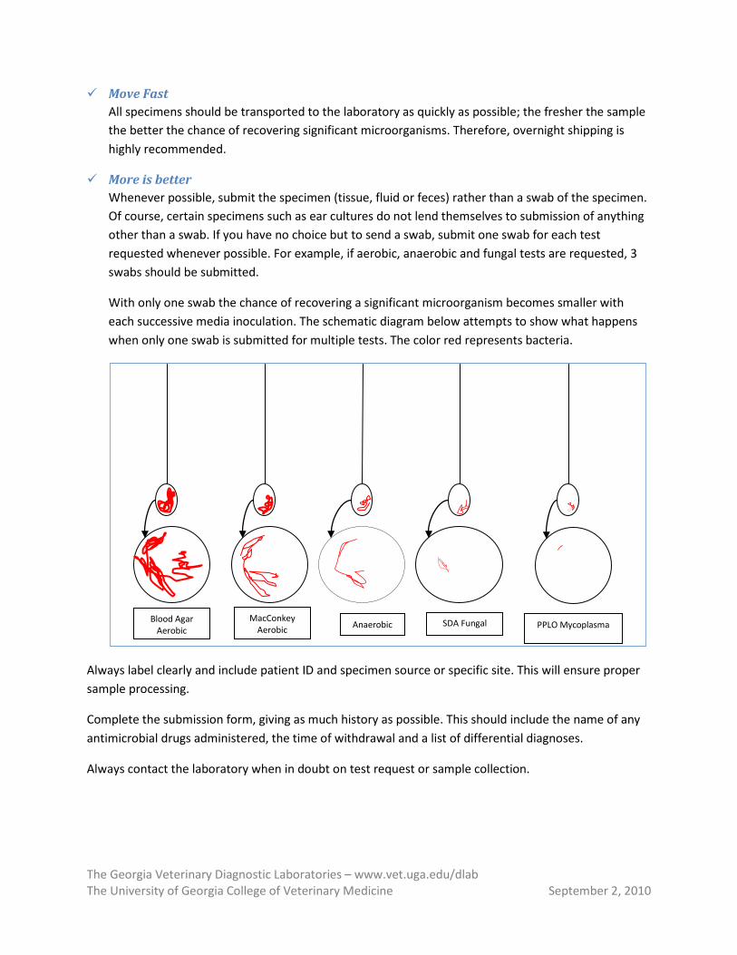
More is better Whenever possible, submit the specimen (tissue, fluid or feces) rather than a swab of the specimen. Of course, certain specimens such as ear cultures do not lend themselves to submission of anything other than a swab. If you have no choice but to send a swab, submit one swab for each test requested whenever possible. For example, if aerobic, anaerobic and fungal tests are requested, 3 swabs should be submitted.
With only one swab the chance of recovering a significant microorganism becomes smaller with each successive media inoculation. The schematic diagram below attempts to show what happens when only one swab is submitted for multiple tests. The color red represents bacteria.
Always label clearly and include patient ID and specimen source or specific site. This will ensure proper sample processing.
Complete the submission form, giving as much history as possible. This should include the name of any antimicrobial drugs administered, the time of withdrawal and a list of differential diagnoses.
Always contact the laboratory when in doubt on test request or sample collection.
Anaerobic PPLO Mycoplasma Blood Agar
Aerobic SDA Fungal MacConkey
Aerobic
The Georgia Veterinary Diagnostic Laboratories – www.vet.uga.edu/dlab The University of Georgia College of Veterinary Medicine September 2, 2010
Specimen recommendations
Tissues From field necropsy cases, a 2-5 cm specimen that includes the lesion is more desirable than a swab. Keep intestines separate from other organs. Tie off the ends of intestinal loops if an anaerobic infection is suspected.
Fluids Liquid specimens such as milk and urine should be collected aseptically in a leak-proof plastic container. Cystocentesis is recommended for urine collection although mid-stream is acceptable for culture if samples are processed soon after collection.
Anaerobes Remember that oxygen is toxic to many anaerobes. Aspirates or biopsy specimens are more desirable than swabs. Swabs should be submitted in an anaerobic transport media.
Do not use Whirl-pack® bags, OB sleeves, or syringes capped with needles for submitting liquid specimens.
The Georgia Veterinary Diagnostic Laboratories – www.vet.uga.edu/dlab The University of Georgia College of Veterinary Medicine September 2, 2010
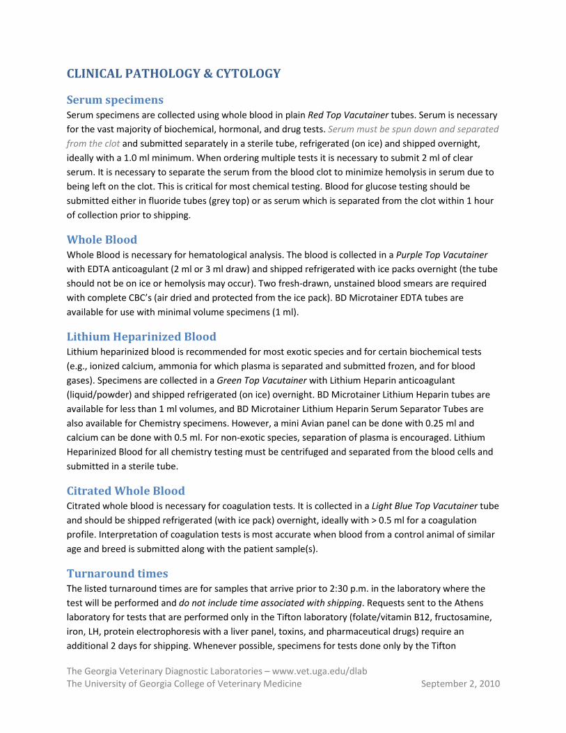
CLINICAL PATHOLOGY & CYTOLOGY
Serum specimens Serum specimens are collected using whole blood in plain Red Top Vacutainer tubes. Serum is necessary for the vast majority of biochemical, hormonal, and drug tests. Serum must be spun down and separated from the clot and submitted separately in a sterile tube, refrigerated (on ice) and shipped overnight, ideally with a 1.0 ml minimum. When ordering multiple tests it is necessary to submit 2 ml of clear serum. It is necessary to separate the serum from the blood clot to minimize hemolysis in serum due to being left on the clot. This is critical for most chemical testing. Blood for glucose testing should be submitted either in fluoride tubes (grey top) or as serum which is separated from the clot within 1 hour of collection prior to shipping.
Whole Blood Whole Blood is necessary for hematological analysis. The blood is collected in a Purple Top Vacutainer with EDTA anticoagulant (2 ml or 3 ml draw) and shipped refrigerated with ice packs overnight (the tube should not be on ice or hemolysis may occur). Two fresh-drawn, unstained blood smears are required with complete CBC’s (air dried and protected from the ice pack). BD Microtainer EDTA tubes are available for use with minimal volume specimens (1 ml).
Lithium Heparinized Blood Lithium heparinized blood is recommended for most exotic species and for certain biochemical tests (e.g., ionized calcium, ammonia for which plasma is separated and submitted frozen, and for blood gases). Specimens are collected in a Green Top Vacutainer with Lithium Heparin anticoagulant (liquid/powder) and shipped refrigerated (on ice) overnight. BD Microtainer Lithium Heparin tubes are available for less than 1 ml volumes, and BD Microtainer Lithium Heparin Serum Separator Tubes are also available for Chemistry specimens. However, a mini Avian panel can be done with 0.25 ml and calcium can be done with 0.5 ml. For non-exotic species, separation of plasma is encouraged. Lithium Heparinized Blood for all chemistry testing must be centrifuged and separated from the blood cells and submitted in a sterile tube.
Citrated Whole Blood Citrated whole blood is necessary for coagulation tests. It is collected in a Light Blue Top Vacutainer tube and should be shipped refrigerated (with ice pack) overnight, ideally with > 0.5 ml for a coagulation profile. Interpretation of coagulation tests is most accurate when blood from a control animal of similar age and breed is submitted along with the patient sample(s).
Turnaround times The listed turnaround times are for samples that arrive prior to 2:30 p.m. in the laboratory where the test will be performed and do not include time associated with shipping. Requests sent to the Athens laboratory for tests that are performed only in the Tifton laboratory (folate/vitamin B12, fructosamine, iron, LH, protein electrophoresis with a liver panel, toxins, and pharmaceutical drugs) require an additional 2 days for shipping. Whenever possible, specimens for tests done only by the Tifton
The Georgia Veterinary Diagnostic Laboratories – www.vet.uga.edu/dlab The University of Georgia College of Veterinary Medicine September 2, 2010
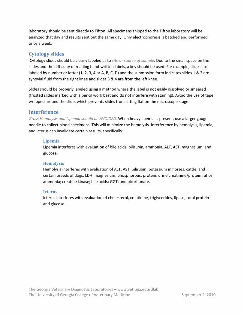
laboratory should be sent directly to Tifton. All specimens shipped to the Tifton laboratory will be analyzed that day and results sent out the same day. Only electrophoresis is batched and performed once a week.
Cytology slides Cytology slides should be clearly labeled as to site or source of sample. Due to the small space on the slides and the difficulty of reading hand-written labels, a key should be used. For example, slides are labeled by number or letter (1, 2, 3, 4 or A, B, C, D) and the submission form indicates slides 1 & 2 are synovial fluid from the right knee and slides 3 & 4 are from the left knee.
Slides should be properly labeled using a method where the label is not easily dissolved or smeared (frosted slides marked with a pencil work best and do not interfere with staining). Avoid the use of tape wrapped around the slide, which prevents slides from sitting flat on the microscope stage.
Interference Gross Hemolysis and Lipemia should be AVOIDED. When heavy lipemia is present, use a larger gauge needle to collect blood specimens. This will minimize the hemolysis. Interference by hemolysis, lipemia, and icterus can invalidate certain results, specifically:
Lipemia Lipemia interferes with evaluation of bile acids, bilirubin, ammonia, ALT, AST, magnesium, and glucose.
Hemolysis Hemolysis interferes with evaluation of ALT; AST; bilirubin; potassium in horses, cattle, and certain breeds of dogs; LDH; magnesium; phosphorous; protein, urine creatinine/protein ratios, ammonia; creatine kinase; bile acids; GGT; and bicarbonate.
Icterus Icterus interferes with evaluation of cholesterol, creatinine, triglycerides, lipase, total protein and glucose.
The Georgia Veterinary Diagnostic Laboratories – www.vet.uga.edu/dlab The University of Georgia College of Veterinary Medicine September 2, 2010
GEORGIA LABORATORY ANIMAL DIAGNOSTIC SERVICE (GLADS) The GLADS program, a recent addition to the Athens Veterinary Diagnostic Laboratory, was implemented to serve both the University Animal Resource Program and the rapidly growing Biotechnology industry in the southeast. Experienced faculty and staff conduct tests with state of the art technology and reagents.
GLADS offers a well rounded diagnostic service for rodents, rabbits, and primates in the following areas: necropsy, histopathology, serology, molecular biology, bacteriology, mycology, parasitology, clinical pathology, and environmental disinfection monitoring. See our INTERACTIVE LAB TESTS & FEE SCHEDULE for a complete list of tests and prices.
A special submission form is required to submit samples to GLADS. GLADS SUBMISSION FORMS are available on our website.
The Georgia Veterinary Diagnostic Laboratories – www.vet.uga.edu/dlab The University of Georgia College of Veterinary Medicine September 2, 2010
MOLECULAR DIAGNOSTICS The Molecular Biology section utilizes Polymerase Chain Reaction (PCR) technology to detect the presence of nucleic acids (DNA/RNA) from pathogenic organisms in clinical samples. Molecular diagnostic tests are fast and more sensitive when compared to other conventional methods such as culture or viral isolation. A wide variety of tests are available for bacteria, fungi, and viruses from many animal species, including pathogens of cats, dogs, horses, marine mammals, exotics, birds, as well as cattle.
Results can be obtained as early as 24 hours from sample receipt in most cases. Please see our INTERACTIVE LAB TESTS & FEE SCHEDULE for available tests, appropriate samples for submission, and turnaround time. Please, check our list on a regular basis as new tests are added frequently.
It is important to remember that a positive PCR test result may indicate an active infection or presence of the organism (live/dead) in the sample submitted. False negative results may occur due to low numbers of the organisms or PCR inhibitory factors present in the sample. Like with any other testing methods, factors such as clinical signs, species of animal tested and sample sites should be considered for reliable interpretation of test results.
The Georgia Veterinary Diagnostic Laboratories – www.vet.uga.edu/dlab The University of Georgia College of Veterinary Medicine September 2, 2010
PATHOLOGY/HISTOPATHOLOGY For appropriate interpretation of the samples submitted, a complete history with a summary of clinical course of disease, including treatment, and surgical or necropsy findings is essential and must be included with all submissions.
Whole Animal submissions Whole animals submitted for necropsy should not be frozen, but should be shipped overnight on ice within an insulated container. Frozen carcasses may take a long time to thaw and greatly increase turn-around time. Freezing also kills many bacteria, often preventing accurate bacterial culture, and creates serious tissue artifacts on histopathology.
Histopathology Specimen submissions Specimens for histopathology should be submitted in neutral buffered 10% formalin solution in wide-mouthed, leak- and shatter-proof containers. The volume ratio of tissue to formalin must be at least 1:10. Tissues fixed for 24 hours in an appropriate amount of formalin prior to submission may be subsequently submitted in a smaller container with a reduced amount of fixative, thus decreasing the size and weight of the container used for shipping.
Containers must be labeled with names of the veterinarian, owner, and animal. The label must also have the date collected and information about the sample submitted. Pencil should be used if there is a chance that the label will come in contact with formalin.
When multiple biopsies are submitted with one case, each tissue should be submitted in a separate container and labeled appropriately.
For neoplasms, inking of surgical margins with India ink, for example, is recommended for the most accurate assessment of completeness of excision.
Improper Handling Improper handling or fixation of tissues can induce artifacts that may result in non-diagnostic specimens. Examples of improper handling include:
Failure to place tissues in formalin immediately after collection Unfixed specimens are subject to dehydration, autolysis, and proliferation of saprophytic bacteria. Refrigeration slows, but does not prevent these changes.
Inadequate fixation Using an inadequate volume of fixative, or attempting to fix large specimens (whole kidney, large mass), will result in incomplete fixation and autolysis. Processing is then delayed because unfixed tissues must be placed in a larger container with fresh formalin for additional fixation prior to routine processing. Formalin penetrates tissue at roughly 18 mm per day, and the rate of penetration slows with the thickness of the tissue. It takes approximately 100 hours (4+ days) for formalin to penetrate 36 mm.
Improper fixatives Alcohol, disinfectants, and saline are unsuitable for transport or fixation of specimens for histopathology and usually result in non-diagnostic samples.
The Georgia Veterinary Diagnostic Laboratories – www.vet.uga.edu/dlab The University of Georgia College of Veterinary Medicine September 2, 2010
Thermal dehydration Electrocautery and laser surgery cooks tissues and may destroy small tumors and skin biopsies. A scalpel or punch should be used instead. It is helpful to include the means of collection on the submission form.
Chemical dehydration Disinfectants and medications applied to the skin or residues on instruments that are chemically sterilized may damage tissues.
Freezing Ice formation in or around cells causes cell lysis and disrupts normal tissue architecture, resulting in non-diagnostic samples. Additionally, autolysis may occur during thawing.
Excessive Pressure Excessive pressure from forceps or digits can rupture and/or compress cells (e.g., crush artifact), making them unidentifiable. Small pieces of tissue, such as skin punch biopsies, can be manipulated with a needle rather than forceps to prevent crush artifact.
Skin biopsies for dermatopathology (skin conditions affecting more than one area) Punch biopsies, at least 6mm in size, of multiple affected areas are recommended. There is a single charge for dermatophology cases, regardless of the number of punch biopsies submitted. We encourage the submission of at least 4-6 skin punch biopsies with primary dermatological disease. Primary lesions are most diagnostic, so biopsy early lesions (e.g., papules, pustules, vesicles, bullae, wheals, etc) rather than chronic ones. Ulcerated lesions are typically not rewarding. If you are unsure how to most appropriately sample a lesion, please call with questions prior to biopsy.
Report types
Brief/routine report (available only in the Athens Lab) This report includes diagnosis and comment. All information required for prognosis (e.g. mitotic rate, completeness of excision, vascular invasion, behavior of lesions, etc.) is included in the comment.
Extended report This report includes the information in the brief/routine report, as well as a detailed histologic description of the lesion.
FFT (formalin fixed and fresh tissue/practitioner necropsy) Submission of a complete set of tissues is recommended. Both formalin-fixed and fresh tissues should be submitted. Alternatively, fresh tissues may be saved, refrigerated, or frozen, if microbial culture or toxicological testing is not needed. Definitive diagnosis of infectious diseases and toxins often requires ancillary testing (i.e., culture, virus isolation, fluorescent antibody testing, electron microscopy, etc.) that cannot be performed on formalin-fixed tissues.
The following is a list of suggested tissues to be collected during practitioner necropsies:
• Brain and/or spinal cord
The Georgia Veterinary Diagnostic Laboratories – www.vet.uga.edu/dlab The University of Georgia College of Veterinary Medicine September 2, 2010
• Heart • Lungs • Liver • Spleen • Kidney • Stomach • Small intestine • Large intestine • Lymph nodes • Urinary bladder • Thymus on young animals • Bone marrow • Skeletal muscle • Adrenal glands • Any affected tissues based on history, clinical signs, and/or gross findings not listed above such
as thyroid gland, reproductive organs, etc.
The Georgia Veterinary Diagnostic Laboratories – www.vet.uga.edu/dlab The University of Georgia College of Veterinary Medicine September 2, 2010
VIROLOGY/SEROLOGY & ELECTRON MICROSCOPY The Virology/Serology and Electron Microscopy sections test specimens to obtain evidence of viral, rickettsial, fungal and protozoal infection. These sections conduct diagnostic testing for the major viruses of cattle, horses, dogs, and cats, and selected testing for viruses of pigs, sheep, goats, wildlife, llamas, alpacas, amphibians and marine mammals. The sections also conduct fluorescent antibody testing and serological testing for various viral, bacterial, rickettsial, protozoal and fungal pathogens of veterinary importance.
Two main groups of tests are offered: (1) tests that determine the actual presence of the infectious disease agent or its components (antigens and toxins) including virus isolation (VI), direct fluorescent antibody (FA) examination, electron microscopy (EM), antigen-ELISA, and cytotoxic assays; and (2) tests that provide evidence of exposure such as various serological assays.
The fluorescent antibody (FA) test is the most widely used antigen detection system for rapid laboratory diagnoses. The test is usually done on frozen sections of fresh tissue; formalin-fixed specimens cannot be used. Fluorescent antibody test results are reliable if fresh, appropriate tissues are tested. Other rapid diagnostic tests include ELISA and electron microscopy (EM). When at all possible, separate specimens should be submitted for EM.
Specimens Specimens from live animals may include nasal or ocular secretions, feces, and whole blood. Specimens collected at necropsy should include the organs or tissues affected by the disease process. For details on appropriate specimens to submit for specific tests, please refer to our INTERACTIVE LAB TESTS & FEE
SCHEDULE.
Virus Isolation Specimens collected early in the acute stage of illness are preferred for virus isolation. These should be fresh with no preservative or fixative added. Materials can sometimes be conveniently submitted on sterile swabs if a viral transport medium is used. Tissue samples should be shipped refrigerated on ice packs.
Serology Serology detects the presence of antibodies in serum samples. Results of serologic tests provide useful information for regulatory and export purposes, and planning herd-health vaccination programs. With the exception of certain life-long viral infections (equine infectious anemia, bovine leukemia, caprine arthritis encephalitis), the presence of antibodies in an animal does not necessarily indicate the animal is infected with that virus at the time of sampling. Therefore, serologic results are of limited use in the diagnosis of most infectious diseases unless the results of acute and convalescent sera are compared. The acute sample should be collected as early in the illness as possible and the convalescent sample 2-4 weeks later. More definitive results can usually be obtained faster if samples are submitted for other tests at the same time, (e.g. fluorescent antibody, electron microscopy, PCR, and virus isolation).
The Georgia Veterinary Diagnostic Laboratories – www.vet.uga.edu/dlab The University of Georgia College of Veterinary Medicine September 2, 2010
In the event of special requests or large numbers of samples, please contact the laboratory prior to submission. Blood samples for serology should be collected in sterile plain tubes without anticoagulants. Bangs’ tubes may contain residues which may be toxic and thus produce non-diagnostic results. Do not freeze blood or allow it to become overheated. Submit serum rather than whole clotted blood if samples will not be received by the laboratory within 48 hours.
Electron Microscopy Electron microscopy is used to detect and identify viruses in clinical specimens, primarily feces (rotavirus, coronavirus, parvovirus, calicivirus, etc.), but is also used with other specimens such as skin lesions (poxvirus, papillomaviruses) and tracheal or conjunctival swabs and scrapings (herpesvirus). At least 1 ml of feces, more if possible, should be submitted in leak-proof containers under refrigeration. Do not freeze! Do not use plastic gloves or OB sleeves! Plastic bags are acceptable if specimens are double bagged. Fecal swabs are often acceptable for detection of canine parvovirus/coronavirus. Swabs and excised skin lesions should be placed in tightly capped containers with a few drops of saline to prevent drying.
Samples should be collected early in the course of the disease as the concentration of virus shed often decreases rapidly. In the case of herd problems, samples should be collected from more than one animal including an apparently healthy individual and several others in different stages of the disease. Multiple samples will also help in detection of viruses that are shed intermittently, as is frequently the case with infection by rota/reoviruses. Results are usually available the same day when specimens are received by the EM lab before 3:00 p.m.
Rabies Suspects The brain of an animal suspected of having rabies and associated with human exposure should be sent directly to the nearest Georgia Department of Human Resources Laboratory. Most of these laboratories request that only the brain be submitted. The brain should be cut in half longitudinally so each half includes parts of the cerebrum, midbrain, cerebellum, and brainstem. One half should be submitted to the nearest official public health rabies laboratory chilled with ice packs; the other half should be placed in formalin and saved or shipped to one of the diagnostic laboratories. The diagnostic laboratories do not perform the official fluorescent antibody test for rabies.
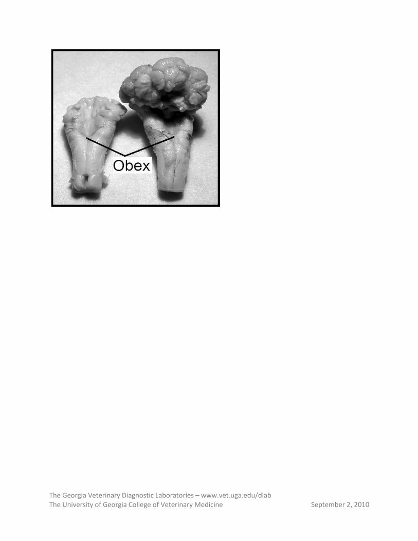
In situations where human exposure has occurred, it is suggested that you hand deliver the brain directly to the official public health rabies laboratories. Additionally, all cattle suspected of rabies should be tested for bovine spongiform encephalopathy (mad cow disease) if the rabies result is negative. To accomplish this, a piece of the brain stem (at the level of the obex, as shown below) should be submitted in a 50-ml capped tube in a box with ice packs to the Athens laboratory with the instruction “TEST FOR BSE IF RABIES NEGATIVE”.
The Georgia Veterinary Diagnostic Laboratories – www.vet.uga.edu/dlab The University of Georgia College of Veterinary Medicine September 2, 2010
The Georgia Veterinary Diagnostic Laboratories – www.vet.uga.edu/dlab The University of Georgia College of Veterinary Medicine September 2, 2010
TOXICOLOGY The University of Georgia Diagnostic Laboratory system no longer has a Veterinary Toxicologist or full service toxicology laboratory. The Tifton laboratory still has limited toxicology testing for heavy metals, cyanide, nitrates, copper and selenium. Most toxicology related samples will be shipped to the Michigan State University Toxicology Laboratory for analysis. Fees charged by the Michigan State Toxicology Laboratory for each test and sample associated shipping fees will be added to the overall cost of the specific case.
What samples are to be submitted for testing? The most critical component for a successful toxicological analysis is selection of appropriate specimen. Sample selection depends on the type of toxin or poison, duration and level of exposure, dead or live animal, etc. Tested materials include food suspected of poisoning in the stomach of affected animals, bait, animal bedding, tissues and biological fluids from poisoned animals including serum, plasma, whole blood, stomach contents, urine, bile, cerebral spinal fluid, etc. Fresh or frozen liver, kidneys, brain, and adipose tissue (fat) is also suitable. The submitting veterinarian can send appropriate samples based on his/her choice or in consultation with laboratory personnel. The samples must be submitted along with detailed history, clinical signs and information about the environment of the animal which are critical components in selecting the appropriate samples for testing and appropriate test(s) to be conducted.
For appropriate testing results, the following components are also critical: (1) adequate sample sizes, (2) appropriate packaging, and (3) proper shipping conditions. Both frozen and fresh tissue samples are accepted based on the test to be conducted. Whole blood samples should not be frozen. An adequate volume/size of sample must be submitted or results may be unreliable. Further information for shipping, packaging and volume/size of sample is provided in our INTERACTIVE LAB TESTS & FEE SCHEDULE.