Mitochondria These are 0.5 -1.5 µm x 3-10µm They can be seen under a good light microscope Mitochondria are the site of aerobic respiration They are found in all cells There are high numbers In Metabolicall y active cells
Transcript
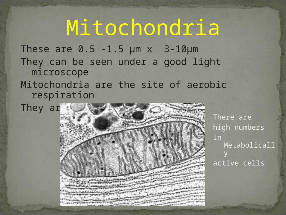
MitochondriaThese are 0.5 -1.5 µm x 3-10µmThey can be seen under a good light microscopeMitochondria are the site of aerobic respirationThey are found in all cells
There arehigh numbersIn Metabolicallyactive cells
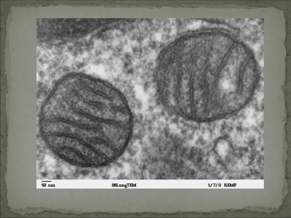
They are surrounded by a double membrane
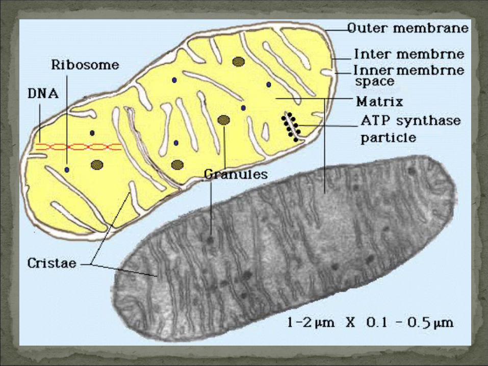
The outer membrane is smooth and the inner is folded into cristae
These folds increase the surface area for the enzymes involved in ATP synthesis
The matrix contains enzymes used in the Krebs cycle of respiration
In the matrix are 70S ribosomes and circular strands of DNA
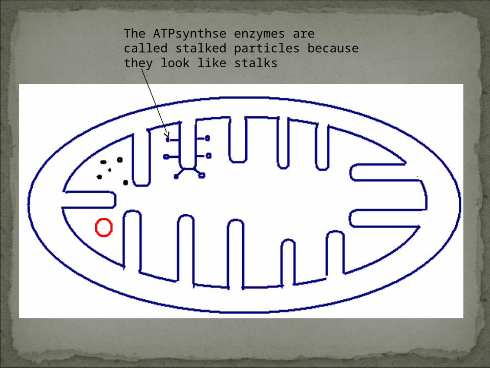
The ATPsynthse enzymes are called stalked particles because they look like stalks
Vacuoles
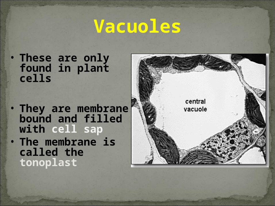
• These are only found in plant cells
• They are membrane bound and filled with cell sap
• The membrane is called the tonoplast
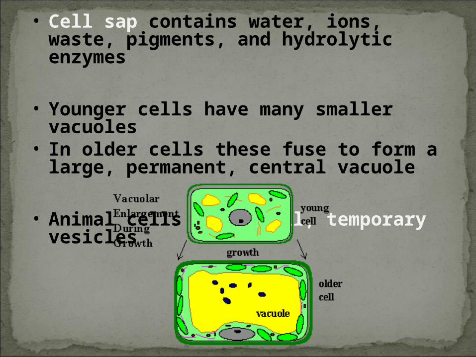
• Cell sap contains water, ions, waste, pigments, and hydrolytic enzymes
• Younger cells have many smaller vacuoles
• In older cells these fuse to form a large, permanent, central vacuole
• Animal cells have small, temporary vesicles
Chloroplasts
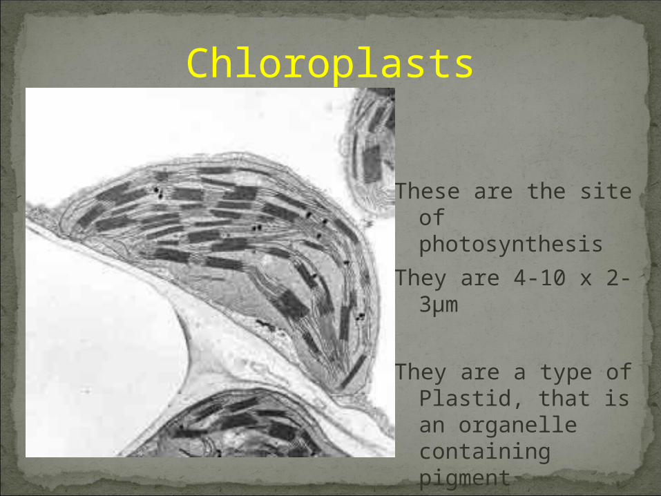
These are the site of photosynthesis
They are 4-10 x 2-3µm
They are a type of Plastid, that is an organelle containing pigment
They are biconvex discs
There is a double membrane
The inner membrane is formed
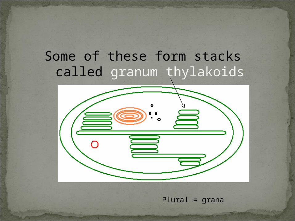
into ling flat sacks called thylakoids
Some of these form stacks called granum thylakoids
Plural = grana
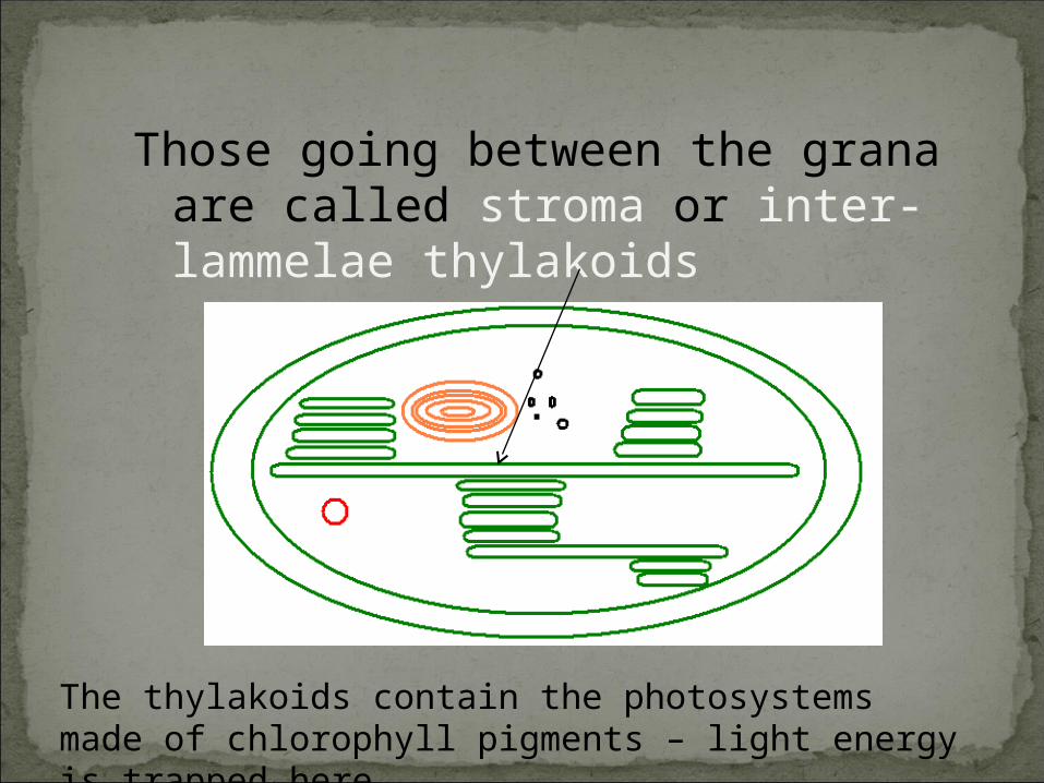
Those going between the grana
are called stroma or inter-lammelae thylakoids
The thylakoids contain the photosystems made of chlorophyll pigments – light energy is trapped here
The fluid filled centre is the stroma
The stroma contains the enzymes needed for the Calvin Cycle which converts carbon dioxide into a carbohydrate
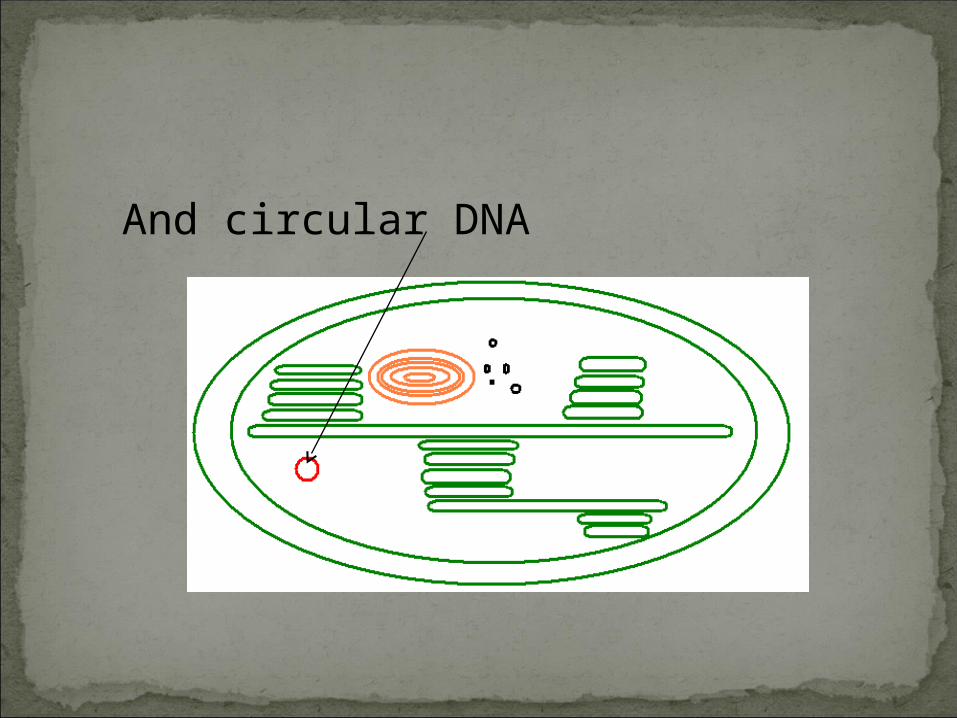
There are 70S ribosomes
And circular DNA
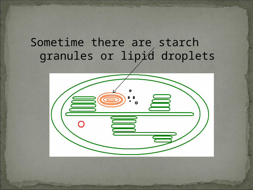
Sometime there are starch
granules or lipid droplets
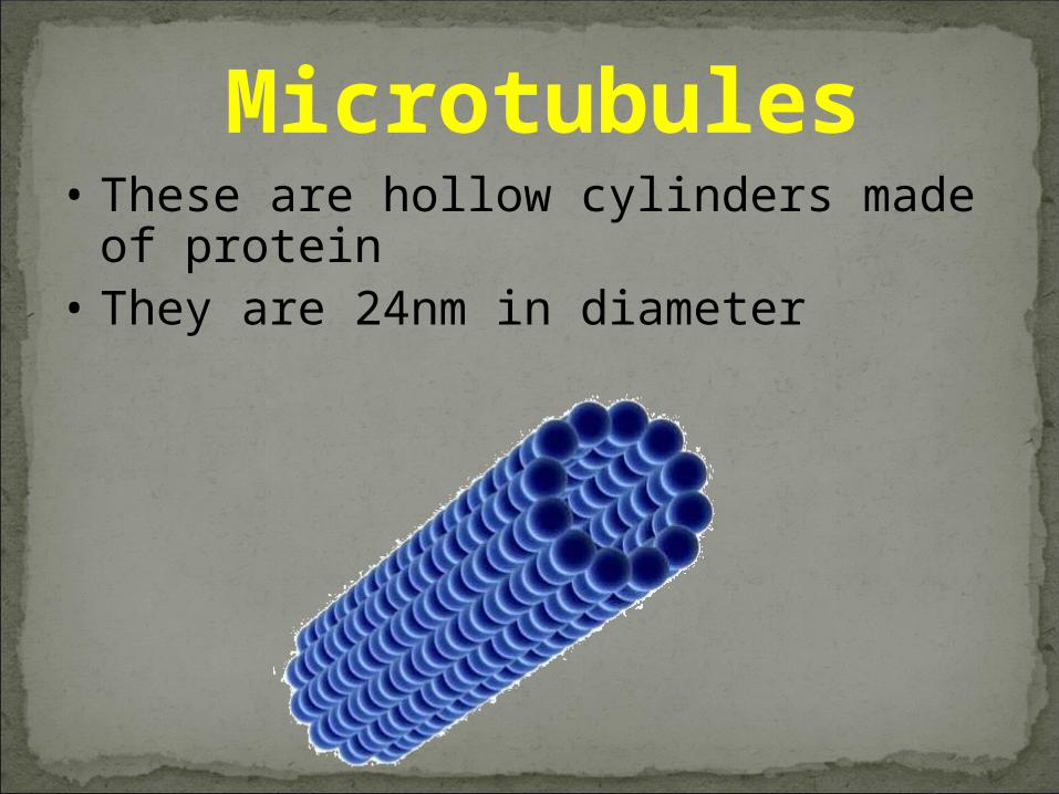
Microtubules• These are hollow cylinders made of
protein• They are 24nm in diameter
They occur throughout the cytoplasm and bring about movement of cell contents, such as lysosomes
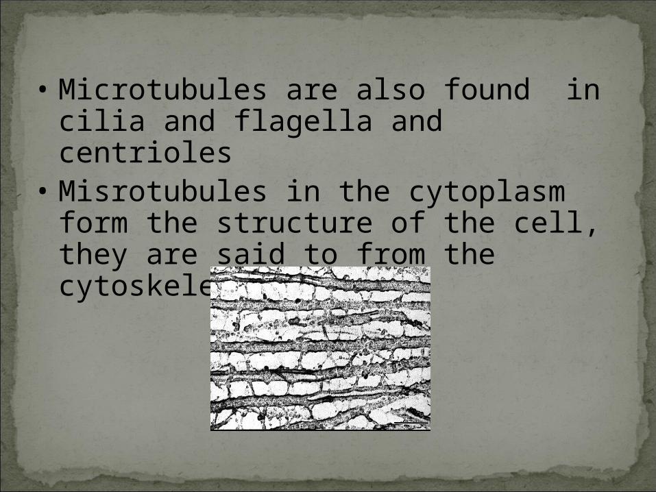
• Microtubules are also found in cilia and flagella and centrioles
• Misrotubules in the cytoplasm form the structure of the cell, they are said to from the cytoskeleton
MicrofilamentsThese are single stranded proteins They are involved in cytoplasmic
movement
Centrioles• These are only found in animal cells• They consist of two hollow cylinders at
right angles• They are about 0.3-0.5µm long and 0.2 µm
wide• Centrioles contain microtubules in the
same structure as seen in cilia and flagella basal bodies
• The Centrioles move to opposite poles during cell division
Centrioles• These are only found in animal cells• They consist of two hollow cylinders at
right angles
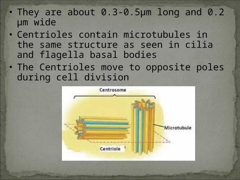
• They are about 0.3-0.5µm long and 0.2 µm wide
• Centrioles contain microtubules in the same structure as seen in cilia and flagella basal bodies
• The Centrioles move to opposite poles during cell division
Cilia and FlagellaCilia are many short cytoplasmic projectionsFlagella are one or a few much longer
cytoplasmic projections
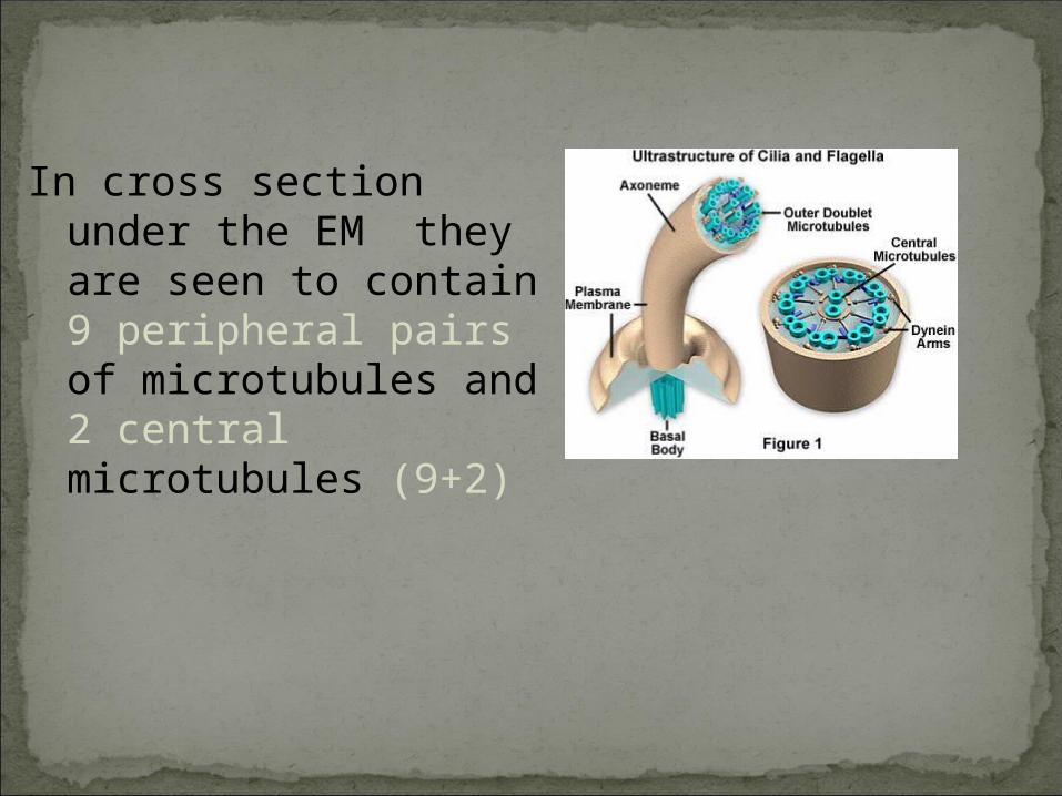
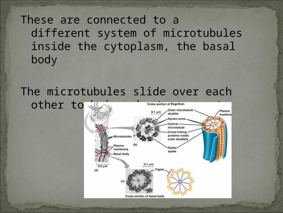
In cross section under the EM they are seen to contain 9 peripheral pairs of microtubules and 2 central microtubules (9+2)
These are connected to a different system of microtubules inside the cytoplasm, the basal body
The microtubules slide over each other to bring about movement


![KLEZMER SUITE [Dedicated to clarinet virtuoso MICHELE GINGRAS] · 2 #' d d d d d d d d d d d d d@ / < d ddd dd d d d d d d@ / < d d d d d d d d d d d](https://static.documents.pub/doc/80x56/5e08f4a7e0170576bd2af37a/klezmer-suite-dedicated-to-clarinet-virtuoso-michele-gingras-2-d-d-d-d-d-d.jpg)


