Page 1
53
DAFTAR PUSTAKA
1. World Health Organization. Pneumonia [internet]. c2012. [ updated Nov
2012; cited 2012 Dec 3]. Available from :
http://www.who.int/mediacentre/factsheets/fs331/en/index.html
2. Pusat data dan surveilans epidemiologi. Situasi pneumonia balita di Indonesia.
Buletin Jendela Epidemiologi. Jakarta : Pusat Data dan Surveilans;2010.
3. Lakhanpaul M, Atkinson M, Stephenson T. Community acquired pneumonia
in children: a clinical update. Arch Dis Child Educ Pract: group.bmj.com,
2004; v. 89.
4. Ostapchuk M, Roberts DM, Haddy R. Community-acquired pneumonia in
infants and children. Am Fam Physician 2004;70(5):899-908.
5. Masria S. Pattern of bacteria causing pneumonia in children and its sensitivity
to some antibiotics. Proc ASEAN Congr Trop Med Parasitol 2008;3:121-4.
6. Ko W-C, Patterson DL, Sagnimeni AJ, et al. Community-acquired Klebsiella
pneumoniae bacteremia: global differences in clinical patterns. Emerging
Infection Disease, 2002; v. 8.
7. Bogaert D, De Groot R, Hermans PW. Streptococcus pneumoniae
colonisation: the key to pneumococcal disease. Lancet Infect Dis
2004;4(3):144-54.
8. Cardozo DM, Nascimento-Carvalho CM, Andrade AL, et al. Prevalence and
risk factors for nasopharyngeal carriage of Streptococcus pneumoniae among
adolescents. J Med Microbiol 2008;57(Pt 2):185-9.
9. M. Cömert M, B. H. Uçan M, Begendik F, et al. The Relationship Between
Pneumonia and Gastric Colonization in Surgical Intensive-Care Unit Patients.
The Journal of applied research 2003;3(2).
10. Singh YD. Pathophysiology of community acquired pneumonia. J Assoc
Physicians India 2012;60 Suppl:7-9.
Page 2
54
11. Faden H, Duffy L, Wasielewski R, et al. Relationship between nasopharyngeal
colonization and the development of otitis media in children.
Tonawanda/Williamsville Pediatrics. J Infect Dis 1997;175(6):1440-5.
12. Podschun R, Ullmann U. Klebsiella spp. as nosocomial pathogens:
epidemiology, taxonomy, typing methods, and pathogenicity factors. Clin
Microbiol Rev 1998;11(4):589-603.
13. Baltimore RS, Duncan RL, Shapiro ED, et al. Epidemiology of pharyngeal
colonization of infants with aerobic gram-negative rod bacteria. J Clin
Microbiol 1989;27(1):91-5.
14. Wang S, Li D, Chu YZ, et al. Determination of oropharyngeal pathogenic
colonization in the elderly community. Chin Med J (Engl) 2009;122(3):315-8.
15. Setiawan DS. Faktor risiko kolonisasi Enterobactericeae pada nasofaring
dewasa. Semarang : Universitas Diponegoro; 2010.
16. Irwanti G. Faktor risiko kolonisasi Enterobactericeae pada nasofaring anak.
Semarang : Universitas Diponegoro; 2010.
17. Garcia-Rodriguez JA, Fresnadillo Martinez MJ. Dynamics of nasopharyngeal
colonization by potential respiratory pathogens. J Antimicrob Chemother
2002;50 Suppl S2:59-73.
18. Jain A, Kumar P, Awasthi S. High nasopharyngeal carriage of drug resistant
Streptococcus pneumoniae and Haemophilus influenzae in North Indian
schoolchildren. Trop Med Int Health 2005;10(3):234-9.
19. Casewell M, Phillips I. Food as a source of Klebsiella species for colonisation
and infection of intensive care patients. Journal of clinical pathology
1978;31:845-9.
20. Djaja IM. Kontaminasi E. coli pada makanan dari tiga jenis tempat
pengelolaan makanan (TPM) di Jakarta Selatan 2003. Makara, Kesehatan
2003;12(1):36-41.
21. Hidayati E, Juli N, Marwani E. Isolasi Enterobacteriaceae patogen dari
makanan berbumbu dan tidak berbumbu kunyit (Curcuma longa L.) serta uji
pengaruh ekstrak kunyit (Curcuma longa L.) terhadap [ertumbuhan bakteri
yang diisolasi. Jurnal Matematika dan Sains 2001;7(2):43-52.
Page 3
55
22. Okuwaki Y, Fujita K, Sugiyama M, et al. Bacteriological and chemical study
of the drinking water in indonesia. Japan J Trop Med Hyg 1981;10:33-9.
23. Chiu SS, Ho PL, Chow FK, et al. Nasopharyngeal carriage of antimicrobial-
resistant Streptococcus pneumoniae among young children attending 79
kindergartens and day care centers in Hong Kong. Antimicrob Agents
Chemother 2001;45(10):2765-70.
24. Hikmawati. Perbedaan pola kolonisasi bakteri potensial patogen respiratori
pada nasofaring anak dan orangtua sehat. Semarang: Universitas Diponegoro;
2010.
25. Wolf B, Gama A, Rey L, et al. Striking differences in the nasopharyngeal
flora of healthy Angolan, Brazilian and Dutch children less than 5 years old.
Ann Trop Paediatr 1999;19(3):287-92.
26. Cruickshank R, Duguid J, Marmion B, et al. Medical microbiology :The
practice of medical microbiologi II, 12 ed. Vol. 2. Edinburgh: Churchill
Livingstone; 1975.
27. Podschun R, Pietsch S, Holler C, et al. Incidence of Klebsiella species in
surface waters and their expression of virulence factors. Appl Environ
Microbiol 2001;67(7):3325-7.
28. LeVan A, Jacob D. Gram Stain : Gram-negative rods. In: Gram negative r,
Klebsiella pneumoniae. (Adriana LeVan and Deena Jacob, University of
Maryland, College Park, MD), ed.: ASM MicrobeLibrary.org, 2010.
29. Brooks GF, Butel JS, Morse SA. Mikrobiologi Kedokteran Jawetz, Melnick &
Adelberg, Ed, 23. Vol. 23. Jakarta: EGC; 2007.
30. Highsmith AK, Jarvis WR. Klebsiella pneumoniae: selected virulence factors
that contribute to pathogenicity. Infect Control 1985;6(2):75-7.
31. Mizuta K, Ohta M, Mori M, et al. Virulence for mice of Klebsiella strains
belonging to the O1 group: relationship to their capsular (K) types. Infect
Immun 1983;40(1):56-61.
32. Hart T, Shears P. Atlas berwarna mikrobiologi kedokteran, 1 ed. Jakarta:
Hipokrates, 1997; 316.
Page 4
56
33. Baron EJ, Peterson LR, Finegold SM. Bailey's & Scott's Diagnostic
Microbiology, 9 ed. St Louis: Mosby Year Book; 1994.
34. Gama-hemolysis on Blood Agar (Klebsiella pneumoniae)[internet], c2012
[cited 2013 Feb 10]. Available from :
http://bacteriainphotos.com/agar%20cultivation%20media.html
35. Buxton R. Mac Conkey Agar Plates. In: Klebsiella pneumoniae [internet].
ASM Conference for Undergraduate Education; 2005 [updated 2005 Sep 30;
cited 2013 Feb 6]. Available from : http://lib.jiangnan.edu.cn/ASM/112-
Introduce2.html.
36. Bagley ST, Seidler RJ. Primary Klebsiella identification with MacConkey-
inositol-carbenicillin agar. Appl Environ Microbiol 1978;36(3):536-8.
37. Dutka BJ, Jones K, Bailey H. Enumeration of Klebsiella spp. in cold water by
using MacConkey-inositol-potassium tellurite medium. Appl Environ
Microbiol 1987;53(7):1716-7.
38. Samra Z, Bahar J, Madar-Shapiro L, et al. Evaluation of CHROMagar KPC
for rapid detection of carbapenem-resistant Enterobacteriaceae. J Clin
Microbiol 2008;46(9):3110-1.
39. Gupte S. Mikrobiologi Dasar, 3 ed. Jakarta: Binarupa Aksara; 1990.
40. Staf pengajar FK UI. Buku Ajar Mikrobiologi Kedokteran Edisi Revisi.
Jakarta: Binarupa Aksara, 1994.
41. Fankhauser DB. Triple sugar iron agar and its use [internet]. 1987 [updated
2001; cited 2013 jan 28]. Available from :
http://biology.clc.uc.edu/fankhauser/labs/microbiology/Triple_Sugar_Iron/TSI
_Use.htm
42. Lehman D. Triple Sugar Iron Agar Protocols[internet.] ACM Microbe
Library, 2005 [cited 2013 jan 28]. Available from :
http://www.microbelibrary.org/component/resource/laboratory-test/2842-
triple-sugar-iron-agar-protocols
Page 5
57
43. Lima ABM, Leão L, Oliveira L, et al. Nasopharyngeal Gram-negative bacilli
colonization in Brazilian children attending day-care centers. Brazilian Journal
of Microbiology 2009;41:24-7.
44. Mengistu Y, Gedebou M. Aerobic gram-negative pharyngeal bacilli of adult
Ethiopians: carrier rates and antibiograms. J Hyg (Lond) 1986;97(2):247-53.
45. Snell RS. Anatomi Klinik untuk mahasiswa kedokteran, 6 ed. Jakarta:
Penerbit buku kedokteran EGC, 2006; 968.
46. Brueggemann AB, Griffiths DT, Meats E, et al. Clonal relationships between
invasive and carriage Streptococcus pneumoniae and serotype- and clone-
specific differences in invasive disease potential. J Infect Dis
2003;187(9):1424-32.
47. Johnson AW, Osinusi K, Aderele WI, et al. Etiologic agents and outcome
determinants of community-acquired pneumonia in urban children: a hospital-
based study. J Natl Med Assoc 2008;100(4):370-85.
48. Pollack M, Charache P, Nieman RE, et al. Factors influencing colonisation
and antibiotic-resistance patterns of gram-negative bacteria in hospital
patients. Lancet 1972;2(7779):668-71.
49. Tuon FF, Rocha JL, Toledo P, et al. Risk factors for KPC-producing
Klebsiella pneumoniae bacteremia. Braz J Infect Dis 2012;16(5):416-9.
50. Casewell M, Phillips I. Hands as route of transmission for Klebsiella species.
Br Med J 1977;2(6098):1315-7.
51. Klebsiella pneumonia in Healthcare settings. Centers for Disease Control and
Prevention; v. 2012.
52. Ebringer A. The relationship between Klebsiella infection and ankylosing
spondylitis. Baillieres Clin Rheumatol 1989;3(2):321-38.
53. Fung CP, Chang FY, Lee SC, et al. A global emerging disease of Klebsiella
pneumoniae liver abscess: is serotype K1 an important factor for complicated
endophthalmitis? Gut 2002;50(3):420-4.
54. Yagupsky P, Porat N, Fraser D, et al. Acquisition, carriage, and transmission
of pneumococci with decreased antibiotic susceptibility in young children
Page 6
58
attending a day care facility in southern Israel. J Infect Dis 1998;177(4):1003-
12.
55. Harrison LM, Morris JA, Telford DR, et al. The nasopharyngeal bacterial
flora in infancy: effects of age, gender, season, viral upper respiratory tract
infection and sleeping position. FEMS Immunol Med Microbiol 1999;25(1-
2):19-28.
56. Faden H. The microbiologic and immunologic basis for recurrent otitis media
in children. Eur J Pediatr 2001;160(7):407-13.
57. Syrjanen RK, Kilpi TM, Kaijalainen TH, et al. Nasopharyngeal carriage of
Streptococcus pneumoniae in Finnish children younger than 2 years old. J
Infect Dis 2001;184(4):451-9.
58. Ramirez-Ronda CH, Fuxench-Lopez Z, Nevarez M. Increased pharyngeal
bacterial colonization during viral illness. Arch Intern Med
1981;141(12):1599-603.
59. Principi N, Marchisio P, Schito GC, et al. Risk factors for carriage of
respiratory pathogens in the nasopharynx of healthy children. Ascanius Project
Collaborative Group. Pediatr Infect Dis J 1999;18(6):517-23.
60. Kvaerner KJ, Tambs K, Harris JR, et al. Otitis media: relationship to
tonsillitis, sinusitis and atopic diseases. Int J Pediatr Otorhinolaryngol
1996;35(2):127-41.
61. Borer A, Meirson H, Peled N, et al. Antibiotic-resistant pneumococci carried
by young children do not appear to disseminate to adult members of a closed
community. Clin Infect Dis 2001;33(4):436-44.
62. Sung RY, Ling JM, Fung SM, et al. Carriage of Haemophilus influenzae and
Streptococcus pneumoniae in healthy Chinese and Vietnamese children in
Hong Kong. Acta Paediatr 1995;84(11):1262-7.
63. Leach AJ, Boswell JB, Asche V, et al. Bacterial colonization of the
nasopharynx predicts very early onset and persistence of otitis media in
Australian aboriginal infants. Pediatr Infect Dis J 1994;13(11):983-9.
Page 7
59
64. Mthwalo M, Wasas A, Huebner R, et al. Antibiotic resistance of
nasopharyngeal isolates of Streptococcus pneumoniae from children in
Lesotho. Bull World Health Organ 1998;76(6):641-50.
65. Ariyani D, Anwar F. Mutu mikrobiologis minuman jajanan di sekolah dasar
wilayah Bogor Tengah. Jurnal Gizi dan makanan;1(1):44-50.
66. Leach AJ, Shelby-James TM, Mayo M, et al. A prospective study of the
impact of community-based azithromycin treatment of trachoma on carriage
and resistance of Streptococcus pneumoniae. Clin Infect Dis 1997;24(3):356-
62.
67. Korona-Glowniak I, Malm A. Characteristics of Streptococcus pneumoniae
strains colonizing upper respiratory tract of healthy preschool children in
Poland. ScientificWorldJournal 2012;2012:732901.
68. Marchisio P, Gironi S, Esposito S, et al. Seasonal variations in nasopharyngeal
carriage of respiratory pathogens in healthy Italian children attending day-care
centres or schools. J Med Microbiol 2001;50(12):1095-9.
69. Utah Department of Health. Nasopharyngeal swab collecting [pamphlet].
Utah: Utah Department of Health, Goverment of Utah, 2005.
70. Gilman RH, Brown KH, Gilman JB, et al. Colonization of the oropharynx
with Gram-negative bacilli in children with severe protein-calorie
malnutrition. Am J Clin Nutr 1982;36(2):284-9.
71. O'Brien KL, Bronsdon MA, Dagan R, et al. Evaluation of a medium (STGG)
for transport and optimal recovery of Streptococcus pneumoniae from
nasopharyngeal secretions collected during field studies. J Clin Microbiol
2001;39(3):1021-4.
Page 8
63
JUDUL PENELITIAN : FAKTOR RISIKO KOLONISASI KLEBSIELLA SP. PADA
NASOFARING BALITA
Instansi pelaksana : Fakultas Kedokteran Undip Semarang
Lembar INFORMED CONSENT
Berikut ini naskah yang akan dibacakan kapada responden/orangtua responden penelitian
Selamat sore/malam, Bapak/Ibu….
Kami dari mahasiswa Kedokteran Umum Fakultas Kedokteran Universitas Diponegoro ingin memohon
waktu dan kesediaan Bapak/Ibu untuk menjadi responden penelitian kami yang berjudul :
FAKTOR RISIKO KOLONISASI KLEBSIELLA SP. PADA BALITA
Penelitian ini bertujuan untuk mendapatkan data tentang bagaimana pola bakteri yang menempati/
hidup di saluran napas atas balita sehat di Indonesia.
Manfaat dari penelitian ini adalah : apabila kita telah mengetahui pola bakteri yang menempati/hidup di
saluran nafas atas balita, kita bisa memperkirakan pola bakteri yang menyebabkan infeksi paru khususnya
pada bayi dan anak balita. Sebab telah banyak dibuktikan, bahwa infeksi paru, yang ada di Indonesia
merupakan penyebab kematian kedua terbanyak selalu didahului dengan berbiaknya bakteri tersebut di
saluran napas atas terlebih dahulu.
Sebenarnya dokter bisa mengetahui bakteri penyebab infeksi paru pada seseorang penderita
dengan melakukan tes biakan kuman pada saat penderita datang dalam keadaan sudah sakit. Tetapi tes ini
membutuhkan waktu beberapa hari sehingga selama tes biakan belum ada hasilnya, dokter tidak
mempunyai pedoman untuk mengobati pasien pada hari-hari pertama pasien dirawat dirumah sakit.
Lagipula, bila pemeriksaan seperti ini dilakukan pada penderita infeksi paru, hasilnya bisa jadi
menyesatkan, khususnya bila pasien sudah mendapat obat-obatan tertentu.
Dengan penelitian ini, kami berharap bisa memperoleh data pola bakteri penyebab penyakit yang
“menghuni” (mengkolonisasi) tubuh orang Indonesia, dan faktor-faktor risiko kolonisasi itu, sehingga
bila seseorang pasien dengan infeksi paru datang ke RS, dan dapat diidentifikasi adanya faktor-faktor
risiko tertentu pada penderita tersebut, maka dokter yang merawat
dapat memperkirakan bakteri penyebab infeksi paru tersebut, dan
memberikan obat dengan lebih tepat walaupun tes biakan belum
selesai dikerjakan
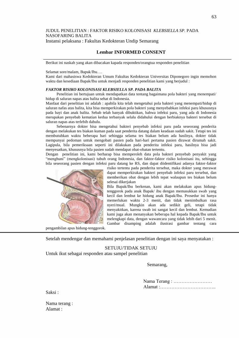
Bila Bapak/Ibu berkenan, kami akan melakukan apus hidung-
tenggorok pada anak Bapak/ ibu dengan memasukkan swab yang
kecil dan lembut ke hidung anak Bapak/Ibu. Prosedur ini hanya
memerlukan waktu 2-3 menit, dan tidak menimbulkan rasa
nyeri/mual. Mungkin akan ada sedikit geli, tetapi tidak
menyakitkan, karena swab ini sangat kecil dan lembut. Kemudian
kami juga akan menanyakan beberapa hal kepada Bapak/Ibu untuk
melengkapi data, dengan wawancara yang tidak lebih dari 5 menit.
Gambar disamping adalah ilustrasi gambar tentang cara
pengambilan apus hidung-tenggorok.
Setelah mendengar dan memahami penjelasan penelitian dengan ini saya menyatakan :
SETUJU/TIDAK SETUJU
Untuk ikut sebagai responden atau sampel penelitian
Semarang,
Nama Terang : ……………………
Alamat :……………………………..
Saksi :
Nama terang :
Alamat :
Page 9
64
KUESIONER KOLONISASI FARING BALITA
DEMOGRAFI
1. Nama L/P
2 Tempat/ tanggal lahir
3 Alamat
4 Riwayat perawatan di RS dalam 3
bulan terakhir
0. Tidak ada 1. Ada : sakit……. , …..hari
5 Minum antibiotik saat ini 0. Tidak 1. Ya 2. Tidak Tahu
6 Minum antibiotik dalam 3 bulan
terakhir
0. Tidak 1. Ya 2. Tidak Tahu
7 Batuk/ pilek saat ini 0. Tidak 1. Ya
MAKANAN *) Makan pagi Makan siang Makan
malam
Makanan ringan
Dimasak dirumah
Restoran
Warung/kantin
Penjaja keliling
AIR *) Minum Menyiapkan makanan Mandi Air ledeng (PDAM)
Air sumur
Air kemasan
Air sungai
Mobil tangki air
Penjual air keliling
SANITASI RUMAH Air ledeng di rumah 0. Ada 1. Tidak
HIGIENE & KESEHATAN PERORANGAN Paparan dengan lansia (>65 tahun) di rumah Di rumah : ……….lansia
*) Dua yang paling sering
CRF : ……………………….
NAMA :……………….........
Kecamatan : ………………..
Page 10
LOG BOOK PENELITIAN
No. subyek
No. Koloni BA CA MC cat gram Optochin XV faktor SIM cat kapsul Diagnosis
Kuman
24
48
NB : jangan lupa tuliskan tanggal setiap melakukan percobaan.
Page 11
66
LAMPIRAN 6. Hasil output data menggunakan SPSS
Karakteristik subyek penelitian
Kecamatan tempat tinggal
86 49.4 49.4 49.4
88 50.6 50.6 100.0
174 100.0 100.0
gayamsari
gunungpati
Total
Valid
Frequency Percent Valid Percent
Cumulative
Percent
Usia_1 * Kecamatan tempat tinggal Crosstabulation
9 9 18
5.2% 5.2% 10.3%
77 79 156
44.3% 45.4% 89.7%
86 88 174
49.4% 50.6% 100.0%
Count
% of Total
Count
% of Total
Count
% of Total
Bayi
Anak balita
Usia_1
Total
gayamsari gunungpati
Kecamatan tempat
tinggal
Total
Jenis Kelamin * Kecamatan tempat tinggal Crosstabulation
55 45 100
31.6% 25.9% 57.5%
31 43 74
17.8% 24.7% 42.5%
86 88 174
49.4% 50.6% 100.0%
Count
% of Total
Count
% of Total
Count
% of Total
Laki-laki
Perempuan
Jenis Kelamin
Total
gayamsari gunungpati
Kecamatan tempat
tinggal
Total
Page 12
67
Distribusi faktor risiko pada kedua lokasi penelitian
higiene_makanan * Kecamatan tempat tinggal Crosstabulation
17 24 41
9.8% 13.8% 23.6%
69 64 133
39.7% 36.8% 76.4%
86 88 174
49.4% 50.6% 100.0%
Count
% of Total
Count
% of Total
Count
% of Total
higiene bagus
tidak bagus
higiene_
makanan
Total
gayamsari gunungpati
Kecamatan tempat
tinggal
Total
Chi-Square Tests
1.360b 1 .243
.975 1 .323
1.366 1 .242
.285 .162
1.352 1 .245
174
Pearson Chi-Square
Continuity Correctiona
Likelihood Ratio
Fisher's Exact Test
Linear-by-Linear
Association
N of Valid Cases
Value df
Asymp. Sig.
(2-sided)
Exact Sig.
(2-sided)
Exact Sig.
(1-sided)
Computed only for a 2x2 tablea.
0 cells (.0%) have expected count less than 5. The minimum expected count is 20.
26.
b.
higiene_air * Kecamatan tempat tinggal Crosstabulation
47 7 54
27.0% 4.0% 31.0%
39 81 120
22.4% 46.6% 69.0%
86 88 174
49.4% 50.6% 100.0%
Count
% of Total
Count
% of Total
Count
% of Total
higiene baik
higiene tidak baik
higiene_air
Total
gayamsari gunungpati
Kecamatan tempat
tinggal
Total
Chi-Square Tests
44.312b 1 .000
42.158 1 .000
48.199 1 .000
.000 .000
44.058 1 .000
174
Pearson Chi-Square
Continuity Correctiona
Likelihood Ratio
Fisher's Exact Test
Linear-by-Linear
Association
N of Valid Cases
Value df
Asymp. Sig.
(2-sided)
Exact Sig.
(2-sided)
Exact Sig.
(1-sided)
Computed only for a 2x2 tablea.
0 cells (.0%) have expected count less than 5. The minimum expected count is 26.
69.
b.
Page 13
68
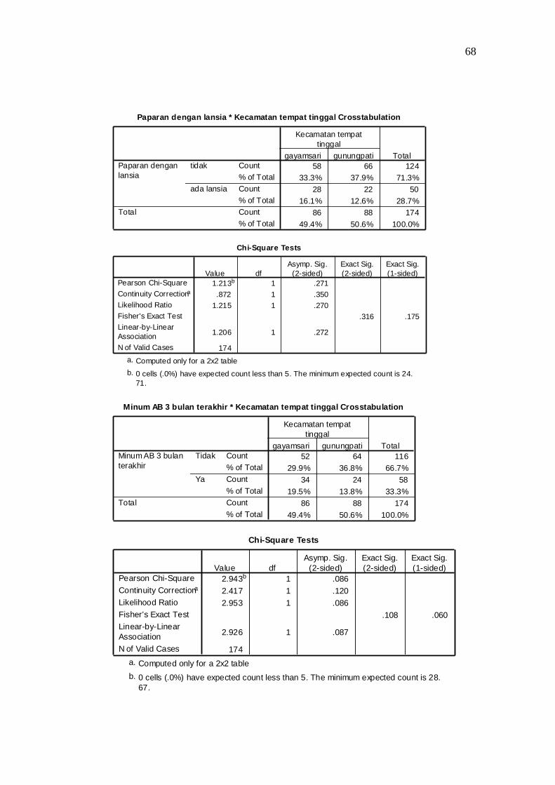
Paparan dengan lansia * Kecamatan tempat tinggal Crosstabulation
58 66 124
33.3% 37.9% 71.3%
28 22 50
16.1% 12.6% 28.7%
86 88 174
49.4% 50.6% 100.0%
Count
% of Total
Count
% of Total
Count
% of Total
tidak
ada lansia
Paparan dengan
lansia
Total
gayamsari gunungpati
Kecamatan tempat
tinggal
Total
Chi-Square Tests
1.213b 1 .271
.872 1 .350
1.215 1 .270
.316 .175
1.206 1 .272
174
Pearson Chi-Square
Continuity Correctiona
Likelihood Ratio
Fisher's Exact Test
Linear-by-Linear
Association
N of Valid Cases
Value df
Asymp. Sig.
(2-sided)
Exact Sig.
(2-sided)
Exact Sig.
(1-sided)
Computed only for a 2x2 tablea.
0 cells (.0%) have expected count less than 5. The minimum expected count is 24.
71.
b.
Minum AB 3 bulan terakhir * Kecamatan tempat tinggal Crosstabulation
52 64 116
29.9% 36.8% 66.7%
34 24 58
19.5% 13.8% 33.3%
86 88 174
49.4% 50.6% 100.0%
Count
% of Total
Count
% of Total
Count
% of Total
Tidak
Ya
Minum AB 3 bulan
terakhir
Total
gayamsari gunungpati
Kecamatan tempat
tinggal
Total
Chi-Square Tests
2.943b 1 .086
2.417 1 .120
2.953 1 .086
.108 .060
2.926 1 .087
174
Pearson Chi-Square
Continuity Correctiona
Likelihood Ratio
Fisher's Exact Test
Linear-by-Linear
Association
N of Valid Cases
Value df
Asymp. Sig.
(2-sided)
Exact Sig.
(2-sided)
Exact Sig.
(1-sided)
Computed only for a 2x2 tablea.
0 cells (.0%) have expected count less than 5. The minimum expected count is 28.
67.
b.
Page 14
69
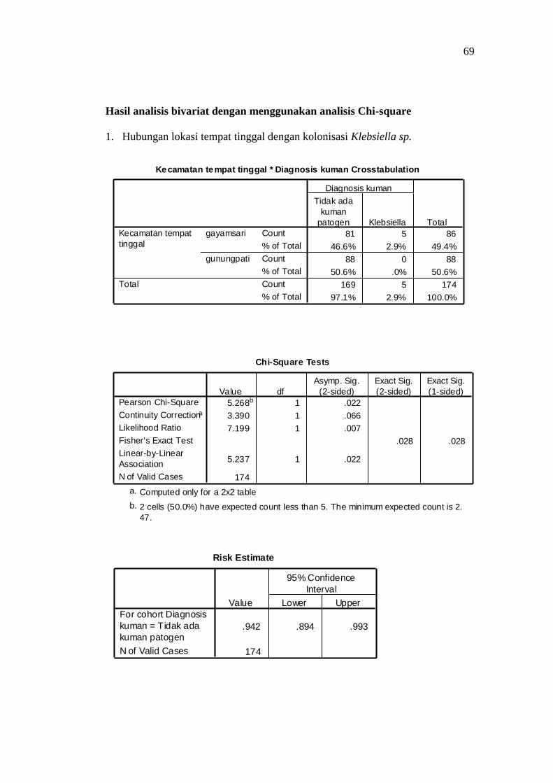
Hasil analisis bivariat dengan menggunakan analisis Chi-square
1. Hubungan lokasi tempat tinggal dengan kolonisasi Klebsiella sp.
Kecamatan tempat tinggal * Diagnosis kuman Crosstabulation
81 5 86
46.6% 2.9% 49.4%
88 0 88
50.6% .0% 50.6%
169 5 174
97.1% 2.9% 100.0%
Count
% of Total
Count
% of Total
Count
% of Total
gayamsari
gunungpati
Kecamatan tempat
tinggal
Total
Tidak ada
kuman
patogen Klebsiella
Diagnosis kuman
Total
Chi-Square Tests
5.268b 1 .022
3.390 1 .066
7.199 1 .007
.028 .028
5.237 1 .022
174
Pearson Chi-Square
Continuity Correctiona
Likelihood Ratio
Fisher's Exact Test
Linear-by-Linear
Association
N of Valid Cases
Value df
Asymp. Sig.
(2-sided)
Exact Sig.
(2-sided)
Exact Sig.
(1-sided)
Computed only for a 2x2 tablea.
2 cells (50.0%) have expected count less than 5. The minimum expected count is 2.
47.
b.
Risk Estimate
.942 .894 .993
174
For cohort Diagnosis
kuman = Tidak ada
kuman patogen
N of Valid Cases
Value Lower Upper
95% Confidence
Interval
Page 15
70
2. Hubungan higiene makanan dengan kolonisasi Klebsiella sp.
higiene_makanan * Diagnosis kuman Crosstabulation
40 1 41
23.0% .6% 23.6%
129 4 133
74.1% 2.3% 76.4%
169 5 174
97.1% 2.9% 100.0%
Count
% of Total
Count
% of Total
Count
% of Total
higiene bagus
tidak bagus
higiene_
makanan
Total
Tidak ada
kuman
patogen Klebsiella
Diagnosis kuman
Total
Chi-Square Tests
.036b 1 .849
.000 1 1.000
.038 1 .846
1.000 .663
.036 1 .849
174
Pearson Chi-Square
Continuity Correctiona
Likelihood Ratio
Fisher's Exact Test
Linear-by-Linear
Association
N of Valid Cases
Value df
Asymp. Sig.
(2-sided)
Exact Sig.
(2-sided)
Exact Sig.
(1-sided)
Computed only for a 2x2 tablea.
2 cells (50.0%) have expected count less than 5. The minimum expected count is 1.
18.
b.
Risk Estimate
1.240 .135 11.418
1.006 .950 1.065
.811 .093 7.054
174
Odds Ratio for higiene_
makanan (higiene
bagus / tidak bagus)
For cohort Diagnosis
kuman = Tidak ada
kuman patogen
For cohort Diagnosis
kuman = Klebsiella
N of Valid Cases
Value Lower Upper
95% Confidence
Interval
Page 16
71
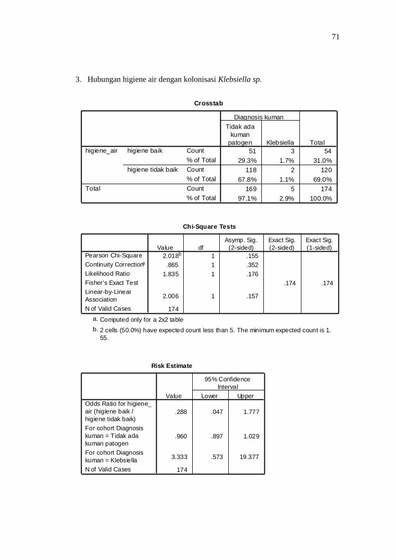
3. Hubungan higiene air dengan kolonisasi Klebsiella sp.
Crosstab
51 3 54
29.3% 1.7% 31.0%
118 2 120
67.8% 1.1% 69.0%
169 5 174
97.1% 2.9% 100.0%
Count
% of Total
Count
% of Total
Count
% of Total
higiene baik
higiene tidak baik
higiene_air
Total
Tidak ada
kuman
patogen Klebsiella
Diagnosis kuman
Total
Chi-Square Tests
2.018b 1 .155
.865 1 .352
1.835 1 .176
.174 .174
2.006 1 .157
174
Pearson Chi-Square
Continuity Correctiona
Likelihood Ratio
Fisher's Exact Test
Linear-by-Linear
Association
N of Valid Cases
Value df
Asymp. Sig.
(2-sided)
Exact Sig.
(2-sided)
Exact Sig.
(1-sided)
Computed only for a 2x2 tablea.
2 cells (50.0%) have expected count less than 5. The minimum expected count is 1.
55.
b.
Risk Estimate
.288 .047 1.777
.960 .897 1.029
3.333 .573 19.377
174
Odds Ratio for higiene_
air (higiene baik /
higiene tidak baik)
For cohort Diagnosis
kuman = Tidak ada
kuman patogen
For cohort Diagnosis
kuman = Klebsiella
N of Valid Cases
Value Lower Upper
95% Confidence
Interval
Page 17
72
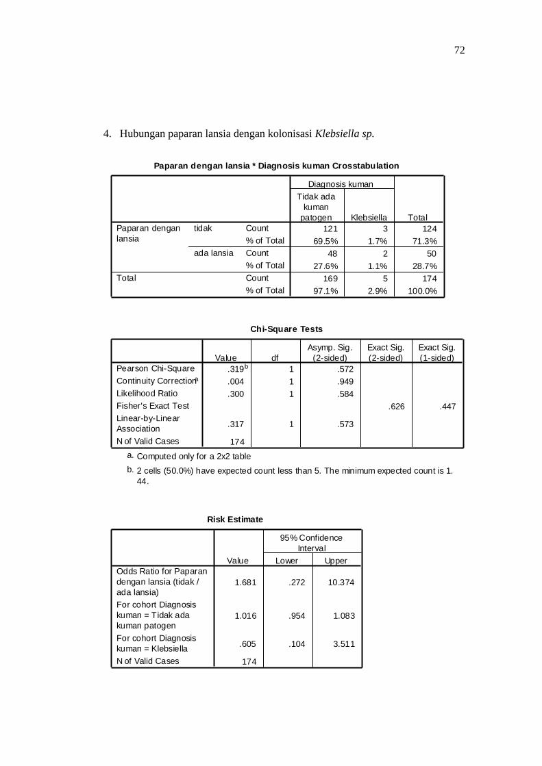
4. Hubungan paparan lansia dengan kolonisasi Klebsiella sp.
Paparan dengan lansia * Diagnosis kuman Crosstabulation
121 3 124
69.5% 1.7% 71.3%
48 2 50
27.6% 1.1% 28.7%
169 5 174
97.1% 2.9% 100.0%
Count
% of Total
Count
% of Total
Count
% of Total
tidak
ada lansia
Paparan dengan
lansia
Total
Tidak ada
kuman
patogen Klebsiella
Diagnosis kuman
Total
Chi-Square Tests
.319b 1 .572
.004 1 .949
.300 1 .584
.626 .447
.317 1 .573
174
Pearson Chi-Square
Continuity Correctiona
Likelihood Ratio
Fisher's Exact Test
Linear-by-Linear
Association
N of Valid Cases
Value df
Asymp. Sig.
(2-sided)
Exact Sig.
(2-sided)
Exact Sig.
(1-sided)
Computed only for a 2x2 tablea.
2 cells (50.0%) have expected count less than 5. The minimum expected count is 1.
44.
b.
Risk Estimate
1.681 .272 10.374
1.016 .954 1.083
.605 .104 3.511
174
Odds Ratio for Paparan
dengan lansia (tidak /
ada lansia)
For cohort Diagnosis
kuman = Tidak ada
kuman patogen
For cohort Diagnosis
kuman = Klebsiella
N of Valid Cases
Value Lower Upper
95% Confidence
Interval
Page 18
73
5. Hubungan riwayat antibiotik 3 bulan terakhir dengan kolonisasi 3 bulan
terakhir
Hasil analisis multivariat
Metode backward
Minum AB 3 bulan te rakhir * Diagnosis kuman Crosstabulation
112 4 116
64.4% 2.3% 66.7%
57 1 58
32.8% .6% 33.3%
169 5 174
97.1% 2.9% 100.0%
Count
% of Total
Count
% of Total
Count
% of Total
Tidak
Ya
Minum AB 3 bulan
terakhir
Total
Tidak ada
kuman
patogen Klebsiella
Diagnosis kuman
Total
Chi-Square Tests
.412b 1 .521
.026 1 .873
.449 1 .503
.666 .459
.409 1 .522
174
Pearson Chi-Square
Continuity Correctiona
Likelihood Ratio
Fisher's Exact Test
Linear-by-Linear
Association
N of Valid Cases
Value df
Asymp. Sig.
(2-sided)
Exact Sig.
(2-sided)
Exact Sig.
(1-sided)
Computed only for a 2x2 tablea.
2 cells (50.0%) have expected count less than 5. The minimum expected count is 1.
67.
b.
Variables in the Equation
18.305 4283.453 .000 1 .997 9E+007 .000 .
.232 .940 .061 1 .805 1.261 .200 7.957
-21.223 4283.453 .000 1 .996 .000
18.418 4284.585 .000 1 .997 1E+008 .000 .
-21.203 4284.585 .000 1 .996 .000
Kecamatan(1)
higiene_air(1)
Constant
Step
1a
Kecamatan(1)
Constant
Step
2a
B S.E. Wald df Sig. Exp(B) Lower Upper
95.0% C.I.for EXP(B)
Variable(s) entered on step 1: Kecamatan, higiene_air.a.
Page 19
74
LAMPIRAN 7. Dokumentasi penelitian
Informed consent dan wawancara kuesioner pengambilan apusan nasofaring
Penanaman bakteri pada media Pembiakan bakteri dengan inkubator
Koloni bakteri pada media Mac Conkey Uji biokimiawi dengan TSIA
Page 20
75
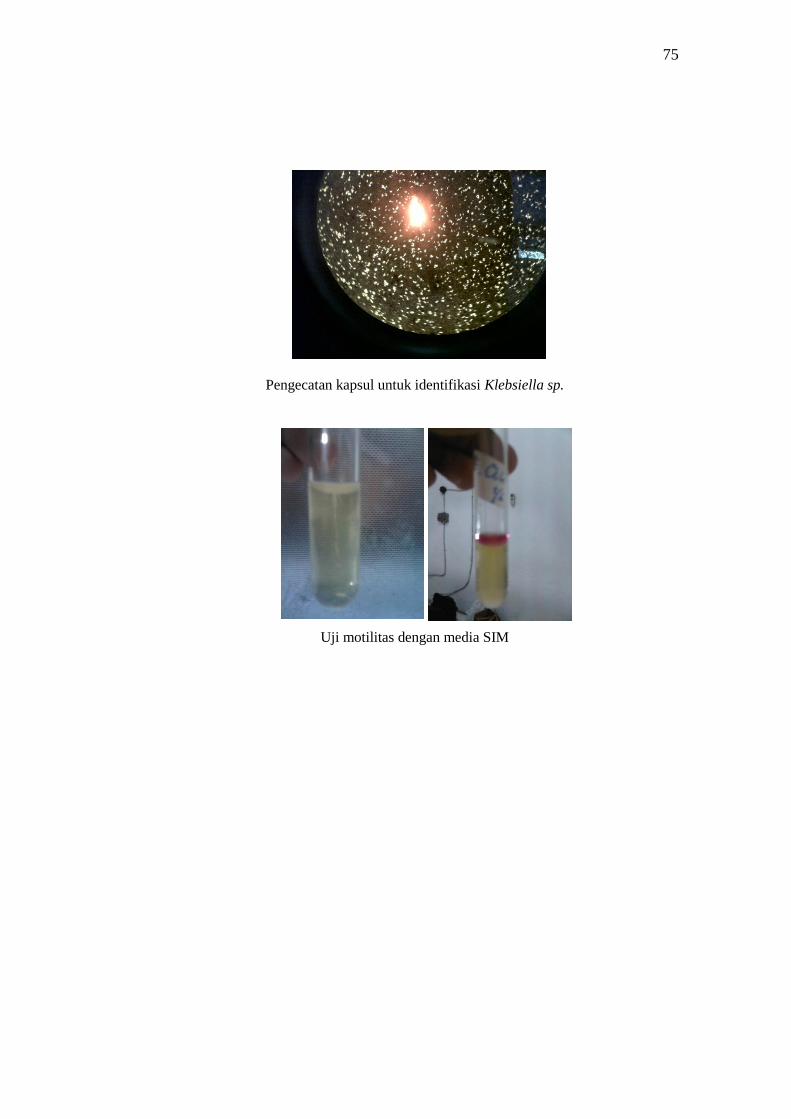
Pengecatan kapsul untuk identifikasi Klebsiella sp.
Uji motilitas dengan media SIM
Page 21
76
LAMPIRAN 8. Biodata mahasiswa
Identitas
Nama : Laurentia Laksmi Ajeng Hatmaningtyas
NIM : G2A009185
Tempat/Tanggal Lahir : Yogyakarta/ 20 Juli 1991
Jenis Kelamin : Perempuan
Alamat : Jl. Sendangguwo Baru IV/13 Semarang
Nomor Telepon : 024 6700004
Nomor HP : 085725810099
E-mail : [email protected]
Riwayat Pendidikan Formal
SD : SD ST Antonius 01 Semarang Lulus Tahun 2003
SMP : SMP Domenico Savio Semarang Lulus Tahun 2006
SMA : SMA Kolese Loyola Semarang Lulus Tahun 2009
Fakultas Kedokteran Universitas Diponegoro Masuk Tahun 2009
Riwayat Organisasi
STAF Kesma BEM KU FK Undip Tahun 2010-2011
Pengurus PRMK FK Undip Tahun 2011-2012
JMKI FK Undip Tahun 2009-2013
PSM FK Undip Tahun 2010-2013
Pengalaman penelitian
1. Prevalensi, Faktor Risiko, dan Pola Kepekaan Antibiotik Kolonisasi
Kuman Respiratori Patogen Pada Nasofaring Bayi dan Balita Sehat Tahun
2013
2. Faktor Risiko Kolonisasi Klebsiella sp. pada Nasofaring Balita Tahun
2013
Pengalaman mengikuti lomba karya tulis ilmiah
Addy S, Laurentia L, Theresia M, Dewi Ayu, Anggara. Prevalensi, Faktor
Risiko, dan Pola Kepekaan Antibiotik Kolonisasi Kuman Respiratori Patogen
Pada Nasofaring Bayi dan Balita Sehat, DIKTI, PKM-P (pendanaan penelitian).