DAIRY FOODS RESEARCH PAPERS Survival of Lactic Acid Bacteria in the Human Stomach and Adhesion to Intestinal Cells P. L. CONWAY 1 CSIRO Division of Food Research North Ryde New South Wales 2113 Australia S. L. GORBACH and B. R. GOLDIN Infectious Disease Service Department of Medicine Tufts-New England Medical Center Hospital Boston, MA 02111 ABSTRACT The survival of four strains of lactic acid bacteria in human gastric juice, in vivo and in vitro, and in buffered saline, pH 1 to 5, has been investigated. The strains studied include two Lactobacillus acidophilus strains, Lactobacillus bul- garicus, and Streptococcus thermophilus. In addition, the adhesion of these strains to freshly collected human and pig small intestinal cells and to pig large intestinal cells has been studied and the effect of milk on both survival and adhesion tested. As a result of these investigations, an in vitro test system for screening potential cultures for use as human dietary adjuncts can be developed. The ability to survive in gastric juice and to adhere varied significantly for the strains tested; L. acidophilus ADH survived and adhered better than the others while S. thermophilus survived and adhered poorly. For all strains, both survival and adhesion was enhanced by milk. As all strains adhered to some extent to both human and pig intestinal cells, the adhesion mechanism is probably a nonspecific attachment as opposed to other reported specific Lactobacillus adhesion to gastric tissue. From the survival and adhesion data it seems feasible to obtain elevated levels of viable Lactobacillus sp. in human intestine by Received March 19, 1986. Accepted July 7, 1986. 1Address for correspondence: P. Conway, Univer- sity of GiSteborg, Marine Microbiology, Carl Skotts- bergs Gata 22, S-413 19 Grteborg, Sweden. careful selection of the bacterial strains ingested. Furthermore, the in vitro methods used here should be valuable to screen potential strains. The data pre- sented here can then be correlated with human in vivo studies monitoring the beneficial effect of ingestion of these Lactobacillus. INTRODUCTION The ingestion of lactic acid bacteria, which was initially proposed by Metchnikoff as a means to reduce intestinal putrification and prolong life (20), has been extensively investi- gated as a beneficial dietary adjunct for gastro- intestinal disorders in humans and animals (16). Some workers (1, 18, 25) have suggested the use of Lactobacillus to prevent and treat diarrhea induced by E. coli, Salmonella, or Shigella. This work has paralleled in vitro studies demonstrating bacteriocin production by Lactobacillus strains (3, 19, 24). Other workers (2) have shown a correlation between L. acidopbilus consumption and a decreased need for laxatives in constipated elderly people; Gilliland et al. (8) have demonstrated assimila- tion of cholesterol by L. acidopbilus. Many antitumor properties of Lactobacilli have been reported (7). Goldin and coworkers have shown that oral L. acidopbilus supplements given to rats (9) and humans (11)lowered the fecal activity of the enzymes 3-glucuronidase, nitro- reductase, and azoreductase. Using an animal chemical carcinogenesis model, occurrence of tumours was decreased significantly when L. acidopbilus was administered orally (10). The strains of lactic acid bacteria and the form of bacterial preparation has varied enor- mously for the various studies (26). These 1987 J Dairy Sci 70:1-12 1
Transcript
DAIRY FOODS RESEARCH PAPERS
Survival of Lactic Acid Bacteria in the Human Stomach and Adhesion to Intestinal Cells
P. L. C O N W A Y 1 CSIRO
Division of Food Research North Ryde New South Wales 2113
Australia
S. L. GORBACH and B. R. G O L D I N Infectious Disease Service
Department of Medicine Tufts-New England Medical Center Hospital
Boston, MA 02111
ABSTRACT
The survival of four strains of lactic acid bacteria in human gastric juice, in vivo and in vitro, and in buffered saline, pH 1 to 5, has been investigated. The strains studied include two Lactobacillus acidophilus strains, Lactobacillus bul- garicus, and Streptococcus thermophilus. In addition, the adhesion of these strains to freshly collected human and pig small intestinal cells and to pig large intestinal cells has been studied and the effect of milk on both survival and adhesion tested. As a result of these investigations, an in vitro test system for screening potential cultures for use as human dietary adjuncts can be developed. The ability to survive in gastric juice and to adhere varied significantly for the strains tested; L. acidophilus ADH survived and adhered better than the others while S. thermophilus survived and adhered poorly. For all strains, both survival and adhesion was enhanced by milk. As all strains adhered to some extent to both human and pig intestinal cells, the adhesion mechanism is probably a nonspecific attachment as opposed to other reported specific Lactobacillus adhesion to gastric tissue. From the survival and adhesion data it seems feasible to obtain elevated levels of viable Lactobacillus sp. in human intestine by
Received March 19, 1986. Accepted July 7, 1986. 1Address for correspondence: P. Conway, Univer-
sity of GiSteborg, Marine Microbiology, Carl Skotts- bergs Gata 22, S-413 19 Grteborg, Sweden.
careful selection of the bacterial strains ingested. Furthermore, the in vitro methods used here should be valuable to screen potential strains. The data pre- sented here can then be correlated with human in vivo studies monitoring the beneficial effect of ingestion of these Lactobacillus.
I N T R O D U C T I O N
The ingestion of lactic acid bacteria, which was initially proposed by Metchnikoff as a means to reduce intestinal putrification and prolong life (20), has been extensively investi- gated as a beneficial dietary adjunct for gastro- intestinal disorders in humans and animals (16). Some workers (1, 18, 25) have suggested the use of Lactobacillus to prevent and treat diarrhea induced by E. coli, Salmonella, or Shigella. This work has paralleled in vitro studies demonstrating bacteriocin production by Lactobacillus strains (3, 19, 24). Other workers (2) have shown a correlation between L. acidopbilus consumption and a decreased need for laxatives in constipated elderly people; Gilliland et al. (8) have demonstrated assimila- tion of cholesterol by L. acidopbilus. Many antitumor properties of Lactobacilli have been reported (7). Goldin and coworkers have shown that oral L. acidopbilus supplements given to rats (9) and humans (11) lowered the fecal activity of the enzymes 3-glucuronidase, nitro- reductase, and azoreductase. Using an animal chemical carcinogenesis model, occurrence of tumours was decreased significantly when L. acidopbilus was administered orally (10).
The strains of lactic acid bacteria and the form of bacterial preparation has varied enor- mously for the various studies (26). These
1987 J Dairy Sci 70:1-12 1
2 CONWAY ET AL.
include freeze-dried commercial preparations of L. acidopbilus and L. bulgaricus (23); defined laboratory strains of L. acidopbilus administered with skim milk (10); dairy cultures used rou- t inely to produce commercial fermented milk products including various Streptococcus sp. (15, 21, 22). More recently, emphasis has been placed on the selection and preparation of Lactobacillus strains (12, 13, 17). Kleeman and Klaenhammer (13) reviewed the need to select strains that can survive and establish within an environment as hostile as the gastro- intestinal t ract and have tested the ability of various strains to adhere to a human fetal intestinal cell line. For successful implantat ion o f ingested Lactobacillus, bacteria must be viable within the gastrointestinal tract and also have adhesive propert ies to avoid the transient passage as in an in vivo human study (23).
The aim of this work was to test, both in vivo and in vitro, survival of several lactic acid bacteria when exposed to human gastric juice and the effects of an additive, such as milk, on survival. In addit ion, adhesion of various strains to freshly collected human and pig intestinal ceils has been studied. Correlation between in vivo and in vitro data demonstrated the reliabil- i ty of the in vitro methodology.
M A T E R I A L S A N D M E T H O D S
Bacterial Cu Itures
The following bacterial cultures were a gift f rom T. R. Klaenhammer at the North Carolina State University, Depar tment of Food Science. All strains were supplied and maintained in skimmed milk in 1-ml vials in liquid nitrogen. They were thawed immediately before use and not refrozen. Lactobacillus acidopbilus ADH was a human isolate reported as strain MSO2 (13); L. acidopbilus N2, originally given to us by M. L. Speck, was the strain used by Goldin and Gorbach (10); L. bulgaricus and S. tber- mopbilus strain were dairy culture strains. The frozen vials contained the following viable cells: L. acidopbilus ADH 1.2 × 101°/ml; L•
Lactobacillus cultures were grown anaero- bically in MRS broth (Difco) at 37°C overnight
and transferred to fresh MRS broth for a further 24 h. Streptococcus tbermopbitus was similarly subcultured using brain-heart infusion bro th (BHI) (Difco). Cultures were centrifuged
• O at 3000 × g/lO mm/4 C, washed once in sterile phosphate-buffered saline [(PBS) NaC1, .8%; .1 M, pH 7.2], and resuspended to one-tenth of the culture volume. These suspensions were used for the in vitro survival studies. The number of viable Lactobacillus cells was deter- mined by serial 10-fold dilution in PBS and • l -ml aliquots were spread evenly on MRS agar. Plates were incubated anaerobically at 37°C for 72 h and the colony forming units estimated• Verification of the ident i ty of the colonies was by Gram stain and the catalase reaction. The Streptococcus tberrnopbilus PBS serial 10-fold dilutions were enumerated on blood agar plates (BHI plus 5% blood) incubated anaerobically at 37°C for 72 h. Colonies were verified by Gram stain morphology.
Effect of pH on Survival
The survival of each of the four bacterial suspensions was studied by the addit ion of .1 ml of the suspension into a series of 2-ml volumes of sterile PBS at pH 1, 3, and 5 (ad- justed using NaOH). The incubation mix was maintained at 37°C and the viable organisms enumerated at 0, .5, 1.0, 1.5, 2.0, 3.0, and 4.0 h.
In Vitro Survival in Gastric Juice
Gastric juice was obtained by aspiration through a nasogastric tube from patients after at least 4 h fasting. For each of the four strains, a .1 ml volume of the bacterial suspension was added to 1.0 ml of gastric juice. To study the effect of the presence of milk, parallel tubes were prepared; .1 ml PBS was added to one series and .1 ml skim milk was added to the other series. The pH was measured at 0 and 4 h. Viable bacterial cells were enumerated at 0, .5, 1.0, 1.5, 2.0, 3, and 4 h.
Stability of Aspirated Gastric Juice pH
The pH of gastric juice was measured 30 min after aspiration and again at 1, 2, 5, and 20 d after storage at - 2 0 ° C . The pH of 1-ml aliquots of the gastric juice also was studied at 0 and 3 h at 37°C after the addit ion of the following compounds: skim milk (10%); NaOH to take
Journal of Dairy Science Vol. 70, No. 1, 1987
SURVIVAL AND ADHESION OF LACTIC ACID BACTERIA 3
pH initially to 6; mylanta (3 and 6%) (Parke Davis Pty. Ltd.).
Survival in Gastric Juice In Vivo
Survival of the three Lactobacillus cultures, in the absence and presence of skim milk, was studied in vivo using healthy human volunteers aged 21 to 31 (except subject D, 42 yr) with conventional American-style eating habits. Streptococcus tbermopbilus was not tested due to poor survival in the in vitro studies. All subjects completed medical and dietary his- tory forms. A standard light low fat breakfast was completed 4 h before commencement of the study. The breakfast consisted of 125 ml Special K cereal (Kelloggs), one slice of white bread with jelly, 250 ml skim milk, and black tea or coffee (with sugar if desired). The bacterial cultures used were the 1-ml stock vials maintained at - 7 0 ° C and thawed just prior to use. A nasogastric tube was inserted and 10 ml of gastric juice was aspirated. The LactobaciIlus suspension (1.0 ml) was introduced into the stomach via the tube. An additional 20 ml of sterile saline was added to ensure all of the culture reached the stomach (tube volume = 20 ml). At 0, 20, 40, 60, 90, 120, and 140 min after addit ion of the culture, a 25-ml aspirate was collected. After the 140-min aspirate was collected, subjects were allowed to rest with the tube remaining in place unti l 240 rain. The procedure was then repeated with the addi t ion of skim milk prior to the introduct ion of the bacterial culture. An addit ional 25 ml of aspirate was taken and then sterile skim milk (250 mi) was added via the tube. Another aspirate was immediately taken and then a further 1 ml of the appropriate bacterial culture was added. Aspirates of 25 ml were collected at 0, 20, 40, 60, 90, and 180 min after addit ion of the culture. The pH and number of viable cells were determined for all aspirates immediately after collection. A Radiometer pH meter was used and the number of viable Lactobacillus enumerated by serial 10-fold dilution of 1 ml samples into PBS; then aliquots were spread on MRS agar as for the in vitro studies. Three subjects received L. acidopbilus N2 and two subjects each received L. acidopbilus ADH and L. bulgaricus.
Cell Wall Stability
Overnight 5-ml MRS broth cultures of L. acidopbilus ADH and N 2 were centrifuged at
• O 3000 x g/lO mm/4 C, washed in 4 ml TE buffer [50 mM Tris base plus 50 mM ethylene- diaminetetraacetate (EDTA) pH 8.5] , and after centrifuging again, were resuspended in .2 ml of a sucrose solution (25% in TE buffer). The lysozyme solution (.05 ml of 10 mg/mt solution in .25 M Tris) was added and the mixture incubated at 37°C for 1 h. Tubes were observed for visible lysis during this incubation. Ethyl- enediaminetetraacetate (.08 ml of .25 M pH 8.0) was added (final concentrat ion 1%) and the tubes were held at room temperature for 10 min. To lyse the cells further, .3 ml of Triton X-100 (.2% in 50 mad Tris-HC1 and 50 mM Na EDTA at pH 8.5) was added and the tubes held at 65°C for 15 rain. The tubes were observed for visible lysis.
Preparation of Bacterial Cells for Adhesion Study
Lactobacill i cultures were grown in MRS broths overnight at 37°C in an anaerobic chamber and cultures were then transferred to fresh MRS broth containing 25 #Ci each of [3H]alanine and [3H]leucine (New England Nuclear Corp., Boston, MA) and then incubated in an anaerobic chamber overnight at 37°C. The Streptococcus tbermopbilus culture was grown in BHI broth overnight at 37°C and subcultured into fresh BHI broth containing the 25/~Ci each of [3 HI alanine and [3 H] leucine and incubated at anaerobically 37°C overnight• All bacterial suspensions were centrifuged at 3000 x g/lO
• O mm/4 C, washed once in cold PBS, and resus- pended in cold PBS to give an optical densi ty of • 35 using a Coleman model 44 spectrophotom- eter. Bacterial suspensions were used immedi- ately in the binding study. The loss of the tri t iated label from the bacterial cells into the fluid phase of the bacterial suspension was moni tored at .5-h intervals for 3 h and found not to increase.
Binding Assay
The binding of all four bacterial suspensions to either human or pig ileal cells and pig cecum or colon cells was carried out according to the method of Deneke et al. (6). A 1-ml sample of
Journal of Dairy Science Vol. 70, No. 1, 1987
4 CONWAY ET AL.
the radioactively labelled bacterial suspension, filtered through 5/am filter, was incubated with 1 ml of the intestinal cell suspension or 1 ml of buffer in glass test tubes in a 37°C water bath for 2 rain; then three samples of .5 ml from each were filtered through a polycarbonate filter (pore size, 2 /am; Nucleopore); each filter was washed with 5 ml PBS. Duplicate .5-ml samples of the bacterial suspension were similarly filtered and washed. The radioactivity (counts per min) on each filter was determined in Aquasol II scintillation fluid (New England Nuclear) using a Beckman LS-3133P liquid scintillation counter.
The number o f bacteria bound per filter, and subsequently per intestinal cell, was estimated from the bacterial specific activity, based on the radioactivity of the bacterial suspension filters and the corresponding optical density measurement; the number o f bacteria was estimated from a standard graph prepared for the optical density and the colony forming units per milliliter. The colony forming units per milliliter were determined by serial dilution and quanti ta t ion on agar plates as for viable counts.
Each fresh supply of intestinal cells was initially tested using a known positive and negative binding E. coli strain, B2C and B44, respectively (5). Only cell preparations produc- ing binding patterns consistent with those of Deneke et al. (5, 6) with human and pig ileal cell preparations were used.
Preparation of Intestinal Suspension
All intestinal cell suspensions, collected as detailed, were maintained at 4°C in tissue culture medium NCTC 135 (Gibco Laboratories, Grand Island, NY) containing penicillin (100 /ag/ml), s t reptomycin (100 /ag/ml), and genta- mycin (100/ag/ml) diluted to 25% with buffer 2 (5) and used in the binding assay as described by Deneke et al. (5, 6).
Ileal Cell Suspension
The human and pig ileal cell suspensions were collected as described by Deneke et al. (5, 6). A concentrat ion of 10 ileal cells/ml was used in the binding assay.
Pig Cecum and Colon Cell Suspension
The cecum and colon were individually
removed from the freshly sacrificed suckling pig (4 to 5 weeks old). Buffer 2 with the mannose omit ted was used throughout. As gentle lavag- ing failed to release epithelial cells (observed by direct microscopy), the cecum and colon were opened longitudinally. The inner surfaces were washed gently with 2 × 20 ml cold buffer and the washing discarded ; then inner surfaces were gently scraped with the end of a glass slide at an angle of 45 ° and epithelial cells collected in cold buffer. The dish containing the open cecum or colon was held on ice during the harvesting. A concentrat ion of 10 cells/ml was used in the binding assay.
Effect of Mannose on Binding to Pig ileal Cells
Pig and human ileal cells were routinely harvested and collected in buffer 2 (which contained 1% mannose). As mannose may influence adhesion, 1-m sections of pig small intestine were lavaged and the ileal cells har- vested, maintained, and used in buffer 2 with- out any mannose. The binding assay was conducted for the four bacterial strains with the ileal cells either in buffer 2 without man- nose or with 1% mannose included with the ileal cells for 5 min before the addition of the bacteria.
Effect of Milk on Binding to Human Ileal Calls
The lactobacilli cultures were transferred twice in MRS broth and the Streptococcus thermophilus were transferred twice in BHI broth before inoculation into the radioactively labelled broths. This addit ional subculture was carried out to reduce the presence of milk from the maintenance fluid in the bacterial suspen- sion used in the binding assay. Skim milk (1%) was added to a second series of radio- actively labelled broths prior to inoculation and the binding compared with milk free cultures.
R ESU LTS
Survival Studies
Effect of Physiological Saline pH. The viable count of all four bacterial strains decreased rapidly at pH 1.0 and more slowly at pH 3 and 5 (Table 1). The L. acidophilus ADH survived bet ter than all the other strains, L. acidophilus N2 bet ter than L. bulgaricus, which in turn was bet ter than S. thermophilus.
Journal of Dairy Science Vol. 70, No. 1, 1987
SURVIVAL AND ADHESION OF LACTIC ACID BACTERIA 5
TABLE 1. Survival of bacterial strains in phosphate-buffered saline at various pH, as determined by viable counts.
Stability o f pH o f Aspirated Gastric Juice. The pH of f reshly aspira ted gastric juice varied f r o m 1 to 2.5 and remained unchanged w h e n s tored at - 2 0 ° C for the 20 d s tudied. When gastric juice o f pH 1 was t rea ted wi th 1) skim milk (10%); 2) NaOH; 3) mylan ta 3%; 4) my- lanta 6%; pH o f 3, 6, 2.5, and 4.5, respect ively , were recorded . When these gastric juice samples were incubated at 37°C for 3 h, no shif t in pH was recorded , which d e m o n s t r a t e d the s tabi l i ty o f the aspirated gastric juice samples.
Effect o f Gastric Juice In Vitro. Survival pa t te rns similar to t h o s e observed using physio- logical saline pH 1 and 3, were n o t e d fo r the four bacter ia l strains w h e n exposed to gastric juice in vitro (Table 2). A significant decrease in the viable cell coun t was n o t e d b e t w e e n .5 and 1 h wi th L. acidophilus ADH surviving b e t t e r than L. acidophilus N, which was b e t t e r t han L. bulgaricus and S. tberrnopbilus. The presence o f skim milk raised the pH and increased the survival o f all strains.
TABLE 2. In vitro survival of bacteria in human gastric juice from donor A (pH 1) and donor B (pH 2.5) as determined by viable counts. The addition of skim milk (10%) to juice from donor A raised the pH to 3.
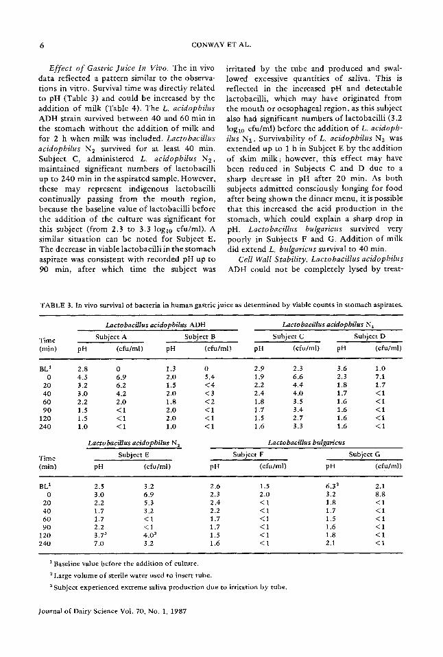
Effect o f Gastric Juice In Vivo. The in vivo data ref lected a pat tern similar to the observa- t ions in vi tro. Survival t ime was di rec t ly related to pH (Table 3) and could be increased by the addi t ion o f milk (Table 4). The L. acidophilus A D H strain survived be tween 40 and 60 rain in the s tomach wi thou t the addi t ion o f milk and for 2 h when milk was included. Lactobacillus acidopbilus N2 survived for at least 40 min. Subjec t C, administered L. acidopbilus N2, mainta ined significant numbers o f lactobacil l i up to 240 min in the aspirated sample. However , these may represent indigenous lactobacil l i cont inua l ly passing f rom the m o u t h region, because the baseline value o f lactobacil l i before the addi t ion o f the cul ture was significant for this subject ( f rom 2.3 to 3.3 logl0 cfu/ml) . A similar s i tuat ion can be noted for Subject E. The decrease in viable lactobaci l l i in the s tomach aspirate was consis tent wi th recorded pH up to 90 min, af ter which t ime the subject was
irri tated by the tube and p roduced and swal- lowed excessive quant i t ies o f saliva. This is ref lected in the increased pH and detectable lactobacill i , which may have originated f rom the m o u t h o r oesophageal region, as this subject also had significant numbers o f lactobacil l i (3.2 log10 cfu /ml) be fo re the addi t ion of L. acidoph- ilus N2. Survivabil i ty o f L. acidopbilus N 2 was ex tended up to 1 h in Subject E by the addi t ion o f skim mi lk ; however , this ef fec t may have been reduced in Subjects C and D due to a sharp decrease in pH after 20 min. As bo th subjects admi t t ed consciously longing for food af ter being shown the d inner menu , it is possible tha t this increased the acid p roduc t ion in the s tomach, which could explain a sharp drop in pH. Lactobacillus bulgaricus survived very poor ly in Subjects F and G. Addi t ion o f milk did ex tend L, bulgaricus survival to 40 min.
Cell Wall Stability. Lactobacillus acidopbilus ADH could no t be comple te ly lysed by treat-
TABLE 3. In vivo survival of bacteria in human gastric juice as determined by viable counts in stomach aspirates.
Lacto bacillus acidopbiIus ADH Lactobacillus acidopbilus N 2
Time Subject A Subject B Subject C Subject D (min) pH (cfu/ml) pH (cfu/ml) pH (cfu/ml) pH (cfu/ml)
1 Baseline value before the addition of culture. 2 Large volume of sterile water used to insert tube.
Subject experienced extreme saliva production due to irritation by tube.
Journal of Dairy Science Vol. 70, No. 1, 1987
SURVIVAL AND ADHESION OF LACTIC ACID BACTERIA 7
TABLE 4. In vivo survival of the three Lactobacillus sp. in human gastric juice. Viable counts expressed as logto colony forming units per milliliter of stomach aspirate. Sterile skim milk (250 ml) added via nasogastric tube before addition of the culture.
Lactobacillus acidopbilus ADH Lactobacillus acidopbilus N 2
Time Subject A Subject B Subject C Subject D (rain) pH (cfu/ml) pH (cfu/ml) pH (cfu/ml) pH (cfu/ml)
1 BBM = Baseline value before addition of milk or culture. 2 BAM = Baseline value after addition of milk and culture. 3 Subjects showed menu for meal at completion of study. 4 Subject experienced excessive saliva production which ceased with repositioning of the tube.
m e n t wi th l y s o z y m e and t h e n T r i t i on X-100, b u t the L. acidopbihts N2 cu l tu re s h o w e d comple t e lysis a f te r i n c u b a t i o n w i t h t he lyso- z y m e for 10 m i n at 37°C. The d i f f i cu l ty in lysing the L. acidopbilus ADH suggests t he p resence o f a m u c h more res i s tan t cell wall.
Adhesion Studies
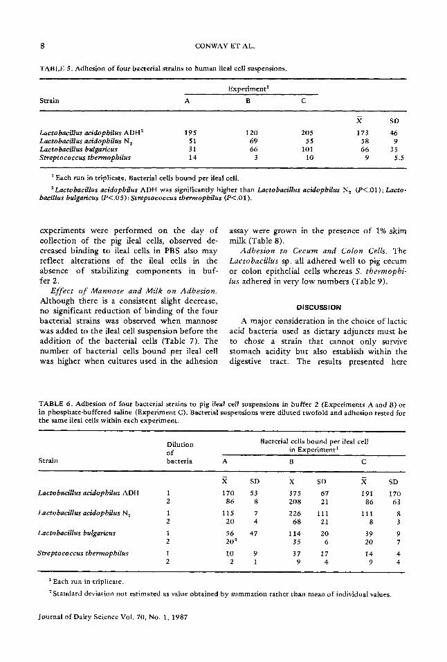
Adhesion to Ileal Cells. Lactobacillus aci- dopbilus ADH b o u n d s igni f icant ly b e t t e r to h u m a n ileal cells t h a n did L. acidopbilus N2 or L, bulgaricus, whereas S. tbermopbilus b o u n d very poor ly (Tab le 5). The da ta are expressed as n u m b e r s o f bac t e r i a b o u n d per ileal cell, assuming l 0 s ileal cells were used. A l t h o u g h th is is an a p p r o x i m a t i o n , the same ileal cell prepara- t i o n was used w i t h i n each e x p e r i m e n t and all
four bac te r i a l s t ra ins were s tud ied at t h e same t ime . A s imilar b i n d i n g p a t t e r n was o b t a i n e d for t he fou r bac te r i a l s t ra ins using pig ileal cells (Table 6). A d i rec t c o m p a r i s o n o f h u m a n and pig ileal cells m a y be m a d e using E x p e r i m e n t A (Tab le 5) and d i lu t ion 1 of E x p e r i m e n t B (Tab le 6). These e x p e r i m e n t s were carr ied o u t at t he same t i m e using t he same bac te r i a l cell suspens ions . All bac te r ia l s t ra ins b o u n d in h igher n u m b e r s to pig ileal cells b u t in t he same p a t t e r n as for h u m a n cells. C o m p o n e n t s of b u f f e r 2 m a y e n h a n c e adhes ion o f t he bac te r ia l s t ra ins as E x p e r i m e n t B and E x p e r i m e n t C (Tab le 6) were car r ied o u t s imu l t aneous ly us ing t he same bac te r i a l suspens ions . B ind ing was s igni f icant ly less w h e n t he pig ileal cells were s u s p e n d e d in PBS ( E x p e r i m e n t C) r a t h e r t h a n b u f f e r 2 ( E x p e r i m e n t B). A l t h o u g h these
Journal of Dairy Science Vol. 70, No. 1, 1987
8 CONWA¥ ET AL.
TABLE 5. Adhesion of four bacterial strains to human ileal cell suspensions.
Each run in triplicate. Bacterial cells bound per ileal cell. 2Lactobacillus acidopbilus ADH was significantly higher than Lactobacillus acidopbilus N 2 (P<.01); Lacto-
expe r imen t s were pe r fo rmed on the day o f col lec t ion of t he pig ileal cells, observed de- creased b inding to ileal cells in PBS also may ref lect a l tera t ions of the ileal ceils in the absence o f stabilizing c o m p o n e n t s in buf- fer 2.
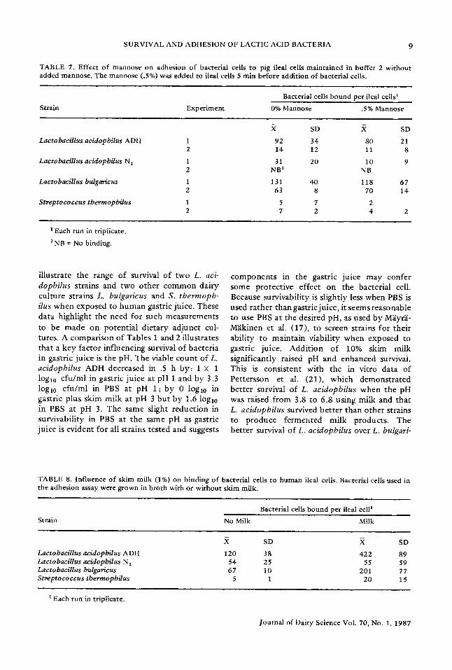
Effect o f Mannose and Milk on Adhesion. Al though there is a cons is ten t slight decrease, no significant r educ t ion of b ind ing of the four bacter ia l strains was observed w h e n mannose was added to the ileal cell suspens ion before the add i t ion of the bacter ial cells (Table 7). The n u m b e r o f bacter ial cells b o u n d per ileal cell was higher w h e n cultures used in the adhesion
assay were g rown in the presence o f 1% skim milk (Table 8).
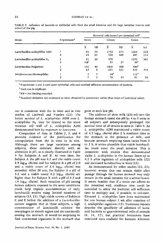
Adhesion to Cecum and Colon Cells. The Lactobacillus sp. all adhered well to pig cecum or colon epithelial cells whereas S. tbermopbi- lus adhered in very low number s (Table 9).
DISCUSSION
A major cons idera t ion in the choice o f lactic acid bacter ia used as d ie ta ry ad junc ts mus t be to chose a strain tha t canno t only survive s t o mach acidi ty bu t also establish wi th in the digestive tract . The results p resen ted here
TABLE 6. Adhesion of four bacterial strains to pig ileal cell suspensions in buffer 2 (Experiments A and B) or in phosphate-buffered saline (Experiment C). Bacterial suspensions were diluted twofold and adhesion tested for the same ileal cells within each experiment.
Dilution Bacterial cells bound per ileal cell of in Experiment 1
2 Standard deviation not estimated as value obtained by summation rather than mean of individual values.
Journal of Dairy Science Vol. 70, No. 1, 1987
SURVIVAL AND ADHESION OF LACTIC ACID BACTERIA 9
TABLE 7. Effect of mannose on adhesion of bacterial ceils to pig ileal cells maintained in buffer 2 without added mannose. The mannose (.5%) was added to ileal cells 5 min before addition of bacterial cells.
SD X SD 1 92 34 80 21 2 14 12 11 8 1 31 20 10 9 2 NB 2 NB
1 131 4O 118 67 2 63 8 70 14 1 5 7 2 2 7 2 4 2
1 Each run in triplicate. 2 NB = No binding.
i l lus t ra te t he range o f survival of t w o L. aci- dopbilus s t ra ins and t w o o t h e r c o m m o n da i ry cu l tu re s t ra ins L. bulgaricus and S. thermopb- ilus w h e n exposed to h u m a n gastr ic ju ice . These da ta h igh l igh t t he need for such m e a s u r e m e n t s to be m a d e o n p o t e n t i a l d i e t a ry a d j u n c t cul- tures. A c o m p a r i s o n of Tab les 1 and 2 i l lus t ra tes t h a t a key f ac to r in f luenc ing survival o f b a c t e r i a in gastr ic ju i ce is the pH. T h e viable c o u n t o f L, acidophilus ADH decreased in .5 h b y : 1 × 1 log10 c f u / m l in gast r ic ju ice at pH 1 and b y 3.3 log10 c f u / m l in PBS at pH 1; b y 0 log10 in gastr ic plus skim mi lk at pH 3 b u t b y 1.6 log10 in PBS at pH 3. The same sl ight r e d u c t i o n in surv ivabi l i ty in PBS a t t h e same pH as gastr ic ju ice is ev iden t for all s t ra ins tes ted and suggests
c o m p o n e n t s in t he gastr ic ju ice m a y c o n f e r some p ro tec t ive e f fec t on t he bac ter ia l cell. Because surv ivabi l i ty is s l ight ly less w h e n PBS is used r a t h e r t h a n gast r ic ju ice , it seems r easonab le to use PBS a t t he desi red pH, as used b y M~yr~- M~k inen et al. (17) , to screen s t ra ins fo r t h e i r ab i l i ty to m a i n t a i n v iab i l i ty w h e n exposed to gastr ic juice. A d d i t i o n o f 10% skim mi lk s ign i f ican t ly raised pH and e n h a n c e d survival. This is cons i s t en t w i th the in vi t ro da ta of Pe t t e r s son et al. (21) , w h i c h d e m o n s t r a t e d b e t t e r surv iva l o f L. acidopbilus w h e n t h e pH was raised f r o m 3.8 to 6.8 us ing mi lk and t h a t L. acidopbilus survived b e t t e r t h a n o t h e r s t ra ins to p r o d u c e f e r m e n t e d mi lk p roduc t s . The b e t t e r survival o f L. acidopbilus over L. bulgari-
TABLE 8. Influence of skim milk (1%) on binding of bacterial cells to human ileal cells. Bacterial cells used in the adhesion assay were grown in broth with or without skim milk.
Bacterial cells bound per ileal cell I Strain No Milk Milk
Lactobacillus acidopbilus A DH Lactobacillus acidopbilus N 2 Lactobacillus bulgaricus Stmptococcus tbermopbilus
SD .X SD 120 38 422 89
54 25 55 59 67 10 201 77
5 1 20 15
1 Each run in triplicate.
Journal of Dairy Science Vol. 70, No. 1, 1987
10 CONWAY ET AL.
TABLE 9. Adhesion of bacteria to epithelial cells from the small intestine and the large intestine (cecum and colon) of the pig.
Bacterial cells bound per intestinal cell 2 Strain Experiment t Ileum Cecum Colon
a Experiment 1 and 2 used same epithelial cells and twofold different concentration of bacteria. 2 Each run in triplicate. a NB = No binding recorded. 4 Standard deviation not estimated as value obtained by summation rather than mean of individual values.
cus is consistent with the in vitro and in vivo studies of Lindwall and Fonden (15). The better survival of L. acidopbilus ADH over L. acidopbilus NR may be related to the more resistant cell wall of L. acidopbilus ADH demonstrated here by exposure to lysozyme.
Comparison of data in Tables 2, 3, and 4 provides evidence of the justification for extrapolation from the in vitro to in vivo. Although there are large variations among subjects, these correlate directly with an alteration in pH, as is clearly illustrated in Table 4 for Subjects A and B. At zero time, for Subject A the pH was 4.5 and the viable count 6.9 log10 cfu/ml and for Subject B a pH of 2.0 and a viable count of 5.4 log10 cfu/ml was recorded. After 20 min, for Subject A a pH of 3.2 and a viable count 6.2 loglo cfu/ml are higher than for Subject B with a pH of 1.5 and <4 log10 cfu/ml. Such variation o f pH among human subjects exposed to the same conditions could help explain inconsistencies of early lactobacilli studies using limited numbers of subjects (13, 23). High viable counts of Subjects C and E before the addition of a Lactobacillus culture suggests that in these subjects, a high concentration of lactobacilli colonize the oesophagus or mouth region and are continually seeding the stomach. It would be surprising to find commensal organisms in the stomach that
grow at such low pH. The addition of skim milk (250 ml) into the
human stomach raised the pH by 4 to 5 units in all subjects and subsequently prolonged the survival t ime of all strains as seen in Table 4. As L. acidopbilus ADH maintained a viable count of 4.3 log10 cfu/ml after 2 h residence time in the stomach in the presence of milk, and because stomach emptying times varies from 0 to 3 h, it seems plausible that viable lactobacil- lus could enter the small intestine. This is consistent with studies that demonstrated viable L. acidopbilus in the human ileum up to 4.5 h after ingestion of acidophilus milk (22) and increased lactobacillus in feces (15).
As demonstrated by other studies (22, 23), bacterial strains that can remain viable after passage through the human stomach may only remain in the small intestine for a several hours. It seems feasible that if a strain can adhere to the intestinal wall, residence time could be extended to allow the bacterial cell sufficient time to multiply and, if possible, to colonize. Elevated L. acidopbilus have been maintained for one human subject 1 wk after cessation o f L. acidopbilus ingestion (15). Numerous reports describe the specificity of adhesion of lactic acid bacteria to gastrointestinal cells of animals (4, 14, 17), but practical limitations have restricted data available for humans. Kleeman
Journal of Dairy Science Vol. 70, No. 1, 1987
SURVIVAL AND ADHESION OF LACTIC ACID BACTERIA 11
and Klaenhammer (13), using a human fetal intestinal celt line, demonstrated specific and calcium-dependent nonspecific adhesion using 32 isolates.
One of the goals of the data presented here was to test a strain of Kleeman and Klaenham- mer (13) that adhered specifically to the fetal intestine cell line plus the other cultures tested in this study for the capacity to adhere to freshly collected human intestinal cells. The intestinal cells were viable, as determined by the t rypan blue exclusion test, and surrounded by an extracellular carbohydrate layer, as demonstrated by periodic acid-Shift test (5). Adhesion to the human ileal ceils has been demonstrated for all strains to various degrees (Table 4). The L. acidopbilus ADH strain, which adhered well to the fetal cell line (13), also adhered well in this system. As a similar binding pat tern for the four bacterial strains was achieved using the pig ileal cells (Table 6), it seems feasible that pig intestinal cells or the fetal cell line may be used to screen routinely adhesive properties of strains for human use. This overlapping of adhesive propert ies also has been demonstrated by M~yr'~-M~kinen et al. (17), because Lactobacillus fermenturn strains isolated from and adhered to the calf intestine could also adhere to pig epithelial cell. But the overlapping of adhesive propert ies conflicts with the results of Barrow et al. (4). The data in Tables 5 and 6 illustrate that the lactic acid bacteria used for the commercial product ion of cultured dairy products can adhere to human and pig ileal cells to various degrees. Although this is the converse of the finding of M~iyr~'- M~kinen et al. (17), Barrow et al. (4) used different commercial strains. As all strains showed some binding to both human and pig intestinal cells, the binding may be nonspecific as compared with that reported for Lactobacil- lus sp. in the murine, (19) chicken, and pig systems (4).
Because adhesion was unaffected by mannose (Table 7), inclusion of mannose in the ileal cell suspension according to Deneke et al. (5) to inhibit mannose sensitive type 1 fimbriae did not alter the adhesion pat terns of the cultures. The promot ion of adhesion by the inclusion of milk (Table 7) is consistent with the enhance- ment of binding in the presence of calcium (13) and could be valuable to increase adhesion in the intestine. The demonstrat ion of adhesion to
both pig small and large intestinal cells strength- ens the suggestion that implantat ion may be possible and need not be restricted to the small intestine.
It has been shown that survival of lactic acid bacteria within the human stomach is closely related to pH, which can vary largely among individuals. Some lactic acid bacteria survive much longer in human gastric juice over the pH range 1 to 5, and it is encouraging that the strain showing best survival in gastric juice, L. acidopbilus ADH, also adhered best to human and pig ileal cells. As the addi t ion of milk both extended survival times of bacteria exposed to gastric juice and enhanced adhesion, the admin- istration of Lactobacillus in milk or as cultured milk products should opt imize Lactobacil- lus levels within the digestive tract. The in vitro system presented here for studying survival and adhesion would be valuable for screening potential lactic acid bacteria for dietary use. Once such conditions are defined it is possible to correlate information on strains with effects produced in vivo which can be monitored quantitatively. Studies continue to determine the effect of L. acidophilus ADH on fecal enzyme activity.
ACKNOWLEDGMENTS
This work was supported by the North Carolina Dairy Foundat ion. As recipient of the Australian Society for Microbiology's Sherris Study Award, P. L. Conway most sincerely thanks John C. Sherris and his wife Elizabeth for funding this award, which resulted in his part icipation in this project. The authors thank both C. F. Deneke for his invaluable assistance with the adhesion assay and supplying the human ileal cells and Fawaz for his assistance with the nasogastric in tubation.
REFERENCES
1 Aim, L. 1983. Survival rate of salmonella and shigella in fermented milk products with and without added human gastric juice. An in vitro study. Prof. Food Nutr. Sci. 7:19.
2 Aim, L., D. Humble, E. Ryd-Kjellen, and G. Setterberg. 1983. The effect of acidophilus milk in the treatment of constipation in hospitalised geriatric patients. Nutrition and the intestinal flora. Page 131 in Symp. Swed. Nutr. Found. 15. Bo Hallgren, ed. Uppsala.
3 Barefoot, S. F., and T. R. Klaenhammer. 1983. Detection and activity of lactacin B, a bacteriocin
Journal of Dairy Science Vol. 70, No. 1, 1987
12 CONWAY ET AL.
produced by Lactobacillus acidopbilus. Appl. Environ. Microbiol. 45 :1808.
4 Barrow, P. A., B. E. Brooker, R. Fuller, and M. J. Newport . 1980. The a t t achmen t of bacteria to the gastric epi thel ium o f the pig and its impor tance in the microecology of the intestine. J. Appl. Bacteriol. 48:147.
5 Deneke, C. F., K. McGowan, G. M. Thorne, and S. L. Gorbach. 1983. A t t a c h m e n t o f entero toxogenic Escbericbia coli to h u m a n intestinal cells. Infect. Immun . 39:1102.
6 Deneke, C. F., K. M. McGowan, A. D. Larson, and S. L. Gorbach. 1984, A t t a c h m e n t o f h u m a n and pig (K88) enterotoxigenic Escbericbia coli strains to ei ther h u m a n or porcine small intestinal cells. Infect. l m m u n . 45:522.
7 Friend, B. A., and K. M. Shahani. 1984. An t i t umor properties o f lactobacilli and dairy products fe rmented by lactobacilli. J. Food Prot. 47:717.
8 Gilliland, S. E., C. R. Nelson, and C. Maxwell. 1985. Assimilation o f cholesterol by Lactobacillus acidopbilus. Appl. Environ. Microbiol. 49:377.
9 Goldin, B. R., and S. L. Gorbach, 1977.Al te ra t ions in fecal microflora enzymes related to diet, age, Lactobacillus supplements , and d imethylhydrazine . Cancer 40:2421.
10 Goldin, B. R., and S. L. Gorbach. 1980. Effect of Lactobacillus acidopbilus dietary supp lements on 1,2-dimethylhydrazine dihydrochloride induced intestinal cancer in rats. J. Natl. Cancer Inst. 64:263.
11 Goldin, B. R., L. Swensson, J. Dwyer, M. Sexton, and S. L. Gorbach. 1980. Effect o f diet and L. acidopbilus supplements on h u m a n fecal bacterial enzymes. J. Natl. Cancer Inst. 64:255.
12 Klaenhammer , T. R. 1982. Microbiological con- siderations in selection and preparation o f Lacto- bacillus strains for use as dietary adjunts. J. Dairy Sci. 65 :1339.
13 Kleeman, E. G., and T. R. Klaenhammer . 1982. Adherence o f Lactobacillus species to h u m a n fetal intestinal ceils. J. Dairy Sci. 65:2063.
14 Kotarski, S. F., and D. C. Savage. 1979. Models for s tudy of the specificity by which endogenous lactobacilli adhere to mur ine gastric epithelia. Infect. l m m u n . 26:966.
15 Lindwall, S., and R. Fonden . 1984. Passage and survival of L. acidopbilus in the human gastrointes- tinal tract . Int. Dairy Fed. Bull. FIL 179. XXI.
16 Luckey, T. D. 1984. Perspectives in intestinal microecology. Microecol. Ther. 14:243.
17 M~iyr~i-M~ikinen, A., M. Manninen, and H. Gyllen- berg. 1983. The adherence o f lactic acid bacteria to the columnar epithelial cells o f pigs and calves. J, Appl. Bacteriol. 55:241.
18 Merson, M. H., G. K. Morris, D. A. Sack et al. 1976. Traveler's diarrhea in Mexico. New England J. Med. 294:1299.
19 McCormick, E. L., and D. C. Savage. 1983. Charac- terization o f Lactobacillus sp. strain 100-37 f rom the mur ine gastrointest inal t ract : ecology, plasmid content , and antagonist ic activity toward Clostridi- urn rarnosurn H1. Appl. Environ. Microbiol. 46 :1103 .
20 Metchnikoff , E. 1907. The prolongation of life. G. P. Pu tman ' s Sons, New York, NY.
21 Pet tersson, L., W. Graf, L. Aim, S. Lindwall, and A. Str6berg. 1983. Survival of Lactobacillus aci- dopbilus NCDO 1748 in the h u m a n gastrointestinal tract . 1. Incubat ion with gastric juice in vitro. Nutr i t ion and the intestinal flora. Page 123 in Symp. Swed. Nutr. Found . 15. Bo Hallgren, ed. Uppsala.
22 Pettersson, L., W. Graf and V. Sewelin. 1983. Survival of Lactobacillus acidopbilus NCDO 1748 in the human gastrointest inal tract . 2. Abili ty to pass the s tomach and intest ine in vivo. Nutri t ion and the intestinal flora. Page 127 in Symp. Swed. Nutr . Found. 15. Bo HaUgren, ed. Uppsala.
23 Robins-Browne, R. M., and M. M. Levine. 1981. The fate of ingested lactobacilli in the proximal small intestine. Am. J. Clin. Nutr . 34:514.
24 Shahani, K. M., J. R. Vakil, and A. Kilara. 1976. Natural antibiotic activity o f Lactobacillus aci- dopbilus and bulgaricus. Cult. Dairy Prod. J. Nov:15.
25 Shore, E. G., A. G. Dean, K. J. Holik, and B. R. Davis. 1974. Ente ro tox inproduc ing Escbericbia coli and diarrheal disease in adult travelers: a prospective study. J. Infect. Dis. 129:577.
26 Speck, M. L. 1980. Preparation o f lactobacilti for die tary uses. J. Food Prot. 42:65.


![Lactic Acid - NIHSLactic Acid 乳酸C3H6O3:90.08 (2RS)-2-Hydroxypropanoic acid [50-21-5] Lactic Acid is a mixture of lactic acid and lactic an-hydride. It contains not less than 85.0z](https://static.documents.pub/doc/80x56/6107906c3f161736212a9570/lactic-acid-nihs-lactic-acid-ec3h6o39008-2rs-2-hydroxypropanoic-acid.jpg)