INSIDE THIS ISSUE 3 Expanding Indications for Reverse Shoulder Arthroplasty 5 Highlight of Recent Scholarly Publication from Mayo Clinic Department of Orthopedic Surgery 6 Double Gracilis Free Muscle Transfer Expands Function in Brachial Plexus Restoration Falls are a well-recognized hazard for the elderly. But it is less well known that people who have undergone a limb amputation—particularly above-knee amputations—face an even greater challenge maintaining their balance. Data show that during a 12-month period, about 30% of the members in an elderly population fall. This compares to an annual fall rate of 50-60% for amputees. Mayo Clinic Orthopedic researchers are striving to improve ambulation by reducing the fall hazard facing US military service members who have undergone unilateral limb amputation. To do this, they are collaborating with a consortium of researchers in a new $2.4 million Applied Research and Advanced Technology Development Award from the US Defense Department. The grant is intended to improve training methods for rehabilitating this group of wounded warriors using advanced technology and novel techniques. Power and Passion for the Cause The program harnesses two key Mayo Orthopedics resources: the power of its research in biomechanics, gait analysis and prosthesis development, and its passion for serving wounded warriors. Kenton R. Kaufman, PhD, head of Mayo’s Motion Analysis Laboratory, is the Mayo specialist on Serving Wounded Warriors Novel Rehabilitation Method for Amputees Focuses on Fall Prevention Vol. 5, No. 2, 2011 Daniel J. Berry, MD Chair, Mayo Clinic Department of Orthopedic Surgery Figure 1. Tagged with retroreflective markers that infrared cameras detect to record position and analyze gait, a war-wounded amputee research subject takes part in a new collaborative study involving Mayo Clinic, the University of Illinois at Chicago and US Navy researchers based at state-of-the-art facilities of the Naval Health Research Center in San Diego and the Naval Medical Center San Diego (NMCSD). The goal is to improve training methods for rehabilitating this group of wounded warriors using advanced technology and novel techniques. continued on page 2
Transcript
INSIDE THIS ISSUE
3 Expanding Indications for Reverse Shoulder Arthroplasty
5 Highlight of Recent Scholarly Publication from Mayo Clinic Department of Orthopedic Surgery
6 Double Gracilis Free Muscle Transfer Expands Function in Brachial Plexus Restoration
Falls are a well-recognized hazard for the elderly. But it is less well known that people who have undergone a limb amputation—particularly above-knee amputations—face an even greater challenge maintaining their balance. Data show that during a 12-month period, about 30% of the members in an elderly population fall. This compares to an annual fall rate of 50-60% for amputees.
Mayo Clinic Orthopedic researchers are striving to improve ambulation by reducing the fall hazard facing US military service members who have undergone unilateral limb amputation. To do this, they are collaborating with a consortium of researchers in a new $2.4 million Applied Research and Advanced Technology Development Award from the US Defense Department. The grant is intended to improve training methods for rehabilitating this group of wounded warriors using advanced technology and novel techniques.
Power and Passion for the CauseThe program harnesses two key Mayo Orthopedics resources: the
power of its research in biomechanics, gait analysis and prosthesis development, and its passion for serving wounded warriors. Kenton R. Kaufman, PhD, head of Mayo’s Motion Analysis Laboratory, is the Mayo specialist on
Serving Wounded WarriorsNovel Rehabilitation Method for Amputees Focuses on Fall Prevention
Vol. 5, No. 2, 2011
Daniel J. Berry, MDChair, Mayo Clinic Department of Orthopedic Surgery
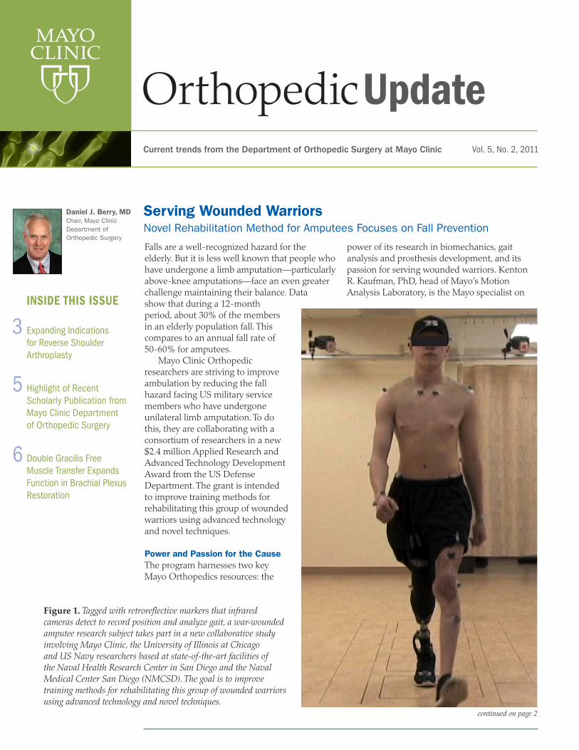
Figure 1. Tagged with retroreflective markers that infrared cameras detect to record position and analyze gait, a war-wounded amputee research subject takes part in a new collaborative study involving Mayo Clinic, the University of Illinois at Chicago and US Navy researchers based at state-of-the-art facilities of the Naval Health Research Center in San Diego and the Naval Medical Center San Diego (NMCSD). The goal is to improve training methods for rehabilitating this group of wounded warriors using advanced technology and novel techniques.
continued on page 2
2 MAYO CLINIC | OrthopedicUpdate
the grant team. His previous work developing a microprocessor-controlled knee orthosis had wide application among wounded warriors and connected Dr Kaufman deeply to improving their rehabilitation. “I’m very passionate about working with wounded warriors, so this opportunity to participate in this research program is really gratifying,’’ he says.
Creative CollaborationIn addition to Dr Kaufman’s command of gait analysis and prosthesis development, Mark D. Grabiner, PhD, from the University of Illinois at Chicago, brings expertise from his specialized training method working with the elderly to prevent falling. Marilynn Wyatt, MA, PT, director of the Biomechanics Laboratory of the US Naval Medical Center in San Diego adds her expertise in rehabilitation training and studies performed
in the state-of-the-art Naval Medical Center’s C5 facility—Center for Comprehensive and Complex Casualty Care. The fall-prevention training is performed in the C5 motion analysis laboratory using a computerized system that allows the researchers to create controlled falls while the amputee walks on a customized treadmill at various paces while wearing a safety harness.
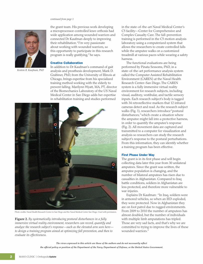
The functional evaluations are being performed by Pinata Sessoms, PhD, in a state-of-the-art performance analysis system called the Computer Assisted Rehabilitation Environment (CAREN) at the Naval Health Research Center–San Diego. The CAREN system is a fully immersive virtual reality environment for research subjects, including visual, auditory, vestibular, and tactile sensory inputs. Each research subject’s body is tagged with 34 retroreflective markers that 12 infrared cameras detect and read. As the research subject walks (Fig. 1), researchers introduce “postural disturbances,’’ which create a situation where the amputee might fall into a protective harness, in order to quantify the amputee’s response (Fig. 2). All movement data are captured and transmitted to a computer for visualization and analysis so researchers can study the research subject’s response to the postural perturbations. From this information, they can identify whether a training program has been effective.
First Phase Under WayThe grant is in its first phase and will begin collecting data later this year from 30 unilateral amputees. Since the grant was written, the amputee population is changing, and the number of bilateral amputees has risen due to casualties in Afghanistan. Compared to Iraq battle conditions, soldiers in Afghanistan are less protected, and therefore more vulnerable to war injuries.
Explains Dr Kaufman: “In Iraq, soldiers were in armored vehicles, so when an IED exploded, they were protected. Now in Afghanistan they are on foot patrol due to rugged environments. From 2009 to 2010 the number of amputees has almost doubled, but the number of individuals with multiple limb amputations has tripled. Those are very sad facts, and that’s why we are committed to trying to improve the lives of these wounded warriors.”
Kenton R. Kaufman, PhD
Figure 2. By systematically introducing postural disturbances in a fully immersive virtual reality environment, researchers can record, quantify and analyze the research subject’s response—such as the elevated arm seen here— to design a training program aimed at optimizing fall prevention, and then to evaluate its effectiveness.
continued from page 1
Photo credits: Naval Health Research Center in San Diego and the Naval Medical Center San Diego. Used with permission.
The views expressed in this article are those of the authors and do not necessarily reflect
the official policy or position of the Department of the Navy, Department of Defense, or the United States Government.
MAYO CLINIC | OrthopedicUpdate 3
The arthritic rotator cuff-deficient shoulder has long been a significant clinical problem due to lack of successful treatment options for this painful and disabling condition. But this situation is rapidly changing with the evolution of implant technology and surgical technique. Since the reverse shoulder arthroplasty (RSA) implant was originally developed nearly 25 years ago in Europe to treat rotator cuff tear arthropathy, it has dramatically improved the treatment of rotator cuff conditions (Fig. 1). Approved in 2004 by the US Food and Drug Administration, it has also engendered controversy related to possible overuse, because indications are expanding beyond rotator cuff tear arthropathy to include a suite of shoulder pathologies. These range from reconstruction after tumor removal to proximal humeral fractures and nonunion (Fig. 2).
After seven years’ experience with RSA, Mayo Clinic orthopedic surgeons remain committed to studying it from both a clinical and basic science perspective. They proceed with expanded indications on an individualized basis, grounded in a comprehensive understanding of the biomechanics of shoulder pathophysiology.
Optimizing Pain Relief and Function“The reverse shoulder arthroplasty has provided an innovative and effective way to relieve pain and restore function in many patients with rotator cuff deficiencies,” explains Mayo Clinic orthopedic surgeon John W. Sperling, MD. In one large outcome study—n=80 patients, mean follow-up of 3.6 years—96% of patients ranked
Expanding Indications for Reverse Shoulder Arthroplasty
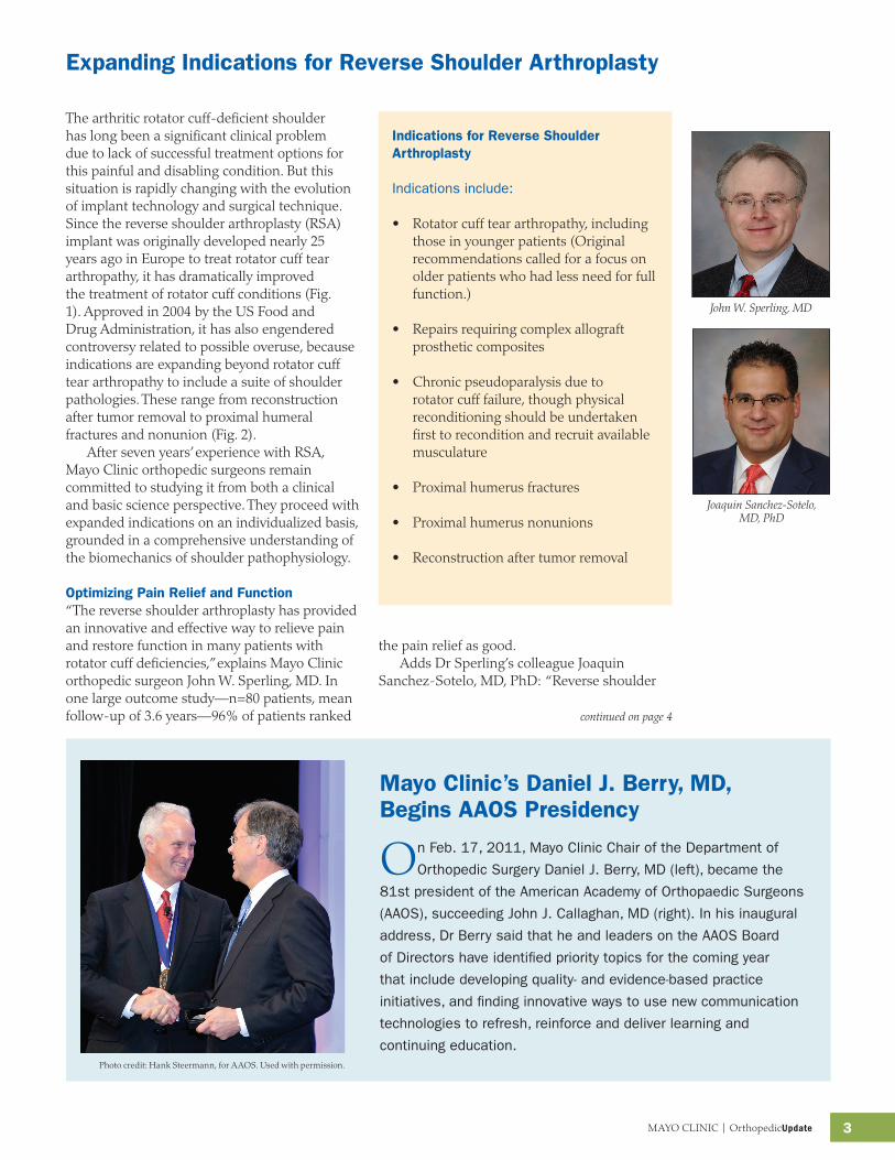
Mayo Clinic’s Daniel J. Berry, MD, Begins AAOS Presidency
On Feb. 17, 2011, Mayo Clinic Chair of the Department of
Orthopedic Surgery Daniel J. Berry, MD (left), became the
81st president of the American Academy of Orthopaedic Surgeons
(AAOS), succeeding John J. Callaghan, MD (right). In his inaugural
address, Dr Berry said that he and leaders on the AAOS Board
of Directors have identified priority topics for the coming year
that include developing quality- and evidence-based practice
initiatives, and finding innovative ways to use new communication
technologies to refresh, reinforce and deliver learning and
continuing education.Photo credit: Hank Steermann, for AAOS. Used with permission.
Indications for Reverse Shoulder Arthroplasty
Indications include:
• Rotator cuff tear arthropathy, including those in younger patients (Original recommendations called for a focus on older patients who had less need for full function.)
• Chronic pseudoparalysis due to rotator cuff failure, though physical reconditioning should be undertaken first to recondition and recruit available musculature
• Proximal humerus fractures
• Proximal humerus nonunions
• Reconstruction after tumor removal
the pain relief as good.Adds Dr Sperling’s colleague Joaquin
Sanchez-Sotelo, MD, PhD: “Reverse shoulder
John W. Sperling, MD
Joaquin Sanchez-Sotelo, MD, PhD
continued on page 4
4 MAYO CLINIC | OrthopedicUpdate
Table 1. Reverse Shoulder Arthroplasty Advantages and Disadvantages
arthroplasty has revolutionized the field of shoulder replacement. At Mayo, our outcomes with RSA have been consistently very encouraging in terms of pain relief, motion and functional outcomes. We have been extremely careful in attempting to perfect our surgical technique, ensure component fixation and determine the optimal soft-tissue tension for each individual.”
Rationale, Advantages, Disadvantages RSA reverses the natural anatomy of the ball-and-socket joint by implanting a concave socket plate into the humeral head, and convex spherical glenoid component into the glenoid fossa. RSA also treats arthritis by resurfacing the glenohumeral joint. The goal is to reduce pain and restore function by overcoming vulnerabilities of traditional shoulder replacements in which the absence of a stabilizing rotator cuff can lead to poor function and mechanical failure of the implant.
By reversing the anatomy, the surgery improves deltoid tension and provides a stable fulcrum that compensates for loss of rotator cuff performance. In the absence of a rotator cuff, attempted arm elevation results in superior migration of the humeral head, with no real fulcrum and poor motion. With RSA, the constrained nature of the implant provides a fulcrum that is particularly advantageous for a better-tensioned deltoid. Disadvantages range from highly variable complication rate to overuse. (Table 1).
IndicationsIn experienced hands, the RSA has the potential to successfully compensate for rotator cuff insufficiency across a broad spectrum of shoulder pathologies. At Mayo Clinic, indications for use of RSA range from the relatively simple to the highly complex, from rotator cuff tear arthropathy to chronic pseudoparalysis.
ContraindicationsPatients for whom RSA may not be indicated include those who have:• Absence of a functioning deltoid to
compensate for the rotator cuff• Presence of considerable elevation abilities,
even with irreparable rotator cuff tear, and absence of glenohumeral joint arthritis
• Active infection
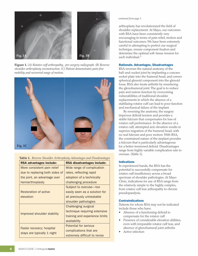
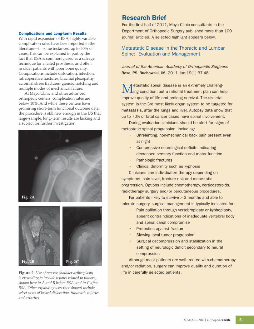
Figure 1. (A) Rotator cuff arthropathy, pre-surgery radiograph. (B) Reverse shoulder arthroplasty reconstruction. (C) Patient demonstrates pain-free mobility and recovered range of motion.
continued from page 3
MAYO CLINIC | OrthopedicUpdate 5
Metastatic Disease in the Thoracic and Lumbar Spine: Evaluation and Management
Journal of the American Academy of Orthopaedic Surgeons
Rose, PS. Buchowski, JM. 2011 Jan;19(1):37-48.
Metastatic spinal disease is an extremely challeng-
ing condition, but a rational treatment plan can help
improve quality of life and prolong survival. The skeletal
system is the 3rd most likely organ system to be targeted for
metastasis, after the lungs and liver. Autopsy data show that
up to 70% of fatal cancer cases have spinal involvement.
During evaluation clinicians should be alert for signs of
metastatic spinal progression, including:
• Unrelenting, non-mechanical back pain present even
at night
• Compressive neurological deficits indicating
decreased sensory function and motor function
• Pathologic fractures
• Clinical deformity such as kyphosis
Clinicians can individualize therapy depending on
symptoms, pain level, fracture risk and metastatic
progression. Options include chemotherapy, corticosteroids,
radiotherapy surgery and/or percutaneous procedures.
For patients likely to survive > 3 months and able to
tolerate surgery, surgical management is typically indicated for:
• Pain palliation through vertebroplasty or kyphoplasty,
absent contraindications of inadequate vertebral body
and spinal canal compromise
• Protection against fracture
• Slowing local tumor progression
• Surgical decompression and stabilization in the
setting of neurologic deficit secondary to neural
compression
Although most patients are well treated with chemotherapy
and/or radiation, surgery can improve quality and duration of
life in carefully selected patients.
Research BriefFor the first half of 2011, Mayo Clinic consultants in the
Department of Orthopedic Surgery published more than 100
journal articles. A selected highlight appears below.Complications and Long-term ResultsWith rapid expansion of RSA, highly variable complication rates have been reported in the literature—in some instances, up to 50% of cases. This can be explained in part by the fact that RSA is commonly used as a salvage technique for a failed prosthesis, and often in older patients with poor bone quality. Complications include dislocation, infection, intraoperative fractures, brachial plexopathy, acromial stress fractures, glenoid notching and multiple modes of mechanical failure.
At Mayo Clinic and other advanced orthopedic centers, complication rates are below 10%. And while these centers have promising short-term functional outcome data, the procedure is still new enough in the US that large-sample, long-term results are lacking and a subject for further investigation.
Figure 2. Use of reverse shoulder arthroplasty is expanding to include repairs related to tumors, shown here in A and B before RSA, and in C after RSA. Other expanding uses (not shown) include select cases of locked dislocation, traumatic injuries and arthritis.
6 MAYO CLINIC | OrthopedicUpdate
free muscle transfer of the gracilis. The concept is that if 1 muscle is good for restoring function, then 2 are better.” Adds Dr. Shin’s reconstructive microsurgeon colleague, hand specialist Allen T. Bishop, MD: “The double gracilis functioning free muscle transfer is an essential tool in the management of brachial plexus injury because it offers the patient the possibility of expanded functionality to include simple grasp, in addition to restoration of shoulder and elbow motion, hand sensation and triceps function.” Neurosurgeon Robert J. Spinner, MD, is the third specialist on the Mayo brachial plexus team: “The key to our approach is the multidisciplinary team,” he says. “We now have vast experience with an algorithm that relies on simultaneous surgeries in a single complex procedure—which is much less traumatic and disruptive for patients.”
Mayo Modified ApproachThe double gracilis FFMT was pioneered as a 2-stage, 2-operation procedure in Japan in 1997. In stage I, the first gracilis transfer, powered by
Double Gracilis Free Muscle Transfer Expands Function in Brachial Plexus Restoration
Complete avulsion of the brachial plexus in adults can be devastating for patients due to loss of function in the upper extremity. Patients with complete avulsion injuries lose shoulder abduction, external rotation, elbow flexion, and animation and sensation in the hand. These injuries are typically caused by high-energy traumatic impact with a stationary object, such as occurs in vehicular accidents—including motorcycle and snowmobile collisions. As more patients survive these serious accidents, there is a greater need for more robust brachial plexus reconstruction options —particularly in the setting of complete avulsion, or when treatment is delayed 12 months or more, resulting in denervation and irreversible muscle atrophy.
Double Gracilis TransferMore robust reconstruction techniques are now emerging and being refined due to enhanced understanding of complex nerve and muscle pathophysiology. Improved results rely on microsurgical techniques including the use of functioning free muscle transfer (FFMT) in recent years. FFMT is indicated when nerve grafting is not possible, when all nerve roots are avulsed from the spinal cord, or when nerve reconnection is unlikely to succeed. Importantly, such repairs seldom restore function to the wrist and hand, explains orthopedic reconstructive microsurgery and hand specialist on Mayo Clinic’s brachial plexus surgical team, Alexander Y. Shin, MD: “At Mayo, we combine traditional brachial plexus reconstruction of nerve transfers or grafts with free tissue transfer options, including double functioning
Allen T. Bishop, MD
Robert J. Spinner, MD
Alexander Y. Shin, MD
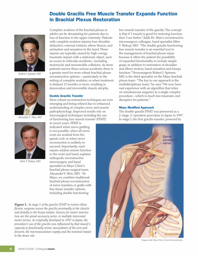
Fig. 1 In stage 1 of the gracilis FFMT to restore elbow flexion, surgeons secure the gracilis proximally to the clavicle and distally to the biceps tendon. Sources for motor innervation are the spinal accessory nerve, or multiple intercostal motor nerves. As originally developed in 1997 in Japan, the procedure’s use of the gracilis was influenced by that muscle’s capacity to functionally mimic musculature of the arm and forearm, its microvasculature supply and the minimal impact harvesting it makes on the donor site.
Figure 1. In stage 1 of the gracilis FFMT to restore elbow flexion, surgeons secure the gracilis proximally to the clavicle and distally to the biceps tendon. Sources for motor innerva-tion are the spinal accessory nerve, or multiple intercostal motor nerves. As originally developed in 1997 in Japan, the procedure’s use of the gracilis was influenced by that muscle’s capacity to functionally mimic musculature of the arm and forearm, the microvasculature supply and the minimal impact to the donor site.
Image credit: Mayo Clinic. Used with permission.
MAYO CLINIC | OrthopedicUpdate 7
Fig. 3 To conduct motor neurotization of the gracilis muscle, the fifth and sixth intercostal motor nerves are used. For tricep neurotization, the third and fourth intercostal motor nerves are used. For sensory neurotization , the lateral cord of the median nerve is used through intercostal sensory nerves three through six.
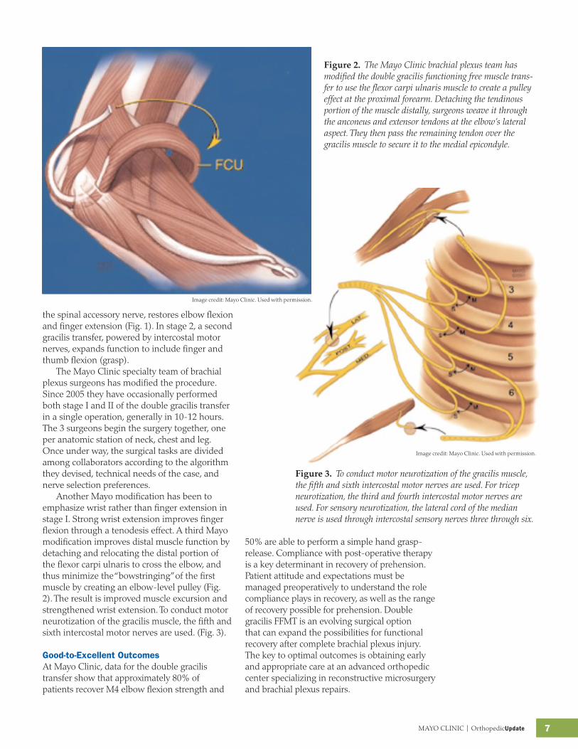
Fig. 2. The Mayo Clinic brachial plexus team has modified the double gracilis functioning free muscle transfer to use the flexor carpi ulnaris muscle to create a pulley effect at the proximal forearm. Detaching the tendinous portion of the muscle distally, surgeons weave it through the anconeous and extensor tendons at the elbow's lateral aspect. They then pass the remaining tendon over the gracilis muscle to secure it to the medial epicondyle.
the spinal accessory nerve, restores elbow flexion and finger extension (Fig. 1). In stage 2, a second gracilis transfer, powered by intercostal motor nerves, expands function to include finger and thumb flexion (grasp).
The Mayo Clinic specialty team of brachial plexus surgeons has modified the procedure. Since 2005 they have occasionally performed both stage I and II of the double gracilis transfer in a single operation, generally in 10-12 hours. The 3 surgeons begin the surgery together, one per anatomic station of neck, chest and leg. Once under way, the surgical tasks are divided among collaborators according to the algorithm they devised, technical needs of the case, and nerve selection preferences.
Another Mayo modification has been to emphasize wrist rather than finger extension in stage I. Strong wrist extension improves finger flexion through a tenodesis effect. A third Mayo modification improves distal muscle function by detaching and relocating the distal portion of the flexor carpi ulnaris to cross the elbow, and thus minimize the “bowstringing” of the first muscle by creating an elbow-level pulley (Fig. 2). The result is improved muscle excursion and strengthened wrist extension. To conduct motor neurotization of the gracilis muscle, the fifth and sixth intercostal motor nerves are used. (Fig. 3).
Good-to-Excellent OutcomesAt Mayo Clinic, data for the double gracilis transfer show that approximately 80% of patients recover M4 elbow flexion strength and
Figure 2. The Mayo Clinic brachial plexus team has modified the double gracilis functioning free muscle trans-fer to use the flexor carpi ulnaris muscle to create a pulley effect at the proximal forearm. Detaching the tendinous portion of the muscle distally, surgeons weave it through the anconeus and extensor tendons at the elbow’s lateral aspect. They then pass the remaining tendon over the gracilis muscle to secure it to the medial epicondyle.
Figure 3. To conduct motor neurotization of the gracilis muscle, the fifth and sixth intercostal motor nerves are used. For tricep neurotization, the third and fourth intercostal motor nerves are used. For sensory neurotization, the lateral cord of the median nerve is used through intercostal sensory nerves three through six.
50% are able to perform a simple hand grasp-release. Compliance with post-operative therapy is a key determinant in recovery of prehension. Patient attitude and expectations must be managed preoperatively to understand the role compliance plays in recovery, as well as the range of recovery possible for prehension. Double gracilis FFMT is an evolving surgical option that can expand the possibilities for functional recovery after complete brachial plexus injury. The key to optimal outcomes is obtaining early and appropriate care at an advanced orthopedic center specializing in reconstructive microsurgery and brachial plexus repairs.
Image credit: Mayo Clinic. Used with permission.
Image credit: Mayo Clinic. Used with permission.
CME Opportunities
7th Mayo Clinic Spine Symposium
March 25-28, 2012
Naples, FL
This course offers surgeons and non-operative clinicians in both orthopedics
and neurology comprehensive clinical care guidance for patients with spinal disor-
ders and deformities. Topics range from current and best-practices use of mini-
mally invasive surgical techniques to use of health quality measures for evaluating
outcomes. The latest developments in the field are presented through an engaging
array of didactic lectures, panel discussions, topical debates and case presenta-
tion. All are designed for optimal comprehension through the use of an audience
response system and interactive discussion format.