Helsinki University of Technology Laboratory of Biomedical Engineering Department of Engineering Physics and Mathematics Teknillinen korkeakoulu L¨ a¨ aketieteellisen tekniikan laboratorio Teknillisen fysiikan ja matematiikan osasto Espoo 2003 DATA REGISTRATION AND FUSION FOR CARDIAC APPLICATIONS Timo M¨ akel¨ a Dissertation for the degree of Doctor of Science in Technology to be presented with due permission of the Department of Engineering Physics and Mathematics for public examina- tion and debate in Auditorium F1 at Helsinki University of Technology (Espoo, Finland) on the 28th of May, 2003, at 12 o’clock noon.
Transcript
Helsinki University of Technology Laboratory of Biomedical Engineering
Department of Engineering Physics and Mathematics
Teknillinen korkeakoulu Laaketieteellisen tekniikan laboratorio Teknillisen fysiikan ja matematiikan osasto
Espoo 2003
DATA REGISTRATION AND FUSION FOR
CARDIAC APPLICATIONS
Timo Makela
Dissertation for the degree of Doctor of Science in Technology to be presented with duepermission of the Department of Engineering Physics and Mathematics for public examina-tion and debate in Auditorium F1 at Helsinki University of Technology (Espoo, Finland) onthe 28th of May, 2003, at 12 o’clock noon.
ISBN (printed) 951-22-6514-1ISBN (pdf) 951-22-6515-X
Picaset OyHelsinki 2003
HELSINKI UNIVERSITY OF TECHNOLOGYP.O. BOX 1000, FIN-02015 HUT
http://www.hut.fi
ABSTRACT OF DOCTORAL DISSERTATION
Author
Name of the dissertation
Date of manuscript Date of the dissertation
Monograph Article dissertation (summary + original articles)
Department
Laboratory
Field of research
Opponent
Supervisor
Instructors
Abstract
Keywords
UDC Number of pages
ISBN (printed) ISBN (pdf)
Publisher
Print distribution
The dissertation can be read at http://lib.hut.fi/Diss/2003/isbn951226515X/
Timo Juhani Mäkelä
Data registration and fusion for cardiac applications
January 27, 2003 May 28, 2003
✔
Department of Engineering Physics and Mathematics
Laboratory of Biomedical Engineering
Medical image processing
Prof. Nicholas Ayache (INRIA, Sophia Antipolis, France)
Prof. Toivo Katila (Helsinki University Of Technology)
D.Sc. (Tech.) Outi Sipilä (HUCH), Dr. Isabelle Magnin and Dr. Patrick Clarysse (INSA Lyon)
The registration and fusion of information from multiple cardiac image modalities such as magnetic resonance imaging (MRI), X-ray computed tomography (CT), positron emission tomography (PET) and single photon emission computed tomography (SPECT) has been of increasing interest to the medical community as tools for furthering physiological understanding and for diagnostic of ischemic heart diseases. Ischemic heart diseases and their consequence, myocardial infarct, are the leading cause of mortality in industrial countries. In cardiac image registration and data fusion, the combina-tion of structural information from MR images and functional information from PET and SPECT is of special interest inthe estimation of myocardial function and viability. Cardiac image registration is a more complex problem than brain image registration. The non-rigid motion of the heart and the thorax structures introduce additional difficulties in registration.
In this thesis the goal was develop methods for cardiac data registration and fusion. A rigid registration method was developed to register cardiac MR and PET images. The method was based on the registration of the segmented thorax structures from MR and PET transmission images. The thorax structures were segmented from images using deformable models. A MR short axis registration with PET emission image was also derived. The rigid registration method was evaluated using simulated images and clinical MR and PET images from ten patients with multivessel coronary arterydiseases. Also an elastic registration method was developed to register intra-patient cardiac MR and PET images and inter-patient head MR images. In the elastic registration method, a combination of mutual information, gradient information and smoothness of transformation was used to guide the deformation of one image towards another image.
An approach for the creation of 3-D functional maps of the heart was also developed. An individualized anatomical heart model was extracted from the MR images. A rigid registration of anatomical MR images and PET metabolic images was carried out using surface based registration, and the registration of MR images with magnetocardiography (MCG) data using external markers.The method resulted in a 3-D anatomical and functional model of the heart that included structural information from the MRI and functional information from the PET and MCG. Different error sources in the registration method of the MR images and MCG data was also evaluated in this thesis. The results of the rigid MR-PET registration method were also used in the comparison of multimodality MR imaging methods to PET.
medical image processing, cardiac image registration, data fusion, MR, PET, MCG, visualization
616.12:616-073:004.92 108
951-22-6514-1 951-22-6515-X
Helsinki University of Technology, Laboratory of Biomedical Engineering
✔
Preface
The work for this thesis was carried out within the image processing group at the Laboratory ofBiomedical Engineering, Helsinki University of Technology and at the CREATIS laboratory ofINSA of Lyon, France. The work was carried out as a part of the graduate school ”FunctionalImaging in Medicine”, the Academy of Finland’s centre of excellence ”Helsinki Brain ResearchCenter” (HBRC) and as a part of several National Technology Agency of Finland (TEKES)funded projects.
I wish to thank Professor Toivo Katila, the supervisor of this thesis, for all the discussionsand for providing excellent research enviroment for this work. I also wish to thank Dr. IsabelleMagnin, the head of the CREATIS laboratory of INSA Lyon, for support and excellent workingconditions during my staying in Lyon. I would also like to thank my instructors D.Sc. (Tech.)Outi Sipila and Dr. Patrick Clarysse for the research ideas and valuable guidance.
I would like to thank the preliminary examiners, Docent Ulla Ruotsalainen and Profes-sor Philippe Cinquin for their corrections and suggestions for improving this thesis. Manythanks are also due to Docent Jyrki Lotjonen, Docent Jukka Nenonen, M.D. Kirsi Lauermaand D.Sc. (Tech.) Eero Salli for their many contributions and help to this work. Also Dr. QuocCuong Pham and M.Sc. Nicoleta Pauna from INSA Lyon own my gratitude for their manycontributions and help to this work.
The people at the Laboratory of Biomedical Engineering, especially the former and currentmembers of the image processing group, deserve my thanks for their assistance and for providingan inspiring atmosphere. Also the people at the CREATIS laboratory of INSA Lyon deservemy thanks for their assistance and friendliness during my totally over one year period in Lyon.
The financial support provided by the Finnish Cultural Foundation, The Jenny and AnttiWihuri Foundation, The Foundation of Technology in Finland, The Research Foundation ofHelsinki University of Technology, the scientific Department of the French Embassy in Finlandand the Region Rhone Alpes, through the ADeMO project is gratefully acknowledged.
Finally, I want to express my gratitude to my parents, relatives and friends for their support.Last, but certainly not least, I thank to my wife Ulla for her support and with her also our sonMikael (2 months) of the patience during the preparation of this work.
3.2 Surface based registration method for cardiac MR and PET images . . . . . . . 113.3 External markers based registration method for MR images and MCG data . . . 163.4 Elastic registration method for cardiac MR and PET images . . . . . . . . . . . 183.5 3-D functional maps . . . . . . . . . . . . . . . . . . . . . . . . . . . . . . . . . 193.6 Applications to cardiac studies . . . . . . . . . . . . . . . . . . . . . . . . . . . . 20
4 Discussion 22
5 Conclusions 27
6 Summary of publications 29
References 32
Appendixes: Publications I - VI
ii
List of publications
This thesis consists of an overview and of the following six publications:
I T.J. Makela, P. Clarysse, O. Sipila, N. Pauna, Q.C. Pham, T. Katila and I.E. Magnin(2002). A review of cardiac image registration methods. IEEE Trans. Med. Imaging21:1011-1021.
II T.J. Makela, P. Clarysse, J. Lotjonen, O. Sipila, K. Lauerma, H. Hanninen, E.-P. Pyok-kimies, J. Nenonen, J. Knuuti, T. Katila and I.E. Magnin (2001). A new method for theregistration of cardiac PET and MR images using deformable model based segmentationof the main thorax structures. In Proc. of the 4nd International conference on Medi-cal Image Computing and Computer Assisted Intervention (MICCAI’01). W. Niessen,M.A. Viergever (Eds.). Springer. Lect. Notes Comput. Sci. 2208:557-564.
III T.J. Makela, Q.C. Pham, P. Clarysse, J. Nenonen, J. Lotjonen, O. Sipila, H. Hanninen,K. Lauerma, J. Knuuti, T. Katila and I.E. Magnin. A 3-D model-based registrationapproach for the PET, MR and MCG cardiac data fusion. Medical Image Analysis,accepted for publication.
IV J. Lotjonen and T.J. Makela (2001). Elastic matching using a deformation sphere.In Proc. of the 4nd International conference on Medical Image Computing and Com-puter Assisted Intervention (MICCAI’01). W. Niessen, M.A. Viergever (Eds.). Springer.Lect. Notes Comput. Sci. 2208:541-548.
V T.J. Makela, J. Lotjonen, O. Sipila, K. Lauerma, J. Nenonen, T. Katila and I.E. Magnin(2002). Error analysis of registering of anatomical and functional cardiac data usingexternal markers. In Proc. 13th Int. Conf. on Biomagnetism (BIOMAG’02). H. Nowak,J. Haueisen, F. Giesler, R. Huonker (Eds.). Verlag. Pages 842-845.
VI K. Lauerma, P. Niemi, H. Hanninen, T. Janatuinen, L-M. Voipio-Pulkki, J. Knuuti,L. Toivonen, T.J. Makela, M. Makijarvi and H.J. Aronen (2000). Multimodality MRimaging assessment of myocardial viability: combination of first-pass and late contrastenhancement to wall motion dynamics and comparison with FDG PET – initial experi-ence. Radiology 217:729-736.
Throughout the overview these publications are referred by their Roman numerals.
iii
List of abbreviations and symbols
2-D two-dimensional3-D three-dimensional4-D four-dimensionalBSPM body surface potential mappingCDE current density estimatesCR correlation ratioCT X-ray computed tomographyECG electrocardiographyFDG 2-[fluorine 18]fluoro-2-deoxy-D-glucoseHUCH Helsinki University Central HospitalLA long axisLV left ventricleMAP maximum a posterioriMCG magnetocardiographyMRI magnetic resonance imagingNMI normalized mutual informationNMR nuclear magnetic resonancePET positron emission tomographyRMS root mean squareRV right ventricleSA short axisSORTEO Simulator of Realistic Tridimensional Emitting ObjectsSPECT single photon emission computed tomographySQUID superconducting magnetometerSDV standard deviationT TeslaUS ultrasonographyVTK Visualization Toolkit Software
1
1 Introduction
The integration of data from different medical imaging modalities is often desired for diagnosticpurposes and in cardiac research. A first step in this integration process is to bring modalitiesinvolved into a spatial alignment, a procedure referred to as registration (Maintz and Viergever,1998). After registration, a fusion step is required to visualize the integrated information fromthe data involved.
In this thesis the main interest was to develop methods to combine information from differentcardiac data modalities. In cardiac image registration and data fusion, the integration ofmultiple complementary information into a common reference allows a more comprehensiveanalysis of cardiac functions and pathologies. Specifically, the interest in multimodality cardiacimage registration and data fusion lies in the determination and quantification of the viabletissues in ischemic heart diseases. A viable tissue is a pathological tissue that can recover aftera blood-flow re-establishment. The quantification of the viability can help to decide whetheror not a patient with coronary artery disease will benefit from a revascularization procedure.Viability estimation, together with ischemic diagnosis, both rely on the joint analysis of theperfusion, metabolism, and contractile function, with each of these being quantified with specificimaging modalities (Fig. 1).
Figure 1: Cardiac data registration and fusion for viability studies (Publ. III).
The registration and fusion of structural and functional information from medical imagesis of special interest to the medical community. Structural information from the heart can beobtained by using magnetic resonance imaging (MRI), X-ray computed tomography (CT) andultrasonography (US). Functional information can be obtained by analyzing the metabolismwith positron emission tomography (PET), perfusion with thallium single photon emissioncomputed tomography (SPECT), MRI, US or PET, and contractile function by using MRIand US. The functional electrical activity of the heart creates both an electric and a magneticfields and these can be measured by using electrocardiography (ECG) and magnetocardiography(MCG), respectively (Siltanen, 1988). ECG, MCG and multichannel ECG mapping techniques,such as body surface potential mapping (BSPM) (Ambroggi et al., 1989; Simelius, 1998), giveunique additional information on the electromagnetic manifestations of myocardial ischemiaand viability.
2 1 INTRODUCTION
In this thesis, MRI, PET and MCG cardiac data modalities were utilized. MRI (Lauterbur,1973) is based on the principles of the nuclear magnetic resonance (NMR). In clinical applica-tions, the NMR signal from the hydrogen nuclei is normally used for imaging (Webb, 1995).In cardiac MR studies short axis (SA) and long axis (LA) images are usually acquired. Scoutimages are first used to define the heart LA and SA orientations. After that SA cardiac cineimages consisting of several slices from the valve level down to the apex can be acquired. ECG-gating and breath holding are commonly used in order to improve image acquisition quality andto reduce registration errors caused by cardiac movement and respiration. Some of the advan-tages of MR imaging are that it is non-invasive, it provides free selection of the imaging planeand provides good contrast between different soft tissue types (Conolly et al., 1995). TaggedMRI is an accurate technique for heart wall deformation analysis (Kerwin, 2000). Heart wallmotion abnormalities are sensitive indicators of disturbed myocardial blood flow (Ratib, 2000).The development of faster MR imaging sequences has also made it possible to determine thefirst pass circulation of the contrast bolus using MRI, which also enables myocardial perfusionstudies (Hartiala and Knuuti, 1995). Compared with US, MR imaging is more accurate forthe viability assessment because MR images can be acquired with reproducible quality that isindependent of the examiner or the patient’s anatomy (Baer et al., 1996).
PET imaging (Sweet, 1951; Wrenn and Handlerp, 1951; Brownell and Sweet, 1953) canprovide information about the perfusion and the metabolic activity of the heart (Budingerand VanBrockling, 1995). PET has been used to assess the benefit of coronary artery bypasssurgery and in viability studies (Hartiala and Knuuti, 1995). The 2-[fluorine 18]fluoro-2-deoxy-D-glucose (FDG) PET imaging provides information on the glucose metabolism of the heart.This method is considered to be the gold standard in determining viable areas of the heart(Hartiala and Knuuti, 1995). PET enables to quantify the regional myocardial perfusion inabsolute terms. In PET acquisitions, the images are usually static, i.e., presents integratedinformation over time. However, with modern scanners, it is also possible to acquire ECG-gatedimages. With dynamic acquisitions, the time course of the radiotracer uptake is followed inregions of interest (Gilardi et al., 1996). In addition to the functional PET emission images,PET transmission images are also acquired for attenuation correction of the emission imageand are obtained using an external radioactive source (e.g., germanium-68). PET transmissionimages resembles a low quality CT image and it offers structural information that can be usedfor segmentation and registration purposes (Kim et al., 1991; Pallotta et al., 1995).
The MCG method allows a comprehensive study of the electromagnetic fields of the heart.In MCG, magnetic fields produced by the electrical activity of the heart are recorded noninva-sively, by using superconducting magnetometers (SQUIDs) (Hamalainen and Nenonen, 1999).Biomagnetic fields measured outside the body are extremely low in magnitude (10 fT to 100pT). Measured signals are generated by the electrical currents in myocardial cells, and thereforethe measurements provide direct real-time functional information about the heart (Hamalainenand Nenonen, 1999). The time scale of the detectable signals ranges from fractions of a millisec-ond to several seconds, or even longer periods. The region of the source activity is calculatedby using data obtained from the SQUID-sensors. In most cases the goal of the data-analysisis to solve the inverse problem, i.e., estimate the source current density underlying measuredexternal fields (Nenonen, 1994). In other words, a current distribution that would yield themeasurement result is calculated. A minimum-norm estimate is often used in estimating theprimary current distribution but, because this inverse problem is ill-posed, regularization hasto be applied (Nenonen, 1997). MCG is currently used at some hospitals to test and furtherdevelop its clinical use (Hamalainen and Nenonen, 1999). Multichannel MCG studies are par-ticularly promising in noninvasively locating abnormal cardiac activities critical for the arousalof arrhythmia (Nenonen, 1997).
3
In this thesis the goal was to develop methods for cardiac data registration and fusionwhich are of increasing interest in the medical community as tools for furthering physiologicunderstanding and for diagnostic of ischemic heart diseases (Publ. I). In particularly, the aimof this thesis was to develop methods to combine cardiac anatomical data from MRI andfunctional data of the metabolism from FDG PET and of the electromagnetic activity fromMCG. Therefore, a new rigid registration method was developed to register cardiac MR andFDG PET images (Publ. II). The developed method is presented and evaluated in Section 3.2.The registration method of cardiac anatomical MR and functional MCG data and evaluationof different error sources in the registration method is described in Section 3.3 (Publ. III andV). Also a new elastic registration method was developed (Publ. IV) and it was applied e.g. tocompensate for heart motion in intra-subject studies (Section 3.4). Last, the results of the rigidMR and FDG PET registrations were utilized in order to build a complete procedure for the 3-Dpatient-specific anatomic functional model of the heart (Publ. III). This is presented in Section3.5. Section 3.6 contains applications for cardiac viability studies. Results are discussed inChapter 4 followed by the general conclusions (Chapter 5). Chapter 6 sum up the publicationsafter which author’s contribution to different publications is detailed. This thesis consists ofan overview, which also includes some new material, and of the six publications.
4 2 REGISTRATION METHODS FOR CARDIAC IMAGES
2 Registration methods for cardiac images
Several survey articles and books have been published in the field of medical image registration(Brown, 1992; Maurer and Fizpatrick, 1993; van den Elsen et al., 1993; Maintz and Viergever,1998; Lester and Arridge, 1998; Fitzpatrick et al., 2000; Audette et al., 2000; Bankman, 2000;Hill et al., 2001; Frangi et al., 2001; Hajnal et al., 2001). Only a few review articles concentrateon cardiac image registration (Habboosh, 1992; Gilardi et al., 1996). Gilardi et al. (1996)presented a review of the techniques and clinical applications for the integration of multi-modal biomedical images of the heart and Habboosh (1992) briefly discussed aspects of cardiacPET and MRI registration. Also, in the review article by Maintz and Viergever (1998), theregistration methods for cardiac images were referred to in a separate section. In Publication I,a review of cardiac image registration methods is presented, including the most recent articles,and implementation and validation issues are also discussed. Registration methods are usuallycomposed of the following main components: a transformation that maps one image ontoanother, a similarity criterion, which indicates when one image mostly resemble another imageand an optimization process which efficiently estimates the best transformation parameters.Evaluation of registration method is also evident and important issue that is often neglected.
2.1 Image transformations
Registration methods are classified into the rigid, affine, projective and curved or elastic meth-ods depending on the nature of the registration transformation (Maintz and Viergever, 1998)(Fig. 2). A transformation is called global if applied to the whole image and local if applied tosubsections of the image that each have their own transformation defined.
Figure 2: Transformations of 2-D images (Maintz and Viergever, 1998).
In global rigid registration only translations and rotations are allowed and the distancesbetween points and angles between lines cannot change during registration (van den Elsenet al., 1993). Affine registration methods map parallel lines onto parallel lines, projectiveregistration lines onto lines and curved or elastic registration method lines onto curves (Maintzand Viergever, 1998; Fitzpatrick et al., 2000).
5
2.2 Similarity criteria
Methods for cardiac and thorax image registration can be divided into two main categories:(1) registration methods based on geometric image features and (2) methods based on voxelsimilarity measures (Publ. I).
The geometric image feature based methods can be divided into registration of a set of pointsand methods which register edges or surfaces. Cardiac image registration methods are oftenvalidated using phantom experiments where a corresponding sets of external marker pointsare registered (Pallotta et al., 1995; Yu et al., 1995; Eberl et al., 1996; Gilardi et al., 1998;Dey et al., 1999). Also, landmark based validations typically uses a registration of sets of thecorresponding landmark points (Kramer et al., 1989; Savi et al., 1995; Sinha et al., 1995; Eberlet al., 1996; Gilardi et al., 1998; Carrillo et al., 2001; Bidaut and Vallee, 2001). Landmarkshave also been utilized for the elastic registration of the thorax MR image to the coordinatesof a CT image (Wirth et al., 1997). In Publication V a skin marker based registration methodwas used to register cardiac MR images and MCG data. A non-iterative least-squares method(Arun et al., 1987) was used to register point sets of the corresponding markers. Edge andsurface based cardiac and thorax registration methods include methods which register heartsurfaces (Faber et al., 1991; Thirion, 1995; Sinha et al., 1995; Andersson et al., 1995; Declercket al., 1997; Thirion, 2001; Nekolla et al., 2000) and thorax surfaces (Pallotta et al. (1995); Yuet al. (1995); Tai et al. (1997); Gilardi et al. (1998); Cai et al. (1999), Publ. II). In registrationmethods in which a transmission image (PET, SPECT) is used as an linking mediator toregister corresponding emission image, the assumption is that patient does not move duringand between transmission and emission image acquisition (Kim et al. (1991); Pallotta et al.(1995); Yu et al. (1995); Tai et al. (1997); Gilardi et al. (1998); Cai et al. (1999), Publ. II).Because the image acquisition times in cardiac PET and SPECT transmission and emissionimages are often several minutes, movement artifacts often occurs. For SPECT imaging it hasbeen presented that if the movement between cardiac SPECT transmission and the emissionimage is more than 2-3 cm, it can also seriously affect the attenuation correction of the emissionimage and, thus, its quality (Stone et al., 1998).
In Publication II a thorax surface based registration method for cardiac MR and FDG PETimages was presented. The method differed from the previously introduced thorax surfacebased registration methods (e.g. Pallotta et al. (1995); Gilardi et al. (1998)), especially as itperformed an automatic deformable model based segmentation of the thorax and lung surfacesfrom both PET transmission and thorax MR images. Another advantage of the developedregistration method was that it not only provided registered transaxial images but that it alsoprovided registered SA images of the heart.
Registration methods based on voxel similarity measures include moments and principal-axes methods, intensity difference and correlation methods and methods based on mutual infor-mation. The moments and principal-axes methods have been mainly used in the initializationsteps of other, more accurate methods (Slomka et al., 1995; Dey et al., 1999). The intensitydifference methods have been mainly used for intramodality registrations (Hoh et al., 1993;Slomka et al., 1995, 2001b; Bidaut and Vallee, 2001; Klein et al., 2002; Klein and Huesman,2002) and also in few cases for intermodality registrations (Eberl et al., 1996; Dey et al., 1999).Further, correlation methods have been mainly used for intramodality registrations (Bettinardiet al., 1993; Bacharach et al., 1993; Turkington et al., 1997; O’Connor et al., 1998; O’Connor,2000; Gallippi and Trahey, 2001) and also for intermodality registration of segmented thoraxstructures (Kim et al., 1991). Cardiac and thorax image registration methods based on mu-tual information are not very numerous in the literature at the moment (Slomka et al. (2000);Carrillo et al. (2001); Slomka et al. (2001a); Zhenghong and Berridge (2002); McLeish et al.
6 2 REGISTRATION METHODS FOR CARDIAC IMAGES
(2003); Shekhar and Zagrodsky (2002); Mattes et al. (2003), Publ. IV, Makela et al. (2003)).In Publication IV an elastic cardiac MR-PET image registration method was developed whichcombined mutual information, gradient information and smoothness of the transformation toguide the deformation of one image towards another image.
Manual methods (Waiter et al., 2000; Behloul et al., 2001) can be considered to be a separatecategory of registration methods. They usually rely on an expert’s ability to interactivelyregister images. Images are often been interpolated to the same voxel dimensions before manualregistration.
2.3 Registration principles
Several types of registration methods have been used to perform the registration. The choice ofa cardiac image registration method is difficult since, at the present time, no general fully auto-matic method exist which could handle the wide variety of the encountered clinical situations(modalities, acquisition protocols etc.).
Methods based on geometric features
The chamfer matching method (Borgefors, 1986, 1988) is often used to register surfaces andpoint sets. In this method the sum of the distances between the transformed points and adistance map built upon the segmented surfaces using the chamfer distance transformation isminimized (Herk, 2000). For cardiac image registration, chamfer matching methods have mainlybeen utilized for registration methods based on the thorax structures (Pallotta et al. (1995);Gilardi et al. (1998); Cai et al. (1999), Publ. II). Also, the ICP algorithm of Besl and McKay(1992) has been used to register lines and surfaces (Declerck et al., 1996, 1997). In the ICPalgorithm, the distances between structures are explicitly computed at every iteration of theregistration and the sum of distances is minimized. The ”head-and-hat” algorithm (Pelizzariet al., 1989; Levin et al., 1988) has also commonly been used to register medical images andwas first used to register brain images. The algorithm models the contours from one image(usually higher resolution image) as a surface (the ”head”) and the contours of the other imageas a series of points (the ”hat”). The ”head-and-hat” method determines the optimum for thetransformation, which minimizes the mean squared deviation between the points of the ”hat”and the surfaces of the ”head” by using Powell’s algorithm for minimization. The method hasbeen applied in the registration of cardiac (Faber et al., 1991; Sinha et al., 1995) and thoraximages (Yu et al., 1995).
Methods based on voxel similarity measures
The principle of the registrations methods using voxel similarity based measures relies on theiterative transformations of the source image to map to the target image at convergence (Maeset al., 1997; Hajnal et al., 2001; Woods, 2000a). These approaches requires the evaluationof the transformation each time a new set of the transformation parameters is selected. Thespeed of a registration algorithm depends on factors such as the need of the preprocessing, thecomplexity of the cost function and the number of the cost function evaluations performed bythe optimization algorithm (Herk, 2000). With voxel similarity based registration methods thenumber of the cost function evaluations is usually high. Fast cost function evaluation techniquesand multi-resolution schemes are likely to speed up the convergence of these kind of registrationalgorithms (Slomka et al. (2001a), Publ. IV).
7
2.4 Obtaining optimal transformation
For the optimization of a rigid 3-D image registration a cost function of 6 parameters (3 trans-lations and 3 rotations) is normally minimized. In affine, projective and curved or elasticregistration methods cost function has more parameters than with rigid registration. A globalsearch to find the minimum (or maximum) of the cost function is usually computationally tooheavy and time consuming. Optimization methods aim at find the optimum faster than moreexhaustive global search. Methods that do not require the evaluation of the gradient of thecost function are usually privileged. Therefore, the Powell optimization method (Powell, 1962;Press et al., 1992) has been widely used in cardiac and thorax registration methods (Faberet al., 1991; Yu et al., 1995; Cai et al., 1999; Dey et al., 1999; Carrillo et al., 2001; Slomka et al.,2000, 2001a) as well as the Simplex optimization method (Nelder and Mead, 1965; Press et al.,1992) for cardiac image registration (Hoh et al., 1993; Slomka et al., 1995; Eberl et al., 1996;Dey et al., 1999; Slomka et al., 2001b). Also, multi-resolution methods can be advantageouslyadopted to increase the probability of finding the global optimum in the parameter space andto make the registration procedure faster (Pallotta et al. (1995); Bidaut and Vallee (2001);Slomka et al. (2001a), Publ. IV).
2.5 Evaluation of the registration
Evaluation of the registration accuracy is a difficult task in medical imaging because the groundtruth (i.e., the gold standard) is generally not available (Fitzpatrick et al., 2000; Woods, 2000b;Hajnal et al., 2001). Evaluation of registration methods is often performed using externalmarkers or anatomical landmarks as gold standards (Woods, 2000b). Visual inspection isthe most obvious method for the qualitative evaluation of the registration accuracy but it isconsidered as an informal and insufficient approach. For registration methods based on thoraxsurfaces, registration accuracy has been evaluated using thorax phantoms (Bettinardi et al.,1993; Pallotta et al., 1995; Yu et al., 1995; Eberl et al., 1996; Gilardi et al., 1998; Krameret al., 1989; Cai et al., 1999; Dey et al., 1999) or a heart phantom (Turkington et al., 1997).Klein et al. (Klein et al., 2002; Klein and Huesman, 2002) utilized a mathematical cardiacphantom (Pretorius et al., 1997) to evaluate a 4-D motion correction algorithm of cardiac PETimages. Simulated images can also be used to estimate cardiac image registration accuracy(Pallotta et al., 1995; Tai et al., 1997; Pauna et al., 2003; Makela et al., 2003). Integratedimaging devices such as combined PET/CT scanners (Beyer et al., 1999; Patton et al., 2000)could also provide gold standards for registration (Goerres et al., 2002). Principal cardiacimage registration methods and their main parameters, including information on accuracy, aresummarized in Table I.
8 2 REGISTRATION METHODS FOR CARDIAC IMAGES
Table 1: OVERVIEW OF EVALUATED CARDIAC AND THORAX IMAGE REGISTRA-TION METHODS (Publ. I)
Reference Modalities Object Trans. Struc. Method Valid. Error Error type
Registration methods based on geometric image features
Point-based registration
Wirth et al.(1997)
CT-MR Thorax Elastic LM int. & - - -
elast.Thorax surface based registration
Yu et al. (1995) CT-PET Thorax Rigid T&L h&h P (x,y) 2.3 mm, (y)3.0 mm
mean (RMS)
Cai et al. (1999) CT-PET Lungs Rigid T&L CM P& (x,y) 2-3 mm, (y) meanPa.&SU 3 - 4 mm,
(rot.)1.5◦
Pallotta et al.(1995)
PET-PET Heart Rigid T&L CM P&SI 3 mm, (rot.) 1 ◦ mean (RMS)
2.19 ± 0.52 mm mean(RMS)±SDV
Gilardi et al.(1998)
SPECT-PET Heart Rigid T&L CM P&Pa. (x,y) 3 mm,5 mm (z)
(x,y)mean(RMS),(z)mean
Publication II MR-PET Heart Rigid T&L CM SU (x,y,z) 2.8 ± 0.5mm
mean±SDV
Makela et al.(2003)
MR-PET Heart Rigid T&L CM SI (x,y,z) 8.2 ± 2.3mm
mean±SDV(RMS)
Heart surface based registration
Faber et al. (1991) MR-SPECT Heart Rigid HS h&h P 2.7 mm mean (RMS)Sinha et al. (1995) MR-PET Heart Rigid HS h&h LM 1.95 mm ± 1.6 mm mean (RMS)Nekolla et al.(2000)
PET-SPECT Heart Rigid HS - SU 2.5 mm mean
Registration methods based on voxel similarity measures
Intensity difference and correlation methods
Gallippi etal.(2001)
MR-MR Heart Rigid & - C M 1.23 ± 0.06 mm left-right(mean)
(time series) Elastic 3.25 ± 1.04 mm ant.-post.(mean)
Bidaut et al.(2001)
MR-MR Heart Rigid - SSD LM 3.0 mm (x), 1.6mm
mean (RMS)
(perfusion) (y), 2.2 mm (z) (maximum)Bacharach et al.(1993)
PET-PET Heart Rigid - CC M (x,y,z) 1 mm,(rot.) 1.5 ◦
mean
Turkington et al.(1997)
PET-PET Heart Rigid - C P (x,y) 1.7 mm, (z)4.2 mm
mean
Klein et al.(2002)
PET-PET Heart Elastic - LS P (x) 1.9 mm, (y)2.4
mean (max.)
(4-D) mm, (z) 6.8 mmHoh et al. (1993) MR-SPECT Heart Rigid - SAD, SSC M (x,y) 0.5 ± 0.5
mm, (z) 1.1 ± 1.1mm, (rot) 0.9 ±1.1 ◦
mean ± SDV
Dey et al. (1999) CT-SPECT Heart& Rigid - SAD P 2.5 ± 1.2 mm mean (RMS)Thorax VIR P 3.3 ± 1.3 mm mean (RMS)
Eberl et al. (1996) SPECT-SPECT Heart Rigid - SAD P 3.1 ± 1.7 mm mean ± SDV1.3 ◦(rot)
Slomka et al.(1995)
SPECT-SPECT Heart Affine - SAD P 1.5 mm(x,y,z),2.0◦ (rot),5.3 % (size)
mean (max.)
Mutual informationCarrillo et al.(2001)
MR-MR Abdom. Rigid - MI LM (x,y,z) 3.05 mm mean
Makela et al.(2003)
MR-PET Heart Rigid - NMI SI (x,y,z) 4.6 ± 1.4mm
mean±SDV(RMS)
Keys to Table I.:Object = Main object to be registered.Trans. = Transformation method (rigid, elastic, affine).Struc. = Registered structures (T=Thorax, L=Lungs, LM=Landmarks, HS=heart surfaces).Method = Method used in registration (C = cross-correlation, CC = correlation coefficient,CM = chamfer matching, elast. = elastic mapping functions, h&h=”head-and-hat”, int. =interpolation, LS = least squares voxel difference, MI = mutual information, NMI = normalizedmutual information, SAD = sum of absolute differences, SSC = stochastic sign change, SSD =sum of squared intensity difference, VIR = varianceof intensity ratio).Valid. = Validation method (P = phantom, Pa. = patient, SI = simulated images, M =misaligned images, LM = landmarks, SU = surfaces).Error: rot. = rotational error.
9
3 Developed methods
In this chapter, cardiac data registration and fusion methods developed in this thesis are pre-sented. The cardiac data utilized in this work is introduced in Section 3.1. A developed rigidregistration method for cardiac MR and FDG PET images is presented and evaluated in Section3.2 (Publ. II), the method for registering cardiac MR images and MCG data is described in Sec-tion 3.3 (Publ. III and Publ. V) and the elastic registration method in Section 3.4 (Publ. IV).A 3-D model based approach for combining information from MR and FDG PET images andMCG data is presented in Section 3.5 (Publ. III), and Section 3.6 contains applications forcardiac viability studies.
3.1 Cardiac data in this work
In this work, MR and FDG PET transmission and emission data (Publications II, III, IVand VI) and MCG data (Publication III) were obtained from ten patients (E1 - E10, meanage was 69, 8 men, 2 women) suffering from multivessel coronary artery disease, diagnosedwith coronary angiography and regional dyskinesia in cineangiograms (Publ. VI). All patientsunderwent MR imaging and FDG PET imaging within 10 days. After this they were treatedby having coronary bypass surgery. Six months later the MRI was repeated for the assessmentof the myocardial response to revascularization. In this work the preoperative MR images wereused.
3.1.1 MRI data
MR data were acquired with a 1.5 T Siemens Magnetom Vision imager (Siemens, Erlangen,Germany) at the Department of Radiology in Helsinki University Central Hospital (HUCH).Usually two images were obtained: a transaxial ECG-gated thorax image, consisting of about40 slices from the neck down to the pelvis, and a SA cardiac cine image consisting of 5 to 10slices from the valve level down to the apex. Transaxial thorax images were acquired during freerespiration using a TurboFLASH sequence (Siemens, 2001; Raichura et al., 2001) with a bodyarray coil. The pixel sizes and the slice thickness for transaxial images were 1.95 x 1.95 mm2
and 10 mm, respectively (Fig. 3a). The SA MR slices (Fig. 3b) consisted of 10 to 15 cardiacphases and were acquired using ECG-gating and breath-hold. The pixel sizes for the SA sliceswere 1.25 mm x 1.25 mm2 and the slice thickness was 7 mm with 15 mm distance betweenslices. The MR data in Publication V was acquired using different parameters (see PublicationV for details).
(a) (b)
Figure 3: (a) Transaxial and (b) SA MR images (Publ. II, Publ. III).
10 3 DEVELOPED METHODS
A typical acquisition protocol with ECG-gated cardiac MRI is illustrated in Fig. 4. Thereis a spatio-temporal image acquisition problem while acquiring ECG-gated cardiac MR images:with sequences the same anatomical region is not observed within the frames at the same slicelevel. Between end-diastole and end-systole (during cardiac cycle) the heart valvular planemoves 9-14 mm towards the apex, and the myocardial walls thicken from approximately 10 mmto over 15 mm (Rogers et al., 1991; O’Dell et al., 1995; Klein and Huesman, 2002).
Figure 4: SA image acquisition with a ECG-gated cine MR sequence. MR SA and LA images(HUCH / Radiology) are presented at end-diastolic and end-systolic time points. Due to the3-D motion of the heart we do not observe the same anatomical region within the same slice(Publ. I).
3.1.2 PET data
The static cardiac FDG PET and PET transmission data were acquired with a Siemens ECAT931/08-12 (Siemens/CTI, Knoxville, USA) PET scanner at the Turku PET Centre. A series of15 contiguous PET transmission and FDG PET emission images were obtained (Fig. 5a, 5b).For both transmission and emission images the pixel sizes and the slice thickness were 2.41 x2.41 mm2 and 6.75 mm, respectively.
PET transmission images were used for the attenuation correction of emission images butthis also gave structural information that was utilized for registration purposes (Publ. II). The
11
(a) (b)
Figure 5: (a) PET transmission and (b) FDG PET emission images (Publ. II).
FDG PET emission image gives information about the glucose metabolism in myocardial tissue.PET emission and transmission images were taken one after each other without moving thepatient and if no serious movement artifacts happened between or during image acquisitions theFDG PET emission image can assumed to be registered with the corresponding transmissionimage (Kim et al., 1991; Bacharach et al., 1993; Bettinardi et al., 1993; Pallotta et al., 1995;Gilardi et al., 1996).
3.1.3 MCG data
The MCG measurements were performed at rest and after stress with a 67-channel cardio-magnetometer (4-D NeuroImaging, Helsinki, Finland) at the BioMag Laboratory at HUCH(Montonen et al., 2000). Acute ischemia was induced by exercise testing with a non-magneticstress ergometer (Hanninen et al., 2001), pedaled in a supine position. The ST-segment differ-ence signals of averaged post-stress and rest recordings were used in computing current densityestimates (CDE) (Nenonen et al., 2001). In this work the depolarization (QRS complex) dataat rest was utilized. Patient-specific boundary-element torso models were acquired from MRimages, including the triangulated thorax and left ventricular (LV) surfaces (Lotjonen et al.,1999; Pham et al., 2001). The torso was assigned a constant electrical conductivity. DiscreteCDE values were computed on the LV at midwall locations. A maximum a posteriori (MAP)estimator was used for the regularization of the inverse-problem (Nenonen et al., 2001).
3.2 Surface based registration method for cardiac MR and PETimages
The rigid registration method for cardiac FDG PET and MR image registration is presented inPublication II. The method is based on registration of the thorax and lung surfaces, which arevisible in both PET transmission and MR transaxial image. The main steps of the registrationmethod are presented in Fig. 6 and described below:
1) Image resizing to get the same isotropic voxel dimensions for transaxial MR and PETtransmission images and FDG PET emission images. Tri-linear interpolation was used to resizePET transmission and emission images in order to correspond to the isotropic MR image voxelsize.
2) Automatic segmentation of the thorax and lungs was performed for the transaxial MRand PET transmission images using a method based on 3-D deformable models (Lotjonen et al.,1999).
12 3 DEVELOPED METHODS
Figure 6: The main steps of the rigid MR and PET image registration method.
13
3) Automatic selection of a set of points from the segmented surfaces of the thorax andlungs in the PET model. The uniformly distributed nodes of the deformed model were used(Lotjonen et al., 1998).
4) Calculation of the rigid registration parameters (3 translations, 3 rotations) to find thebest registration between the point set and the surfaces of the segmented MR image, by usingthe chamfer matching method (Borgefors, 1988).
5) Registration of the FDG PET emission image to the transaxial MR image coordinateswas obtained using the computed registration parameters.
6) Registration of the SA MR images with FDG PET emission data. Information aboutslice positions in the MR image header provided the transformation between transaxial MRand SA MR slices. The SA FDG PET emission slices corresponding to SA MR images werecomputed from the registered transaxial images using the header information.
Segmentation
Segmentation of the thorax structures in Publication II was based on the elastic deformation(free form deformations) of a topologic and geometric prior model using a multi-resolutionapproach as presented in (Lotjonen et al., 1999). A thorax model including triangulated thoraxand lung surfaces was used with transaxial MR images (Fig. 7a). With the transmission PETimages, due to the reduced field of view, a truncated model with only a part of the thorax wasused (Fig. 7b).
(a) (b)
Figure 7: Geometric and topologic prior model of the thorax for (a) transaxial MR and (b)transmission PET image segmentation (Publ. II, Publ. III) .
The deformation algorithm adapted the prior model to locally fit the salient edges in theimage within a minimization process. The energy to be minimized was;
Etotal = Eimage + γEmodel, (1)
where Eimage represents the registration error between the prior model and the partial edgesin the image. The energy term Emodel tends to preserve the model’s shape by restricting thedeformation of the prior model. It describes the deviation of the model’s surface normal fromits original orientation. The parameter γ sets the contribution of the two energy componentsbetween zero and one. A multi-resolution approach was utilized for the minimization of theenergy function. The image energy was calculated using oriented distance maps (Lotjonen et al.,1999) built upon extracted edges obtained from the Canny-Deriche edge detection (Canny,
14 3 DEVELOPED METHODS
1986; Deriche, 1987) or from image thresholding. In practice Canny-Deriche edge detection wasutilized with MR images and with most of the PET transmission images. Thorax and lungsborders were in general well detected (visual inspection) from both MR and PET transmissionimages even though in PET transmission image edges are smoother than in MR images. WithMR images the maximum error of 7-10 voxels (and average error of 1 voxel) has been obtainedfor deformable models based segmentation results while compared to MR volumes segmentedby an expert (Lotjonen et al., 1999).
Computing the optimal registration transformation
The optimization of the rigid registration parameters was performed using a non-standardmethod, referred to here as the grid optimization method (Publ. II). The optimal registrationparameters minimize the distance of a set of node points from the PET transmission image sur-face and the surface of the segmented MR image. The distances between the transformed nodepoints and segmented MR surfaces were calculated using the chamfer distance map algorithm(Borgefors, 1988). In Fig. 8, an example of MR distance maps is presented.
Figure 8: MR distance maps. The registration algorithm minimizes the MR thorax surface dis-tance with points of the PET transmission image surface by using the distance map (Publ. III).
In the grid optimization method a optima of 6 parameter vector was searched iterativelyin the discrete search space. The search space was first divided using a user defined densitygrid (see Publication II for details). Registration error was calculated for each node point ofthe grid. A user-defined number of combinations, having the lowest registration error, wereselected for the new initial parameter vectors for the denser grid. Iterations were repeated untilthe cost function did not decrease by more than a user-predefined value. The algorithm did notnecessarily converge to the global minimum of the cost function. However, the method samplesthe search-space more than the basic gradient descent method and allows the minimum with ahigher probability to be found. The method also allowed limited search space; it was possibleto bound rotations and translations.
15
Evaluation of the rigid MR-PET registration method
The evaluation of the rigid MR-PET registration method in Publication II was mainly presentedin Makela et al. (2003) and is summarized here. In order to evaluate the registration methoda reference PET-MRI data set was build up (Pauna et al., 2003). Transaxial MR imagesfrom the thorax of a healthy volunteer were acquired on a 1.5 T Siemens Magnetom VisionImager (Siemens, Erlangen, Germany) at the Cardiological Hospital of Lyon. A series of 15 T1weighted ECG-gated contiguous transaxial images covering the heart area were acquired duringa breath hold sequence with a body array coil (Fig. 9a). The MR image was segmented andlabeled (Fig. 9b). The result was inputted into a PET simulator. Simulated PET transmission(Fig. 9c) and FDG PET emission (Fig. 9d) images and original MR image provided the goldstandard for registration.
(a) (b) (c) (d)
Figure 9: (a) MR image, (b) segmented MR image, (c) simulated PET transmission and (d)FDG PET emission images (Makela et al., 2003).
The simulation of PET transmission and FDG PET emission images was performed usingthe SORTEO (Simulator of Realistic Tridimensional Emitting Objects) PET simulator (Reilhacet al., 2000, 2002). SORTEO is a Monte Carlo simulator, which uses a realistic 3-D softwarephantom based on MR images segmented into 9 classes (muscle, lungs, liver, fat, spine (bone),heart LV, the cavity of the LV, right ventricle (RV) and the cavity of the RV). It takes intoaccount the specific activity and attenuation of each tissue. FDG PET emission image simu-lation was performed using an F-18 radioactive tracer. A PET transmission image simulationwas obtained using Ge-68 as an external radioactive source. Reconstructed simulated PETtransmission and FDG PET emission images (FBP with Hanning filter) had pixel sizes andslice thickness of 3.52 x 3.52 mm2 and 2.43 mm, respectively. Both MR and simulated PETtransmission and FDG PET emission images were interpolated (trilinear interpolation) to thesame isotropic voxel size of 1.95 mm with 256 x 256 x 58 matrix size. Construction of thereference data set is fully described in Pauna et al. (2003).
Fifty transformations, obtained by randomly sampling the 6 parameter transformation vec-tor, were applied to the PET transmission and FDG PET emission data. The translations werelimited to ± 5 cm along each of the three axis and rotations around each axis ranged between± 5 ◦. The selection of transformations followed Gaussian distribution and obeyed the previousconstraints; N(0, 1.67)) cm for translations and N(0,1.67) degrees for rotations.
The fifty transformed simulated PET transmission images (and emission images) were reg-istered with the reference MR image by using the surface based rigid registration method(Publ. II). The registration accuracy was evaluated by computing a Root Mean Square (RMS)error on all points belonging to the whole image area (number of points, n = 3801088), thethorax area (n = 907481), the heart area (n = 75944) and the LV area (n = 26517). The RMSerror was obtained from the equation;
16 3 DEVELOPED METHODS
RMS =
√√√√ 1
n
n∑i=1
‖ Pi − I(T (Pi)) ‖2, (2)
where Pi is a voxel-point, T (Pi) is the transformed voxel-point using the known transforma-tions T , and I is the evaluated registration transformation calculated using the surface basedregistration method.
Results for the surface based registration of simulated images are presented in Table 2.Mean and standard deviation (SDV) values for RMS-errors were calculated for all four areas ofinterest. Two standard optimization methods, Powell and Simplex (Press et al., 1992), and onenon-standard optimization method, referred to here as the grid optimization method (presentedin the previous section), were tested. In the grid based optimizations the search space waslimited to ± 25 voxel (about ± 5 cm) translations for all 3 directions (x, y, z) and ± 5 degreesof rotations around all three axis.
Table 2: Registration accuracy (RMS-error in mm) for 50 tested transformations with the rigidsurface based registration method. For each of the four areas of interest (whole image, thorax,heart and LV) mean RMS error and SDV using three optimization methods were calculated.
Ten patient cardiac MR-PET image sets were also registered with the rigid surface basedregistration method. For estimating the results, visual analysis of the registered images werecarried out for both the MR and PET transmission image registrations and then for MR andFDG PET emission image registrations. The results were visually satisfying in 9 of the 10cases. In one PET image registration, unexpected artifacts in the FDG PET emission imagewas observed and the visual goodness of that registration result was difficult to confirm; thePET transmission image was also visually well registered in that case.
3.3 External markers based registration method for MR images andMCG data
The registration of MR images and MCG data relies on external markers. It was described inPublication III and in Publication V. The registration method is required for two purposes.Firstly in the inverse problem computations, the individual torso models, obtained from MRthorax images, are needed to be transformed into the coordinate system of the bioelectromag-netic measurement device. Secondly, the MCG solution of functional information of the cardiacelectric excitation is needed to transform in the anatomy of the MR images.
The position of the MCG recording system with respect to the patient was determinedby attaching three marker coils (magnetic dipoles) to the skin (Fig. 10). The magnetic fieldsproduced by the coils were then used to compute the sensor locations relative to the markercoils (Montonen et al., 2000). The three marker coils were also used to define the MCG sensor
17
coordinates with respect to the nine MCG markers which were selected for registering the MCGsensor system to MR images.
Figure 10: Placements of the nine MCG markers and three marker coils on the chest in a typicalpatient study. The round pieces of plastic in two silicone strips of rubber denote the MCGmarker locations. Their centrepoints are digitized, and thereafter the locations are stamped onthe skin. The three marker coils are used to define the MCG sensor coordinates in respect tothe MCG markers (Publ. III and V).
The locations of the MCG markers and the small marker coils were defined with a 3-Ddigitization system (3SPACE ISOTRAK II, Polhemus Inc., Colchester, VT, USA) (Polhemus,1993). The digitized MCG marker positions were stamped on the skin. Prior to MR imaging,the nine MRI markers were placed on the stamped positions on the skin. The MRI markerswere constructed from two perpendicular tubes filled with 1 mmol/l MnCl2 fluid, inserted insidea piece of plastic of 4.0× 4.0× 0.7 cm. The cross-shaped figure of a marker was clearly visiblein the MR images. The MRI markers were located manually from MR images, using dedicatedsoftware. The nine marker coordinate sets (x, y, z) in the MCG and MRI coordinate systemsrespectively, were registered using a non-iterative least-squares method (Arun et al., 1987).Only rigid transformations, including global rotations and translations, were considered.
In Publication V the relative impact of the different error sources in the MR-MCG regis-tration method were evaluated. The objective of Publication V was to analyze the most severeerror sources in the registration method, and to reduce their magnitude if possible. Measure-ments were made with a phantom and on a volunteer. The registration error includes variouserror sources and measurements were divided into five studies: A) the reproducibility of the3-D localization using the digitization pen, B) the error in alignment of the patient, C) theerror arising from repositioning of the MRI markers, D) the effect of different shapes in themeasurement beds, and E) the localization error of the MRI markers from the images. Thesum of all registration error components was about 6 mm and the contribution of different errorsources to the total error was approximately equal.
18 3 DEVELOPED METHODS
3.4 Elastic registration method for cardiac MR and PET images
In Publication IV a novel method was developed for the elastic registration of two images. Acombination of mutual information, gradient information and transformation smoothness wasused to guide the deformation of the one image toward the other. The method was utilized forthe registration of intra-patient cardiac MR and FDG PET emission images and inter-patientMR images of the head.
The elastic registration was achieved by maximizing the total energy, Etotal, of the followingenergy function:
Etotal = EMI + αEgrad + βEmodel, (3)
where EMI and Egrad were respectively the mutual information and the joint gradient energycomponents (Pluim et al., 2000). The Emodel was an energy component which constrainedtransformation so that they remained smooth (Rueckert et al., 1999). The terms α and β areuser-defined weight parameters for the energy components.
The mutual information energy term EMI measures the statistical dependence between tworandom variables or the amount of information that one variable contains about the other(Fitzpatrick et al., 2000; Hajnal et al., 2001; Maes et al., 1997; Wells et al., 1996; Woods,2000a). The mutual information energy term can be qualitatively considered as a measure ofhow well one image explains the other. The mutual information is maximized at the optimalalignment and no assumptions are made regarding the nature of the relation between the imageintensities in the registered images (Hill and Hawkes, 2000; Maes et al., 1997).
The energy component Egrad was derived from the gradients of the edges in the registeredimages. The joint gradient information comes from the assumption that the edges in themodel should match either identical or opposite direction oriented edges in the data. Theenergy component Emodel regularizes the transformation constraining transformation so thatthey remain smooth. Alternatively, if the surface model is available, the smoothness of thetransformation can be controlled by constraining the change in the shape of the model surfaces.
The deformation of the images to be registered was accomplished within a multi-resolutionprocess. The deformation of the images was done inside spheres with a varying position andradius. By default, the locations of the spheres are randomly chosen inside a model volume.The model volume with gray-scale information is elastically matched to a data volume. Ifa surface is not included in the model, only the smoothness of the transformation can becontrolled. However, if the surface information is included in the model, the locations canbe randomly chosen at the positions of the surface points. Also, if the surface is available,the method regulating the normal directions is recommended, because it makes the run timeremarkably shorter. The model should contain surfaces for the regions which are required tobe well matched in the final result. For example, if the thorax borders are to be registered fromthe image, the transformation inside and outside the borders are usually not of great interest.The use of the surfaces locates the deformation to the most interesting regions, and it speedsup the process because a great part of the volume is excluded, such as background. However,the spheres used at the lowest resolutions levels contain usually the whole or the most of themodel volume, and allow therefore global transformation also for the regions far from the edges.
If model volume to be registered is an atlas, i.e. a volume where the tissue classes of thevoxels are known, the result of elastic matching provides also a segmentation. The use of elasticmodels or deformable models, such as snakes, in the segmentation of cardiac images is a widelystudied field in medical image processing (Frangi et al., 2001).
19
3.5 3-D functional maps
An approach for generating 3-D functional maps of the heart was presented in Publication III.The 3-D maps included information from registered MRI, FDG PET and MCG. To create maps,the SA MR image was segmented by using a bi-ventricular deformable heart model (Phamet al., 2001; Pham, 2002). The endocardial and epicardial boundaries were simultaneouslyextracted from SA MR images by a 3-D elastic deformation template. The medial surface wasautomatically calculated between the LV endo- and epicardial surfaces of the model. Calculationof the medial surface was done by firstly computing a normal to each node of the endocardialsurface and secondly by calculating the middle point between the endocardial surface and thenormal intersection with the epicardial surface (Fig. 11).
Figure 11: The medial surface calculation (Publ. III).
The model with the medial surface was then transformed from the SA MR image into theregistered PET-FDG emission image and a FDG uptake mean value was computed at eachsurface node of the medial surface in a 5 x 5 x 5 neighborhood (Fig. 12). As a result theFDG metabolic activity over the LV medial surface together with the right ventricular andpericardial surfaces was combined to form a 3-D representation of the heart anatomy togetherwith FDG uptake values. To obtain corresponding 3-D MCG functional maps, MCG inversecurrent-density estimates were calculated for the nodes of the medial surface.
Figure 12: (a) Intersection of endocardial (innermost contour), epicardial (outermost contour)and calculated medial (middle contour) surfaces using MR SA image, (b) the same contoursare shown in the corresponding registered FDG PET SA image (medial contour in middle) and(c) the mean value of the FDG-uptake was computed at the medial surface nodes in a smallneighborhood (Publ. III).
20 3 DEVELOPED METHODS
3.6 Applications to cardiac studies
Data sets of ten MR and PET transmission and FDG PET emission images were rigidly regis-tered in Publication II. In Figs. 13 and 14, rigidly registered end-diastolic MR and FDG PETemission images are presented for the case E1 in the transaxial and in the SA planes respectively(see Publication II for details).
Figure 13: Registered transaxial end-diastolic MR (top) and FDG PET emission (bottom) imageslices for the E1 case (Publ. II).
Figure 14: Registered end-diastolic SA MR (top) and FDG PET emission (bottom) image slicesfor the E1 case (Publ. II).
Fig. 15 (left) illustrates the FDG PET metabolic activity and MCG results (middle) overthe LV medial surface for case E1. Right ventricular and pericardial surfaces are shown intransparency. The low LV FDG uptake areas can be seen in dark blue and low MCG values indark green colors in the 3-D displays. For the initial evaluation of the method, the results werecompared to manually made polarmap (bull’s eye) presentations (Fig. 15) (right) (Publ. III).
Fig. 16 presents the result of the elastic registration of SA MR and SA FDG PET emissionimages. As an initialization, the SA FDG PET emission images were interpolated to SA MRvoxel dimensions.
21
Figure 15: 3-D representations of the FDG PET uptake (left) and MCG values (middle) onthe biventricular heart model of the patient E1. For comparison and evaluation of the methodthe manually made polarmap presentation (Siemens -software, Turku PET Centre) of the cor-responding FDG PET image is presented on the right. A postero-laterobasal scar area can beseen coloured in blue (dark) in the basal level of the 3-D representation (left arrow) and ingreen (gray) in the polarmap image (right arrow). In the MCG data (middle) highest currentmagnitudes can be seen in red at the basal level of the presentations (arrow) and lowest currentmagnitudes in dark green (Publ. III).
Figure 16: Elastic registration of SA FDG PET emission and SA MR images for patient E3.After coarse registration result (up) and elastic registration result (down). Arrows in the imagesshow one area where elastic registration clearly improved registration (Publ. V).
22 4 DISCUSSION
4 Discussion
The registration of cardiac images is a complex problem, partly due to image acquisition con-ditions. Some acquisition constrictions and possible solutions are reviewed:
1) In the rigid registration method in this thesis an assumption was made that the patientdid not move in the MR imaging scanner during or between transaxial and SA MR imageacquisitions. It was also assumed that the patient did not move during or between PETtransmission and FDG PET emission imaging. In the cardiac MR imaging, the fixing of bodyrelative to the surface coil might prevent the motion of the patient. Patient fixing systems suchas vacuum cushions could also be used to reduce movement artifact during PET transmissionand FDG PET emission imaging. Acquisition of the PET transmission images before and afterthe FDG PET emission image would also give an estimation of patient movement during andbetween the image acquisitions (Bettinardi et al., 1993). This would enable motion correction,but it would unfortunately also increase the radiation dose to the patient. Methods have alsobeen presented for the registration of FDG PET emission and transmission images (Bettinardiet al., 1993; Costa et al., 1994). Also improved algorithms have been presented for shorteningPET transmission imaging time and thus reduce the likelihood of movements artifacts duringtransmission imaging (Alenius et al., 1999). In general perfect match between transmission andemission studies is critical in whole-body studies where attenuation correction is crucial due tothe presence of heterogenous tissues (mucle, bone, lung, etc.) (Bettinardi et al., 1993).
2) ECG-gated transaxial MR images of the ten patients were obtained using a snapshottechnique during free respiration, and imaging of the whole thorax took about 1 minute. Incomparison, PET transmission images can be seen as an integral image over the acquisitiontime (about 20 minutes). This might lead to differences in the thorax shape and diaphragmposition between these two imaging methods. Registration in the rigid method was basedon the delineation of the thorax and lung surfaces and therefore, the diaphragm should havesimilar positions in both imaging modalities. A possible solution could be similar breath gating(or breath holding in short sections) procedures in both imaging modalities or the use of theelastic registration methods. In general the bottom of the lungs were well visible in all of the 10patient PET transmission images. In five images there were very small portition of the bottomof other or both lungs which were not visible in the PET transmission images. In these casesthe deformable model approximates bottom of the small part of the lung bottom and this cancause error in registrations.
3) The registration of SA MR and FDG PET emission images was performed between end-diastolic cine MR images and ungated FDG PET images. The end-diastolic time point ofcine MR images was selected because the ungated FDG PET images resulted from the uptakeintegration over several heart cycles, and mostly represented the diastolic phase. Gated PETemission images would help the registration by reducing the effect of the cardiac motion on theregistration method. Gated PET emission images would also provide functional informationduring the heart cycle.
4) As the patients arms would attenuate the signal in PET transmission and emissionimaging, they were held above the head during the imaging. This might change the position ofthe heart and lungs compared to MR imaging, where the patients keep their hands down besidethe body (Dahlbom and Huang, 2000). It was not possible to use the same patient positionin the MR imager as was used in the PET scanner because the duration of MR imaging forviability studies can last up to 60 minutes. Also, the diameter of the MR imagers tube is toosmall for some patients to lie in this position. Elastic registration techniques could help to solvesmall anatomical differences in these situations (Tai et al., 1997).
5) The utilized MR and PET images pose several requirements for the registration. The
23
registration algorithm did not necessarily converge to the correct result if the initial parametervector was too far from the optimal result. With the PET transmission and MR transaxialimages used for the calculation of the rigid transformation the initial parameter vectors wereclose enough for the algorithm to converge, and, for example, manual repositioning of theimages before registration was not needed. The surfaces extracted should not present rotationalsymmetries (e.g. for cylindrical objects the solution is not unique) which was usually not thecase with the thorax including lungs. Every surface point used in registration should have aunique corresponding point in the other image. In patient PET transmission and FDG PETemission images there was smaller field of view than in MR images. Therefore in some casespart of the PET transmission image surface points were excluded semi-automatically from thecalculation of the registration parameters if they had no corresponding edges in MR images.
6) Patient MR images covered the whole thorax area while MR, PET transmission and FDGPET emission images used for registration method evaluation only covered the heart area. Inpatient PET transmission images the bottom of the lungs were in general more clearly visiblethan in simulated transmission images and thus helped the surface based registration of thepatient images. Also, the larger imaging area of patient MR images (the whole thorax) couldalso help with registration when compared to simulated images where only the heart area wasimaged. However, simulated images did not included movement artifacts caused by breathing,cardiac movement and patient movement which are all present in patient images. In patient MRimages the imaging time was too long (about 1 minute) for patient breath holding because thewhole thorax area was imaged. For MR images used in the PET simulator only the heart areawas covered and breath holding was used. The hands were segmented from MR images used inthe PET simulator as during patient PET transmission and FDG PET emission imaging handsare normally hold above the head to avoid extra attenuation in the cardiac area. Simulationsof PET images (emission, transmission) were done from segmented MR images as an input.The segmentation of the MR images (into 9 classes) is prone to errors and can therefore causesome differences in simulated images when compared to the original MR image.
The rigid surface based registration method in MR and FDG PET images was evaluatedwith simulated images and with data obtained from ten patients. Surface based methods areefficient registration methods when registered structures are clearly visible and thus easy tosegment. They in principally do not require a priori knowledge of the nature of registeredimages or information from intensity distribution of images. The accuracy of the surface basedmethods is mainly depended how accurately surfaces used for registration can be extractedfrom images. The results of the surface based registration with simulated images (Table 2)showed that the RMS error was greater than the voxel size of the registered images (isotropic1.95 mm) with all optimization methods in all four studied areas (whole image, thorax, heartand LV). The best results for the surface based registration of 50 cases looking at the heartarea were obtained with the grid optimization method with a RMS error of 8.2 ± 2.3 mm.In the surface based registration method with grid optimization the search space was limitedto ± 25 voxel (about ± 5 cm) translations from the original position for all directions and± 5 degrees for rotations around all three axis. With the registration of patient images gridbased optimization with limited search space gave also visually satisfying results in 9 of the 10cases. In one patient case the registration of transmission image was successful but there wereunexpected artifacts in the FDG PET emission data. Probable reason for the problems in FDGPET emission data was movement artifacts between (or during) PET transmission and FDGPET emission imaging. With patient images grid optimization method was the most robustwhile considering visual inspections; two cases of ten registration with both Powell and Simplexminimizations were visually unsatisfactorily registered. One probable reason for robustness ofgrid method was that the limited search space allowed only reasonable results.
24 4 DISCUSSION
The developed rigid registration method in FDG PET and MR images differs from thepreviously introduced methods (e.g. Pallotta et al. (1995); Gilardi et al. (1998)), especially asit has an automatic deformable model based segmentation of thorax and lung surfaces derivedfrom both the PET transmission and thorax MR images. Another advantage of the registrationmethod was that it not only provided registered transaxial images but also SA images, whichhave clinical importance in cardiology. The registration method could be applied to MR-SPECT or PET-SPECT image registration as well if also the SPECT transmission image isavailable. Gated cardiac PET emission images would also improve registration results, allowingthe similar cardiac phases to be registered between MR and PET emission images.
The speed of registration algorithm depends on factors like the need of the preprocessing, thecomplexity of the cost function and the number of the cost function evaluations performed by theoptimization algorithm (Bankman, 2000). Compared to the ICP algorithm (Besl and McKay,1992) the presented surface based MR-PET registration approach also requires the segmentationof the data. In surface based registration method, the distance map is computed once as apreprocessing step and after that the estimation of the distances between the model and thedata points is immediate. In the ICP algorithm the distances between surfaces are explicitlycomputed at every iteration. Thus when chamfer distance map algorithm is compared to ICPalgorithm it has lower cost function complexity but it requires more preprocessing (Bankman,2000). With patient images automatic segmentation of the MR and PET transmission imageswith the size of 256 x 256 x 217 voxels took less than 3 minutes on a PC workstation (PIII, 800MHz). With the same image size, the execution time for registration (grid optimization) wasabout 50 seconds when using about 1000 points to compute the rigid transformation parameters.In our experiments the proposed registration parameter search strategy of grid optimizationmethod provided a fast and reliable results.
The rigid surface based registration method (Publ. II) was also compared with rigid voxelsimilarity based registrations with mutual information (Maes et al., 1997; Wells et al., 1996),normalized mutual information (NMI) (Studholme et al., 1997, 1999) and a correlation ratio(CR) (Roche et al., 1998, 2001) in Makela et al. (2003). Image intensity based methods donot need a priori extraction of registered structures (e.g. segmentation of surfaces) and arethus promising methods for the automatic registration of images. Mutual information is aninformation theory measure of the statistical dependence between two random variables orthe amount of information that one variable contains about the other (Fitzpatrick et al., 2000;Hajnal et al., 2001; Maes et al., 1997; Wells et al., 1996; Woods, 2000a). Mutual information canbe qualitatively considered as a measure of how well one image explains the other. The mutualinformation is maximized at the optimal alignment (Hill and Hawkes, 2000). No assumptionsare made regarding the nature of the relation between the image intensities in the registeredimages (Maes et al., 1997). Therefore, the mutual information method is promising in particularfor intermodality registration. In CR measure (Roche et al., 1998, 2001) one image is consideredas an model of other image and functional dependency is assumed between image intensities.The registration results of image intensity based methods using simulated MR-PET images wereevaluated in similar manner than with the surface based registration method (Section 3.2).Most accurate results (when also compared with surface based registrations) were obtainedwith NMI and CR voxel similarity based registration methods. For NMI and CR with simplexoptimization, the RMS error for the heart area with 50 isotropic simulated images was 2.9±0.5mm and 2.9 ± 0.4 mm, respectively. Registration with mutual information based registrationfailed (visually checked) in several cases and the RMS error was more than ten times biggerthan with NMI and CR. While the RMS error of rigid surface based registration methodwith limited search space was 8.2 ± 2.3 mm, the comparison of registration accuracy withvoxel similarity based registration methods showed that the surface based registration method
25
performed better than mutual information based method, and that the registration accuracy ofthe surface based registration method was lower than the accuracies of NMI and CR methods.When registering emission images directly with MR images using mutual information, NMI andCR image intensity based methods, only about fifth of the cases succeeded (visual inspection)while considering registrations with both simulated images and patient images. Thus, resultsindicated that the use of transmission image as a linking mediator is more accurate and reliableway to register FDG PET emission images and MR images.
In Publication III an approach for the 3-D visualization of anatomical and functional infor-mation from registered MR and FDG PET images and MCG data was developed. The datafusion method combined structural information from MR images and functional informationfrom FDG PET and MCG. With presented approach, the localization of metabolic and con-duction defects is straightforward since the biventricular model with LV medial surface allowsfor the unambiguous identification of myocardial territories. In the literature, electrical activ-ity and metabolism have usually been studied separately, principally because the acquisitionsystem are quite different and involve different specialists. From the physiopathological pointof view, it is clear that all the activities are related one to each other. Therefore, our ob-jective here was to allow for the study of the correlation between the electrical (MCG data)and metabolic (FDG PET imaging) activities which may reveal new aspects of hearts complexpathological processes. The data fusion method gave good spatial understanding of myocardialFDG uptake information and MCG values of the heart and might help to make better clini-cal diagnosis e.g. in viability studies (Nenonen et al., 2001). The presented 3-D method givesgood possibilities especially for visual comparison of data from different imaging modalities.Within the same framework, it is natural to envisage the inclusion of other complementaryfunctional data such as information related to the myocardial deformation or perfusion andalso to temporal information by using e.g. gated FDG PET image acquisitions (Behloul et al.,2001). The reliability of the method depends on the accuracy of the rigid PET-MRI registra-tion and the accuracy of the segmentation of the heart structures and extraction of LV medialsurface. Methods accuracy also depends on the data modalities utilized. PET imaging deviceshave typically 4 - 10 mm spatial resolution (Hartiala and Knuuti, 1995). In MCG, accuraciesof 5 to 25 mm have been reported by comparing the MCG localization results to: i) cardiacsurgery, ii) catheter ablation, iii) the results of invasive electrophysiological studies, iv) ECGlocalization results, and v) physiological knowledge (X-ray or MRI) (Fenici et al., 1998; Pesolaet al., 1999). The presented the 3-D visualization method with LV medial surface seems themost straightforward way to obtain LV functional cartographies preventing from border effectsthat could occur from slight mis-registration.
The elastic registration method was developed in Publication IV. In the elastic registrationmethod qualitative visual analysis of the elastically registered cardiac SA MR-PET imageswere satisfactory for the tested cases. Elastic registration methods can, for example, reduce theeffect of the small differences in thorax and heart shapes between registered images. However,the evaluation of the method was only carried out visually, and more extensive validation with,for example, simulated images has to undergone.
The error analysis for the external marker based registration method for anatomical MRdata with functional MCG data was presented in Publication V. The relative strengths of thedifferent error sources were evaluated. The sum of all the registration error components was6 mm. The error value explained well the RMS error of the nine registered markers in our 50patient studies where the error was obtained also to be about 6 mm (Pesola et al., 2000). Adrastic reduction of the total RMS error would not be easy to accomplish because any errorsources appeared to dominate. However, the effect of the differences in measurement bed shapescould be reduced by making a firm support which copied the shape of the MRI bed and could
26 4 DISCUSSION
be used during MCG digitizations. Also, the use of oblique slices appeared to be superior toorthogonal slices when defining MR markers from images. The use of breath holding duringthe MR imaging (e.g. in short sections) and the 3-D localization of the MCG markers couldalso help to reduce registration error.
In Publication VI the combined results of three MR imaging modalities (dobutamine stresscine, first pass and late contrast material-enhanced T1-weighted imaging) were compared withFDG PET results in the assessment of unviable myocardium in coronary artery diseases. TheFDG PET data were used as reference material in order to analyze the ability of the MRmethods to detect unviable myocardium areas. The visualization of the registered MR-PETimages clearly showed decreased activity in the possible infarction areas. The combination offirst-pass enhancement analysis and wall motion assessment with stress significantly increasedthe specificity of MR imaging in the detection of unviable sectors.
27
5 Conclusions
Several aspects related to the cardiac image registration and data fusion methods were pre-sented and discussed in this thesis from methods developments and implementation to clinicalcardiac studies. This thesis consisted of an overview of the above including also new material,and of six publications. New methods were developed, particularly for the registration and datafusion of MR and FDG PET images and MCG data. Registration and data fusion methodsperformed well in general, but generating 3-D functional maps presenting anatomical and func-tional information was very time-consuming; it needed registration, segmentation, calculationof MCG values and then data fusion with 3-D visualizations. Alternative methods do exist,especially in the area of registration of multimodality data.
The choice of a cardiac image registration method is a difficult one since a generally appli-cable method for registering images does not exists. External skin marker based registration ofcardiac images does not guarantee registration of the heart within the body because of heartmovement and movement of thorax structures. Landmark based registration of the heart isdifficult because there are not many spatially accurate anatomical landmarks in cardiac imagesand they can also be less visible with certain modalities and in some pathological conditions,such as ischemia. Surface based registration methods utilizing the thorax outline can some-times be used if it is not possible to obtain structural information from the heart surfacesdirectly. With thorax surface based cardiac image registration methods using both thorax andlungs surfaces, which are clearly visible in thorax MR and CT images and in PET and SPECTtransmission images, is recommend (Pallotta et al., 1995). Nevertheless, these methods areprone to errors induced by respiration and different movement artifacts (Goerres et al., 2002).Direct registration of the heart surfaces can result in better registration of the area of interest,but the choice of the surfaces to be registered is important and depends on the application andmodalities used. The voxel similarity measures, compared to geometric image feature based reg-istration methods, have an important advantage in that they do not require a priori extractionof the features (e.g. segmentation). In modern information theoretic voxel similarity methods,such as mutual information, little assumptions are made regarding the nature of the relationbetween the image intensities in the registered images (Maes et al., 1997). These methods areparticularly promising for multimodality cardiac image registration. Taking into account ourpreliminary experiments (Makela et al., 2003) it would seem that multimodal cardiac MR-PETimage registration, with NMI and with CR, gave more accurate results than the rigid thoraxsurface based registration of the images. Because of the very recent development of the mutualinformation based registration methods, applications to cardiac image registration are still rare.
Rigid cardiac image registration generally does not describe the spatial relationship betweenimages adequately. Elastic cardiac image registration is needed largely as a consequence ofcardiac motion; between end-diastole and end-systole (during cardiac cycle) the heart valvularplane moves 9-14 mm towards the apex, and the myocardial walls thicken from approximately10 mm to over 15 mm (Rogers et al., 1991; O’Dell et al., 1995; Klein and Huesman, 2002).Further, the problems due to imaging conditions, different movement artifacts and elasticityof the body, lungs and the heart cause different tissue deformations which are not possible tocompensate for with rigid registration methods. Elastic methods would also be useful in atlasbased methods, where large datasets would be registered to the same atlas, allowing statisticalinformation relating to the functional and structural parameters to be collected (Thirion, 2001).
In this thesis methods for data registration and fusion in cardiac applications were developed.A rigid registration method was developed to combine anatomical information from MR, andmetabolic information from FDG PET. The registration method was evaluated with simulatedimages and applied to 10 patient cases. The method has been applied to research purposes
28 5 CONCLUSIONS
in (Publ. III, Nenonen et al. (2001)) as well as clinical studies (Publ. VI). A developed elasticregistration method was applied for the registration of intra-patient cardiac MR and FDG PETimages and inter-subject MR images of the head. Method gave visually satisfactory resultsfor both cases. The elastic registration method can compensate small differences caused byheart motion and shape variability’s in inter-subject studies. The 3-D model based approachwas developed to combine anatomical information from MR and functional information fromFDG PET and MCG. The data fusion method gave good spatial understanding of myocardialFDG uptake information and MCG values of the heart, and can help to make better clinicalevaluations, e.g. in viability studies. For registering MR images and MCG data external markersbased rigid registration method was utilized. In this work also different error sources for MRimage and MCG data registration methods were evaluated with volunteer studies and phantomexperiments. Obtained results can help to reduce registration error in the MR-MCG registrationmethod. Finally, improved analysis of MR and FDG PET images and MCG data was obtainedemploying the presented methodology.
29
6 Summary of publications
I T.J. Makela, P. Clarysse, O. Sipila, N. Pauna, Q.C. Pham, T. Katila and I.E. Magnin(2002). A review of cardiac image registration methods. IEEE Trans. Med. Imaging21:1011-1021.
In this review the current status of cardiac image (MRI, CT, PET, SPECT and US) regis-tration methods was presented, and implementation and validation issues were discussed.Registration of these modalities has become of increasing interest in the medical commu-nity for physiologic understanding and diagnostic purposes. There are numerous specificproblems in cardiac image registrations, mainly related to the different motion sources(patient, respiration and heart) and to the specificity of each imaging modality. To date,no general method exists to automatically register any modality with any other modality.Modern information theoretic voxel similarity based registration methods, such as mutualinformation, are particularly promising for the intramodality cardiac image registration.Elastic cardiac image registration methods are also needed because rigid cardiac imageregistration generally does not describe the spatial relationship between cardiac imagesadequately.
II T.J. Makela, P. Clarysse, J. Lotjonen, O. Sipila, K. Lauerma, H. Hanninen, E.-P. Pyok-kimies, J. Nenonen, J. Knuuti, T. Katila and I.E. Magnin (2001). A new method forthe registration of cardiac FDG PET and MR images using deformable model basedsegmentation of the main thorax structures. In Proc. of the 4nd International confer-ence on Medical Image Computing and Computer Assisted Intervention (MICCAI’01).W. Niessen, M.A. Viergever (Eds.). Springer. Lect. Notes Comput. Sci. 2208:557-564.
A method for the rigid registration of cardiac MR and FDG PET images was developed.The method was based on the registration of the surfaces of thorax structures extractedby a deformable model from PET transmission and MR transaxial images. A MR shortaxis registration with a FDG PET emission image was also derived and used to studyviability in proper anatomical conditions. The method was tested in 10 patient casesand the registration results were visually satisfying in 9 of the 10 cases. In one case,registration was successful with transmission images but there were unexpected artifactsin the FDG PET emission data.
III T.J. Makela, Q.C. Pham, P. Clarysse, J. Nenonen, J. Lotjonen, O. Sipila, H. Hanninen,K. Lauerma, J. Knuuti, T. Katila and I.E. Magnin (2002). A 3-D model-based registrationapproach for the PET, MR and MCG cardiac data fusion. Medical Image Analysis,accepted for publication.
An approach for creating 3-D functional maps of the heart was developed. The data fusionmethod combines structural information from MR images, and functional informationfrom FDG PET images and MCG data. The method utilized surface based registrationfrom MR and PET images and the external marker based registration of MR imagesand MCG data. The extraction of an individualized anatomical heart model from MRimages was done using deformable models. The 3-D data visualizations gave good spatialunderstanding of myocardial FDG uptake information and MCG values of the heart, andcan better help make clinical evaluations, e.g. in viability studies.
30 6 SUMMARY OF PUBLICATIONS
IV J. Lotjonen and T.J. Makela (2001). Elastic matching using a deformation sphere.In Proc. of the 4nd International conference on Medical Image Computing and Com-puter Assisted Intervention (MICCAI’01). W. Niessen, M.A. Viergever (Eds.). Springer.Lect. Notes Comput. Sci. 2208:541-548.
A novel method was developed for the elastic registration of two images. A combinationof mutual information, gradient information and smoothness of the transformation wasused to guide the deformation of one image towards another image. The deformation wasaccomplished within a multi-resolution process by spheres containing a vector field. Thefeasibility of the elastic registration method was demonstrated in two cases: registrationof intra-patient cardiac MR and FDG PET images and inter-patient MR images of thehead. Qualitative visual analysis of the elastically registered images were satisfactory forthe tested cases.
V T.J. Makela, J. Lotjonen, O. Sipila, K. Lauerma, J. Nenonen, T. Katila and I.E. Magnin(2002). Error analysis of registering of anatomical and functional cardiac data usingexternal markers. In Proc. 13th Int. Conf. on Biomagnetism (BIOMAG’02). H. Nowak,J. Haueisen, F. Giesler, R. Huonker (Eds.). Verlag. Pages 842-845.
In this work the relative strengths of the different error sources in a skin marker basedregistration method for functional MCG data and anatomical MR images of the heart wereevaluated. The objective was to analyze the most severe error sources in the registrationmethod and to reduce their magnitude if possible. The error sources were divided intofive studies: A) the reproducibility of the 3-D localization using the digitization pen, B)the error in alignment of the patient, C) the error arising from repositioning of the MRImarkers, D) the effect of different shapes in the measurement beds, and E) the localizationerror of the MRI markers from the images. Measurements were made with a phantom andon a volunteer. The sum of all registration error components was 6 mm. No specific errorsources dominated and their contribution to the total error was approximately equal.
VI K. Lauerma, P. Niemi, H. Hanninen, T. Janatuinen, L-M. Voipio-Pulkki, J. Knuuti,L. Toivonen, T.J. Makela, M. Makijarvi and H.J. Aronen (2000). Multimodality MRimaging assessment of myocardial viability: combination of first-pass and late contrastenhancement to wall motion dynamics and comparison with FDG PET – initial experi-ence. Radiology 217:729-736.
Three MR imaging modalities (dobutamine stress cine, first pass, and late contrastmaterial-enhanced T1-weighted imaging) and FDG PET results were compared in theassessment of unviable myocardium in coronary artery disease. The registered FDG PETdata was seen as a reference which could be used to analyze the ability of MR methodsto detect myocardial viability. The combination of first-pass enhancement analysis andwall motion assessment with stress significantly increased the specificity of MR imagingin the detection of unviable sectors.
31
Author’s contribution
All publications included in this thesis are result of a group effort. The majority of the workin Publication I was carried out by the author. For Publication II, the majority of the workconcerning the design of the developed rigid registration method for FDG PET and MR im-ages was done by the author. The author applied the deformable model based segmentationmethod and rigid registration algorithm for the 10 patient MR-PET studies. The deformablemodel based segmentation method was not developed in this work but the author implementedalgorithms for the automatization of different steps in the rigid MR-PET registration method.In Chapter 3 the author evaluated the rigid cardiac MR-PET registration method by applyingsimulated cardiac MR-PET images. In Publications III the author designed the 3-D modelbased approach for obtaining structural information from MR and functional information fromFDG PET and MCG, and registered MR and FDG PET images for this purpose. Additionally,in Publication III, the author participated in the design and implementation of most of thealgorithms used to obtain the 3-D visualizations from the registered images, except for thesegmentation method used to obtain the 3-D model and for the calculations of MCG values.For the visualizations in Publication III, the Visualization Toolkit Software (VTK) was used.In Publication IV the author applied the elastic registration method for cardiac MR and FDGPET images. In Publication V the author participated in the performance of the methodsfor estimating different sources of registration errors and for the calculations of results. Theregistration method for MR images and MCG data was not developed in this work. The rigidregistration of MR and FDG PET images in Publication VI were carried out by the author.Publication I and II were written by the author and Publication III and V was mostly writtenby the author. The author also participated in the writing of Publication IV.
32 REFERENCES
References
Alenius, S., Ruotsalainen, U., and Astola, J. (1999). Attenuation correction for PET using count-limited transmission images reconstructed with median root prior. IEEE Trans. Nucl. Science,46(3):1011–1021.
Ambroggi, L., Musso, E., and Taccardi, B. (1989). Comprehensive electrocardiology, volume 2, chapterBody-Surface Mapping, pages 1015–1049. Pergamon Press.
Andersson, J. L. R., Bagnhammar, B., and Schneider, H. (1995). Accurate attenuation correctiondespite movement during PET imaging. J. Nucl. Med., 36(4):670–678.
Arun, K. S., Huang, T. S., and Blostein, S. D. (1987). Least-squares fitting of two 3-D point sets.IEEE Trans. Pattern Anal. Machine Intell., 9(5):698–700.
Audette, M., Ferrie, F., and Peters, T. (2000). An algorithmic overview of surface registration tech-niques for medical imaging. Med. Image Anal., 4(4):201–217.
Bacharach, S., Douglas, M., Carson, R., Kalkowski, P., Freedman, N., Perrone, P., and Bonow, R.(1993). Three-dimensional registration of cardiac positron emission tomography attenution scans.Computer Vision, Graphics and Image Processing, 34(2):311–321.
Baer, F., Voth, E., and LaRosee, K. (1996). Comparison of dobutamine transesophageal echocardio-graphy and dobutamine magnetic resonance imaging for detection of residual myocardial viability.Am. J. Cardiol., 78:417–419.
Bankman, I. N. (2000). Handbook of Medical Imaging: Processing and Analysis. Academic Press.
Behloul, F., Lelieveldt, B. P. F., Boudraa, A., Janier, M., Revel, D., and Reiber, J. H. C. (2001). Neuro-fuzzy systems for computer-aided myocardial viability assessment. IEEE Trans. Med. Imaging,20(12):1302–1313.
Besl, P. J. and McKay, N. D. (1992). A method for registration of 3-D shapes. IEEE Trans. PatternAnal. Machine Intell., 14(2):239–256.
Bettinardi, V., Gilardi, M., Lucignani, G., Landoni, C., and Rizzo, G. (1993). A procedure for patientrepositioning and compensation for misalignment between transmission and emission data in PETheart studies. J. Nucl. Med., 34(1):137–142.
Beyer, T., Townsend, D., Brun, T., Kinahan, P., Charron, M., Roddy, R., and Jerin, J. (1999). Acombined PET/CT scanner for clinical oncology. J. Nucl. Med., 41(8):1369–1379.
Bidaut, L. and Vallee, J.-P. (2001). Automated registration of dynamic MR images for the quantifi-cation of myocardial perfusion. Journal of magnetic resonance imaging, 13:648–655.
Borgefors, G. (1986). Distance transformation in digital images. Computer Vision Graphics and ImageProcessing, 48:344–371.
Borgefors, G. (1988). Hierarchical chamfer matching: A parametric edge matching algorithm. IEEETrans. Pattern Anal. Machine Intell., 10(6):849–865.
Brown, L. (1992). A survey of image registration techniques. ACM Computing Surveys, 24(4):325–376.
Brownell, G. and Sweet, W. (1953). Localization of brain tumors with positron emitters. Nucleonics,11:40–45.
REFERENCES 33
Budinger, T. and VanBrockling, H. (1995). The biomedical engineering handbook, chapter PositronEmission Tomography PET, pages 1134–1150. CRC Press.
Cai, J., Chu, J., Recine, D., Sharma, M., Nguyen, C., Rodebaugh, R., Saxena, A., and Ali, A. (1999).CT and PET lung image registration and fusion in radiotherapy treatment planning using thechamfer-matching method. Int. J. Radiation Oncology Biol. Phys., 43(4):883–891.
Canny, J. (1986). A computational approach to edge detection. IEEE Trans. Pattern Anal. MachineIntell., 8(6):679–698.
Carrillo, A., Duerk, J., Lewin, J., and Wilson, D. (2001). Semiautomatic 3-D image registration asapplied to interventional MRI liver cancer treatment. IEEE Trans. Med. Imaging, 19(3):175–185.
Conolly, S., Macovski, A., and Pauly, J. (1995). The biomedical engineering handbook, chapter Mag-netic resonance imaging, pages 1006–1014. CRC Press.
Costa, W., Haynor, D., and Lewellen, T. (1994). Registration of segmented attenuation and emissiondata in PET. In Proc. of Nucl. Science Symposium and Medical Imaging Conference, pages 1407 –1411.
Dahlbom, M. and Huang, S.-C. (2000). Handbook of Medical Imaging: Processing and Analysis,chapter Physical and biological bases of spatial distortions in positron emission tomography images,pages 439–448. Academic Press.
Declerck, J., Feldmar, J., Goris, M., and Betting, F. (1996). Automatic registration and alignment ona template of cardiac stress and rest SPECT images. Research Report 2770, INRIA.
Declerck, J., Feldmar, J., Goris, M., and Betting, F. (1997). Automatic registration and alignmenton a template of cardiac stress and rest reoriented SPECT images. IEEE Trans. Med. Imaging,16(6):727–737.
Deriche, R. (1987). Using Canny’s criteria to derive a recursively implemented optimal edge detector.International Journal of Computer Vision, 1:167–187.
Dey, D., Slomka, P., Hahn, L., and Kloiber, R. (1999). Automatic three-dimensional multimodalityregistration using radionuclide transmission CT attenuation maps: A phantom study. J. Nucl.Med., 40(3):448–455.
Eberl, S., Kanno, I., Fulton, R., Ryan, A., Hutton, B., and Fulham, M. (1996). Automated interstudyimage registration technique for SPECT and PET. J. Nucl. Med., 37(1):137–145.
Faber, T., McColl, R., Opperman, R., Corbett, J., and Peshock, R. (1991). Spatial and temporalregistration of cardiac SPECT and MR images: methods and evaluation. Radiology, 179(3):857–861.
Fenici, R., Nenonen, J., Pesola, K., Korhonen, P., Lotjonen, J., Makijarvi, M., Poutanen, V. P., Keto,P., and Katila, T. (1998). Non-fluoroscopic localization of an amagnetic stimulation catheter bymultichannel magnetocardiography. PACE, 22:1210–1220.
Fitzpatrick, J., Hill, D., and Maurer, C. (2000). Handbook of Medical Imaging, volume 2, chapterImage registration, pages 447–513. SPIE Press.
Frangi, A. F., Niessen, W. J., and Viergever, M. A. (2001). Three-dimensional modeling for functionalanalysis of cardiac images, a review. IEEE Trans. Med. Imaging, 20(1):2–25.
34 REFERENCES
Gallippi, C. M. and Trahey, G. E. (2001). Automatic image registration for MR and ultrasoundcardiac images. In Insana, M. F. and Leahy, R. M., editors, Lecture Notes in Computer Science2082: Information Processing in Medical Imaging, IPMI01, pages 141–147. Springer.
Gilardi, M., Rizzo, G., Savi, A., and Fazio, F. (1996). Registration of multi-modal biomedical imagesof the heart. Q. J. Nucl. Med., 40(1):142–150.
Gilardi, M. C., Rizzo, G., Savi, A., Landoni, C., Bettinardi, V., Rossetti, C., Striano, G., and Fazio,F. N. (1998). Correlation of SPECT and PET cardiac images by a surface matching registrationtechnique. Computerized Medical Imaging and graphics, 22:391–398.
Goerres, G. W., Kamel, E., Heidelber, T.-N. H., Schwitter, M. R., Burger, C., and von Schulthess,G. K. (2002). PET-CT image co-registration in the thorax: influence of respiration. Eur. J. Nucl.Med., 29(3):351–360.
Habboosh, A. W. (1992). A review of MRI and PET correlation. In Proc. IEEE Conf. in Bioengi-neering, pages 16–17.
Hajnal, J., Hill, D., and Hawkes, D. (2001). Medical image registration. CRC Press.
Hamalainen, M. and Nenonen, J. (1999). Encyclopedia of Electrical Engineering, volume 12, chapterMagnetic Source Imaging, pages 464–479. New York: Wiley & Sons.
Hanninen, H., Takala, P., Makijarvi, M., Montonen, J., Korhonen, P., Oikarinen, L., Simelius, K.,Nenonen, J., Katila, T., and Toivonen, L. (2001). Recording locations in multichannel magneto-cardiography and body surface potential mapping sensitive for regional exercise-induced ischemia.Basic Res. Cardiol., 96:405–414.
Hartiala, J. and Knuuti, J. (1995). Imaging of heart by MRI and PET. Ann. Med., 27:35–45.
Herk, M. V. (2000). Handbook of Medical Imaging: Processing and Analysis, chapter Image registrationusing chamfer matching, pages 515–527. Academic Press.
Hill, D. and Hawkes, D. (2000). Handbook of Medical Imaging: Processing and Analysis, chapterAcross-modality registration using intensity-based cost functions, pages 537–553. Academic Press.
Hill, D. L. G., Batchelor, P. G., Holden, M. H., and Hawkes, D. J. (2001). Medical image registration.Physics in medicine and biology, 46(1):1–45.
Hoh, C., Dahlbom, M., Harris, G., Choi, Y., Hawkins, R., Philps, M., and Maddahi, J. (1993).Automated iterative three-dimensional registration of positron emission tomography images. J.Nucl. Med., 34(11):2009–2018.
Kerwin, W. (2000). Handbook of Medical Imaging: Processing and Analysis, chapter Image processingand analysis in tagged cardiac MRI, pages 375–391. Academic Press.
Kim, R., Aw, T., Bacharach, S., and Bonow, R. (1991). Correlation of cardiac MRI and PET imagesusing lung cavities as landmarks. In Proc. IEEE Conf. Computers in Cardiology, pages 49–52.
Klein, G. J. and Huesman, R. H. (2002). Four-dimensional processing of deformable cardiac PETdata. Medical Image Analysis, 6:29–46.
Klein, G. J., Reutter, B. W., and Huesman, R. H. (2002). Four-dimensional affine registration modelsfor respiratory-gated PET. IEEE Trans. Medical Imag., 48(3):756–760.
Kramer, E., Noz, M., Sanger, J., Megibow, A., and Maguire, G. (1989). CT-SPECT fusion to correlateradiolabeled monoclonal antibody uptake with abdominal CT findings. Radiology, 172(10):861–865.
REFERENCES 35
Lauterbur, P. (1973). Image formation by induced local interactions: Examples employing nuclearmagnetic resonance. Nature, 242:190–191.
Lester, H. and Arridge, S. (1998). A survey of hierarchical non-linear medical image registration.Pattern recognition, 32:129–149.
Levin, D., Pelizzari, C., Chen, G., Chen, C.-T., and Cooper, M. D. (1988). Retrospective geometriccorrelation of MR, CT, and PET images. Radiology, 169(3):817–823.
Lotjonen, J., Reissman, P.-J., Magnin, I. E., and Katila, T. (1999). Model extraction from magneticresonance volume data using the deformable pyramid. Med. Image Anal., 3(4):387–406.
Lotjonen, J., Reissman, P.-J., Magnin, I. E., Nenonen, J., and Katila, T. (1998). A triangulationmethod of an arbitrary point set for biomagnetic problems. IEEE Trans. Magn., 34(4):2228–2233.
Maes, F., Collignon, A., Vandermeulen, D., Marchal, G., and Suetens, P. (1997). Multimodality imageregistration by maximization of mutual information. IEEE Trans. Med. Imaging, 16(2):187–198.
Maintz, J. B. A. and Viergever, M. A. (1998). A survey of medical image registration. Med. ImageAnal., 2(1):1–36.
Makela, T., Pollari, M., Lotjonen, J., Pauna, N., Reilhac, A., Clarysse, P., Magnin, I. E., and Katila,T. (2003). Evaluation and comparison of surface and intensity based rigid registration methods forthorax and cardiac MR and PET images. In Second International Workshop on Functional Imagingand Modeling of the Heart, FIMH 2003, accepted for publication.
Mattes, D., Haynor, D., Vesselle, H., Lewellen, T., and Eubank, W. (2003). PET-CT image registrationin the chest using free-form deformations. IEEE Trans. Med. Imaging, 22(1):120 –128.
Maurer, C. and Fizpatrick, J. M. (1993). A review of medical image registration. Interactive image-guided neurosurgery, pages 17–44.
McLeish, K., Hill, D., Atkinson, D., Blackall, J., and Razavi, R. (2003). A study of the motion anddeformation of the heart due to respiration. IEEE Trans. Med. Imaging, 21(9):1142 –1150.
Montonen, J., Ahonen, A., Hamalainen, M., Ilmoniemi, R., Laine, P., Nenonen, J., Paavola, M.,K. Simelius, K., Paavola, M., Simola, K. S. J., and Katila, T. (2000). Magnetocardiographic func-tional imaging studies in biomag laboratory. In Aine, C., editor, Biomag96, Proc. Tenth Internat.Conf. on Biomagnetism, pages 494–497.
Nekolla, S., Ibrahim, T., Balbach, T., and Klein, C. (2000). Understanding cardiac imaging techniques- from basic pathology to image fusion, chapter Coregistration and fusion of cardiac magnetic reso-nance and positron emission tomography studies, pages 144–154. IOS Press, Amsterdam.
Nelder, J. and Mead, R. (1965). A simplex method for function minimization. Comp. J., 7:308–313.
Nenonen, J. (1994). Solving the inverse problem in magnetocardiography. IEEE Eng. Med. Biol.,13:487–496.
Nenonen, J., Pesola, K., Hanninen, H., K. Lauerma, K., Takala, P., Makela, T. J., Makijarvi, M.,Knuuti, J., Toivonen, L., and Katila, T. (2001). Current-density estimation of exercise-inducedischemia in patients with multivessel coronary artery disease. Journal of Electrocardiography,34(suppl.):37–42.
Nenonen, J. T. (1997). Multimodal cardiac source imaging in the biomag laboratory. Biomedizinischetechnik, 42(1):29–32.
36 REFERENCES
O’Connor, M. (2000). Evaluation of motion-correction techniques in cardiac SPECT. J. Nucl. Med.,41(7):1298.
O’Connor, M., Kanal, K., Gebhard, M., and Rossman, P. (1998). Comparison of four motion correctiontechniques in SPECT imaging of the heart: a cardiac phantom study. J. Nucl. Med., 39:2027–2034.
O’Dell, W. G., Moore, C. C., Hunter, W. C., Zerhouni, E. A., and McViegh, E. R. (1995). Three-dimensional myocardial defomations: calculation with displacement field fitting to tagged MR im-ages. Radiology, 195:829–835.
Pallotta, S., Gilardi, M. C., Bettinardi, V., Rizzo, G., Landoni, C., Striano, G., Masi, R., and Fazio, F.(1995). Application of a surface matching image registration technique to the correlation of cardiacstudies in positron emission tomography by transmission images. Physics in Medicine and Biology,40:1695–1708.
Patton, J., Delbeke, D., and Sandler, M. (2000). Image fusion using an integrated, dual-head coinci-dence camera with X-ray tube-based attenuation maps. J. Nucl. Med., 41(8):1364–1368.
Pauna, N., Croisille, P., Costes, N., Reilhac, A., Makela, T., Cozar, O., Janier, M., and Clarysse,P. (2003). A strategy to quantitatively evaluate MRI/PET cardiac registration methods using amonte carlo simulator. In Second International Workshop on Functional Imaging and Modeling ofthe Heart, FIMH 2003, accepted for publication.
Pelizzari, C. A., Ghen, G. T. Y., Spelbring, D. R., Weichselbaum, R. R., and Chen, C. T. (1989).Accurate three-dimensional registration of CT, PET and/or MR images of the brain. J ComputAssist Tomogr, 13(1):20–26.
Pesola, K., Lotjonen, J., Nenonen, J., Magnin, I., Lauerma, K., Fenici, R., and Katila, T. (2000). Theeffect of geometry and topology differences in boundary element models on magnetocardiographiclocalization accuracy. IEEE Trans. Biomedical Eng., 47(9):1237–1247.
Pesola, K., Nenonen, J., Fenici, R., Lotjonen, J., Makijarvi, M., Fenici, P., Korhonen, P., Lauerma,K., Valkonen, M., Toivonen, L., and Katila, T. (1999). Bio-electromagnetic localization of a pacingcatheter in the heart. Phys. Med. Biol, 44:2565–2578.
Pham, Q. (2002). Segmentation et mise en correspondance en imagerie cardiaque multimodale con-duites par un modele anatomique bi-cavites du coeur. PhD thesis, Institut National Polytechniquede Grenoble, France.
Pham, Q. C., Vincent, F., Clarysse, P., Croisille, P., and Magnin, I. E. (2001). A FEM-based de-formable model for the 3-D segmentation and tracking of the heart in cardiac MRI. In Image andSignal Processing and Analysis ISPA 2001, pages 250–254.
Pluim, J., Maintz, J., and Viergever, M. (2000). Image registration by maximization of combinedmutual information and gradient information. IEEE Trans. Med. Imaging, 19(8):809–814.
Powell, M. (1962). An iterative method of finding stationary values of a function of several variables.Comp. J., 5:147–151.
Press, W. H., Teukolsky, S. A., Vetterling, W. T., and Flannery, B. P. (1992). Numerical Recipes inC: The art of scientific computing 2nd Edition. Cambridge Univ. Press, Cambridge.
Pretorius, P. H. ., Xia, W., Kinag, M. A., Tsui, B. M. W., Pan, T. S., and Villegas, B. J. (1997).Evaluation of right and left ventricular volume and ejection fraction using a mathematical cardiactorso phantom. J. Nucl. Med., 38(10):1528–1535.
REFERENCES 37
Raichura, N., Entwisle, J., Leverment, J., and Beardsmore, C. (2001). Breath-hold MRI in evaluatingpatients with pectus excavatum. British Journal of Radiology, 74(884):701–708.
Ratib, O. (2000). Handbook of Medical Imaging: Processing and Analysis, chapter Quantitative anal-ysis of cardiac function, pages 359–374. Academic Press.
Reilhac, A., Gregoire, M.-C., Costes, N., Levanne, F., Pierre, C., Diou, A., and Pujol, J.-F. (2000). APET monte carlo simulateur from numerica phantom: Validation against the EXACT ECAT HR+scanner. In Proc. IEEE Medical Imaging Conference, pages 1527–1531.
Reilhac, A., Lartizien, C., Costes, N., Sans, S., Comtat, C., and Evans, A. (2002). Accounting forsinges rates related phenomena in PET monte-carlo based simulations. In IEEE Nuclear SciencesSymposium, accepted for publication.
Roche, A., Pennec, X., Malandain, G., and Ayache, N. (1998). The correlation ratio as a new simi-larity measure for multimodal image registration. In Wells, W. M., Colchester, A., and Delp, S.,editors, Lecture Notes in Computer Science 1496: Medical Image Computing and Computer-AssistedIntervention, MICCAI98, pages 1115–1124. Springer.
Roche, A., Pennec, X., Malandain, G., and Ayache, N. (2001). Rigid registration of 3-D ultrasoundwith MR images : A new approach combining intensity and gradient information. IEEE Trans.Med. Imaging, 20(10):1038–1049.
Rogers, W. J., Shapiro, E. P., Weiss, J. L., Buchalter, M. B., Rademakers, F. E., Weisfeldt, M. L.,and Zerhouni, E. A. (1991). Quantification of and correction for left ventricular systolic long-axisshortening by magnetic resonance tissue tagging and slice isolation. Circulation, 84(2):721–731.
Rueckert, D., Sonoda, L., Hayes, C., Hill, D., Leach, M., and Hawkes, D. (1999). Nonrigid registra-tion using free-form deformations: Application to breast MR images. IEEE Trans. Med. Imaging,18(8):712–721.
Savi, A., Gilardi, M., Rizzo, G., Pepi, M., Landoni, C., Rossetti, C., Lucignani, G., Bartonelli, A.,and Fazio, F. (1995). Spatial registration of echocardiographic and positron emission tomographicheart studies. Eur. J. Nucl. Med., 22(3):243–247.
Shekhar, R. and Zagrodsky, V. (2002). Mutual information-based rigid and nonrigid registration ofultrasound volumes. IEEE Trans. Med. Imaging, 21(1):9–22.
Siemens (2001). Applications guide, MAGNETOM Vision to MAGNETOM Sonata, chapter Fastimaging with TurboFLASH, pages B.5.1–B.5.10. Siemens AG, Erlanger, Germany.
Siltanen, P. (1988). Comprehensive Electrocardiology, chapter Magnetocardiography. Oxford: Perga-mon Press.
Simelius, K. (1998). Development of cardiographic mapping techniques for clinical use. Licenciatethesis, Helsinki university of Technology.
Sinha, S., Sinha, U., Czernin, J., Porenta, G., and Schelbert, H. (1995). Noninvasive assessment ofmyocardial perfusion and metabolism: feasibility of registering gated MR and PET images. Am. J.Roentgenol., 36:301–307.
Slomka, P., Gilbert, A., Stephenson, J., and Cradduc, T. (1995). Automated alignment and sizing ofmyocardial stress and rest scans to three-dimensional normal templates using an image registrationalgorithm. J. Nucl. Med., 36:1115–1122.
Slomka, P. J., Dey, D., Przetak, C., and Baum, R. (2000). Automated 3-D spatial integration of 18-FFDG wholebody PET with CT. J. Nucl. Med., 41(6):59P.
38 REFERENCES
Slomka, P. J., Dey, D., Przetak, C., and Baum, R. (2001a). Automated nonlinear 3-D registration of18-F FDG wholebody PET with thoracic CT. J. Nucl. Med., 42(5):11P.
Slomka, P. J., Radau, P., Hurwitz, G. A., and Dey, D. (2001b). Automated three-dimensional quan-tification of myocardiac perfusion and brain SPECT. Computerized Medical Imaging and graphics,(25):153–164.
Stone, C., McCormick, J., Gilland, D., Greer, K., Coleman, R., and Jaszczak, R. (1998). Effect ofregistration errors between transmission and emission scans on a SPECT system using sequentialscanning. J. Nucl. Med., 39(2):365–373.
Studholme, C., Hill, D. L., and Hawkes, D. J. (1997). Automated three-dimensional registration ofmagnetic resonance and positron emission tomography brain images by multiresolution optimasationof voxel mesures. Medical Physics, 24(1):25–35.
Studholme, C., Hill, D. L., and Hawkes, D. J. (1999). An overlap invariant entropy mesure of 3dmedical image aligment. Pattern Recognition, 32:71–86.
Sweet, W. (1951). The use of nuclear disintegration in the diagnosis and treatment of brain tumor.New England Journal of Medicine, (247):875–878.
Tai, Y.-C., Lin, K., Hoh, C., Huang, S., and Hoffman, E. (1997). Utilization of 3-D elastic trans-formation in the registration of chest X-ray CT and whole body PET. IEEE Trans. Nucl. Med,44(4):1606–1612.
Thirion, J.-P. (1995). Fast non-rigid matching of 3-D medical images. Research Report 2547, INRIA.
Thirion, J.-P. (2001). Understanding cardiac imaging techniques - from basic pathology to image fusion,chapter Perfusion and motion from gated SPECT, pages 84–93. IOS Press, Amsterdam.
Turkington, T., DeGrado, T., Hanson, M., and R.E.Coleman (1997). Alignment of dynamic cardiacPET images for correction of motion. IEEE Trans. Nucl. Sci., 44(2):235–242.
van den Elsen, P., Pol, E.-J., and Viergever, M. (1993). Medical image matching - A review withclassification. IEEE Engineering in Medicine and Biology, 12(2):16–39.
Waiter, G. D., Al-Mohammad, A., Norton, M. Y., Redpath, T. W., Welch, A., and Walton, S. (2000).Regional myocardial wall thickening assessed at rest by ECG gated (18)F- FDG positron emissiontomography and by magnetic resonance imaging. Heart, 84:332–333.
Webb, S. (1995). The Physics of Medical Imaging. Academic Press.
Wells, W., Viola, P., Atsumi, H., Nakajima, S., and Kikinis, R. (1996). Multi-modal volume registra-tion by maximization of mutual information. Med. Image Anal., 1(1):35–51.
Wirth, M. A., Choi, C., and Jennings, A. (1997). Point-to-point registration of non-rigid medicalimages using local elastic transformation methods. In IEEE Int. Conf. on Image Processing and itsApplications, pages 780–784.
Woods, R. (2000a). Handbook of Medical Imaging: Processing and Analysis, chapter Within-modalityregistration using intensity-based cost functions, pages 529–536. Academic Press.
Woods, R. (2000b). Handbook of Medical Imaging: Processing and Analysis, chapter Validation ofregistration accuracy, pages 491–497. Academic Press.
Wrenn, F. and Handlerp, M. P. (1951). The use of positron emitting radioisotopes for localization ofbrain tumors. Science, 113:153–164.
REFERENCES 39
Yu, J. N., Fahey, F. H., Gage, H. D., Eades, C. G., Harkness, B. A., and Pelizzari, C. A. (1995).Intermodality, retrospective image registration in the thorax. J. Nucl. Med., 36(12):2333–2338.
Zhenghong, L. and Berridge, M. (2002). PET imaging-based evaluation of aerosol drugs and theirdelivery devices: nasal and pulmonary studies. IEEE Trans. Med. Imaging, 21(10):1324–1331.


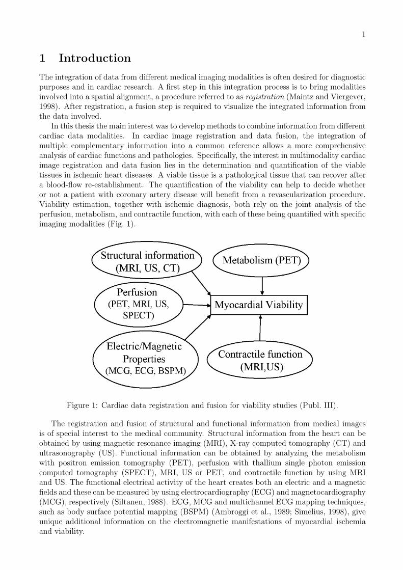

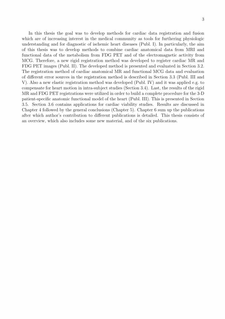

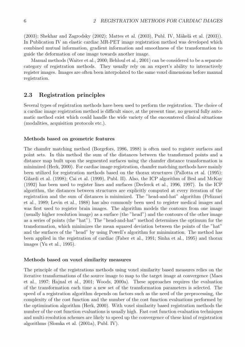


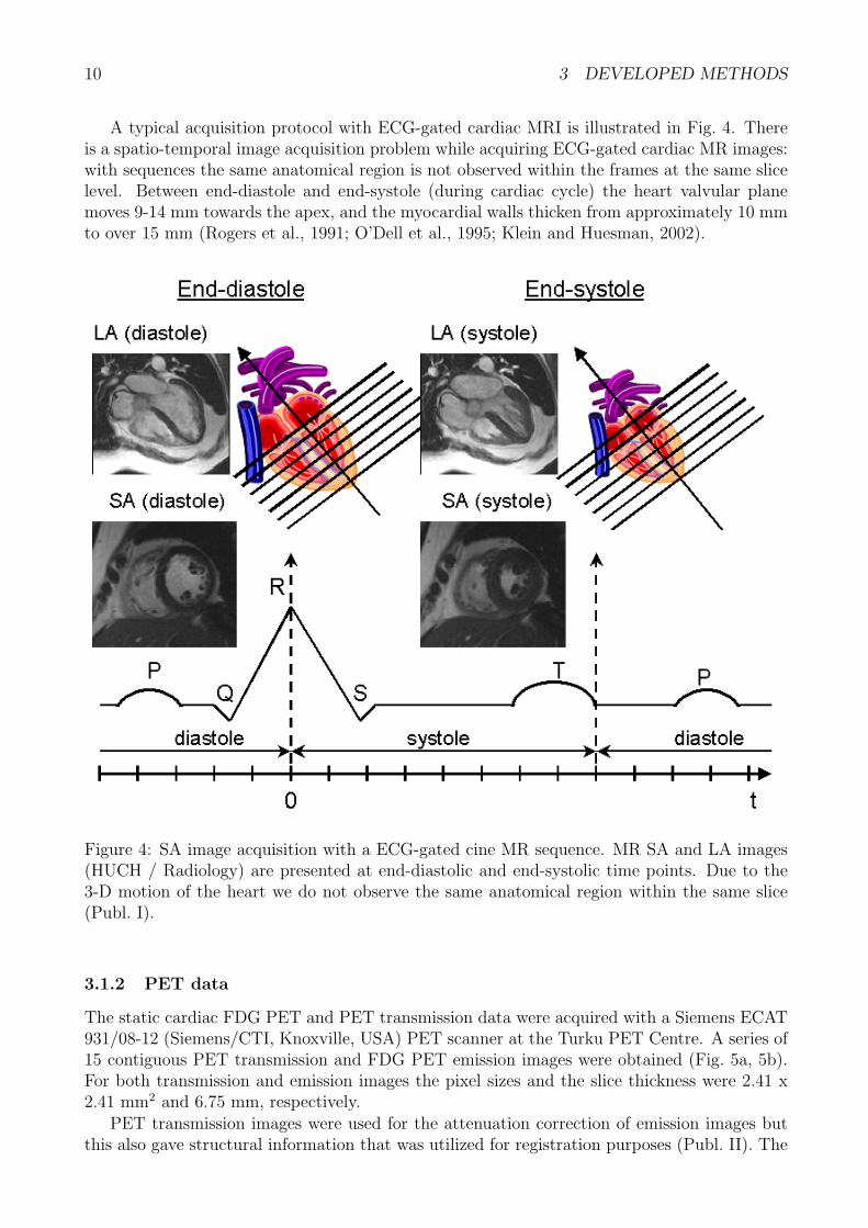
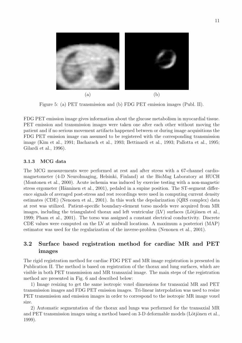


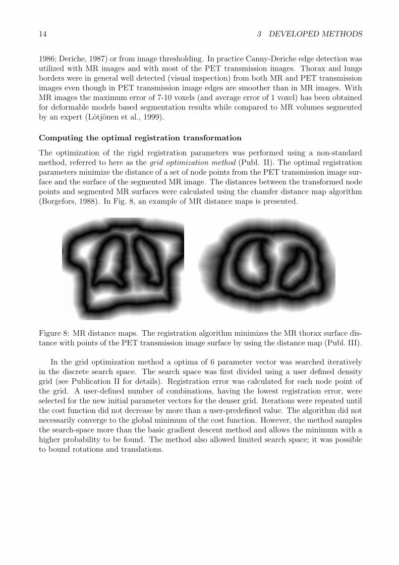

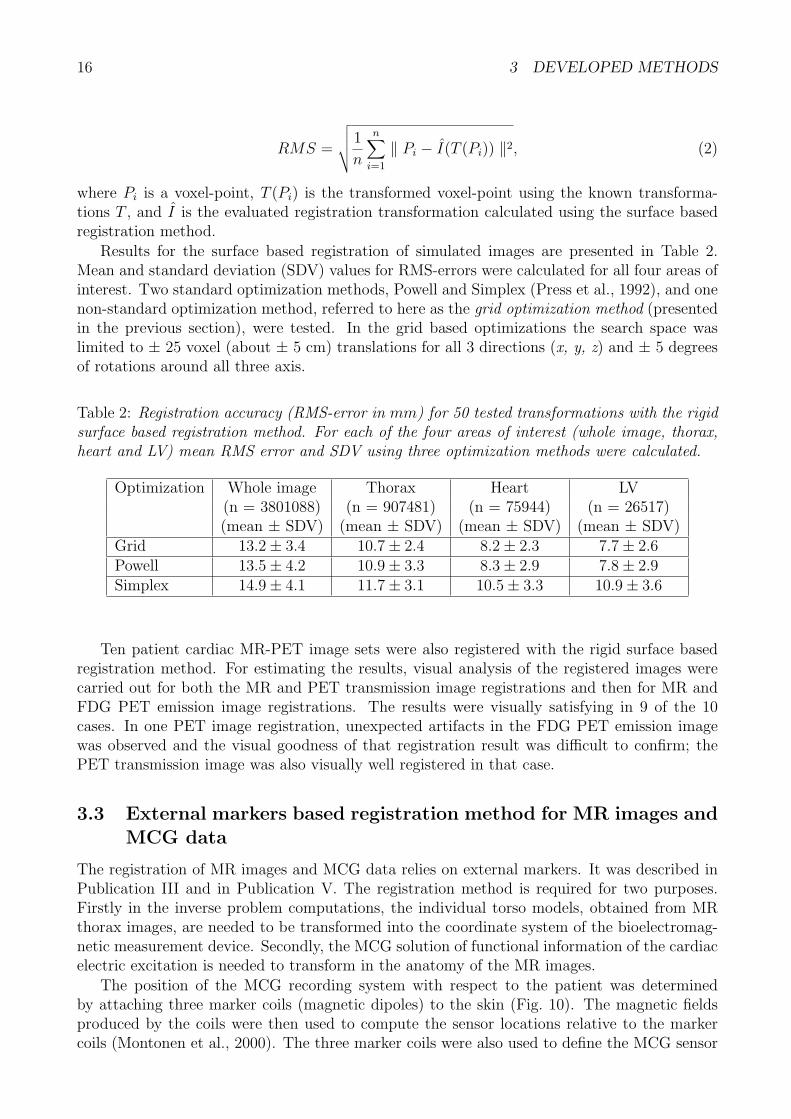


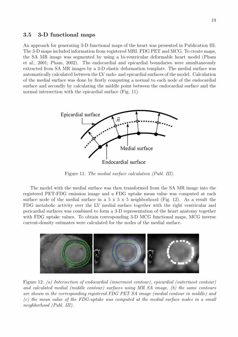

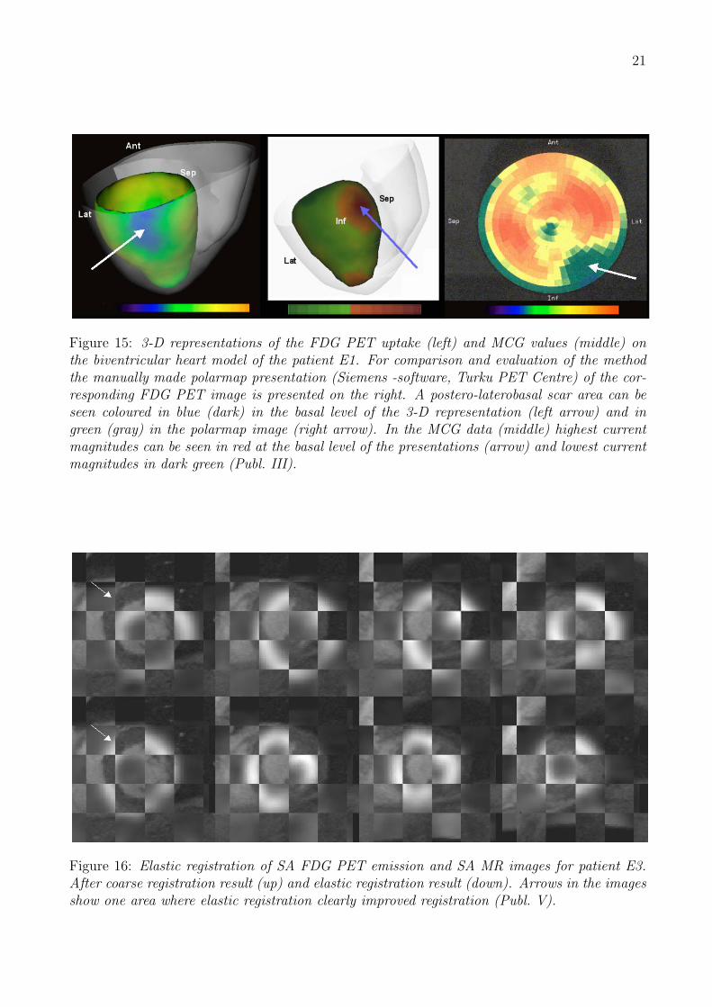

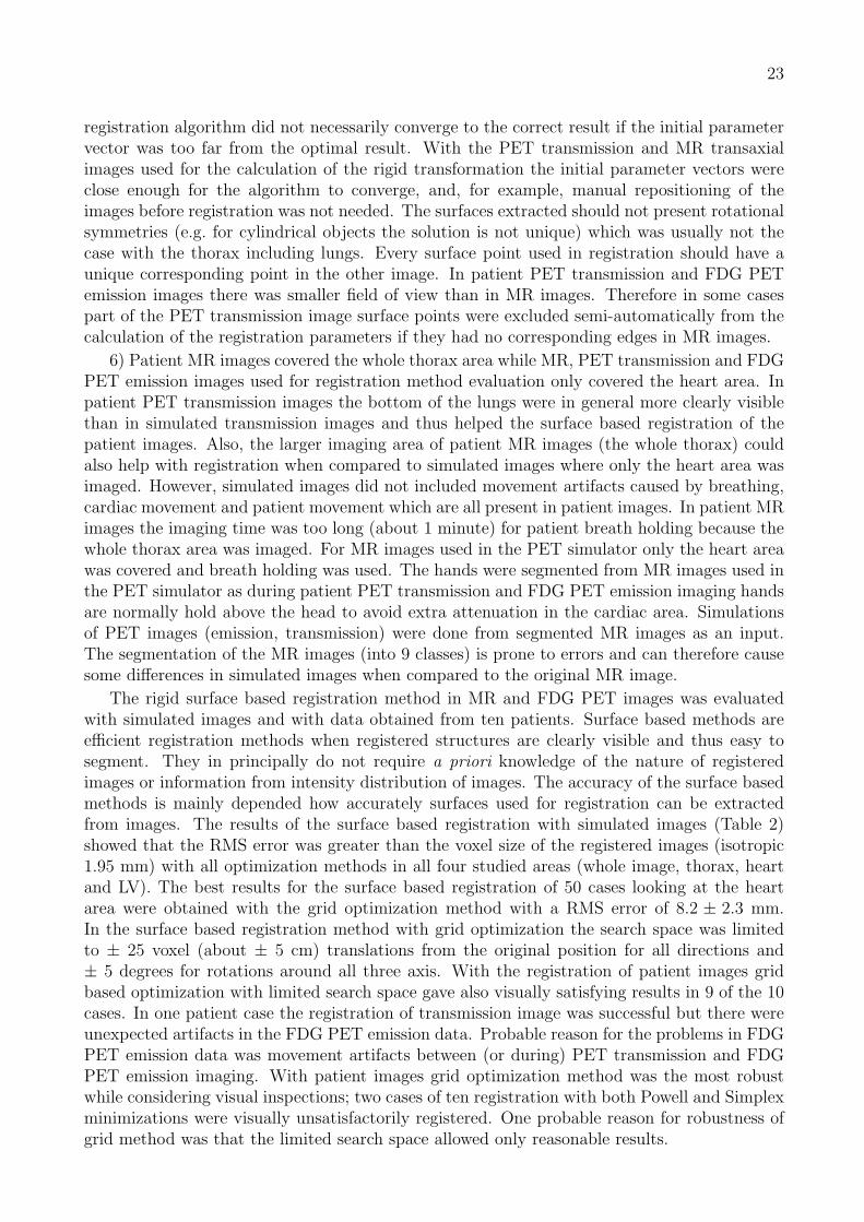


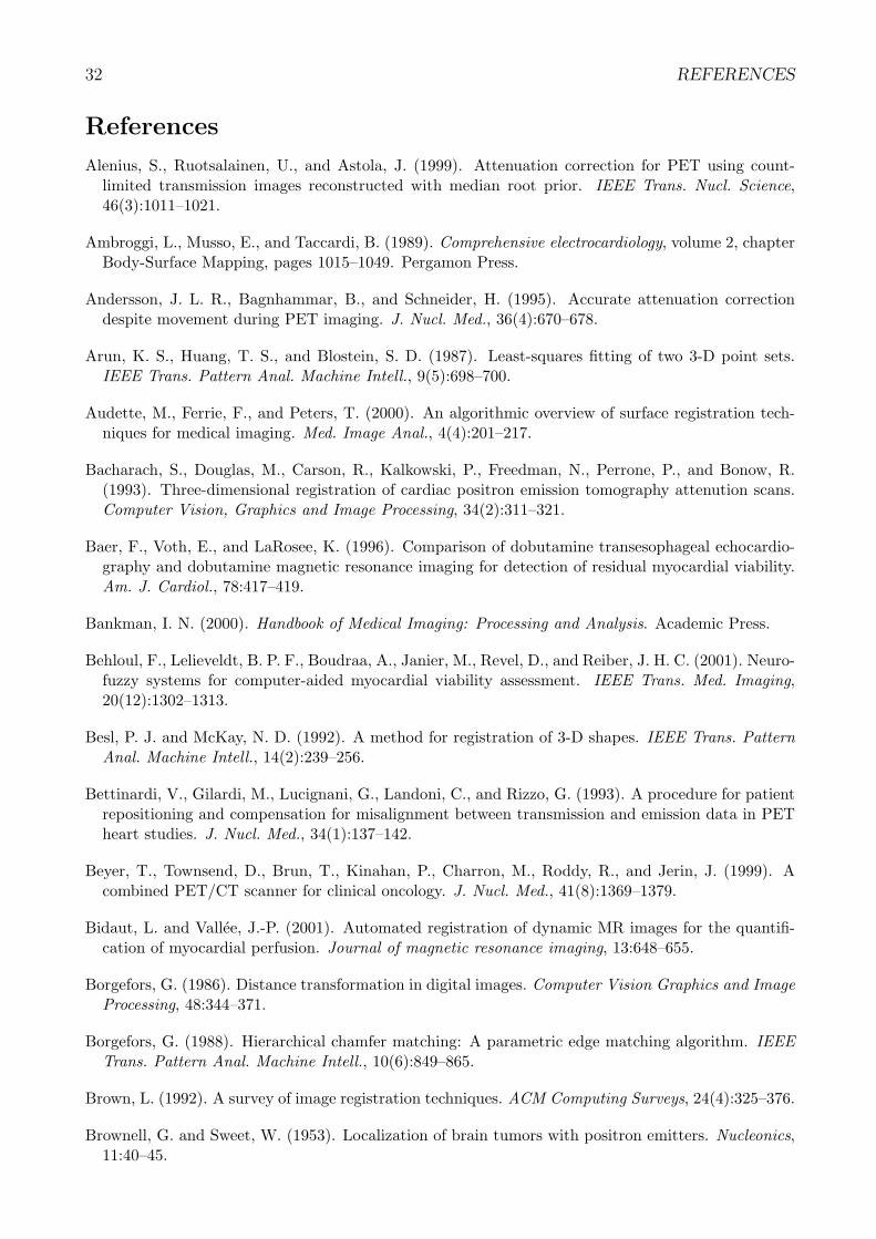

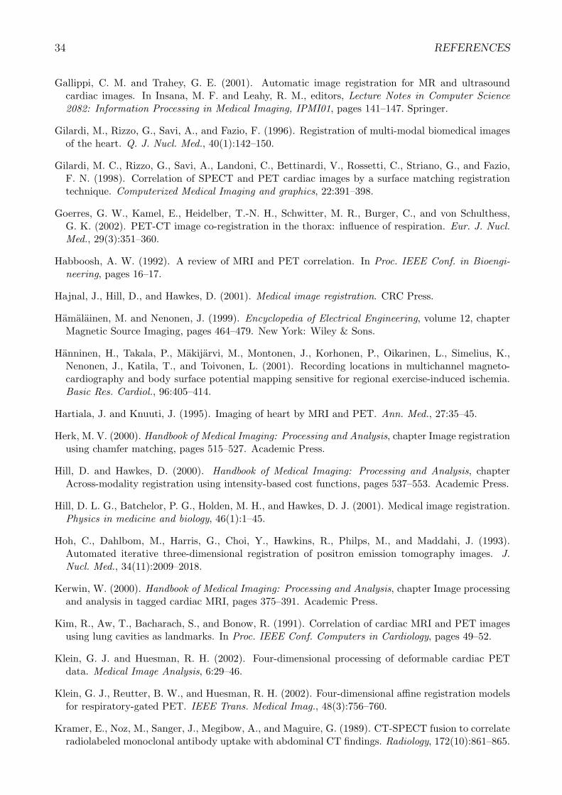


![J.Lund´enandV.Koivunen, “Automaticradarwaveformrecognition ...lib.tkk.fi/Diss/2009/isbn9789522480545/article8.pdf · recognition of communication signals, e.g., [5]–[7]. Automatic](https://static.documents.pub/doc/80x56/5f85c66b4ae7a045744098d4/jlund-aoeautomaticradarwaveformrecognition-libtkkfidiss2009isbn9789522480545article8pdf.jpg)


