Introduction In human renal transplantation, a significant proportion of grafts is lost due to chronic renal transplant failure (CRTF). More than 50% of the grafts that do survive for 1 year fail within 10 years through progressive loss of renal function and histopathological structures, i.e., in- terstitial fibrosis, intimal thickening of the arteries, and focal and segmental glomerulosclerosis (FGS). The vascular changes consist of dense intimal fibrosis mainly in the cortical arteries and result in renal ischemia manifested by glomerular ischemic simplification and obsolescence, interstitial fibrosis, and tubule atrophy. The latter process results in a decline in renal function and slowly rising serum creatinine levels. Ischemic injury may play a significant role in the pathogenesis of CRTF since it causes local production of reactive oxygen species (ROS), which may subsequently damage the graft. Ischemia also leads to cellular adenosine triphosphate (ATP) release [13], which can be degraded by ecto-adenosinetriphospha- tase (ATPase). It has been shown that glomerular ORIGINAL ARTICLE Transpl Int (2002) 15: 602–609 DOI 10.1007/s00147-002-0470-5 Annemieke Smit-van Oosten Winston W. Bakker Harry van Goor De-novo expression of vascular ecto-5¢-nucleo- tidase and down-regulation of glomerular ecto-ATPase in experimental chronic renal transplant failure Received: 24 August 2001 Revised: 3 June 2002 Accepted: 8 July 2002 Published online: 26 October 2002 ȑ Springer-Verlag 2002 A. Smit-van Oosten (&) W.W. Bakker H. van Goor Department of Pathology and Laboratory Medicine, University Hospital Groningen, P.O. Box 30.001, 9700 RB Groningen, The Netherlands E-mail: [email protected]Tel.: +31-50-3614179 Fax: +31-50-3632510 Abstract Ischemic injury plays an important role in chronic renal transplant failure (CRTF). Down- regulation of ecto-adenosine triphosphatase (ATPase) in combi- nation with up-regulation of ecto- 5¢-nucleotidase is a hallmark of ischemic injury. We studied the expression of renal ecto-5¢-nucleoti- dase and ecto-ATPase in experi- mental renal transplantation. Fisher 344-to-Lewis allografted rats were either treated with an angiotensin- converting enzyme inhibitor (ACEi) or left untreated. Lewis-to-Lewis syngrafted rats served as controls. Untreated allografted rats developed proteinuria, glomerulosclerosis, and mild intimal hyperplasia. ACEi completely prevented focal and segmental glomerulosclerosis (FGS) and proteinuria, but significantly enhanced intimal hyperplasia. Untreated allografted rats revealed marked vascular ecto-5¢-nucleotidase activity, which increased with ACEi. Vascular ecto-5¢-nucleotidase activi- ty was absent in syngrafted animals. Ecto-5¢-nucleotidase activity corre- lated well with intimal hyperplasia. Glomerular ecto-ATPase expression was significantly reduced in untreat- ed allografted rats compared to syn- grafted rats and correlated well with the extent of FGS. ACEi prevented reduction in glomerular ecto-AT- Pase. We found de-novo expression of ecto-5¢-nucleotidase at sites of re- nal intimal hyperplasia. Glomerular ecto-ATPase expression was mark- edly reduced in allografted rats and was prevented by ACEi. These en- zyme expression patterns suggest lo- cal ischemic damage in experimental CRTF. Keywords Kidney transplanta- tion Rat Ecto-adenosinetriphos- phatase Ecto-5¢-nucleotidase Ischemia
Transcript
Introduction
In human renal transplantation, a significant proportionof grafts is lost due to chronic renal transplant failure(CRTF). More than 50% of the grafts that do survivefor 1 year fail within 10 years through progressive loss ofrenal function and histopathological structures, i.e., in-terstitial fibrosis, intimal thickening of the arteries, andfocal and segmental glomerulosclerosis (FGS). Thevascular changes consist of dense intimal fibrosis mainlyin the cortical arteries and result in renal ischemia
manifested by glomerular ischemic simplification andobsolescence, interstitial fibrosis, and tubule atrophy.The latter process results in a decline in renal functionand slowly rising serum creatinine levels.
Ischemic injury may play a significant role inthe pathogenesis of CRTF since it causes localproduction of reactive oxygen species (ROS), whichmay subsequently damage the graft. Ischemia also leadsto cellular adenosine triphosphate (ATP) release [13],which can be degraded by ecto-adenosinetriphospha-tase (ATPase). It has been shown that glomerular
ORIGINAL ARTICLETranspl Int (2002) 15: 602–609DOI 10.1007/s00147-002-0470-5
Annemieke Smit-van Oosten
Winston W. Bakker
Harry van Goor
De-novo expression of vascular ecto-5¢-nucleo-tidase and down-regulation of glomerularecto-ATPase in experimental chronic renaltransplant failure
Received: 24 August 2001Revised: 3 June 2002Accepted: 8 July 2002Published online: 26 October 2002� Springer-Verlag 2002
A. Smit-van Oosten (&) Æ W.W. BakkerH. van GoorDepartment of Pathology and LaboratoryMedicine, University Hospital Groningen,P.O. Box 30.001, 9700 RB Groningen,The NetherlandsE-mail: [email protected].: +31-50-3614179Fax: +31-50-3632510
Abstract Ischemic injury plays animportant role in chronic renaltransplant failure (CRTF). Down-regulation of ecto-adenosinetriphosphatase (ATPase) in combi-nation with up-regulation of ecto-5¢-nucleotidase is a hallmark ofischemic injury. We studied theexpression of renal ecto-5¢-nucleoti-dase and ecto-ATPase in experi-mental renal transplantation. Fisher344-to-Lewis allografted rats wereeither treated with an angiotensin-converting enzyme inhibitor (ACEi)or left untreated. Lewis-to-Lewissyngrafted rats served as controls.Untreated allografted rats developedproteinuria, glomerulosclerosis, andmild intimal hyperplasia. ACEicompletely prevented focal andsegmental glomerulosclerosis (FGS)and proteinuria, but significantlyenhanced intimal hyperplasia.Untreated allografted rats revealedmarked vascular ecto-5¢-nucleotidase
activity, which increased with ACEi.Vascular ecto-5¢-nucleotidase activi-ty was absent in syngrafted animals.Ecto-5¢-nucleotidase activity corre-lated well with intimal hyperplasia.Glomerular ecto-ATPase expressionwas significantly reduced in untreat-ed allografted rats compared to syn-grafted rats and correlated well withthe extent of FGS. ACEi preventedreduction in glomerular ecto-AT-Pase. We found de-novo expressionof ecto-5¢-nucleotidase at sites of re-nal intimal hyperplasia. Glomerularecto-ATPase expression was mark-edly reduced in allografted rats andwas prevented by ACEi. These en-zyme expression patterns suggest lo-cal ischemic damage in experimentalCRTF.
Keywords Kidney transplanta-tion Æ Rat Æ Ecto-adenosinetriphos-phatase Æ Ecto-5¢-nucleotidase ÆIschemia
Verwendete Distiller 5.0.x Joboptions
Dieser Report wurde automatisch mit Hilfe der Adobe Acrobat Distiller Erweiterung "Distiller Secrets v1.0.5" der IMPRESSED GmbH erstellt. Sie koennen diese Startup-Datei für die Distiller Versionen 4.0.5 und 5.0.x kostenlos unter http://www.impressed.de herunterladen. ALLGEMEIN ---------------------------------------- Dateioptionen: Kompatibilität: PDF 1.2 Für schnelle Web-Anzeige optimieren: Ja Piktogramme einbetten: Ja Seiten automatisch drehen: Nein Seiten von: 1 Seiten bis: Alle Seiten Bund: Links Auflösung: [ 600 600 ] dpi Papierformat: [ 595.276 785.197 ] Punkt KOMPRIMIERUNG ---------------------------------------- Farbbilder: Downsampling: Ja Berechnungsmethode: Bikubische Neuberechnung Downsample-Auflösung: 150 dpi Downsampling für Bilder über: 225 dpi Komprimieren: Ja Automatische Bestimmung der Komprimierungsart: Ja JPEG-Qualität: Mittel Bitanzahl pro Pixel: Wie Original Bit Graustufenbilder: Downsampling: Ja Berechnungsmethode: Bikubische Neuberechnung Downsample-Auflösung: 150 dpi Downsampling für Bilder über: 225 dpi Komprimieren: Ja Automatische Bestimmung der Komprimierungsart: Ja JPEG-Qualität: Mittel Bitanzahl pro Pixel: Wie Original Bit Schwarzweiß-Bilder: Downsampling: Ja Berechnungsmethode: Bikubische Neuberechnung Downsample-Auflösung: 600 dpi Downsampling für Bilder über: 900 dpi Komprimieren: Ja Komprimierungsart: CCITT CCITT-Gruppe: 4 Graustufen glätten: Nein Text und Vektorgrafiken komprimieren: Ja SCHRIFTEN ---------------------------------------- Alle Schriften einbetten: Ja Untergruppen aller eingebetteten Schriften: Nein Wenn Einbetten fehlschlägt: Warnen und weiter Einbetten: Immer einbetten: [ ] Nie einbetten: [ ] FARBE(N) ---------------------------------------- Farbmanagement: Farbumrechnungsmethode: Alles für Farbverwaltung kennzeichnen (keine Konvertierung) Methode: Standard Arbeitsbereiche: Graustufen ICC-Profil: Dot Gain 10% RGB ICC-Profil: sRGB IEC61966-2.1 CMYK ICC-Profil: R705-Noco-gl-01-220499-ICC Geräteabhängige Daten: Einstellungen für Überdrucken beibehalten: Ja Unterfarbreduktion und Schwarzaufbau beibehalten: Ja Transferfunktionen: Anwenden Rastereinstellungen beibehalten: Ja ERWEITERT ---------------------------------------- Optionen: Prolog/Epilog verwenden: Nein PostScript-Datei darf Einstellungen überschreiben: Ja Level 2 copypage-Semantik beibehalten: Ja Portable Job Ticket in PDF-Datei speichern: Nein Illustrator-Überdruckmodus: Ja Farbverläufe zu weichen Nuancen konvertieren: Nein ASCII-Format: Nein Document Structuring Conventions (DSC): DSC-Kommentare verarbeiten: Nein ANDERE ---------------------------------------- Distiller-Kern Version: 5000 ZIP-Komprimierung verwenden: Ja Optimierungen deaktivieren: Nein Bildspeicher: 524288 Byte Farbbilder glätten: Nein Graustufenbilder glätten: Nein Bilder (< 257 Farben) in indizierten Farbraum konvertieren: Ja sRGB ICC-Profil: sRGB IEC61966-2.1 ENDE DES REPORTS ---------------------------------------- IMPRESSED GmbH Bahrenfelder Chaussee 49 22761 Hamburg, Germany Tel. +49 40 897189-0 Fax +49 40 897189-71 Email: [email protected] Web: www.impressed.de
ecto-ATPase is extremely sensitive for oxygen stress,resulting in down-regulation of this enzyme afterexposure to ROS [2, 19]. Glomerular ecto-ATPase ismarkedly decreased in patients with acute and chronicrejection [3] as well as in subjects with delayed diuresisfollowing renal transplantation [19] and after renalxenotransplantation [17].
Further degradation of nucleotides by ecto-5¢-nucle-otidase promotes the formation of adenosine, whichexerts potent vaso-active, anti-inflammatory, and anti-thrombotic activity [16]. In vivo, glomerular expressionof ecto-5¢-nucleotidase is found in human CRTF and insubjects with glomerular ischemia due to malignanthypertension [3] as well as in experimental glomerulo-sclerosis [16]. Therefore, we feel that down-regulation oflocal ecto-ATPase expression concomitant with up-reg-ulation of ecto-5¢-nucleotidase is characteristic of is-chemic injury [3].
In the Fisher-to-Lewis rat model for human CRTF,structural changes such as FGS, interstitial fibrosis andinflammation, and vascular intimal fibrosis develop inthe transplanted organ. Treatment with angiotensin-converting enzyme inhibitor (ACEi) in this model pre-vents FGS and proteinuria without affecting interstitialchanges [4, 18]. However, this treatment regimen causesmassive increments in vascular intimal hyperplasia whenused for more than 20 weeks [18].
In the present study, we document the presence oflocal ischemic damage in the renal vasculature oftransplanted rats with CRTF, using specific stainingmethods for ecto-ATPase and ecto-5¢-nucleotidase.Furthermore, we assessed whether intervention withACE inhibition modulates these expression patterns.
Materials and methods
Experimental techniques
Inbred male rats, weighing 215±9 g were used (Harlan, Horst, TheNetherlands). Lewis SsN rats (Lew) served as recipients, Fisher 344NHsd rats (F344) as donors. Lew kidneys placed in Lew recipientsserved as controls. The rats were anesthetized with isoflurane; leftdonor kidneys were flushed with saline, preserved in saline on icefor approximately 20 min, and transplanted orthotopically. Theright donor kidney was also removed and used to determine basalexpression of ecto-ATPase and ecto-5¢-nucleotidase. The recipientrat underwent left-sided nephrectomy, and subsequently, the leftrenal vessels and ureter were anastomosed end-to-end to the vesselsand ureter of the donor kidney with 10-0 Prolene sutures (Johnson& Johnson, Brussels, Belgium). Vascular clamps were released afterthe vascular anastomosis was completed, with a warm ischemiatime of 15 to 18 min. The right native kidney of the recipient wasremoved 10 days after transplantation, and the transplanted kidneywas checked at that time. One out of ten transplanted kidneys wasconsidered a technical failure due to hydronephrosis. These ratswere excluded from the study. A short course of cyclosporin A (1.5mg/kg per day; Sandimmun, Sandoz Pharma, Basle, Switzerland)was administered subcutaneously to allografted and syngrafted ratsover the first 10 post-operative days to reverse acute rejection.
Principles of laboratory animal care were followed; all animalprocedures were approved by the animal research ethics committeeof the Faculty of Medical Sciences of the University of Groningen.
Experimental groups
After cessation of cyclosporine therapy, allografted rats receivedeither no treatment and served as controls (F-L, n=9) or receivedthe ACEi lisinopril (Merck, Sharp & Dohme Research Laborato-ries, Rahway, N.J., USA) (F-L+ACEi, n=8, 75 mg/l drinkingwater). This regimen prevents clinical signs of CRTF and preservesglomerular morphology, although long-term treatment inducesmassive vascular thickening [18]. Syngrafted rats were not treated(L-L, n=8). Lisinopril was dissolved in drinking water. Seventy-five milligrams of lisinopril were dissolved in 100 ml of a saturatedNaHCO3 solution (Merck, Darmstadt, Germany) and then dilutedin 3.9 l 0.5% (w/v) methylcellulose suspension (Sigma-AldrichChemie, Steinheim, Germany). Untreated allografted and syn-geneically grafted rats received only the methylcellulose suspensionwith NaHCO3 in the same concentration. The rats were housedunder standard conditions with free access to drinking solution andstandard rat chow.
Clinicopathological parameters
The total observation period was 34 weeks. Body weight was de-termined every 4 weeks. We determined urinary protein excretion,measured by the pyrogallol red-molybdate method, every 4 weeksand at the end of the study by placing the rats for 24 h in metaboliccages with access to medication solution only. At the end of theobservation period, rats were anesthetized with isoflurane, andsystolic blood pressure (SBP) was measured by the tail-cuff meth-od. Subsequently, the aorta was cannulated and the kidney graftwas perfused in situ for 1 min with saline.
Tissue processing and histochemical staining procedures
After perfusion, two coronal tissue slices of the kidney wereobtained. One slice was snap-frozen in liquid isopentane (–80 �C)and stored at –80 �C. The other specimen was fixed in 4%paraformaldehyde and processed for paraffin embedding. Theparaffin sections were stained with periodic acid-Schiff (PAS)reagent and Verhoeff stain for assessment of glomerular andvascular damage.
Immunohistochemistry and enzyme histochemistry
We performed immunohistological examination for rat ecto-ATPase on frozen sections using a monoclonal anti-ecto-ATPaseantibody [5] followed by peroxidase-conjugated rabbit anti-mouseantibody and peroxidase-conjugated goat anti-rabbit antibody.Peroxidase activity was visualized with 3-amino-9-ethylcarbazole(AEC). For the demonstration of renal ecto-5¢-nucleotidase activ-ity, we used conventional enzyme histochemistry [21] with adeno-sine monophosphate (AMP) as a substrate and lead as capture ion.The precipitated reaction product (lead phosphate) was visualizedwith sodium disulfide, showing a dark-brown reaction product [21].Since macrophage-derived cytokines can up-regulate the expressionof 5¢-nucleotidase, we immunostained sections with the ED1 anti-body [9] to determine whether these cells were present at sites ofincreased ecto-5¢-nucleotidase. We performed additional immuno-staining according to standard procedures for smooth muscle a-actin and endothelial cells to determine the cellular composition ofvascular intimal hyperplasia. Sections in which the first antibodywas replaced with phosphate-buffered saline served as controls andwere consistently negative.
603
Analysis of structural, immunohistochemical,and enzyme histochemical changes
Morphological changes, i.e., severity of FGS, were scored semi-quantitatively on a scale of 0 to 4+. FGS was scored positive ifmesangial matrix expansion and adhesion formation were presentin the same quadrant. If 25% of the glomerulus was affected, ascore of 1+ was adjudged; 50% was scored as 2+; 75% as 3+;and 100% as 4+. The ultimate score was obtained by multiplica-tion of the degree of change by the percentage of glomeruli with thesame degree of injury, and addition of these scores (maximum score400). A total number of 40–50 glomeruli per animal were scoredmoving from cortex to medulla and vice versa.
Arterial intimal surface area was measured by morphometry.Verhoeff-stained paraffin-embedded sections were screened at amagnification of 200· by a light microscope with a camera attachedand a drawing prism. The image of a given transversally cut arterypresent on a computer screen was traced over the surface of agraphic tablet connected to a computer. The image was tracedalong the lamina elastica and the lumen. The surface area of thearterial intima was calculated by computer analysis. To comparelarger and smaller vessels, we calculated the ratio between intimalsurface area and intimal circumference.
Vascular ecto-5¢-nucleotidase staining was scored semi-quanti-tatively. When staining was present in one quadrant of the vessel, ascore of 1+ was adjudged; staining in 2 quadrants was scored as2+; in 3 quadrants as 3+; and when staining was present in 4quadrants, a 4+ score was given. Final scores were determined asdescribed above for FGS. All visible vessels present in the sectionwere scored.
Glomerular ecto-ATPase staining was measured by computer-assisted morphometry. Thirty glomeruli were screened with a lightmicroscope equipped with a camera device connected to a videoscreen. The image of a given glomerulus present on the screen wastraced with a cursor along the glomerular tuft over the surface of agraphic tablet connected to the computer. Subsequently, the totalsurface with red precipitate was measured and divided by the totalsurface of the glomerulus. An average score was calculated persection.
Statistical analysis
Reported values are the group mean ± standard deviation. Dif-ferences between untreated allografted and syngrafted rats weretested for significance with the Mann-Whitney U-test or the t-test.Differences were considered statistically significant with a P valueof less than 0.05. We log-transformed the values for vascular ecto-5¢-nucleotidase activity and FGS to normalize their distribution.The Pearson correlation coefficient was calculated between FGSscore and ecto-ATPase staining for untreated allografts, and cor-relation between ecto-5¢-nucleotidase and intimal hyperplasia wascalculated for all groups.
Results
Clinical and structural changes
No differences in body weight were observed betweenthe experimental groups throughout the study. In theuntreated allografted rats, proteinuria was significantlyelevated at 12 weeks (31±16 mg/24 h) compared withthe syngrafted animals (13±3 mg/24 h, P<0.01), fur-ther increasing to 93±75 mg/24 h at 34 weeks. Thesyngrafted rats did not develop proteinuria (22±13 vs
93±75 mg/24 h, P<0.01) at 34 weeks. Treatment of theallografted group with lisinopril completely preventedproteinuria at the end of the study (8±4 vs 93±75 mg/24 h, P<0.0001).
SBP was increased in the untreated allograft group(145±19), although not significantly different from thesyngrafted rats (125±30). ACE inhibition resulted in asignificantly lower SBP than in untreated allografts(91±10 vs 145±19 mg/24 h, P<0.0001). FGS wasprominent in the untreated allografted rats, whereassyngrafts revealed no signs of FGS (137±138 vs 6±11mg/24 h, P<0.01). ACE inhibition completely prevent-ed the development of FGS when compared with theuntreated allografted animals (2±3 vs 137±138 mg/24h, P<0.001).
Data on intimal surface area are shown in Fig. 1.Intimal surface area (lm2/lm circumference) was sig-nificantly lower in syngrafted rats compared with allo-grafted rats (P<0.01). ACE inhibition for 34 weeksdramatically increased intimal hyperplasia comparedwith untreated allografted rats (P<0.0001).
Ecto-5¢-nucleotidase activity
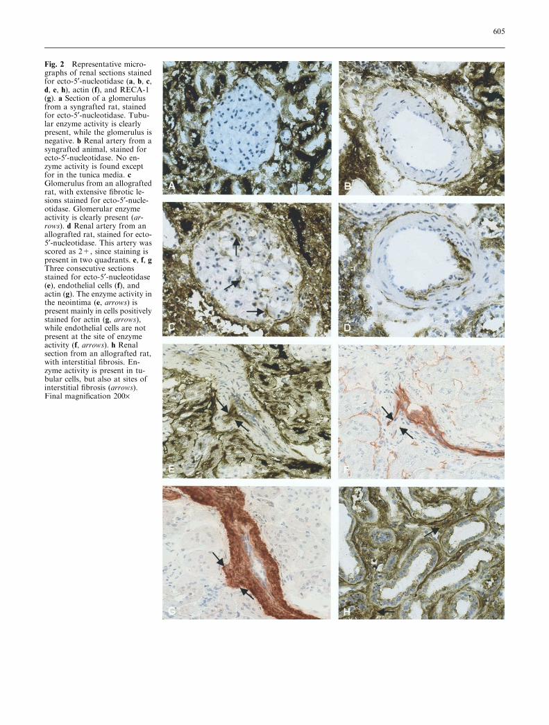
No differences were observed in ecto-5¢-nucleotidaseactivity between non-transplanted F344 and Lew controlkidneys. Both in syngrafted and allografted rats, ecto-5¢-nucleotidase activity was predominantly localized onthe brush border of the renal tubules (Fig. 2a and c).
Except for strong staining in the tunica media oflarger renal arteries, ecto-5¢-nucleotidase was absent inglomeruli and arteries from syngrafted rats (Fig. 2a and
Fig. 1 The presence of vascular intimal hyperplasia in untreatedallografted rats (F-L), allografted rats receiving ACEi (F-L+ACEi) and syngrafted rats (L-L). Values are presented as mean ±SD. **P<0.01, L-L vs F-L; ***P<0.001, F-L+ACEi vs F-L
604
Fig. 2 Representative micro-graphs of renal sections stainedfor ecto-5¢-nucleotidase (a, b, c,d, e, h), actin (f), and RECA-1(g). a Section of a glomerulusfrom a syngrafted rat, stainedfor ecto-5¢-nucleotidase. Tubu-lar enzyme activity is clearlypresent, while the glomerulus isnegative. b Renal artery from asyngrafted animal, stained forecto-5¢-nucleotidase. No en-zyme activity is found exceptfor in the tunica media. cGlomerulus from an allograftedrat, with extensive fibrotic le-sions stained for ecto-5¢-nucle-otidase. Glomerular enzymeactivity is clearly present (ar-rows). d Renal artery from anallografted rat, stained for ecto-5¢-nucleotidase. This artery wasscored as 2+, since staining ispresent in two quadrants. e, f, gThree consecutive sectionsstained for ecto-5¢-nucleotidase(e), endothelial cells (f), andactin (g). The enzyme activity inthe neointima (e, arrows) ispresent mainly in cells positivelystained for actin (g, arrows),while endothelial cells are notpresent at the site of enzymeactivity (f, arrows). h Renalsection from an allografted rat,with interstitial fibrosis. En-zyme activity is present in tu-bular cells, but also at sites ofinterstitial fibrosis (arrows).Final magnification 200·
605
b). Glomeruli in untreated allografted animals with se-vere FGS did reveal some ecto-5¢-nucleotidase activity(Fig. 2c). In the untreated allograft group, ecto-5¢-nu-cleotidase activity was clearly present in the renal ar-teries (Fig. 2d). Staining was predominantly localized inthe hyperplastic intima and occasionally in the media. Insome cases vessels were positively stained for ecto-5¢-nucleotidase in the absence of morphological signs ofintimal hyperplasia. In addition, marked staining wasobserved in the tunica media of larger arteries.
Chronic ACE inhibition further increased the ecto-5¢-nucleotidase staining in the thickened vascular areas(Fig. 2e). Macrophages were absent at sites of de-novoecto-5¢-nucleotidase staining. To analyze which celltypes express ecto-5¢-nucleotidase in the vasculature, weperformed additional immunostaining on consecutivesections using antibodies directed against smooth muscleactin and endothelial cells. Endothelial cells lining theluminal surface were still present, despite marked intimalthickening (Fig. 2f). All cells in the intima and mediastained positive for actin antibody, suggesting that thecells expressing ecto-5¢-nucleotidase were smooth musclecells or myofibroblasts (Fig. 2g). The area positive for5¢-nucleotidase and actin and negative for endothelialcells is marked by arrows. In addition, the fibrotic areasin the renal interstitium and some inflammatory cells inall allografted rats revealed marked ecto-5¢-nucleotidaseactivity (Fig. 2h). To determine the extent of vascularecto-5¢-nucleotidase activity, we semi-quantitatively an-alyzed renal tissue sections. The scores are presented inFig. 3. In syngrafted animals, 4% of the renal arteriesrevealed traces of ecto-5¢-nucleotidase activity. Incontrast, 25% of the arteries of allografted rats revealed
intensive ecto-5¢-nucleotidase activity, which was sig-nificantly increased when compared with syngrafted rats(P<0.01). Treatment of the allograft group with anACE inhibitor further increased ecto-5¢-nucleotidaseactivity (P<0.02 vs untreated allografts) in 40% of thearteries. Vascular ecto-5¢-nucleotidase expression corre-lated well with the extent of intimal hyperplasia in al-lografted rats (r=0.71, P<0.01).
Ecto-ATPase expression
By immunohistochemistry, ecto-ATPase staining wasfound in the brush border of the tubuli, the peri-tubularareas, and the renal arteries in both syngrafted andallografted animals (Fig. 4a). Using morphometricalanalysis, we found no significant difference in glomeru-lar ecto-ATPase expression between non-transplantedkidneys of F344 (n=8) and Lew (n=8) rats (42%±6%and 37%±3%, respectively, P=n.s.). Syngrafted ratsrevealed strong ecto-ATPase expression along thecapillary wall of the glomeruli (Fig. 4b, Fig. 5).Morphological analysis, however, revealed lower valuesfor ecto-ATPase expression in syngrafted animalscompared with non-transplanted controls. Glomerularstaining was markedly reduced in the untreated allo-graft group (Fig. 4c, Fig. 5) and was significantlylower compared with syngrafts (P<0.02). In contrast,treatment of allografts with ACE inhibitor preservedglomerular ecto-ATPase staining at the level seenin syngrafted rats (Fig. 4d, Fig. 5). Ecto-ATPasestaining in untreated rats was significantly reducedwhen compared with ACEi-treated rats (P<0.05).Ecto-ATPase staining strongly correlated with FGS(r=0.87, P<0.01) in the untreated allograft group.Both in treated and untreated allografts, areas of inter-stitial fibrosis were ecto-ATPase-positive. These inter-stitial cells had the morphological characteristics ofinterstitial fibroblasts.
Discussion
The goal of the present study was to localize renal is-chemic injury in experimental CRTF as reflected by theexpression of vascular or glomerular ecto-5¢-nucleoti-dase and ecto-ATPase. Secondly, we assessed the effectof intervention with ACE inhibition in this model withrespect to these parameters.
It was shown that intimal thickening, which is knownto occur in CRTF, is associated with de-novo expressionof vascular ecto-5¢-nucleotidase. In addition, this de-novo expression occurs in localized foci of intimal cellsand enhances significantly with the increase in intimalthickening following ACE inhibition. The presence ofecto-5¢-nucleotidase in vessels that do not have the
Fig. 3 The presence of ecto-5¢-nucleotidase activity in untreatedallografted rats (F-L), allografted animals receiving ACEi (F-L+ACEi), and syngrafts (L-L). Values are presented as mean ± SD.*P<0.02, F-L+ACEi vs F-L; **P<0.01, L-L vs F-L
606
morphological characteristics of intimal hyperplasiadoes suggest that enzyme expression occurs early in thedisease. The marked aggravation of intimal thickeningcaused by ACE inhibition in this model has beendescribed previously and is believed to be the result oflong-term (>20 weeks) treatment [18]. Since all the cellspresent in the thickened intima express smooth musclecell a-actin, the expression of ecto-5¢-nucleotidase mostprobably originates from smooth muscle cells. It is likelythat local up-regulation of ecto-5¢-nucleotidase relates tothe production of adenosine at sites of ischemic damage,as is described in experimental hypoxia [12]. Thus, ahallmark of cellular ischemic tissue damage is release ofATP and extracellular hydrolysis of ATP to AMP.Conversion of AMP to adenosine by ecto-5¢-nucleoti-dase may serve as a counter-mechanism to ischemiaproducing adenosine, which exerts various beneficialfunctions including anti-inflammatory and anti-throm-botic activities, vasodilatation, and scavenging of ROS[7, 8, 14, 16]. It is obvious that obliteration of renalarteries promoting an ischemic micro-environment inthis model may enhance hypoxia. Although it is tempt-ing to ascribe an anti-ischemic function to ecto-5¢-nu-cleotidase in proliferating intimal cells, as similarlydescribed for the renal interstitium by Le Hir et al. [12],such a putative function remains to be proven in thepresent model. The function of ecto-5¢-nucleotidase ex-pression in the neointima is unknown. Studies with
chicken gizzard ecto-5¢-nucleotidase suggest that it alsofunctions as a potent ligand of fibronectin and laminin[20] and that it is involved in the spreading of variousmesenchyme-derived cells on a laminin substrate [6].
Fig. 4 Representative micro-graphs of renal sections fromsyngrafted (a, b), untreated al-lografted (c), and ACEi-treatedallografted rats (d), stained forecto-ATPase. a Ecto-ATPasestaining is clearly present on thetubular brush border (arrows)and also in the peri-tubularareas (arrowhead). b Ecto-ATPase staining is clearlypresent along the glomerularcapillary wall of kidneys fromsyngrafted rats (arrows) andfrom allografted rats after ACEtreatment (d). c Allografted ratsshowed diminished glomerularecto-ATPase staining whencompared with syngrafted rats.Final magnification 200·
Fig. 5 The presence of glomerular ecto-ATPase staining inuntreated allografted rats (F-L), allografted animals receivingACEi (F-L+ACEi), and syngrafted rats (L-L). Values arepresented as mean ± SD. *P<0.02, F-L vs L-L; *P<0.05, F-L+ACEi vs F-L
607
Thus, in addition to its anti-inflammatory and anti-thrombotic properties, ecto-5¢-nucleotidase may also beinvolved in the spreading of cells in the neointima of theobliterated arteries in our model.
With respect to glomerular expression of ecto-5¢-nu-cleotidase, we noted that focal expression of this ecto-enzyme occurs exclusively in severely damaged glomeruliof allografted rats. This observation in combination withdiminished expression of glomerular ecto-ATPase inallografted rats may point to ischemic injury of theglomerular microvasculature. An identical staining pat-tern has been observed in human CRTF as well as inischemic glomerular injury due to malignant hyperten-sion [3]. Down-regulation of glomerular ecto-ATPaseexpression may be mediated by the local ROS, since thisglomerular ecto-enzyme is extremely sensitive to toxicoxygen products [15]. X-irradiation of rat kidneys, giv-ing rise to local release of ROS, results in diminishedexpression of glomerular ecto-ATPase [1, 15]. Similaroxidant-mediated loss of glomerular ecto-ATPase ex-pression has been demonstrated in adriamycin nephrosis[15]. The significant correlation of the magnitude ofsclerotic lesions on the one hand and diminished ecto-ATPase in glomeruli of allografted rats on the otherhand suggests that local ischemia within the capillarytuft plays a role in the pathogenesis of FGS in thismodel.
The pathogenesis of the glomerular lesions in CRTFis unknown. Following an acute inflammatory allograftresponse in the initial phase after graft rejection, it seemsobvious that intimal hyperplasia of intra-renal arteriesmay contribute to downstream ischemia, giving rise tolocal release of ROS as well as subsequent down-regu-lation of glomerular ecto-ATPase and collapse ofglomerular capillaries. In other experimental models ofglomerulosclerosis, up-regulation of ecto-5¢-nucleotidasewas found as well [16].
Interestingly, long-term ACE inhibition enhancedvascular intimal proliferation and intimal ecto-5¢-nucle-
otidase activity, whereas this treatment significantly pro-tected the glomerular microvasculature, showing normalglomerular ecto-ATPase expression concomitant with asignificant reduction of glomerulosclerosis in allograftedrats. This is in line with evidence obtained from otherinvestigators, suggesting that the reno-protective actionof ACE inhibition depends on improving renal interstitialoxygenation [11]. Interstitial oxygenationmay counteractlocal ischemic injury in the present model, explaining thereno-protective effect of ACE inhibition on the glomeru-lar microvasculature as reflected by normal expression ofglomerular ecto-ATPase. In contrast, ACE inhibition isknown to impair reparative angiogenesis in murine limbischemia, which may explain the absence of beneficialeffects ofACE inhibition on vascular lesions in the presentstudy. Repair of ischemia-reperfusion injury and allo-related damage to the vasculature may be inhibited bylong-term ACE inhibition [10].
In summary, in the present allograft model, we lo-calized ischemic-like injury in renal arteries as well as inthe glomerular microvasculature, as reflected by up-regulation of ecto-5¢-nucleotidase and down-regulationof glomerular ecto-ATPase. Increased intimal thickeningin renal vessels correlated well with enhanced ecto-5¢-nucleotidase expression, whereas glomerulosclerosis wasassociated with decreased expression of glomerular ecto-ATPase. Finally, while long-term ACE inhibition causedincreased intimal thickening with signs of ischemia inrenal vessels, this treatment caused protection of glom-erular ecto-ATPase, which correlated with fewer glom-erular lesions in these animals. Further studies areneeded to analyze the mechanisms underlying the en-zyme changes observed in this model.
Acknowledgements The authors wish to thank Feiko Kiestra andTheo Borghuis for their excellent technical assistance and PieterKlok for biotechnical support. The compound lisinopril was a kindgift from Merck, Sharp & Dohme Research Laboratories, Rahway,N.J., USA. This study was supported by a grant (no. C97-1553)from the Dutch Kidney Foundation.
References
1. Bakker WW, Kalicharan D, Donga J,Hulstaert CE, Hardonk MJ (1987) De-creased ATPase activity in adriamycinnephrosis is independent of proteinuria.Kidney Int 31:704–709
2. Bakker WW, Poelstra K, Hardonk MJ(1993) Relevance of adenine nucleotid-ases in the glomerular filtration barrier.Nephron 64:338–342
3. Bakker WW, Mui KW, van Son WJ(2000) Detection of glomerular isch-emia in chronic graft failure by quant-itation of glomerular ecto-5¢-nucleotidase and ecto-ATPase. In:Vanduffel L, Lemmens R (eds) Ecto-ATPases and related ectonucleotidases.Shaker, Maastricht, pp 192–200
4. Benediktsson H, Chea R, Davidoff A,Paul LC (1996) Antihypertensive drugtreatment in chronic renal allograft re-jection in the rat. Transplantation62:1634–1642
6. Codogno P, Doyennette-Moyne MA,Aubery M, Dieckhoff J, Lietzke R,Mannherz HG (1988) Polyclonal andmonoclonal antibodies against chickengizzard 5¢-nucleotidase inhibit thespreading process of chicken embryonicfibroblasts on laminin substratum. ExpCell Res 174:344–354
608
7. Cronstein BN, Kramer SB, WeissmannG, Hirschhorn R (1983) Adenosine: aphysiological modulator of superoxideanion generation by human neutroph-ils. J Exp Med 158:1160–1170
8. Cronstein BN, Levin RI, Belanoff J,Weissmann G, Hirschhorn R (1986)Adenosine: an endogenous inhibitor ofneutrophil-mediated injury to endothe-lial cells. J Clin Invest 78:760–770
9. Dijkstra CD, Dopp EA, Joling P, KraalG (1984) The heterogeneity of mono-nuclear phagocytes in lymphoid organs:distinct macrophage subpopulations inrat recognized by monoclonal antibod-ies ED1, ED2 and ED3. Immunology54:589–599
11. Fine LG, Bandyopadhay D, NormanJT (2000) Is there a common mecha-nism for the progression of differenttypes of renal diseases other than pro-teinuria? Towards the unifying theme ofchronic hypoxia. Kidney Int 57 [Suppl75]:S22–S26
12. Le Hir M, Kaissling B (1993) Distri-bution and regulation of renal ecto-5¢-nucleotidase: implications for physio-logical functions of adenosine. Am JPhysiol 264:F377–F387
13. Mandel LJ, Takano S, Soltoff SP,Murdaugh S (1988) Mechanismswhereby adenine nucleotides improverabbit renal proximal function duringand after anoxia. J Clin Invest 81:1255–1264
14. Murray RD, Churchill PC (1985)Concentration dependency of the renalvascular and renin secretory responsesto adenosine receptor agonists. J Phar-macol Exp Ther 232:189–193
15. Poelstra K, Hardonk MJ, Bakker WW(1988) Adriamycin-induced decrease ofecto–ATPase activity in the glomerularbasement membrane of the rat kidney ismediated by oxygen free radical species.In: Gubler MC, Sternberg M (eds)Progress in basement membrane re-search, renal and related aspects inhealth and disease. John Libbey Euro-text, London, pp 259–264
16. Poelstra K, Heynen ER, Baller JF,Hardonk MJ, Bakker WW (1992)Modulation of anti-Thy1 nephritis inthe rat by adenine nucleotides. Evi-dence for an anti-inflammatory role fornucleotidases. Lab Invest 66:555–563
17. Roush W (1995) New ways to avoidorgan rejection buoy hopes. Science270:234–235
18. Smit-van Oosten A, Navis G, StegemanCA, Joles JA, Klok PA, Kuipers F,Tiebosch AT, van Goor H (2001)Chronic blockade of angiotensin II ac-tion prevents glomerulosclerosis, butinduces graft vasculopathy in experi-mental kidney transplantation. J Pathol194:122–129
19. Son WJ van, Wit F, van Balen OL,Tegzess AM, Ploeg RJ, Bakker WW(1997) Decreased expression of glom-erular ecto-ATPase in kidney graftswith delayed graft function. TransplantProc 29:352–354
20. Stochaj U, Richter H, Mannherz HG(1990) Chicken gizzard 5¢-nucleotidaseis a receptor for the extracellular matrixcomponent fibronectin. Eur J Cell Biol51:335–338
21. Wachstein M, Meisel W (1957) Histo-chemistry of hepatic phosphatases at aphysiologic pH, with special referenceto the demonstration of bile canaliculi.Am J Clin Pathol 27:13–23