35
Deanna Protocol Program for ALS: Substantiation and Putative Mechanisms January 2016

Deanna Protocol Program for ALS: Substantiation and Putative Mechanisms January 2016

1 | P a g e Copyright © Simplesa LLC
Deanna Protocol Program for ALS: Substantiation and Putative Mechanisms
Executive Summary
Amyotrophic Lateral Sclerosis (ALS) is a progressive neurodegenerative disease that primarily affects
upper and lower motor neurons. Upper motor neuron disease causes slowness, hyperreflexia, and spasticity
while lower motor neuron disease causes weakness, muscle atrophy, and fasciculations. Four out of five
people with ALS will present with asymmetric limb weakness, while the remaining 20% will first exhibit
dysarthria/dysphagia. ALS may negatively affect cognition in up to 50% of patients, though this is usually
mild. Autonomic symptoms of ALS include constipation, excessive sweating, and urinary urgency without
incontinence. The disease usually becomes life-threatening when respiratory muscles/motor units are
involved. In most cases, death occurs within two to five years of diagnosis.
The exact etiology of ALS is unknown; however, a number of abnormalities have been discovered.
Alterations in RNA and protein processing likely play a major role in the pathogenesis of ALS. Genetic studies
of families with ALS have revealed a number of mutations in genes coding for RNA binding proteins.
Moreover, abnormalities in the superoxide dismutase-1 enzyme likely contribute to ALS neuropathology.
There are also inflammatory and excitotoxic components of the disease. Primary dorsal motor neuron cultures
and transgenic mouse models of ALS have been extensively used to uncover these pathophysiological
mechanisms and test prospective treatments.
While many medications have been clinically tested, few have been successful in treating ALS. In fact, the
only prescription medication that improves survival in patients with ALS is riluzole—and this effect is modest.
The median increased survival time in patients taking riluzole is about two to three months. Riluzole is
believed to reduce glutamate-induced excitotoxicity by inhibiting glutamate release from excitatory neurons,
noncompetitively blocking NMDA receptors, and directly acting on voltage-dependent sodium channels.
However, the precise mechanism by which riluzole extends survival in ALS is unknown.
The Deanna Protocol is a combination of nutritional supplements that are available over-the-counter that
have been organized into a daily treatment regimen. The original Deanna Protocol was developed by an

2 | P a g e Copyright © Simplesa LLC
orthopedic surgeon, Dr. Vincent Tedone, who formulated the regimen for his daughter, Deanna, and is now
administered by the non-profit organization Winning the Fight. The current document describes the
components of the Deanna Protocol and additional supplements that complement the Deanna Protocol,
collectively called the Deanna Protocol Program for ALS. Many of the ingredients in the Deanna Protocol and
the dosing scheme have been streamlined for ease of use. This document describes the theoretical foundations
of the Deanna Protocol Program for ALS and provides possible mechanisms by which each of the ingredients
could beneficially affect patients with ALS. Importantly, the statements contained in this document have not
been evaluated by the FDA. This product is not intended to diagnose, treat, cure, or prevent any disease.

3 | P a g e Copyright © Simplesa LLC
Deanna Protocol Program for ALS: Substantiation and Putative Mechanisms
Amyotrophic Lateral Sclerosis Clinical Features Amyotrophic Lateral Sclerosis (ALS) is the most
common motor neuron disease in adults. The most notable
feature of the disease is that it afflicts motor neurons
specifically. When upper motor neurons (i.e., neurons
traveling from the cortex in the brain to the spinal cord) are
affected, it results in spasticity, exaggerated reflexes, and
reduced coordination. Conversely, the death and
dysfunction of lower motor neurons (i.e., neurons traveling
from the spinal cord to muscles) causes muscle cramps,
atrophy, fasciculations, and generalized weakness (Fig. 1).
ALS is a progressive, neurodegenerative disease. The
disease progresses rapidly—most people will die within
two to five years of symptom onset—though there are
notable exceptions to this, such as Stephen Hawking. The
risk of developing ALS increases with each decade of life,
peaking at age 74.1 Approximately 5 to 10 percent of people
who develop ALS will have a familial form of the disease,
while the rest have sporadic ALS.2 Several mutations are
present in familial ALS and, in some cases, sporadic ALS.
Figure 1. Upper motor neurons extend from the motor cortex in the brain. Lower motor neurons originate in the spinal cord. Adapted from Gray's Anatomy of the Human Body.

4 | P a g e Copyright © Simplesa LLC
Putative genetic associations to ALS include mutations of SOD1, TARDBP, ANG, FUS, C9ORF72, OPTN, and
SETX genes.3 ATXN2 mutations are present in a small portion of patients with sporadic ALS.3 Duplication of
the survival motor neuron 1 (SMN1) gene may be a risk factor for sporadic ALS in adults.4
Age and family history are the only confirmed risk factors for ALS, though dozens of others have been
explored. Other (unconfirmed) potential risk factors for ALS are environmental exposures (e.g., heavy metal,
vaporized plastics) or occupational interactions (e.g., military service, agricultural work, welders). Pathophysiology The main pathological feature of ALS is motor neuron degeneration and death. As the nerve cells die, glia
increase in number and replace lost neurons. Affected areas of the cortex and spinal cord undergo atrophy,
especially the frontal cortex and ventral roots of the spinal cord. Why or how these cells specifically die is
unknown; however, several pathological features of the disease suggest possible causative mechanisms. Inclusions Most neurons that eventually die in the cortex are large (pyramidal) neurons.
These dysfunctional and dying neurons commonly develop intracellular
inclusions. These inclusions include Bunina bodies5, neurofilamentous inclusions,
and aggregates of an RNA-binding protein called TDP-43 (Fig. 2). While it is
unclear whether these inclusions are simply a byproduct of the disease or a
primary toxic substance, their presence is common in various neurodegenerative
diseases, including ALS. There are potential mechanisms by which these
aggregates are neurotoxic. For example, the presence of abnormal levels of TDP-
43 suggests that abnormal RNA processing plays a role in the pathophysiology of
ALS. Superoxide Dismutase Type 1 Abnormalities Superoxide Dismutase Type 1 or SOD1 mutations are common in people with familial ALS and occur
infrequently among those with sporadic ALS.6 Abnormal SOD1 proteins could be toxic to nerve cells for a
variety of reasons. Since SOD1 knockout mice do not exhibit motor neuron disease, it is more likely that ALS
patients acquire a pro-oxidant, gain-of-function SOD1 mutation, which leads to a harmful production of
reactive oxygen species.7 Mutant SOD1 may also lead to abnormal protein folding and subsequent protein

5 | P a g e Copyright © Simplesa LLC
aggregation within neuronal cell bodies. While this is more frequently a feature of late-stage disease, even
small accumulations of SOD1 aggregates can be neurotoxic.8 Inflammation Inflammation may also play a role in ALS disease progression.9,10,11 Non-neuronal cells such as astrocytes
and microglia are activated in the disease. Through cytokine release and other cellular attractants, these cells
recruit immune cells such as natural killer cells and monocytes to cross the blood-brain barrier. Moreover,
activated microglia release a number of substances that are toxic to neurons including nitric oxide, oxygen
radicals and glutamate.7,12,13 Excitotoxicity and Mitochondrial Dysfunction Excitotoxicity is the well-characterized process of neuron death that follows excessive stimulation by
excitatory neurotransmitter ligands. Excessive stimulation of neurons causes abnormally high levels of calcium
to enter the cell body. Excessive intracellular calcium initiates a cascade of events that kills the cell. Specifically,
high levels of intracellular calcium lead to peroxidation of membrane lipids, damage to RNA and DNA, and
disruption of mitochondria. One effect on mitochondria is that pores in the mitochondrial membrane
(mitochondrial transition pore) open and release reactive oxygen species into the cytoplasm. This is one trigger
apoptosis (programmed cell death).
Several lines of evidence suggest that excitotoxicity plays a role in the pathophysiology of ALS. Glutamate
levels are abnormally high in patients with sporadic ALS14 perhaps due to deficiencies in glutamate reuptake
transporters.15 Abnormal glutamate receptors on nerve cell membranes may also contribute to excessive
excitatory stimulation. In addition, trials with riluzole lend clinical support for an excitotoxic mechanism in
ALS. Riluzole is the only prescription medication shown to improve survival in patients with ALS.16,17,18 While
the precise therapeutic mechanism of action in ALS is not known, riluzole inhibits glutamate release,
inactivates voltage-dependent sodium channels, slows potassium channel inactivation, and inhibits protein
kinase C19,20,21,22—all of which contribute to excitotoxic cell death.
Mitochondria may be affected by the disease before and apart from excitotoxicity, however. Cells collected
from patients with ALS exhibit impaired mitochondrial function, specifically in mitochondrial complex I.23,24
This deficit can be counteracted by the addition of ketone bodies, which are used by complex II.25 In other
words, ALS impairs complex I function, but ketone bodies seem to act as a “fuel” source in the mitochondrial
membrane at complex II and later.

6 | P a g e Copyright © Simplesa LLC
Current Treatments and Future Targets The only prescription drug approved for the treatment of ALS is riluzole (Fig. 3). Riluzole is generally
administered twice a day at a dose of 50 mg. The most common side effects are gastrointestinal problems and
elevated liver enzymes. Rarely, neutropenia may occur. While riluzole is the
only medication that can prolong survival in people with ALS, the median
additional time of survival is only between two to three months (Fig. 4).16
While various other treatments have shown promise in laboratory and
animal studies, clinical trials have been disappointing. Celecoxib (Cox-2
inhibitor), gabapentin (GABA analog), lamotrigine (antieplileptic, mood
stabilizer), lithium (mood stabilizer), topiramate and valproic acid (antieplileptics), verapamil (calcium
channel blocker), and minocycline (antibiotic with neurological effects) have failed to meet clinical endpoints
in ALS trials.
Future research is focused on several avenues, but definitive results are several years off, if they are fruitful
at all. One promising approach is the use of antisense oligonucleotide therapy for familial ALS with SOD-1
mutations. If this
approach is successful,
it will only be useful for
a fraction of patients
with ALS. Trials are
underway with
memantine, a drug that
presumptively blocks
glutamatergic
neurotransmission
through NMDA-type
glutamate receptors.
Phase II trials of creatine
are currently underway.
The use of stem cells
and gene therapy remains promising, but these technologies will require considerable laboratory research
before any agents can be trialed.

7 | P a g e Copyright © Simplesa LLC
The Deanna Protocol Program for ALS
The original Deanna Protocol was developed by an orthopedic surgeon, Dr. Vincent Tedone, who wanted
to formulate a treatment regimen for his daughter, Deanna, when she was diagnosed with ALS. Dr. Tedone
had anecdotally reported that his daughter had good experience with various versions of the protocol. The
current Deanna Protocol Program for ALS includes supplements that have been proven in the laboratory to be
effective in ALS and slow the progression of the disease. These substances, known as the DPTM Plan Essentials,
include AAKG, AKG, GABA, Ubiquinol (CoQ10), Niacin (non-flush) and 5-HTP (5-hydroxtrytophane). The
Deanna Protocol Program for ALS also includes ancillary substances which go beyond the Deanna
Protocol® DP™ Plan Essentials in offering potential benefits to ALS patients. They are not included in the DPTM
Plan Essentials because they have not been proven in a lab to be effective in ALS patients. The Deanna Protocol DPTM Plan Essentials The Deanna Protocol® DP™ Plan Essentials consists of the necessary supplements to follow the Deanna
Protocol. These include AAKG, AKG, GABA, Ubiquinol (CoQ10), Niacin (non-flush) and 5-HTP (5-
hydroxtrytophane). Simplesa AAKG+ Core Powder contains the full recommended daily dosages of GABA,
Ubiquinol and Niacin and the minimum recommended daily dose of AAKG. Higher dosages of AAKG can be
obtained with AAKG Powder, used to increase the AAKG level above the minimum daily recommended
dosage of 9 grams to a maximum daily dosage of 18 grams. 5-HTP is taken every evening, and AKG is taken
every hour between three daily doses of the Core Powder. The Winning the Fight Program for ALS The Winning the Fight Program for ALS, also known as the Deanna Protocol Comprehensive Approach,
consists of ancillary supplements that have been found to be effective in Deanna, but have not been proven in
a lab to be effective in ALS patients, and additional ingredients which the National Institutes of Health (NIH)
claims maintain the health of the nervous system and muscles. Many of these ancillary substances can be
supplied by a healthy diet. However, individuals with ALS may need more of them than healthy individuals,
which is why taking the ancillary substances makes sense for those who can afford to cover the cost. The NIH
has published a manuscript "Nutrition and Supplements in Motor Neuron Diseases" which emphasizes the
importance of nutrition to support quality of life for ALS patients.

8 | P a g e Copyright © Simplesa LLC
While adherence with the Deanna Protocol Comprehensive Approach may still be challenging for some
patients, the current protocol and product offerings streamline treatment as much as possible while taking into
account the pharmacokinetics of the supplements.
One of the chief features of the Deanna Protocol Comprehensive Approach is that it is all-inclusive. In other
words, the Deanna Protocol Comprehensive Approach has been formulated to include a multitude of agents
that could potentially work to improve ALS symptoms, improve functioning, or slow progression of the
disease. It is unlikely that any one agent included in this protocol will exert a clinically meaningful effect on
the disease. Indeed, the only approved drug for ALS that lengthens survival is riluzole, which only provides a
median of 2-3 months of additional life. Thus, the approach of the Deanna Protocol® DP™ Plan Essentials and
the Deanna Protocol Comprehensive Approach is to include several agents, all of which are generally
considered safe at the doses provided. In this way, the risk of omitting a potentially useful ingredient or
under-dosing one of the ingredients in minimized.
Just as no single, known agent is likely to significantly alter the course of ALS, it is possible that some of the
ingredients of the Deanna Protocol Comprehensive Approach may not add additional benefit. However, when
attempts have been made to alter the Deanna Protocol and eliminate some components, anecdotal evidence
suggests that the efficacy has been reduced. In other words, when ingredients are removed, patients report less
beneficial effect. Consequently, the Deanna Protocol Comprehensive Approach has been developed to be more
inclusive but to increase the ease of consumption and to augment the dosages of certain ingredients.

9 | P a g e Copyright © Simplesa LLC

10 | P a g e Copyright © Simplesa LLC
Synergistic Effects: The Sum is Greater Than any Component Virtually all clinical trials and laboratory studies in ALS have tested single ingredients. Some of the more
ambitious trials have tested two or three ingredients simultaneously. This is unfortunate because the greatest
potential therapeutic benefit of the Deanna Protocol® DP™ Plan Essentials and the Deanna Protocol
Comprehensive Approach is the additive or synergistic effect of the ingredients taken together.
One potential synergism is that of N-acetylcysteine and glutathione. Glutathione is critically important for
detoxification. Molecules that must be removed from the body are conjugated with glutathione to facilitate
excretion. Since each molecule destined for elimination requires at least one molecule of glutathione, the
detoxification process is limited by the amount of glutathione in cells. One obvious and effective solution is to
supplement with glutathione. While glutathione supplementation can provide increased metabolic capacity,
there are inherent limitations (e.g. there is a maximal amount of glutathione that can enter cells). N-
Acetylcysteine, however, can rapidly and profoundly replenish glutathione reserves in cells. Thus, N-
acetylcysteine supplementation can work in conjunction with glutathione to maintain a constant pool of
glutathione.
Another potential synergism comes from CoQ10 and
alpha-ketoglutarate. CoQ10 performs a key role in the
electron transport chain in mitochondria, serving as an
electron carrier. Alpha-ketoglutarate, on the other hand, is a
key substrate in the Kreb’s/citric acid cycle. It generates the
energy required to maintain the proton gradient across the
inner mitochondrial membrane, through NAD+/NADH).
Thus, alpha-ketoglutarate supports the action of CoQ10 and
energy creation in the electron transport chain requires
CoQ10 (Fig. 5). Inadequate amounts of either substance or an
overabundance of either substance renders cellular energy
production less efficient. Therefore, the Deanna Protocol® DP™ Plan Essentials and the Deanna Protocol
Comprehensive Approach includes both ingredients.
Another combination is folic acid and Vitamin B12. These vitamins both participate in the conversion of
homocysteine to methionine. They are commonly replenished at the same time, mainly because replacing one
can mask deficiencies in the other. For example, folic acid supplementation can partially reverse some of the
11 | P a g e Copyright © Simplesa LLC
hematologic abnormalities of Vitamin B12 deficiency, but neurologic abnormalities will continue to progress.
Thus, both molecules are required in adequate amounts for proper neurologic function.
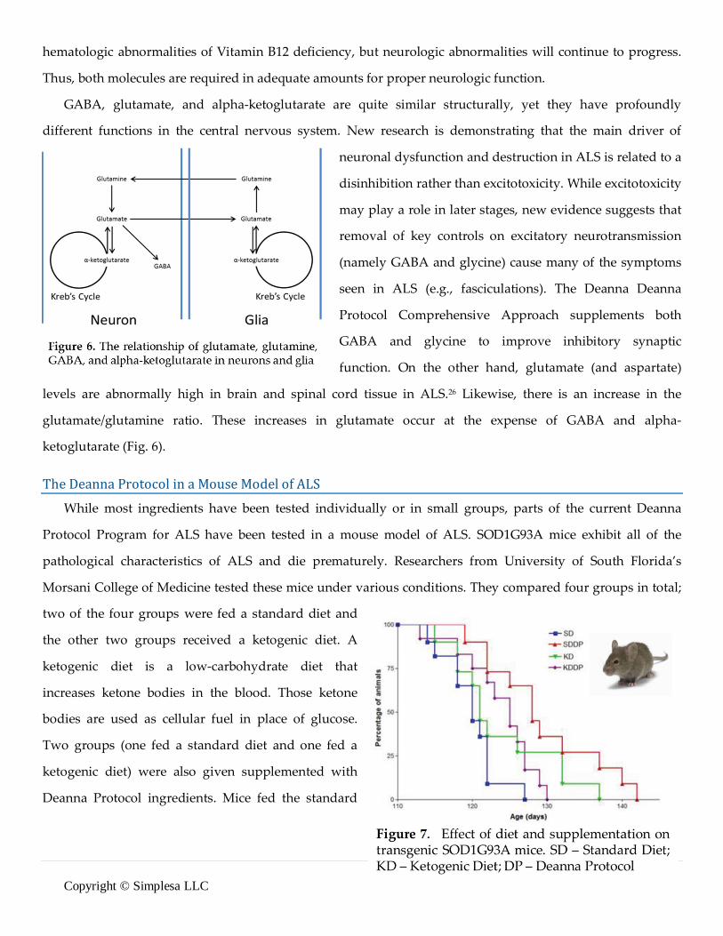
GABA, glutamate, and alpha-ketoglutarate are quite similar structurally, yet they have profoundly
different functions in the central nervous system. New research is demonstrating that the main driver of
neuronal dysfunction and destruction in ALS is related to a
disinhibition rather than excitotoxicity. While excitotoxicity
may play a role in later stages, new evidence suggests that
removal of key controls on excitatory neurotransmission
(namely GABA and glycine) cause many of the symptoms
seen in ALS (e.g., fasciculations). The Deanna Deanna
Protocol Comprehensive Approach supplements both
GABA and glycine to improve inhibitory synaptic
function. On the other hand, glutamate (and aspartate)
levels are abnormally high in brain and spinal cord tissue in ALS.26 Likewise, there is an increase in the
glutamate/glutamine ratio. These increases in glutamate occur at the expense of GABA and alpha-
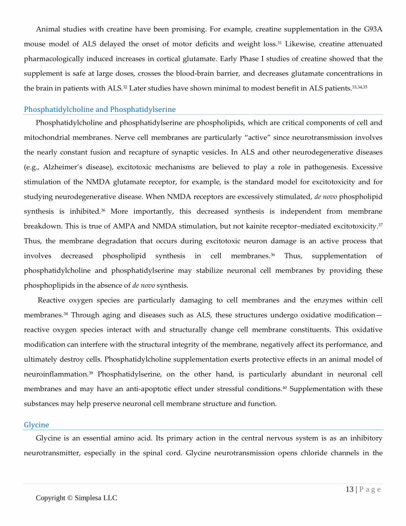
ketoglutarate (Fig. 6). The Deanna Protocol in a Mouse Model of ALS While most ingredients have been tested individually or in small groups, parts of the current Deanna
Protocol Program for ALS have been tested in a mouse model of ALS. SOD1G93A mice exhibit all of the
pathological characteristics of ALS and die prematurely. Researchers from University of South Florida’s
Morsani College of Medicine tested these mice under various conditions. They compared four groups in total;
two of the four groups were fed a standard diet and
the other two groups received a ketogenic diet. A
ketogenic diet is a low-carbohydrate diet that
increases ketone bodies in the blood. Those ketone
bodies are used as cellular fuel in place of glucose.
Two groups (one fed a standard diet and one fed a
ketogenic diet) were also given supplemented with
Deanna Protocol ingredients. Mice fed the standard

12 | P a g e Copyright © Simplesa LLC
diet and the Deanna Protocol lived significantly longer than mice in the other groups (Fig. 7).
While formal clinical trials using the Deanna Protocol have not been performed, there are numerous
anecdotal reports that the Deanna Protocol decreased fasciculations, spasms, tremors, salivary secretions,
speech impediments and weakness along with improved respiratory symptoms, swallowing capacity, and
balance. Creatine Creatine is a molecule that is formed endogenously by several organs including the kidneys and liver. It is
synthesized from the amino acids arginine and glycine with the addition of a methyl group from methionine
(Fig. 8). The molecule is actively taken up into brain, muscle, and heart tissue through transporter proteins.
Creatine is critical for normal neurological functioning. Most notably, people who lack the enzymes to create
creatine have several neurological problems including developmental delay, extrapyramidal movement
disorders, and seizures, which can be partially or fully ameliorated with creatine supplementation.
Creatine supplementation may be helpful in the treatment of ALS for several reasons.27 Increasing creatine
levels during periods of muscle relaxation increases the reserve of phosphocreatine and helps protect muscles
against fatigue.28 Creatine also enhances anaerobic metabolism and may increase lean muscle mass.27 Thus,
creatine may be of functional/symptomatic benefit in ALS.
However, the cellular energy benefits of creatine may be outweighed by the molecule’s potential role in
neuroprotection. The energy buffering ability of creatine and its ability to stabilize mitochondrial membranes
may partially counteract the mitochondrial dysfunction seen in ALS.27 For example, creatine/phosphocreatine
blocks the opening of the mitochondrial permeability transition pore, interrupting apoptosis-signaling
pathways.29 Likewise, creatine has
direct and indirect antioxidant
activity, which could dampen
damage caused by reactive oxygen
species. Creatine may help protect
against excitotoxic cell death in two
separate ways. The supplement can
enhance glutamate uptake into
synaptic vesicles without ATP30, which may help remove glutamate from the synaptic cleft without requiring
additional cellular energy. In addition, creatine helps normalize aberrant intracellular calcium systems by
increasing the uptake of calcium into the sarcoplasmic reticulum.
13 | P a g e Copyright © Simplesa LLC
Animal studies with creatine have been promising. For example, creatine supplementation in the G93A
mouse model of ALS delayed the onset of motor deficits and weight loss.31 Likewise, creatine attenuated
pharmacologically induced increases in cortical glutamate. Early Phase I studies of creatine showed that the
supplement is safe at large doses, crosses the blood-brain barrier, and decreases glutamate concentrations in
the brain in patients with ALS.32 Later studies have shown minimal to modest benefit in ALS patients.33,34,35 Phosphatidylcholine and Phosphatidylserine Phosphatidylcholine and phosphatidylserine are phospholipids, which are critical components of cell and
mitochondrial membranes. Nerve cell membranes are particularly “active” since neurotransmission involves
the nearly constant fusion and recapture of synaptic vesicles. In ALS and other neurodegenerative diseases
(e.g., Alzheimer’s disease), excitotoxic mechanisms are believed to play a role in pathogenesis. Excessive
stimulation of the NMDA glutamate receptor, for example, is the standard model for excitotoxicity and for
studying neurodegenerative disease. When NMDA receptors are excessively stimulated, de novo phospholipid
synthesis is inhibited.36 More importantly, this decreased synthesis is independent from membrane
breakdown. This is true of AMPA and NMDA stimulation, but not kainite receptor–mediated excitotoxicity.37
Thus, the membrane degradation that occurs during excitotoxic neuron damage is an active process that
involves decreased phospholipid synthesis in cell membranes.36 Thus, supplementation of
phosphatidylcholine and phosphatidylserine may stabilize neuronal cell membranes by providing these
phosphoplipids in the absence of de novo synthesis.
Reactive oxygen species are particularly damaging to cell membranes and the enzymes within cell
membranes.38 Through aging and diseases such as ALS, these structures undergo oxidative modification—
reactive oxygen species interact with and structurally change cell membrane constituents. This oxidative
modification can interfere with the structural integrity of the membrane, negatively affect its performance, and
ultimately destroy cells. Phosphatidylcholine supplementation exerts protective effects in an animal model of
neuroinflammation.39 Phosphatidylserine, on the other hand, is particularly abundant in neuronal cell
membranes and may have an anti-apoptotic effect under stressful conditions.40 Supplementation with these
substances may help preserve neuronal cell membrane structure and function. Glycine Glycine is an essential amino acid. Its primary action in the central nervous system is as an inhibitory
neurotransmitter, especially in the spinal cord. Glycine neurotransmission opens chloride channels in the

14 | P a g e Copyright © Simplesa LLC
postsynaptic cell causing an inhibitory postsynaptic potential (IPSP) and further polarizing the cell membrane.
Glycine is also a “co-agonist” at NMDA glutamate receptors in the brain.
Glycine levels are reduced in the lumbar ventral and dorsal horns of the spinal cord in patients with ALS.41
It has been suggested that decreases in the level of the major inhibitory neurotransmitter in the spinal cord,
glycine, leads to increased excitatory activity and even excitotoxicity.41 Indeed, transgenic mice that express
mutant SOD1 have poor glycine receptor currents, smaller clusters of receptors on their cell membranes, and
lower expression of glycine receptor mRNA in their motor neurons.42 Since selectively targeting NMDA
receptors with pharmacological agents can be challenging, researchers have postulated that enhancing glycine
inhibitory activity may be a promising therapeutic approach.43
L-Taurine Taurine is an organic acid that has several important biological functions (Fig. 9).44 By the strictest of
definitions, taurine is not an amino acid and is not incorporated into proteins. Nonetheless, taurine is a major
constituent of bile salts and is found abundantly in brain, retina, muscle, and
other organs. It is still a matter of speculation whether taurine is a true
neurotransmitter; however, when it is applied to neurons it exerts an inhibitory
effect.45 Taurine appears to be a potent, endogenous cytoprotectant. This cell-
protecting effect is presumably due to its antioxidant actions,46 its ability to
stabilize mitochondrial enzymes involved in the electron transport chain, and its capacity to reduce reactive
oxygen species generation.47 Taurine protects neurons from several different destructive mechanisms
including dibromoacetonitrile-induced neurotoxicity48 and acrylonitrile-induced oxidative stress.49 When
combined with vitamins C and E, taurine blocked the pro-oxidant effects of 3-nitropropionic acid.50
Taurine levels are higher in the brains and spinal cords of people with ALS.41,51 Indeed, G93A mice (ALS
mouse model) have higher levels of brain taurine and higher levels of the taurine transporter.52 Expression of
this transporter increases under periods of oxidative stress and is controlled by heat shock factor 1, a heat
shock protein. This work strongly suggests that increased expression of taurine transporters and uptake of
taurine in motor neurons in ALS is a compensatory mechanism.52 In other words, neurons affected by the ALS
disease process attempt to recruit the neuroprotectant taurine to partially attenuate the oxidative stress of the
disease. It is conceivable that augmented levels of taurine (e.g., through supplementation) would enhance this
protective effect.
L-Carnitine
15 | P a g e Copyright © Simplesa LLC
L-Carnitine is a naturally occurring compound found in virtually every type of human cell. It is derived
from the amino acids methionine and lysine. Its main role in the cell is in the cellular energy process—carnitine
shuttles long-chain fatty acids from the cytosol to the mitochondrial matrix for beta-oxidation.53 The molecule
is instrumental in converting fats into cellular energy. L-Carnitine also inhibits apoptosis and can minimize the
effects of oxidative stress-induced damage to mitochondria.54
Enhancing any or all of these actions could be advantageous in ALS. For example, lipid peroxidation
increases during the course of ALS with a concomitant increase in free radical production.55 As a result, free
fatty acids levels increase in cells. Additional carnitine could enhance normal intracellular shuttling, but also
stem the increase in cytosolic free fatty acids and prevent the release of cytochrome c or activation of caspases
(i.e., apoptotic signals).56
In laboratory studies of male G93A animals (ALS disease model), L-carnitine supplementation significantly
delayed the onset of disease phenotypes such as hind limb muscle and spinal cord abnormalities.
Supplementation also reduced markers of oxidative stress and prevented muscle cell apoptosis.57 Interestingly,
L-carnitine supplementation prolonged the lifespan of ALS model animals.57
Acetyl-L-carnitine performed favorably when compared to placebo in patients with definite/probable
ALS.58 In this trial, all patients were taking riluzole and receiving standard medical care during the yearlong
trial. Patients in the carnitine group were more likely to remain self-sufficient during the study period. Also,
forced vital capacity (i.e., a test of pulmonary function) remained higher in the carnitine group. Median
survival time was also significantly longer in the carnitine group (45 months) versus placebo (22 months).58
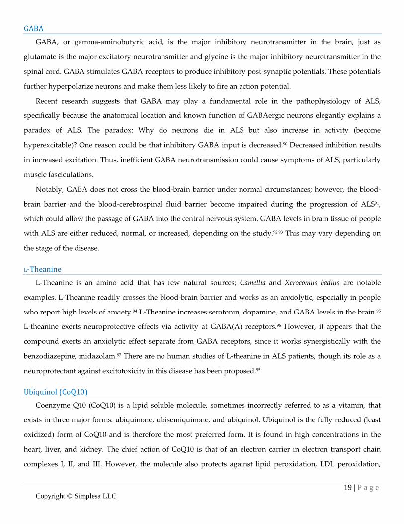
Moreover, patients tolerated carnitine supplementation well—adverse event rates were similar in both groups. B Vitamins: Thiamine, Riboflavin, Niacin, Pyridoxine, and Biotin The B vitamins, namely thiamine (Vitamin B1), riboflavin (Vitamin B2), niacin (Vitamin B3), pyridoxine
(Vitamin B6), and biotin (Vitamin B7) are water-soluble vitamins that are critical cofactors in various cellular
processes. Deficiencies in any of these vitamins can have deleterious effects on the body as a whole. For
example, Vitamin B1 deficiency causes Beriberi, a disease that causes profound changes in carbohydrate and
lipid metabolism, and deficits in glutamate and GABA neurotransmitter synthesis.

16 | P a g e Copyright © Simplesa LLC
There is some evidence to suggest that patients with ALS have deficits in enzymes that require B vitamins
as cofactors. Expression of riboflavin kinase, which catalyzes the formation of flavin mononucleotide from
riboflavin, is reduced in ALS.59 Human motor neurons are particularly susceptible to decreases in oxidative
metabolism, which results from reduced expression of enzymes in the pathway.59 People with ALS also have
diminished thiamine levels in their cerebrospinal fluid.60 In fact, decreases of thiamine with respect to its
related intracellular compound, thiamine monophosphate, are very specific to sporadic ALS; so much so that
some have proposed using the thiamine/thiamine monophosphate ratio as a biochemical marker of the
disease.61 Methylcobalamin and Methyl-Folic Acid Methylcobalamin (Vitamin B12) and folic acid (Vitamin B9) are often discussed together because they
participate together in various cellular functions. Moreover, deficits in one of these vitamins can mimic or
mask deficits in the other. They are also usually administered together for purposes of repletion.
These two vitamins are also described together because they both play a role in homocysteine metabolism.
Homocysteine is an amino acid related to cysteine, but is not incorporated into proteins. High levels of
homocysteine have been detected in various diseases, including heart disease and ALS.62,63 Since the
conversion of homocysteine to methionine requires the presence of Vitamin B12 and folic acid, deficiencies in
these vitamins lead to accumulation of the toxic substance. Conversely, methylcobalamin was able to rescue
neurons from the homocysteine toxicity in vitro.64 Curiously, methylfolate did not rescue these neurons;
however, folic acid did protect motor neurons from homocysteine, inflammation, and apoptosis in a SOD1
G93A transgenic mouse model of ALS.65 Moreover, Zhang and colleagues suggest that decreased 5-
methyltetrahydrofolate is sufficiently specific to people with pre-symptomatic ALS that it should be
considered a biomarker for early stage disease.66 Cholecalciferol (Vitamin D3) Cholecalciferol or Vitamin D3 is one of the main compounds within the Vitamin D family. Vitamin D3 can
be synthesized from cholesterol in sun-exposed skin (photoreaction). Nonetheless, Vitamin D deficiencies are
surprisingly common in developed nations. Vitamin D is critical for calcium and phosphorus homeostasis in
the body—it controls absorption from the gut, deposition in bones, and excretion in the kidney.
Supplementation with Vitamin D also increases the expression of calcium-binding proteins parvalbumin
and calbindin-D28K.67 This is potentially important in ALS for several reasons. Calcium influx at synaptic
buttons is a critical part of excitotoxic processes presumed to contribute to ALS neuropathology. Increased

17 | P a g e Copyright © Simplesa LLC
expression of calcium binding proteins (in this case, by Vitamin D supplementation) could act as a buffer to
chelate/bind excess calcium. This could conceivably halt the excitotoxic process.68 Indeed, decreased expression
of calbindin-D28K and/or parvalbumin occurs in motor neurons that are particularly vulnerable in ALS.69
Thus, in addition to combating demineralization of bone that can occur in ALS70, Vitamin D supplementation
may increase expression of calcium-binding proteins in motor neurons and subsequently interfere with
excitotoxic processes. Dietary Vitamin D3 supplementation improves functional performance in G93A
transgenic mice; mice treated with high levels of Vitamin D3 had greater paw grip endurance and motor
performance.71 Conversely, Vitamin D3 deficiency impairs motor performance in these animals after disease
onset.72
L-Glutathione and N-Acetylcysteine L-Glutathione is a chain of three amino acids (tripeptide) that
can be synthesized from cysteine, glutamate, and glycine. It is
present in various tissues, especially the liver, where it participates
in various metabolic processes. The molecule is a reductant (i.e.,
antioxidant) at the thiol group, where it quenches hydrogen
peroxide and other reactive oxygen species. Glutathione can also be
conjugated with various exogenous compounds so that they can be
more readily eliminated from the body.
N-Acetylcysteine is very similar to cysteine, structurally, with
an acetyl group covalently bound at the nitrogen atom. It has a
number of clinical uses, such as acting as an antidote in
acetaminophen/paracetamol overdose and for kidney protection
prior to intravenous contrast administration. N-acetylcysteine is a
precursor of glutathione—supplementation with N-acetylcysteine
increases the concentration of glutathione in cells.
Glutathione is abnormally low in patients with ALS as are the
activities of the enzymes that produce glutathione.73 Moreover,
certain ALS animal models display naturally and greatly reduced
levels of glutathione (70 to 80%).74 This deficiency in glutathione
may contribute to motor neuron vulnerability in ALS75 and may

18 | P a g e Copyright © Simplesa LLC
also increase aggregation of TDP-43 proteins (Fig. 10).76
This suggests that glutathione supplementation is potentially beneficial in reversing or halting these
pathological consequences.77 Indeed, N-acetylcysteine treatment prolonged survival and delayed symptom
onset in G93A transgenic mice.78 In a small clinical trial, there was a trend for N-acetylcysteine to improve
survival and delay limb onset of symptoms; however, the results did not reach statistical significance.79
Similarly, glutathione administration resulted in a slight, but not statistically significant slowing of ALS
progression.80 In both trials, small numbers of patients may have limited the power of the statistical analyses,
so additional studies are warranted. Alpha-Lipoic Acid While not a vitamin per se, alpha-lipoic acid is a key cofactor for multi-enzyme complexes, e.g., alpha-
ketoglutarate dehydrogenase, pyruvate dehydrogenase, and glycine decarboxylase.81 It also has the rather
unique property of acting as an antioxidant in both aqueous
and lipid phases.81 Lipoic acid is inert in its native form, but
it is readily converted in the body to its active form,
dihydrolipoic acid. In addition to being a potent antioxidant,
alpha-lipoic acid supplementation also restores glutathione
levels (Fig. 11).82 When aged rats were given lipoic acid
supplementation, they exhibited decreased oxidative
damage and improved mitochondrial function, which was
associated with enhanced ambulation.83 The molecule also
enhances blood flow to small vessels around nerves and
reduces lipid peroxidation.83 Magnesium The element magnesium exerts several actions on cells. It is well known that magnesium blocks NMDA
excitatory glutamate receptors. Excessive stimulation at these receptors leads to excitotoxic cell death.
Likewise, magnesium also inhibits calcium influx at calcium channels.84 Magnesium protects neurons from
glutamate-mediated cell death.85 Magnesium is particularly high in brain tissue of people with ALS.86 Results
from animal87 and human88 studies suggest that magnesium supplementation does not decrease the risk of
developing ALS symptoms or ALS. On the other hand, magnesium depletion may accelerate the uptake of
aluminum into brain and contribute to degenerative processes.89
19 | P a g e Copyright © Simplesa LLC
GABA GABA, or gamma-aminobutyric acid, is the major inhibitory neurotransmitter in the brain, just as
glutamate is the major excitatory neurotransmitter and glycine is the major inhibitory neurotransmitter in the
spinal cord. GABA stimulates GABA receptors to produce inhibitory post-synaptic potentials. These potentials
further hyperpolarize neurons and make them less likely to fire an action potential.
Recent research suggests that GABA may play a fundamental role in the pathophysiology of ALS,
specifically because the anatomical location and known function of GABAergic neurons elegantly explains a
paradox of ALS. The paradox: Why do neurons die in ALS but also increase in activity (become
hyperexcitable)? One reason could be that inhibitory GABA input is decreased.90 Decreased inhibition results
in increased excitation. Thus, inefficient GABA neurotransmission could cause symptoms of ALS, particularly
muscle fasciculations.
Notably, GABA does not cross the blood-brain barrier under normal circumstances; however, the blood-
brain barrier and the blood-cerebrospinal fluid barrier become impaired during the progression of ALS91,
which could allow the passage of GABA into the central nervous system. GABA levels in brain tissue of people
with ALS are either reduced, normal, or increased, depending on the study.92,93 This may vary depending on
the stage of the disease.
L-Theanine L-Theanine is an amino acid that has few natural sources; Camellia and Xerocomus badius are notable
examples. L-Theanine readily crosses the blood-brain barrier and works as an anxiolytic, especially in people
who report high levels of anxiety.94 L-Theanine increases serotonin, dopamine, and GABA levels in the brain.95
L-theanine exerts neuroprotective effects via activity at GABA(A) receptors.96 However, it appears that the
compound exerts an anxiolytic effect separate from GABA receptors, since it works synergistically with the
benzodiazepine, midazolam.97 There are no human studies of L-theanine in ALS patients, though its role as a
neuroprotectant against excitotoxicity in this disease has been proposed.95 Ubiquinol (CoQ10) Coenzyme Q10 (CoQ10) is a lipid soluble molecule, sometimes incorrectly referred to as a vitamin, that
exists in three major forms: ubiquinone, ubisemiquinone, and ubiquinol. Ubiquinol is the fully reduced (least
oxidized) form of CoQ10 and is therefore the most preferred form. It is found in high concentrations in the
heart, liver, and kidney. The chief action of CoQ10 is that of an electron carrier in electron transport chain
complexes I, II, and III. However, the molecule also protects against lipid peroxidation, LDL peroxidation,

20 | P a g e Copyright © Simplesa LLC
mitochondrial toxins, and DNA damage.104,105 The reduced form ubiquinol is a potent, lipid-soluble
antioxidant. Interestingly, CoQ10 functions as an extracellular superoxide dismutase106, one of the enzymes
that are abnormal in ALS.7,107
CoQ10 administration in an ALS mouse model (mutant SOD1 overexpression) starting at 70 days
significantly increased their lifespan compared to control animals.108 While high-dose CoQ10 is well tolerated
by patients with ALS109, phase II results with the compound were disappointing.110 CoQ10 supplementation
did not significantly change forced vital capacity, daily functioning scores, or biochemical markers of oxidative
stress.110 The authors of the Phase II trial suggest that Phase III evaluation is unwarranted.110 Moreover, it is
unlikely that CoQ10 alone is useful in treating humans with ALS; the supplement should probably be
combined with other agents for clinical effect. Propolis Propolis is a natural product that is collected from various plants by honeybees. It contains over 300
compounds including polyphenols, amino acids, steroids and
inorganic compounds.111 One of the constituents of propolis that
has been extensively studied as a potential neuroprotectant is
caffeic acid phenethyl ester (CAPE). CAPE is a potent antioxidant
that can prevent several types of neuronal injury including
neurotoxicity from MPTP112, 6-hydroxydopamine113,114, kainate115,
glutamate116, and permanent focal ischemia (Fig. 13).117 CAPE is also
a potent anti-inflammatory agent and has inhibitory effects on
myeloperoxidase and NADPH oxidase.118 In a screening test of over
2000 small molecules for the ability to rescue dorsal motor neurons
from imminent cell death (in vitro ALS model), CAPE was one of the top three most successful and potent
substances.119 Interestingly, mice expressing mutant SOD (ALS mouse model) lived significantly longer when
they were given CAPE than those fed simply a standard diet.120 Caprylic Acid Ketogenic diets consist of high fat and low carbohydrate meals and are intended to increase circulating
levels of ketone bodies. The ketogenic diet has demonstrated beneficial effects in several neurological
diseases—perhaps none more profound than in epilepsy. When SOD1-G93A transgenic mice are fed a
21 | P a g e Copyright © Simplesa LLC
ketogenic diet, they have increased survival and improved motor function compared to mice fed a standard
diet.121
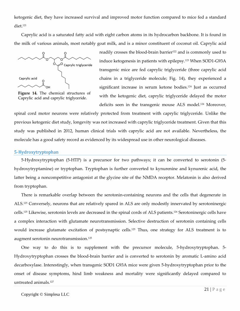
Caprylic acid is a saturated fatty acid with eight carbon atoms in its hydrocarbon backbone. It is found in
the milk of various animals, most notably goat milk, and is a minor constituent of coconut oil. Caprylic acid
readily crosses the blood-brain barrier122 and is commonly used to
induce ketogenesis in patients with epilepsy.123 When SOD1-G93A
transgenic mice are fed caprylic triglyceride (three caprylic acid
chains in a triglyceride molecule; Fig. 14), they experienced a
significant increase in serum ketone bodies.124 Just as occurred
with the ketogenic diet, caprylic triglyceride delayed the motor
deficits seen in the transgenic mouse ALS model.124 Moreover,
spinal cord motor neurons were relatively protected from treatment with caprylic triglyceride. Unlike the
previous ketogenic diet study, longevity was not increased with caprylic triglyceride treatment. Given that this
study was published in 2012, human clinical trials with caprylic acid are not available. Nevertheless, the
molecule has a good safety record as evidenced by its widespread use in other neurological diseases. 5-Hydroxytryptophan 5-Hydroxytryptophan (5-HTP) is a precursor for two pathways; it can be converted to serotonin (5-
hydroxytryptamine) or tryptophan. Tryptophan is further converted to kynurenine and kynurenic acid, the
latter being a noncompetitive antagonist at the glycine site of the NMDA receptor. Melatonin is also derived
from tryptophan.
There is remarkable overlap between the serotonin-containing neurons and the cells that degenerate in
ALS.125 Conversely, neurons that are relatively spared in ALS are only modestly innervated by serotoninergic
cells.125 Likewise, serotonin levels are decreased in the spinal cords of ALS patients.126 Serotoninergic cells have
a complex interaction with glutamate neurotransmission. Selective destruction of serotonin containing cells
would increase glutamate excitation of postsynaptic cells.125 Thus, one strategy for ALS treatment is to
augment serotonin neurotransmission.125
One way to do this is to supplement with the precursor molecule, 5-hydroxytryptophan. 5-
Hydroxytryptophan crosses the blood-brain barrier and is converted to serotonin by aromatic L-amino acid
decarboxylase. Interestingly, when transgenic SOD1 G93A mice were given 5-hydroxytryptophan prior to the
onset of disease symptoms, hind limb weakness and mortality were significantly delayed compared to
untreated animals.127

22 | P a g e Copyright © Simplesa LLC
Alpha-Ketoglutarate and Arginine Alpha-Ketoglutarate Alpha-ketoglutarate is one of the main constituents of the Kreb’s cycle/citric acid cycle. As such, it
participates in the cell’s energy making process. Alpha-ketoglutarate is integral to the conversion of ingested
sugar into adenosine triphosphate. Within the citric acid cycle, alpha-ketoglutarate is converted to succinyl-
Coenzyme A (succinyl-CoA) through the enzyme alpha-ketoglutarate dehydrogenase. With this enzymatic
conversion, a molecule of NAD+ (nicotinamide adenine dinucleotide) is protonated to become NADH. NADH
is a high-energy molecule and can convert ADP to ATP, the main energy carrier of cells.
α-ketoglutarate + NAD+ + CoA→ succinyl CoA + CO2 + NADH + H+
Alpha-ketoglutarate dehydrogenase also appears to be a major participant in mitochondrial function and
neurodegenerative disease. For example, there are reduced levels of alpha-ketoglutarate dehydrogenase in the
brains of people with Parkinson’s disease.128 Likewise, alpha-ketoglutarate dehydrogenase activity is reduced
in Alzheimer’s disease.129 Reductions in the enzyme complex can make neurons vulnerable and reduced
activity causes apoptosis without mitochondrial involvement.130 Thus, as enzyme levels decrease, one
corrective intervention may be to supplement with additional amounts of the substrate, alpha-ketoglutarate, to
compensate for this loss of enzyme. Activity of alpha-ketoglutarate dehydrogenase relates to the production of
reactive oxygen species and fatty acid metabolism.130
Arginine alpha-ketoglutarate is the water-soluble salt of the amino acid arginine and alpha-ketoglutarate.
In addition to the effects of alpha-ketoglutarate listed above, arginine may exert separate beneficial effects.
Arginine is an amino acid that is a key substrate for various enzymes. The amino acid is also abnormally low
in patients with ALS patients, a state that is exacerbated by malnutrition in more advanced states of the
disease. However, the supplementation of arginine may not simply be useful in reversing deficiencies. When
SOD1G93A mice are given arginine, they exhibit significantly less motor neuron destruction and glial
activation. Furthermore, the amino acid slows the progression lumbar spinal cord degeneration and delays the
onset of motor dysfunction in SOD1G93A mice. Importantly, arginine also significantly prolonged life in these
mice.
Safety
All of the ingredients contained in the Deanna Protocol® DP™ Plan Essentials and the Deanna Protocol
Comprehensive Approach are available over-the-counter, without a prescription. The ingredients in the

23 | P a g e Copyright © Simplesa LLC
Deanna Protocol® DP™ Plan Essentials and the Deanna Protocol Comprehensive Approach that have been
evaluated by the US Food and Drug Administration were deemed Generally Recognized as Safe (GRAS). The
dosages of ingredients included in the formulations have been derived from analogous dosages used in
laboratory work, published clinical studies, and commercially available dosages.
Disclaimer
The statements contained within have not been evaluated by the FDA. This product is not intended to
diagnose, treat, cure, or prevent any disease. You should contact a qualified medical provider before starting
any dietary regimen. While information in this document has been compiled from peer-reviewed journal
articles, the clinical action of the combined ingredients of the Deanna Protocol® DP™ Plan Essentials and the
Deanna Protocol Comprehensive Approach have not been tested in clinical trials.
References
1. Worms PM. The epidemiology of motor neuron diseases: a review of recent studies. J Neurol Sci. Oct 15 2001;191(1-2):3-9.
2. Byrne S, Walsh C, Lynch C, et al. Rate of familial amyotrophic lateral sclerosis: a systematic review and meta-analysis. J Neurol Neurosurg Psychiatry. Jun 2011;82(6):623-627.
3. Lattante S, Conte A, Zollino M, et al. Contribution of major amyotrophic lateral sclerosis genes to the etiology of sporadic disease. Neurology. Jul 3 2012;79(1):66-72.
4. Blauw HM, Barnes CP, van Vught PW, et al. SMN1 gene duplications are associated with sporadic ALS. Neurology. Mar 13 2012;78(11):776-780.
5. Momeni P, Schymick J, Jain S, et al. Analysis of IFT74 as a candidate gene for chromosome 9p-linked ALS-FTD. BMC Neurol. 2006;6:44.
6. Andersen PM. Amyotrophic lateral sclerosis associated with mutations in the CuZn superoxide dismutase gene. Curr Neurol Neurosci Rep. Jan 2006;6(1):37-46.
7. Boillee S, Cleveland DW. Revisiting oxidative damage in ALS: microglia, Nox, and mutant SOD1. J Clin Invest. Feb 2008;118(2):474-478.
8. Karch CM, Prudencio M, Winkler DD, Hart PJ, Borchelt DR. Role of mutant SOD1 disulfide oxidation and aggregation in the pathogenesis of familial ALS. Proc Natl Acad Sci U S A. May 12 2009;106(19):7774-7779.
9. Philips T, Robberecht W. Neuroinflammation in amyotrophic lateral sclerosis: role of glial activation in motor neuron disease. Lancet Neurol. Mar 2011;10(3):253-263.
10. Nagai M, Re DB, Nagata T, et al. Astrocytes expressing ALS-linked mutated SOD1 release factors selectively toxic to motor neurons. Nat Neurosci. May 2007;10(5):615-622.
11. Boillee S, Yamanaka K, Lobsiger CS, et al. Onset and progression in inherited ALS determined by motor neurons and microglia. Science. Jun 2 2006;312(5778):1389-1392.
12. Hall ED, Oostveen JA, Gurney ME. Relationship of microglial and astrocytic activation to disease onset and progression in a transgenic model of familial ALS. Glia. Jul 1998;23(3):249-256.
13. Harraz MM, Marden JJ, Zhou W, et al. SOD1 mutations disrupt redox-sensitive Rac regulation of NADPH oxidase in a familial ALS model. J Clin Invest. Feb 2008;118(2):659-670.
14. Shaw PJ, Forrest V, Ince PG, Richardson JP, Wastell HJ. CSF and plasma amino acid levels in motor neuron disease: elevation of CSF glutamate in a subset of patients. Neurodegeneration. Jun 1995;4(2):209-216.

24 | P a g e Copyright © Simplesa LLC
15. Rothstein JD, Van Kammen M, Levey AI, Martin LJ, Kuncl RW. Selective loss of glial glutamate transporter GLT-1 in amyotrophic lateral sclerosis. Ann Neurol. Jul 1995;38(1):73-84.
16. Miller RG, Mitchell JD, Moore DH. Riluzole for amyotrophic lateral sclerosis (ALS)/motor neuron disease (MND). Cochrane Database Syst Rev. 2012;3:CD001447.
17. Lacomblez L, Bensimon G, Leigh PN, Guillet P, Meininger V. Dose-ranging study of riluzole in amyotrophic lateral sclerosis. Amyotrophic Lateral Sclerosis/Riluzole Study Group II. Lancet. May 25 1996;347(9013):1425-1431.
18. Bensimon G, Lacomblez L, Meininger V. A controlled trial of riluzole in amyotrophic lateral sclerosis. ALS/Riluzole Study Group. N Engl J Med. Mar 3 1994;330(9):585-591.
19. Xu L, Enyeart JA, Enyeart JJ. Neuroprotective agent riluzole dramatically slows inactivation of Kv1.4 potassium channels by a voltage-dependent oxidative mechanism. J Pharmacol Exp Ther. Oct 2001;299(1):227-237.
20. Noh KM, Hwang JY, Shin HC, Koh JY. A novel neuroprotective mechanism of riluzole: direct inhibition of protein kinase C. Neurobiol Dis. Aug 2000;7(4):375-383.
21. Doble A. The pharmacology and mechanism of action of riluzole. Neurology. Dec 1996;47(6 Suppl 4):S233-241. 22. Hubert JP, Delumeau JC, Glowinski J, Premont J, Doble A. Antagonism by riluzole of entry of calcium evoked by
NMDA and veratridine in rat cultured granule cells: evidence for a dual mechanism of action. Br J Pharmacol. Sep 1994;113(1):261-267.
23. Martin LJ. Mitochondrial pathobiology in ALS. J Bioenerg Biomembr. Dec 2011;43(6):569-579. 24. Swerdlow RH, Parks JK, Cassarino DS, et al. Mitochondria in sporadic amyotrophic lateral sclerosis. Exp Neurol.
Sep 1998;153(1):135-142. 25. Tieu K, Perier C, Caspersen C, et al. D-beta-hydroxybutyrate rescues mitochondrial respiration and mitigates
features of Parkinson disease. J Clin Invest. Sep 2003;112(6):892-901. 26. Plaitakis A, Constantakakis E, Smith J. The neuroexcitotoxic amino acids glutamate and aspartate are altered in
the spinal cord and brain in amyotrophic lateral sclerosis. Ann Neurol. 1988;24(3):446-449. 27. Ellis AC, Rosenfeld J. The role of creatine in the management of amyotrophic lateral sclerosis and other
neurodegenerative disorders. CNS Drugs. 2004;18(14):967-980. 28. Guerrero-Ontiveros ML, Wallimann T. Creatine supplementation in health and disease. Effects of chronic
creatine ingestion in vivo: down-regulation of the expression of creatine transporter isoforms in skeletal muscle. Mol Cell Biochem. Jul 1998;184(1-2):427-437.
29. Fontaine E, Bernardi P. Progress on the mitochondrial permeability transition pore: regulation by complex I and ubiquinone analogs. J Bioenerg Biomembr. Aug 1999;31(4):335-345.
30. Xu CJ, Klunk WE, Kanfer JN, Xiong Q, Miller G, Pettegrew JW. Phosphocreatine-dependent glutamate uptake by synaptic vesicles. A comparison with atp-dependent glutamate uptake. J Biol Chem. Jun 7 1996;271(23):13435-13440.
31. Andreassen OA, Jenkins BG, Dedeoglu A, et al. Increases in cortical glutamate concentrations in transgenic amyotrophic lateral sclerosis mice are attenuated by creatine supplementation. J Neurochem. Apr 2001;77(2):383-390.
32. Atassi N, Ratai EM, Greenblatt DJ, et al. A phase I, pharmacokinetic, dosage escalation study of creatine monohydrate in subjects with amyotrophic lateral sclerosis. Amyotroph Lateral Scler. Dec 2010;11(6):508-513.
33. Shefner JM, Cudkowicz ME, Schoenfeld D, et al. A clinical trial of creatine in ALS. Neurology. Nov 9 2004;63(9):1656-1661.
34. Rosenfeld J, King RM, Jackson CE, et al. Creatine monohydrate in ALS: effects on strength, fatigue, respiratory status and ALSFRS. Amyotroph Lateral Scler. Oct 2008;9(5):266-272.
35. Pastula DM, Moore DH, Bedlack RS. Creatine for amyotrophic lateral sclerosis/motor neuron disease. Cochrane Database Syst Rev. 2012;12:CD005225.
36. Gasull T, Sarri E, DeGregorio-Rocasolano N, Trullas R. NMDA receptor overactivation inhibits phospholipid synthesis by decreasing choline-ethanolamine phosphotransferase activity. J Neurosci. May 15 2003;23(10):4100-4107.
37. Gasull T, DeGregorio-Rocasolano N, Trullas R. Overactivation of alpha-amino-3-hydroxy-5-methylisoxazole-4-propionate and N-methyl-D-aspartate but not kainate receptors inhibits phosphatidylcholine synthesis before excitotoxic neuronal death. J Neurochem. Apr 2001;77(1):13-22.

25 | P a g e Copyright © Simplesa LLC
38. Zaidi A, Michaelis ML. Effects of reactive oxygen species on brain synaptic plasma membrane Ca(2+)-ATPase. Free Radic Biol Med. Oct 1999;27(7-8):810-821.
39. Tokes T, Eros G, Bebes A, et al. Protective effects of a phosphatidylcholine-enriched diet in lipopolysaccharide-induced experimental neuroinflammation in the rat. Shock. Nov 2011;36(5):458-465.
40. Kim HY, Akbar M, Lau A, Edsall L. Inhibition of neuronal apoptosis by docosahexaenoic acid (22:6n-3). Role of phosphatidylserine in antiapoptotic effect. J Biol Chem. Nov 10 2000;275(45):35215-35223.
41. Malessa S, Leigh PN, Bertel O, Sluga E, Hornykiewicz O. Amyotrophic lateral sclerosis: glutamate dehydrogenase and transmitter amino acids in the spinal cord. J Neurol Neurosurg Psychiatry. Nov 1991;54(11):984-988.
42. Martin LJ, Chang Q. Inhibitory synaptic regulation of motoneurons: a new target of disease mechanisms in amyotrophic lateral sclerosis. Mol Neurobiol. Feb 2012;45(1):30-42.
43. Sasabe J, Aiso S. Aberrant control of motoneuronal excitability in amyotrophic lateral sclerosis: excitatory glutamate/D-serine vs. inhibitory glycine/gamma-aminobutanoic acid (GABA). Chem Biodivers. Jun 2010;7(6):1479-1490.
44. Ripps H, Shen W. Review: taurine: a "very essential" amino acid. Mol Vis. 2012;18:2673-2686. 45. Curtis DR, Watkins JC. The pharmacology of amino acids related to gamma-aminobutyric acid. Pharmacol Rev.
Dec 1965;17(4):347-391. 46. Aruoma OI, Halliwell B, Hoey BM, Butler J. The antioxidant action of taurine, hypotaurine and their metabolic
precursors. Biochem J. Nov 15 1988;256(1):251-255. 47. Jong CJ, Azuma J, Schaffer S. Mechanism underlying the antioxidant activity of taurine: prevention of
mitochondrial oxidant production. Amino Acids. Jun 2012;42(6):2223-2232. 48. Sayed RH, Salem HA, El-Sayeh BM. Potential protective effect of taurine against dibromoacetonitrile-induced
neurotoxicity in rats. Environ Toxicol Pharmacol. Nov 2012;34(3):849-857. 49. Mahalakshmi K, Pushpakiran G, Anuradha CV. Taurine prevents acrylonitrile-induced oxidative stress in rat brain.
Pol J Pharmacol. Nov-Dec 2003;55(6):1037-1043. 50. Rodriguez-Martinez E, Rugerio-Vargas C, Rodriguez AI, Borgonio-Perez G, Rivas-Arancibia S. Antioxidant effects
of taurine, vitamin C, and vitamin E on oxidative damage in hippocampus caused by the administration of 3-nitropropionic acid in rats. Int J Neurosci. Sep 2004;114(9):1133-1145.
51. Yoshino Y, Koike H, Akai K. Free amino acids in motor cortex of amyotrophic lateral sclerosis. Experientia. Feb 15 1979;35(2):219-220.
52. Jung MK, Kim KY, Lee NY, et al. Expression of taurine transporter (TauT) is modulated by heat shock factor 1 (HSF1) in motor neurons of ALS. Mol Neurobiol. Apr 2013;47(2):699-710.
53. Bremer J. Carnitine--metabolism and functions. Physiol Rev. Oct 1983;63(4):1420-1480. 54. Hagen TM, Liu J, Lykkesfeldt J, et al. Feeding acetyl-L-carnitine and lipoic acid to old rats significantly improves
metabolic function while decreasing oxidative stress. Proc Natl Acad Sci U S A. Feb 19 2002;99(4):1870-1875. 55. Farooqui AA, Yang HC, Rosenberger TA, Horrocks LA. Phospholipase A2 and its role in brain tissue. J Neurochem.
Sep 1997;69(3):889-901. 56. Kim JS, He L, Lemasters JJ. Mitochondrial permeability transition: a common pathway to necrosis and apoptosis.
Biochem Biophys Res Commun. May 9 2003;304(3):463-470. 57. Kira Y, Nishikawa M, Ochi A, Sato E, Inoue M. L-carnitine suppresses the onset of neuromuscular degeneration
and increases the life span of mice with familial amyotrophic lateral sclerosis. Brain Res. Jan 27 2006;1070(1):206-214.
58. Beghi E, Pupillo E, Bonito V, et al. Randomized double-blind placebo-controlled trial of acetyl-L-carnitine for ALS. Amyotroph Lateral Scler Frontotemporal Degener. Sep 2013;14(5-6):397-405.
59. Lin J, Diamanduros A, Chowdhury SA, Scelsa S, Latov N, Sadiq SA. Specific electron transport chain abnormalities in amyotrophic lateral sclerosis. J Neurol. May 2009;256(5):774-782.
60. Poloni M, Patrini C, Rocchelli B, Rindi G. Thiamin monophosphate in the CSF of patients with amyotrophic lateral sclerosis. Arch Neurol. Aug 1982;39(8):507-509.
61. Poloni M, Mazzarello P, Patrini C, Pinelli P. Inversion of T/TMP ratio in ALS: a specific finding? Ital J Neurol Sci. Jun 1986;7(3):333-335.
62. Valentino F, Bivona G, Butera D, et al. Elevated cerebrospinal fluid and plasma homocysteine levels in ALS. Eur J Neurol. Jan 2010;17(1):84-89.

26 | P a g e Copyright © Simplesa LLC
63. Zoccolella S, Simone IL, Lamberti P, et al. Elevated plasma homocysteine levels in patients with amyotrophic lateral sclerosis. Neurology. Jan 15 2008;70(3):222-225.
64. Hemendinger RA, Armstrong EJ, 3rd, Brooks BR. Methyl Vitamin B12 but not methylfolate rescues a motor neuron-like cell line from homocysteine-mediated cell death. Toxicol Appl Pharmacol. Mar 15 2011;251(3):217-225.
65. Zhang X, Chen S, Li L, Wang Q, Le W. Folic acid protects motor neurons against the increased homocysteine, inflammation and apoptosis in SOD1 G93A transgenic mice. Neuropharmacology. Jun 2008;54(7):1112-1119.
66. Zhang X, Chen S, Li L, Wang Q, Le W. Decreased level of 5-methyltetrahydrofolate: a potential biomarker for pre-symptomatic amyotrophic lateral sclerosis. J Neurol Sci. Jun 15 2010;293(1-2):102-105.
67. Alexianu ME, Robbins E, Carswell S, Appel SH. 1Alpha, 25 dihydroxyvitamin D3-dependent up-regulation of calcium-binding proteins in motoneuron cells. J Neurosci Res. Jan 1 1998;51(1):58-66.
68. Appel SH, Beers D, Smith RG, Wilson JE. Altered calcium homeostasis in ALS as a target for therapy. Amyotroph Lateral Scler Other Motor Neuron Disord. Dec 2000;1 Suppl 4:27-32.
69. Alexianu ME, Ho BK, Mohamed AH, La Bella V, Smith RG, Appel SH. The role of calcium-binding proteins in selective motoneuron vulnerability in amyotrophic lateral sclerosis. Ann Neurol. Dec 1994;36(6):846-858.
70. Ishizaki F, Koyama T, Sunayashiki T, Murata Y, Iida T, Harada T. [Control of bone remodeling by nervous system. Bone metabolic changes in neurological diseases]. Clin Calcium. Dec 2010;20(12):1841-1849.
71. Gianforcaro A, Hamadeh MJ. Dietary vitamin D3 supplementation at 10x the adequate intake improves functional capacity in the G93A transgenic mouse model of ALS, a pilot study. CNS Neurosci Ther. Jul 2012;18(7):547-557.
72. Solomon JA, Gianforcaro A, Hamadeh MJ. Vitamin D3 deficiency differentially affects functional and disease outcomes in the G93A mouse model of amyotrophic lateral sclerosis. PLoS One. 2011;6(12):e29354.
73. Moumen R, Nouvelot A, Duval D, Lechevalier B, Viader F. Plasma superoxide dismutase and glutathione peroxidase activity in sporadic amyotrophic lateral sclerosis. J Neurol Sci. Oct 3 1997;151(1):35-39.
74. Vargas MR, Johnson DA, Johnson JA. Decreased glutathione accelerates neurological deficit and mitochondrial pathology in familial ALS-linked hSOD1(G93A) mice model. Neurobiol Dis. Sep 2011;43(3):543-551.
75. Tartari S, D'Alessandro G, Babetto E, Rizzardini M, Conforti L, Cantoni L. Adaptation to G93Asuperoxide dismutase 1 in a motor neuron cell line model of amyotrophic lateral sclerosis: the role of glutathione. FEBS J. May 2009;276(10):2861-2874.
76. Iguchi Y, Katsuno M, Takagi S, et al. Oxidative stress induced by glutathione depletion reproduces pathological modifications of TDP-43 linked to TDP-43 proteinopathies. Neurobiol Dis. Mar 2012;45(3):862-870.
77. Muyderman H, Hutson PG, Matusica D, Rogers ML, Rush RA. The human G93A-superoxide dismutase-1 mutation, mitochondrial glutathione and apoptotic cell death. Neurochem Res. Oct 2009;34(10):1847-1856.
78. Andreassen OA, Dedeoglu A, Klivenyi P, Beal MF, Bush AI. N-acetyl-L-cysteine improves survival and preserves motor performance in an animal model of familial amyotrophic lateral sclerosis. Neuroreport. Aug 3 2000;11(11):2491-2493.
79. Louwerse ES, Weverling GJ, Bossuyt PM, Meyjes FE, de Jong JM. Randomized, double-blind, controlled trial of acetylcysteine in amyotrophic lateral sclerosis. Arch Neurol. Jun 1995;52(6):559-564.
80. Chio A, Cucatto A, Terreni AA, Schiffer D. Reduced glutathione in amyotrophic lateral sclerosis: an open, crossover, randomized trial. Ital J Neurol Sci. Dec 1998;19(6):363-366.
81. Kagan VE, Shvedova A, Serbinova E, et al. Dihydrolipoic acid--a universal antioxidant both in the membrane and in the aqueous phase. Reduction of peroxyl, ascorbyl and chromanoxyl radicals. Biochem Pharmacol. Oct 20 1992;44(8):1637-1649.
82. Shay KP, Moreau RF, Smith EJ, Smith AR, Hagen TM. Alpha-lipoic acid as a dietary supplement: molecular mechanisms and therapeutic potential. Biochim Biophys Acta. Oct 2009;1790(10):1149-1160.
83. Baker SK, Tarnopolsky MA. Targeting cellular energy production in neurological disorders. Expert Opin Investig Drugs. Oct 2003;12(10):1655-1679.
84. Iseri LT, French JH. Magnesium: nature's physiologic calcium blocker. Am Heart J. Jul 1984;108(1):188-193. 85. Finkbeiner S, Stevens CF. Applications of quantitative measurements for assessing glutamate neurotoxicity.
Proceedings of the National Academy of Sciences. June 1, 1988 1988;85(11):4071-4074.

27 | P a g e Copyright © Simplesa LLC
86. Hozumi I, Hasegawa T, Honda A, et al. Patterns of levels of biological metals in CSF differ among neurodegenerative diseases. J Neurol Sci. Apr 15 2011;303(1-2):95-99.
87. Pamphlett R, Todd E, Vink R, McQuilty R, Cheema SS. Magnesium supplementation does not delay disease onset or increase survival in a mouse model of familial ALS. J Neurol Sci. Dec 15 2003;216(1):95-98.
88. Fondell E, O'Reilly EJ, Fitzgerald KC, et al. Magnesium intake and risk of amyotrophic lateral sclerosis: results from five large cohort studies. Amyotroph Lateral Scler Frontotemporal Degener. Sep 2013;14(5-6):356-361.
89. Mitani K. Relationship between neurological diseases due to aluminium load, especially amyotrophic lateral sclerosis, and magnesium status. Magnes Res. Sep 1992;5(3):203-213.
90. Bae JS, Simon NG, Menon P, Vucic S, Kiernan MC. The puzzling case of hyperexcitability in amyotrophic lateral sclerosis. J Clin Neurol. Apr 2013;9(2):65-74.
91. Garbuzova-Davis S, Hernandez-Ontiveros DG, Rodrigues MC, et al. Impaired blood-brain/spinal cord barrier in ALS patients. Brain Res. Aug 21 2012;1469:114-128.
92. Niebroj-Dobosz I, Janik P. Amino acids acting as transmitters in amyotrophic lateral sclerosis (ALS). Acta Neurol Scand. Jul 1999;100(1):6-11.
93. Foerster BR, Callaghan BC, Petrou M, Edden RA, Chenevert TL, Feldman EL. Decreased motor cortex gamma-aminobutyric acid in amyotrophic lateral sclerosis. Neurology. May 15 2012;78(20):1596-1600.
94. Kimura K, Ozeki M, Juneja LR, Ohira H. L-Theanine reduces psychological and physiological stress responses. Biol Psychol. Jan 2007;74(1):39-45.
95. Nathan PJ, Lu K, Gray M, Oliver C. The neuropharmacology of L-theanine(N-ethyl-L-glutamine): a possible neuroprotective and cognitive enhancing agent. J Herb Pharmacother. 2006;6(2):21-30.
96. Egashira N, Hayakawa K, Osajima M, et al. Involvement of GABA(A) receptors in the neuroprotective effect of theanine on focal cerebral ischemia in mice. J Pharmacol Sci. Oct 2007;105(2):211-214.
97. Heese T, Jenkinson J, Love C, et al. Anxiolytic effects of L-theanine--a component of green tea--when combined with midazolam, in the male Sprague-Dawley rat. AANA J. Dec 2009;77(6):445-449.
98. Kelley BJ, Knopman DS. Alternative medicine and Alzheimer disease. Neurologist. Sep 2008;14(5):299-306. 99. Braquet P, Hosford D. Ethnopharmacology and the development of natural PAF antagonists as therapeutic
agents. J Ethnopharmacol. Apr 1991;32(1-3):135-139. 100. Sastre J, Millan A, Garcia de la Asuncion J, et al. A Ginkgo biloba extract (EGb 761) prevents mitochondrial aging
by protecting against oxidative stress. Free Radic Biol Med. Jan 15 1998;24(2):298-304. 101. Zhu L, Wu J, Liao H, Gao J, Zhao XN, Zhang ZX. Antagonistic effects of extract from leaves of ginkgo biloba on
glutamate neurotoxicity. Zhongguo Yao Li Xue Bao. Jul 1997;18(4):344-347. 102. Kanada A, Nishimura Y, Yamaguchi JY, et al. Extract of Ginkgo biloba leaves attenuates kainate-induced increase
in intracellular Ca2+ concentration of rat cerebellar granule neurons. Biol Pharm Bull. May 2005;28(5):934-936. 103. Ferrante RJ, Klein AM, Dedeoglu A, Beal MF. Therapeutic efficacy of EGb761 (Gingko biloba extract) in a
transgenic mouse model of amyotrophic lateral sclerosis. J Mol Neurosci. Aug 2001;17(1):89-96. 104. Salama M, Yuan TF, Machado S, et al. Co-enzyme Q10 to treat neurological disorders: basic mechanisms, clinical
outcomes, and future research direction. CNS Neurol Disord Drug Targets. Aug 2013;12(5):641-664. 105. Beal MF, Henshaw DR, Jenkins BG, Rosen BR, Schulz JB. Coenzyme Q10 and nicotinamide block striatal lesions
produced by the mitochondrial toxin malonate. Ann Neurol. Dec 1994;36(6):882-888. 106. Littarru GP, Tiano L. Bioenergetic and antioxidant properties of coenzyme Q10: recent developments. Mol
Biotechnol. Sep 2007;37(1):31-37. 107. Allen MJ, Lacroix JJ, Ramachandran S, et al. Mutant SOD1 forms ion channel: implications for ALS
pathophysiology. Neurobiol Dis. Mar 2012;45(3):831-838. 108. Matthews RT, Yang L, Browne S, Baik M, Beal MF. Coenzyme Q10 administration increases brain mitochondrial
concentrations and exerts neuroprotective effects. Proc Natl Acad Sci U S A. Jul 21 1998;95(15):8892-8897. 109. Ferrante KL, Shefner J, Zhang H, et al. Tolerance of high-dose (3,000 mg/day) coenzyme Q10 in ALS. Neurology.
Dec 13 2005;65(11):1834-1836. 110. Kaufmann P, Thompson JL, Levy G, et al. Phase II trial of CoQ10 for ALS finds insufficient evidence to justify
phase III. Ann Neurol. Aug 2009;66(2):235-244. 111. Farooqui T, Farooqui AA. Beneficial effects of propolis on human health and neurological diseases. Front Biosci
(Elite Ed). 2012;4:779-793.

28 | P a g e Copyright © Simplesa LLC
112. Fontanilla CV, Ma Z, Wei X, et al. Caffeic acid phenethyl ester prevents 1-methyl-4-phenyl-1,2,3,6-tetrahydropyridine-induced neurodegeneration. Neuroscience. Aug 11 2011;188:135-141.
113. Ma Z, Wei X, Fontanilla C, et al. Caffeic acid phenethyl ester blocks free radical generation and 6-hydroxydopamine-induced neurotoxicity. Life Sci. Aug 22 2006;79(13):1307-1311.
114. Noelker C, Bacher M, Gocke P, et al. The flavanoide caffeic acid phenethyl ester blocks 6-hydroxydopamine-induced neurotoxicity. Neurosci Lett. Jul 22-29 2005;383(1-2):39-43.
115. Kwon YS, Park DH, Shin EJ, et al. Antioxidant propolis attenuates kainate-induced neurotoxicity via adenosine A1 receptor modulation in the rat. Neurosci Lett. Jan 30 2004;355(3):231-235.
116. Wei X, Ma Z, Fontanilla CV, et al. Caffeic acid phenethyl ester prevents cerebellar granule neurons (CGNs) against glutamate-induced neurotoxicity. Neuroscience. Sep 9 2008;155(4):1098-1105.
117. Altug ME, Serarslan Y, Bal R, et al. Caffeic acid phenethyl ester protects rabbit brains against permanent focal ischemia by antioxidant action: a biochemical and planimetric study. Brain Res. Mar 27 2008;1201:135-142.
118. Lotfy ML. Biological activity of bee propolis in health and disease. Asian Pac J Cancer Prev. Jan-Mar 2006;7(1):22-31.
119. Barber SC, Higginbottom A, Mead RJ, Barber S, Shaw PJ. An in vitro screening cascade to identify neuroprotective antioxidants in ALS. Free Radic Biol Med. Apr 15 2009;46(8):1127-1138.
120. Fontanilla CV, Wei X, Zhao L, et al. Caffeic acid phenethyl ester extends survival of a mouse model of amyotrophic lateral sclerosis. Neuroscience. Mar 15 2012;205:185-193.
121. Zhao Z, Lange DJ, Voustianiouk A, et al. A ketogenic diet as a potential novel therapeutic intervention in amyotrophic lateral sclerosis. BMC Neurosci. 2006;7:29.
122. Spector R. Fatty acid transport through the blood-brain barrier. J Neurochem. Feb 1988;50(2):639-643. 123. Sills MA, Forsythe WI, Haidukewych D, MacDonald A, Robinson M. The medium chain triglyceride diet and
intractable epilepsy. Arch Dis Child. Dec 1986;61(12):1168-1172. 124. Zhao W, Varghese M, Vempati P, et al. Caprylic triglyceride as a novel therapeutic approach to effectively
improve the performance and attenuate the symptoms due to the motor neuron loss in ALS disease. PLoS One. 2012;7(11):e49191.
125. Sandyk R. Serotonergic mechanisms in amyotrophic lateral sclerosis. Int J Neurosci. Jul 2006;116(7):775-826. 126. Bertel O, Malessa S, Sluga E, Hornykiewicz O. Amyotrophic lateral sclerosis: changes of noradrenergic and
serotonergic transmitter systems in the spinal cord. Brain Res. Dec 6 1991;566(1-2):54-60. 127. Turner BJ, Lopes EC, Cheema SS. The serotonin precursor 5-hydroxytryptophan delays neuromuscular disease in
murine familial amyotrophic lateral sclerosis. Amyotroph Lateral Scler Other Motor Neuron Disord. Sep 2003;4(3):171-176.
128. Gibson GE, Kingsbury AE, Xu H, et al. Deficits in a tricarboxylic acid cycle enzyme in brains from patients with Parkinson’s disease. Neurochemistry International. 2003;43(2):129-135.
129. Bubber P, Haroutunian V, Fisch G, Blass JP, Gibson GE. Mitochondrial abnormalities in Alzheimer brain: Mechanistic implications. Ann Neurol. 2005;57(5):695-703.
130. Gibson GE, Blass JP, Beal MF, Bunik V. The alpha-ketoglutarate-dehydrogenase complex: a mediator between mitochondria and oxidative stress in neurodegeneration. Mol Neurobiol. 2005;31(1-3):43-63.
29 | P a g e Copyright © Simplesa LLC
Appendix 1: Nutritional Supplements, Products and Dosages
A. Simplesa AAKG+ Core Powder This formula provides the minimum recommended dosage* of Arginine Alpha Ketoglutarate (AAKG), as well as the recommended dosages of Ubiquinol (CoQ10), GABA (Gamma Aminobutyric Acid) and Niacin (Non-Flush) in every serving. Simplesa AAKG+ Core Protocol Blend has a 1:1 ratio of Arginine to Alpha Ketoglutarate. Taken three times a day, a daily serving includes:
• Arginine Alpha Ketoglutarate (AAKG) – 9 grams per day • Ubiquinol (CoQ10) - 1,200 mg per day • GABA (Gamma Aminobutyric Acid) – 500 mg per day • Niacin (Non-Flush) – 250 mg per day
*Begin with 9 grams of Arginine Alpha Ketoglutarate daily and increase slowly to 18 grams per day. DO NOT exceed 18 grams per day. A daily serving of Simplesa AAKG+ Core Protocol Powder contains 9 grams of AAKG. For those people taking more than 9 grams of AAKG per day, combining Simplesa AAKG+ Core Protocol Powder with pure AAKG (1:1 ratio) increases the amount of AAKG while still providing for the proper amounts of Ubiquinol, GABA and Niacin. B. Alpha Ketoglutarate Acid (AKG) 300 mg of Alpha-Ketoglutarate should be taken once per hour during each waking hour between doses of the Core Powder formula. C. DPS-AM Morning Protocol Blend This 2-ounce liquid supplement should be taken in the morning. It contains the following nutrients:
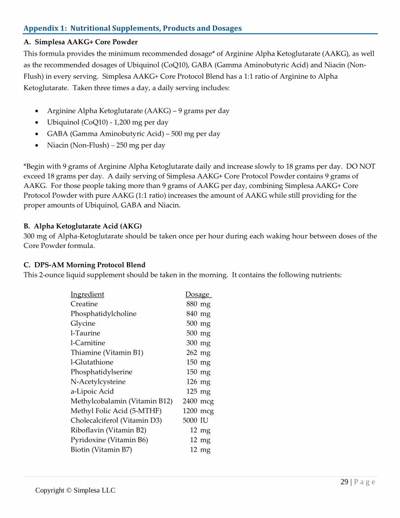
Ingredient Dosage Creatine 880 mg Phosphatidylcholine 840 mg Glycine 500 mg l-Taurine 500 mg l-Carnitine 300 mg Thiamine (Vitamin B1) 262 mg l-Glutathione 150 mg Phosphatidylserine 150 mg N-Acetylcysteine 126 mg a-Lipoic Acid 125 mg Methylcobalamin (Vitamin B12) 2400 mcg Methyl Folic Acid (5-MTHF) 1200 mcg Cholecalciferol (Vitamin D3) 5000 IU Riboflavin (Vitamin B2) 12 mg Pyridoxine (Vitamin B6) 12 mg Biotin (Vitamin B7) 12 mg

30 | P a g e Copyright © Simplesa LLC
D. DPS-PM Evening Protocol Blend This 2-ounce liquid supplement should be taken in the evening. It contains the following nutrients:
Ingredient Dosage Phosphatidylcholine 840 mg N-Acetylcysteine 500 mg Glycine 500 mg l-Taurine 500 mg Propolis Extract 500 mg Magnesium Lactate 400 mg l-Theanine 210 mg Methyl Folic Acid (5-MTHF) 1200 mcg Vitamin E 125 mg
E. Other Nutrients Liposomal Glutathione: Take approximately 425 mg once per day. 5-Hydroxytryptophan: Take 50 mg once per day in the evening with the Evening Protocol Blend. Caprylic Acid: Found in coconut oil or MCT oil. Take to tolerance.

31 | P a g e Copyright © Simplesa LLC
Exhibit 1. Products from Simplesa LLC
A. Simplesa AAKG+ Core Powder

32 | P a g e Copyright © Simplesa LLC
B. Simplesa Alpha Ketoglutarate Acid (AKG) Liquid Concentrate

33 | P a g e Copyright © Simplesa LLC
C. Simplesa DPS-AM Morning Protocol Blend

34 | P a g e Copyright © Simplesa LLC
D. Simplesa DPS-PM Evening Protocol Blend


