+ All Categories
Home > Documents > Dear Author - upcommons.upc.edu

Dear Author - upcommons.upc.edu

Date post: 11-Mar-2022
Category:
Upload: others
View: 0 times
Download: 0 times
Share this document with a friend
10
Dear Author Please use this PDF proof to check the layout of your article. If you would like any changes to be made to the layout, you can leave instructions in the online proofing interface. Making your changes directly in the online proofing interface is the quickest, easiest way to correct and submit your proof. Please note that changes made to the article in the online proofing interface will be added to the article before publication, but are not reflected in this PDF proof. If you would prefer to submit your corrections by annotating the PDF proof, please download and submit an annotatable PDF proof by clicking here and you'll be redirected to our PDF Proofing system.
Transcript

Dear Author

Please use this PDF proof to check the layout of your article. If you would like any changes to

be made to the layout, you can leave instructions in the online proofing interface. Making your

changes directly in the online proofing interface is the quickest, easiest way to correct and submit

your proof. Please note that changes made to the article in the online proofing interface will be

added to the article before publication, but are not reflected in this PDF proof.

If you would prefer to submit your corrections by annotating the PDF proof, please download and

submit an annotatable PDF proof by clicking here and you'll be redirected to our PDF Proofing

system.

ARTICLE IN PRESS

JID: ACTBIO [m5G; December 10, 2019;5:41 ]

Acta Biomaterialia xxx (xxxx) xxx

Contents lists available at ScienceDirect

Acta Biomaterialia

journal homepage: www.elsevier.com/locate/actbio

Self-assembling as regular nanoparticles dramatically minimizes

photobleaching of tumour-targeted GFP

Ugutz Unzueta

a , b , c , 1 , ∗, Mònica Roldán

d , h , 1 , Mireia Pesarrodona

b , c , e , 2 , Raul Benitez

g , h , Q1

Alejandro Sánchez-Chardi f , Oscar Conchillo-Solé e , Ramón Mangues a , b , Antonio Villaverde

b , c , e , ∗, Esther Vázquez

b , c , e , ∗

a Institut d’Investigacions Biomèdiques Sant Pau, Hospital de la Santa Creu i Sant Pau, 08025 Barcelona, Spain b CIBER de Bioingeniería, Biomateriales y Nanomedicina (CIBER-BBN), C/ Monforte de Lemos 3-5, 28029 Madrid, Spain c Departament de Genètica i de Microbiologia, Universitat Autònoma de Barcelona, Bellaterra, 08193 Barcelona, Spain d Unitat de Microscòpia Confocal. Servei d’Anatomia Patològica, Institut Pediàtric de Malalties Rares (IPER), Hospital Sant Joan de Déu, Esplugues de

Llobregat, Barcelona, Spain e Institut de Biotecnologia i de Biomedicina, Universitat Autònoma de Barcelona, Bellaterra, 08193 Barcelona, Spain f Departament de Biologia Evolutiva, Ecologia i Ciències Ambientals, Facultat de Biologia, Universitat de Barcelona, Av. Diagonal 643, 08028 Barcelona, Spain g Biomedical Engineering Research Center and Automatic Control Department, Universitat Politècnica de Catalunya, Av. Eduard Maristany 16, 08019

Barcelona, Spain h Institut de Recerca Sant Joan de Déu, Santa Rosa 39-57, 08950 Esplugues de Llobregat, Spain

a r t i c l e i n f o

Article history:

Received 14 June 2019

Revised 29 November 2019

Accepted 3 December 2019

Available online xxx

Keywords:

Nanoparticles

Fluorescent proteins

Photostability

Self-assembling

Tumour targeting

a b s t r a c t

Fluorescent proteins are useful imaging and theranostic agents, but their potential superiority over alter-

native dyes is weakened by substantial photobleaching under irradiation. Enhancing protein photostabil-

ity has been attempted through diverse strategies, with irregular results and limited applicability. In this

context, we wondered if the controlled oligomerization of Green Fluorescent Protein (GFP) as nanoscale

supramolecular complexes could stabilize the fluorophore through the newly formed protein-protein con-

tacts, and thus, enhance its global photostability. For that, we have here analyzed the photobleaching pro-

file of several GFP versions, engineered to self-assemble as tumour-homing nanoparticles with different

targeting, size and structural stability. This has been done under prolonged irradiation in confocal laser

scanning microscopy and by small-angle X-ray scattering. The results show that the oligomerization of

GFP at the nanoscale enhances, by more than seven-fold, the stability of fluorescence emission. Interest-

ingly, GFP nanoparticles are much more resistant to X-ray damage than the building block counterparts,

indicating that the gained photostability is linked to enhanced structural resistance to radiation. There-

fore, the controlled oligomerization of self-assembling fluorescent proteins as protein nanoparticles is a

simple, versatile and powerful method to enhance their photostability for uses in precision imaging and

therapy.

Statement of significance

Fluorescent protein assembly into regular and highly symmetric nanoscale structures has been identified

to confer enhanced structural stability against radiation stresses dramatically reducing their photobleach-

ing. Being this the main bottleneck in the use of fluorescent proteins for imaging and theranostics, this

protein architecture engineering principle appears as a powerful method to enhance their photostability

for a broad applicability in precision imaging, drug delivery and theranostics.

© 2019 Acta Materialia Inc. Published by Elsevier Ltd. All rights reserved.

Abbreviations: NPs, Nanoparticles; BBs, Building Blocks; FDHM, Full Duration at Half Maximum; DLS, Dynamic Light Scattering; SAXS, Small Angle X-ray Scattering; SDS,

Sodium Dodecyl Sulphate. ∗ Corresponding authors.

E-mail addresses: [email protected] (U. Unzueta), [email protected] (A. Villaverde), [email protected] (E. Vázquez). 1 Equally contributed. 2 Present address: Institute for Research in Biomedicine (IRB Barcelona), The Barcelona Institute of Science and Technology, Barcelona 08028, Spain.

https://doi.org/10.1016/j.actbio.2019.12.003

1742-7061/© 2019 Acta Materialia Inc. Published by Elsevier Ltd. All rights reserved.

Please cite this article as: U. Unzueta, M. Roldán and M. Pesarrodona et al., Self-assembling as regular nanoparticles dramatically mini-

mizes photobleaching of tumour-targeted GFP, Acta Biomaterialia, https://doi.org/10.1016/j.actbio.2019.12.003

2 U. Unzueta, M. Roldán and M. Pesarrodona et al. / Acta Biomaterialia xxx (xxxx) xxx

ARTICLE IN PRESS

JID: ACTBIO [m5G; December 10, 2019;5:41 ]

1. Introduction 1

Among the catalogue of fluorescent proteins with applicability 2

in biomedicine [1] , GFP has been widespread explored for ther- 3

apeutic uses in vitro and in vivo, including full body and tissue 4

imaging. In oncology, lighting up tumours and metastatic foci is 5

appealing for image-assisted surgery [2 , 3] , an application for which 6

GFP has been specifically adapted by the incorporation of tumour- 7

homing peptides [4–8] . Although GFP (and other fluorescent pro- 8

teins) presents advantages over fluorescent dyes and quantum dots 9

[5] , photobleaching, namely the fading of fluorescent emission dur- 10

ing light excitation [9] , impairs the optical detection of tumoral tis- 11

sues at low emission levels such as those expected in micrometas- 12

tasis [10–12] . High-quality imaging and quantitative analyses of 13

fluorescence signals demand robust fluorescence emission [13] . In 14

this context, different approaches have been explored to improve 15

the photostability of fluorescent proteins [14–16] , including di- 16

rected molecular evolution and protein engineering [17] , associa- 17

tion with metals [18] , or manipulation of cell culture media com- 18

position [16 , 19] . 19

In recent studies, we have engineered peptide-tagged recom- 20

binant GFP versions, that self-assemble as nanoparticles (NPs) of 21

sizes ranging from 12 nm to 60 nm [20–22] , to successfully de- 22

liver small molecular weight drugs [23] and human pro-apoptotic 23

factors [24] to metastatic cancer stem cells, in colorectal cancer an- 24

imal models. The biodistribution of these materials, as determined 25

by fluorescence detection, is highly specific and it fulfil s the high 26

standards required for both, drug delivery and imaging [10] . These 27

proteins are internalized in target cells but not in normal organs. 28

Then, the main tumour and metastatic foci selectively become 29

highly fluorescent [10] . In this context, we wondered if the con- 30

trolled self-assembling of GFP as regular nanoscale oligomers, sus- 31

tained by the novo formed protein-protein contacts, might increase 32

the photostability of the protein, making it more suitable for ther- 33

anostics than the standard unassembled forms. If so, protein engi- 34

neering aimed to promote the formation of nanoscale supramolec- 35

ular complexes could be a versatile and promising approach to 36

reduce photobleaching of protein dyes. In this study, we have 37

examined several tumour-homing GFP versions that spontaneously 38

self-assemble as protein-only nanoparticles, with different size and 39

structural stability, regarding their resistance to radiation stress. 40

The comparison of GFP-based NPs with the unassembled ver- 41

sions of the respective forming protein building blocks (BBs) has 42

revealed that the formation of macromolecular complexes is a 43

powerful and versatile approach to enhance phostostability and 44

structural stability of protein imaging tools for theranostics. 45

2. Materials and methods 46

2.1. Protein design, production and purification 47

The synthetic genes encoding modular proteins (GFP-H6, T22- 48

GFP-H6 and A5-GFP-H6) have been designed in-house and were 49

provided by Geneart (ThermoFisher) cloned into pET22b (No- 50

vagen). Gene encoding for R9-GFP-H6 modular protein was pro- 51

duced in-house by directed mutagenesis and cloned into pET21b 52

(Novagen). T22 (MRRWCYRKCYKGYCYRKCR), R9 (MRRRRRRRRR) 53

and A5 (MRLVSYNGIIFFLK) are amino terminal peptide tags that act 54

as tumour targeting agents, binding CXCR4 (T22 and R9, [10 , 21] ) 55

and CD44 (A5, [25] ) cell surface proteins respectively (supplemen- 56

tary data 1). Being cationic, they also promote self-assembling 57

of the fusion protein as nanoparticles. All recombinant vectors 58

were transformed and encoding proteins produced in E scherichia 59

coli BL21 (Novagen) for A5-GFP-H6 and GFP-H6, Origami B (No- 60

vagen) for T22-GFP-H6 and Rosetta (Novagen) for R9-GFP-H6 upon 61

addition of 0.1 mM isopropyl β- d -1-thiogalactopyranoside (IPTG) 62

overnight at 16 °C for A5-GFP-H6, at 20 °C for T22-GFP-H6 and 63

GFP-H6 and at 25 °C for R9-GFP-H6. Cell pellets were then har- 64

vested by centrifugation (10 min at 50 0 0 g ) and resuspended in 65

wash buffer (20 mM Tris, 500 mM NaCl, 10 mM imidazole; pH8) 66

in presence of protease inhibitors (Complete EDTA free, Roche) for 67

further purification. Cell disruption was performed at 1200 psi in a 68

French Press (Thermo) and protein containing supernatants sepa- 69

rated by centrifugation (45 min at 20,0 0 0 g ) for immobilized metal 70

affinity chromatography (IMAC) purification in an Äkta pure system 71

(GE Healthcare), using Hitrap Chelating HP columns (GE Health- 72

care). Proteins were eluted by a linear gradient of elution buffer 73

(20 mM Tris, 500 mM NaCl, 500 mM imidazole; pH 8) and purified 74

proteins dialyzed against carbonate buffer (166 mM NaCO 3 H; pH 75

8) for GFP-H6 and A5-GFP-H6, carbonate with salt buffer (166 mM 76

NaCO 3 H, 333 mM NaCl; pH 8) for T22-GFP-H6 and Tris dextrose 77

buffer (20 mMTris + 5 % dextrose; pH 8) for R9-GFP-H6. Purified 78

protein purity and identity was then analyzed by SDS-PAGE elec- 79

trophoresis and further Western-blot immunodetection using anti- 80

His monoclonal antibodies (Santa Cruz Biotechnology), and protein 81

integrity determined by MALDI-TOF mass spectrometry (Bruker). 82

Finally, protein concentration was determined by Bradford’s assay. 83

The self-assembling proteins T22-GFP-H6, A5-GFP-H6 and R9-GFP- 84

H6 were found as NPs after purification. Protein building blocks 85

(BBs) were obtained from NPs by disassembling upon addition of 86

sodium dodecyl sulphate (SDS) to 0.1 % for 1 h. NPs and BBs for 87

SAXS analysis of T22-GFP-H6 and A5-GFP-H6 were isolated us- 88

ing a Size-exclusion chromatography with a Superdex 200 Increase 89

10/300 GL column. 90

2.2. Dynamic light scattering (DLS) 91

Volume size distribution of self-assembled protein nanoparti- 92

cles and SDS-mediated disassembled BBs were analyzed by DLS 93

at 633 nm in a Zetasizer nano (Malvern Instruments). All samples 94

were analyzed in triplicate at the same concentration (1 mg/mL) 95

and pH (pH 8) to avoid any concentration of pH-dependent varia- 96

tions. 97

2.3. Size exclusion chromatography (SEC) 98

Hydrodynamic size distribution of self-assembled protein 99

nanoparticles was determined by size exclusion chromatography 100

upon injection of 200ug protein sample in a Superdex 200 increase 101

10/300GL column (GE Healthcare) using an Äkta purifier system 102

(GE Healthcare). Samples were run in their respective buffers sup- 103

plemented with 0.1 mM ZnCl2 to avoid losing nanoparticles coor- 104

dinating divalent cations. 105

2.4. Fluorescence emission 106

Specific fluorescence emission was measured at 510 nm in a 107

Varian Cary Eclipse Fluorescence spectrophotometer (Agilent Tech- 108

nologies) upon excitation at 488 nm, and relative fluorescence per- 109

centages were determined and related to control GFP-H6 protein. 110

GFP-H6 protein showed a specific fluorescence of 840 fluorescence 111

units mg/mL when measured in its respective buffer in a 1 cm light 112

path quartz cuvette and the detector was set at medium voltage 113

and excitation and emission slits were set at 5 nm. All fluorescent 114

data were measured at the same moment to avoid device-age re- 115

lated variances and were further normalized by molar concentra- 116

tion for comparison purposes. 117

2.5. Electron microscopy (EM) 118

Size and shape of NPs were determined by EM at nearly native 119

state with two rapid techniques and observed with high resolution 120

Please cite this article as: U. Unzueta, M. Roldán and M. Pesarrodona et al., Self-assembling as regular nanoparticles dramatically mini-

mizes photobleaching of tumour-targeted GFP, Acta Biomaterialia, https://doi.org/10.1016/j.actbio.2019.12.003

U. Unzueta, M. Roldán and M. Pesarrodona et al. / Acta Biomaterialia xxx (xxxx) xxx 3

ARTICLE IN PRESS

JID: ACTBIO [m5G; December 10, 2019;5:41 ]

electron microscopes [26] . Drops of 3 μL of NPs at 0.25 mg/mL 121

were directly deposited on silicon wafers (Ted Pella Inc.) for 1 min, 122

excess of liquid blotted with filter paper, air dried, and imme- 123

diately observed without coating with a high resolution in-lens 124

secondary electron detector in a field emission scanning electron 125

microscope (FESEM) Zeiss Merlin (Zeiss) operating at 1 kV. For 126

negative staining, drops of 3 μL of the same three samples were di- 127

rectly deposited on 200 mesh carbon-coated copper grids (Electron 128

Microscopy Sciences) for 30 s , excess blotted with filter paper, con- 129

trasted with 3 μL of 1 % uranyl acetate (Polysciences Inc.) for 1 min, 130

blotted again and observed in a transmission electron microscope 131

(TEM) Jeol 1400 (Jeol Ltd.) working at 80 kV and equipped with a 132

GatanOrius SC200 CCD camera (Gatan Inc.) [26] . For each sample 133

and technique, representative images of different fields were 134

captured at high magnifications (from 10 0,0 0 0 × to 60 0,0 0 0 ×). 135

2.6. Confocal laser scanning microscopy (CLSM) and photobleaching 136

measurements 137

Photobleaching measurements were performed with a Leica TCS 138

SP8 STED 3 × (Leica Microsystems) using a Plan-Apochromatic 63 139

x objective (NA 1.4, oil). GFP-based structures were illuminated 140

with a 20 mW argon laser 488 nm at a 20 % AOTF output and de- 141

tected on a 500 to 550 nm spectral bandwidth. The laser illumi- 142

nation occurred without intermittence and each measurement was 143

repeated at least four times using the same settings for all sam- 144

ples. Fluorescence intensity image size was fixed to 512 by 512 145

pixels with 12 bits of dynamic range, and the confocal pinhole was 146

2 AU diameter. For a 1600-Hz line scan rate, the total time be- 147

tween frames was 337 ms for 5 min. In the study area, 4 ROIs of 148

30 μm

2 were selected to show the MFI in the region in relation to 149

time. All NPs and BBs samples were compared at the same molar 150

concentration and pH in order to avoid any concentration or pH- 151

dependent variations. Data from all studies were analysed using 152

the LAS X software (Leica Microsystems). 153

2.7. Small- angle X-ray scattering (SAXS) 154

Radiation damage to protein samples was measured by evalu- 155

ating progressive changes in SAXS profiles after multiple frames 156

at 1 –2 s of exposure at 12.4 keV ( λ = 1 A) without attenuation 157

recorded in the non-crystalline diffraction (NCD) beamline at ALBA 158

synchrotron Light Source (Cerdanyola del Valles, Spain) using an 159

imXPAD-S1400 photon-counting detector (imXPAD) placed at 5.9 m 160

from the sample. Samples were measured in a Teflon cell with 161

25 μm thickness mica windows and 3 mm path length and radia- 162

tion damage data was analysed by Microsoft Excel software (Mi- 163

crosoft). 164

2.8. Molecular modelling 165

Models were constructed as presented elsewhere [20] . Interface 166

residues were determined by selecting those residues exposed to 167

the surface of the protein in the monomer which surface accessi- 168

bility changes in the NP. Those residues with 40 % or more surface 169

accessibility were considered exposed as recommended in Had- 170

dock procedures [27] . Surface accessibility was calculated using the 171

Naccess [28] program. 172

2.9. Statistical methods 173

Data were tested for normality and homogeneity of variances 174

with Shapiro-Wilk and Levene tests, respectively. Confocal mi- 175

croscopy measurements are expressed as mean and standard error 176

(x ± SE) and have been represented using SigmaPlot 10.0 software. 177

Pairwise comparisons between protein groups were determined by 178

paired t-tests also using SigmaPlot 10.0, and significant differences 179

were assumed at p < 0.05. 180

Pixelwise analysis of fluorescent decay has been done over 12- 181

bit grayscale images of size 512 × 512 pixels with a physical pixel 182

size of 0.181μm. From each experiment, samples of 525 pixels 183

from a regular grid were fitted against one-, two- and stretched 184

exponential time decay models to their pixel intensity. A two- 185

exponential model a e −bt + c e −dt + e was selected using SSE and 186

adjusted R-squared as goodness-of-fit parameters. From the fit- 187

ted function, we obtained the Full Duration at Half Maximum 188

(FDHM) as the time at which the fluorescence intensity decreased 189

half of its initial amplitude. Differences in decay stability were 190

then characterized using the distribution of FDHM among con- 191

ditions. The comparison of the FDHM between parental GFP-H6 192

and NPs was found to be statistically significative in all cases 193

( p < 0.001, two-sided Wilcoxon rank sum test). A non-parametric 194

test was applied since the samples were not normally distributed 195

( p < 0.001, Kolmogorov-Smirnov test). Similarly, comparison be- 196

tween each NPs and their respective SDS-mediated BBs was found 197

to be statistically significative ( p < 0.001, two-sided Wilcoxon rank 198

sum test). 199

3. Results 200

GFP-H6 is a fluorescent fusion protein that remains unassem- 201

bled, sizing around 5 –6 nm ( Fig. 1 A), compatible with the occur- 202

rence of the dimer, the most common form of recombinant GFP 203

[29 , 30] . T22-GFP-H6, A5-GFP-H6 and R9-GFP-H6 are modular GFP- 204

H6 derivatives that spontaneously self-assemble, upon purification, 205

as regular NPs of sizes ranging from 12 nm (T22-GFP-H6) to 56 nm 206

(A5-GFP-H6) ( Fig. 1 A –C). They only differ in the amino acid se- 207

quence of the amino terminal peptide, which is highly cationic in 208

the case of T22 and R9, and moderately cationic in the case of A5. 209

These three peptides are completely unrelated in sequence and in 210

source [10 , 21 , 25] . When treated with 0.1 % SDS, NPs disassembled 211

as BBs of sizes comparable to that of the parental GFP-H6 ( Fig. 1 A), 212

with exception of R9-GFP-H6. This protein was not completely 213

disassembled and remained partially organized as supramolecular 214

structures, sizing 9 nm ( Fig. 1 A), probably because of the high 215

cationic load of the N-terminal peptide. All the tested modular 216

proteins resulted fluorescent as NPs and as SDS-released BBs, 217

allowing the exploration of their photostability under radiation 218

stresses. The specific emission intensities were comparable to that 219

of the parental GFP-H6 ( Fig. 1 A), being the fluorescence of the 220

protein materials lower, in any case, than that of the unassembled 221

GFP-H6. This fact excluded a potential enhancement of the fluores- 222

cence mediated by intermolecular protein-protein contacts in the 223

NPs, which might interfere with photostability analyses. All NPs 224

were also structurally stable and similarly fluorescent in a range of 225

different physiological pH values, showing only low fluorescence 226

reduction and moderate particles aggregation (only for A5-GFP-H6 227

NPs) at pH = 6.5, the lowest expected pH for a physiological media 228

(Supplementary Figure 1). 229

In the first step, solutions of GFP NPs were irradiated with a 230

20 mW argon laser at 488 nm, under a confocal microscope, to 231

evaluate photobleaching of nanostructured GFP and to compare the 232

differential rate of fluorescence extinction of NPs, SDS-released BBs 233

and parental control GFP-H6. As observed ( Fig. 2 A), the rate of flu- 234

orescence decay was much slower in all NPs than in their respec- 235

tive SDS-generated BBs, which occurred at values comparable to 236

those of control GFP-H6. When comparing to the parental GFP-H6, 237

the photostability of GFP was generically enhanced in all oligomers 238

by factors ranging from 2.5 to 7.7 ( Table 1 ). However, when com- 239

paring to the SDS-generated BBs, R9-GFP-H6 NPs showed only a 240

2-fold factor of improved stability ( Table 1 , Fig. 2 A), fitting with 241

the fact that the true BBs of this protein were not obtained. The 242

Please cite this article as: U. Unzueta, M. Roldán and M. Pesarrodona et al., Self-assembling as regular nanoparticles dramatically mini-

mizes photobleaching of tumour-targeted GFP, Acta Biomaterialia, https://doi.org/10.1016/j.actbio.2019.12.003

4 U. Unzueta, M. Roldán and M. Pesarrodona et al. / Acta Biomaterialia xxx (xxxx) xxx

ARTICLE IN PRESS

JID: ACTBIO [m5G; December 10, 2019;5:41 ]

mAU

80

60

40

20

0

mAU

30

20

10

06 10 14 18 ml

R9-GFP-H6R9 GFP H6L

GFP-H6

-SDS: 5.1 ± 0.09 pdi: 0.702+ SDS 5.15 ± 0.17 pdi: 0.358

100 % ± 0.14

NPs: 31.03 ± 0.21 pdi: 0.197BBs: 9.22 ± 0.08 pdi: 0.260

84.3 % ± 0.07 68,4 % ± 0.33

GFP H6

1 10 100 1000Size (nm)

25

20

15

10

5

0

25

20

15

10

5

01 10 100 1000

Volu

me

(%)

T22-GFP-H6T22 GFP H6L

NPs: 12.78 ± 0.13 pdi: 0.569BBs: 6.15 ± 0.31 pdi: 0.525

80,1 % ± 0.16 84,4 % ± 0.08

1 10 100 1000

25

20

15

10

5

0

A5-GFP-H6A5 GFP H6L

NPs: 56.02 ± 0.82 pdi: 0.274BBs: 6.19 ± 0.02 pdi: 0.285

97,1 % ± 0.31 98,1 % ± 0.24

1 10 100 1000

25

20

15

10

5

0

Size (nm)

Volu

me

(%)

TEM

FE

SEM T22-GFP-H6 A5-GFP-H6 R9-GFP-H6

A

B

CmAU

30

20

10

0

T22-GFP-H6 A5-GFP-H6 R9-GFP-H6

6 10 14 18 ml6 10 14 18 ml

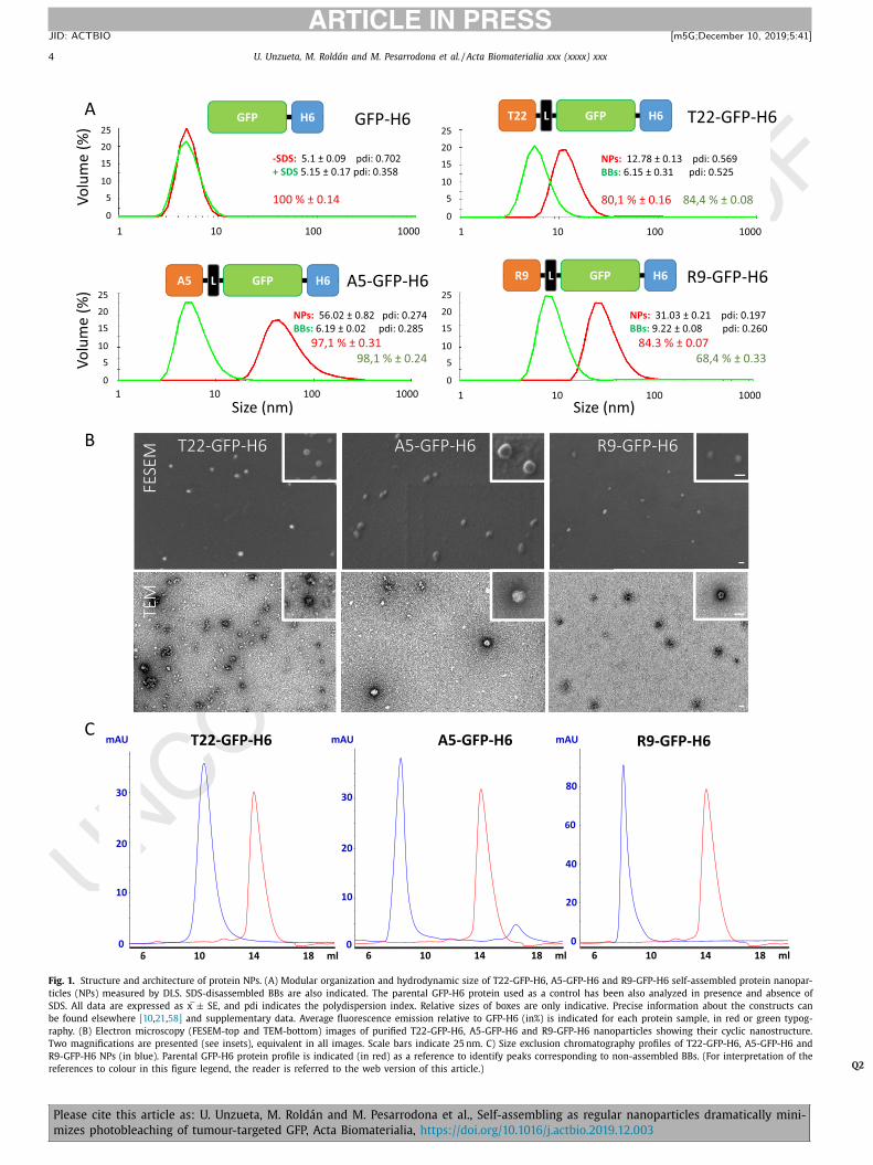
Fig. 1. Structure and architecture of protein NPs. (A) Modular organization and hydrodynamic size of T22-GFP-H6, A5-GFP-H6 and R9-GFP-H6 self-assembled protein nanopar-

ticles (NPs) measured by DLS. SDS-disassembled BBs are also indicated. The parental GFP-H6 protein used as a control has been also analyzed in presence and absence of

SDS. All data are expressed as x ± SE, and pdi indicates the polydispersion index. Relative sizes of boxes are only indicative. Precise information about the constructs can

be found elsewhere [10 , 21 , 58] and supplementary data. Average fluorescence emission relative to GFP-H6 (in%) is indicated for each protein sample, in red or green typog-

raphy. (B) Electron microscopy (FESEM-top and TEM-bottom) images of purified T22-GFP-H6, A5-GFP-H6 and R9-GFP-H6 nanoparticles showing their cyclic nanostructure.

Two magnifications are presented (see insets), equivalent in all images. Scale bars indicate 25 nm. C) Size exclusion chromatography profiles of T22-GFP-H6, A5-GFP-H6 and

R9-GFP-H6 NPs (in blue). Parental GFP-H6 protein profile is indicated (in red) as a reference to identify peaks corresponding to non-assembled BBs. (For interpretation of the

references to colour in this figure legend, the reader is referred to the web version of this article.) Q2

Please cite this article as: U. Unzueta, M. Roldán and M. Pesarrodona et al., Self-assembling as regular nanoparticles dramatically mini-

mizes photobleaching of tumour-targeted GFP, Acta Biomaterialia, https://doi.org/10.1016/j.actbio.2019.12.003

U. Unzueta, M. Roldán and M. Pesarrodona et al. / Acta Biomaterialia xxx (xxxx) xxx 5

ARTICLE IN PRESS

JID: ACTBIO [m5G; December 10, 2019;5:41 ]

NPsBBsGFP-H6

T22-GFP-H6

0 100 200 300

100

80

60

40

20

00 100 200 300 0 100 200 300

A5-GFP-H6 R9-GFP-H6

50% **

*

* 75%**

**

NPs BBs NPs BBs NPs BBsT22-GFP-H6 A5-GFP-H6 R9-GFP-H6GFP-H6

NPs BBs NPs BBs NPs BBsT22-GFP-H6 A5-GFP-H6 R9-GFP-H6GFP-H6

Time (sec)

)%(

ecnecseroulF

60

40

20

0

Tim

e (s

ec)

200

150

100

50

0Ti

me

(sec

)

**

A

B

Fig. 2. Protein nanoparticles photostability. (A) Photobleaching curves for T22-GFP-H6, A5-GFP-H6 and R9-GFP-H6 NPs and BBs. GFP-H6 is added as a control in all the

measures. Data was recorded by confocal laser microscopy upon excitation at 488 nm by a 20 mW argon laser during subsequent frames. Orange dashed lines indicate 50%

of photobleaching while pink dashed lines indicate 75% of photobleaching. The initial intensities of fluorescence were assumed as 100%. B) Graphical quantification of 50%

(left) and 75% (right) photobleaching times for different protein samples. Red dashed line indicates photobleaching times corresponding to control GFP-H6 protein. ∗ indicates

p < 0.01 and ∗∗ indicates p < 0.001. (For interpretation of the references to colour in this figure legend, the reader is referred to the web version of this article.)

GFP-H6 BBs NPs BBs NPs BBs NPs0 50 100 150 200 250 300 350

3500

3000

2500

2000

1500

1000

500

0

120

100

80

60

40

20

0

A B

Time (sec)

ecnecseroulFun

its.

adjR2:- GFP-H6: 0.9671- T22-GFP-H6(NPs): 0.9751- T22-GFP-H6(BBs): 0.9619- A5-GFP-H6 (NPs): 0.9879- A5-GFP-H6 (BBs): 0.9836- R9-GFP-H6 (NPs): 0.8013- R9-GFP-H6 (BBs): 0.7322

** ** **

**

T22-GFP-H6 A5-GFP-H6 R9-GFP-H6

FDH

M

Fig. 3. Nanoparticles fluorescence decay analysis. (A) Double exponential fitting of parental GFP-H6 fluorescence decay analyzed at pixel level. Average adjusted R-squared

for all protein samples are indicated in the inset as goodness of fit parameter. (B) Graphical quantification of Full Duration at Half Maximum (FDHM) for different protein

samples analyzed at pixel level. ∗∗ indicates p < 0.001. (For interpretation of the references to colour in this figure legend, the reader is referred to the web version of this

article.)

increased photostability of the nanostructured forms of GFP was 243

fully supported by the longer time periods required to reach both 244

50 % and 75 % of fluorescence reduction, compared to SDS-generated 245

BBs, with statistically significant differences ( Fig. 2 B). At that point 246

and to exclude any protocol-related protein mixing problems, BBs 247

and NPs of the model T22-GFP-H6, immobilized in polyacrylamide, 248

were also analyzed, rendering results that fully validated our previ- 249

ous data (Supplementary Fig. 2). Also, the impact of divalent cation 250

that coordinate the assembling of NPs or the presence of SDS 251

over the photostability of the samples was discarded by comparing 252

EDTA-generated BBs (in which divalent cations have been removed 253

by EDTA and no SDS was present) with SDS-generated BBs (which 254

still contained divalent cations and SDS was also present). In this 255

context, no significant differences were observed with control GFP- 256

H6 (Supplementary Figure 3). 257

Then, in order to further study the fluorescence decay of our 258

protein samples, pixel intensity data were fitted against different 259

exponential decay models, including single exponential (a ∗exp(- 260

b ∗t)), stretched exponential (a ∗exp(-b ∗x c)) or double exponential 261

(a ∗exp(-b ∗x) + c ∗exp(-d

∗x) + e), being the double exponential decay 262

Please cite this article as: U. Unzueta, M. Roldán and M. Pesarrodona et al., Self-assembling as regular nanoparticles dramatically mini-

mizes photobleaching of tumour-targeted GFP, Acta Biomaterialia, https://doi.org/10.1016/j.actbio.2019.12.003

6 U. Unzueta, M. Roldán and M. Pesarrodona et al. / Acta Biomaterialia xxx (xxxx) xxx

ARTICLE IN PRESS

JID: ACTBIO [m5G; December 10, 2019;5:41 ]

T22-GFP-H6 NPs T22-GFP-H6 BBs

A5-GFP-H6 NPs A5-GFP-H6 BBs

Q (nm-1)0.10 0.15 0.20 0.25 0.30 0.35 0.40 0.45

Q (nm-1)

2.15

1.85

1.45

Log

(I)

2.00

1.70

1.30

Log

(I)

GFP-H6A

BQ (nm-1)

Log

(I)

Time (sec)

15101520

Time (sec)

15101520

Time (sec)

15101520

Time (sec)

210203040

Time (sec)

210203040

2.00

1.70

1.30

2.15

1.85

1.45

0.10 0.15 0.20 0.25 0.30 0.35 0.40 0.45

0.10 0.15 0.20 0.25 0.30 0.35 0.40 0.45

1.9

1.60

1.30

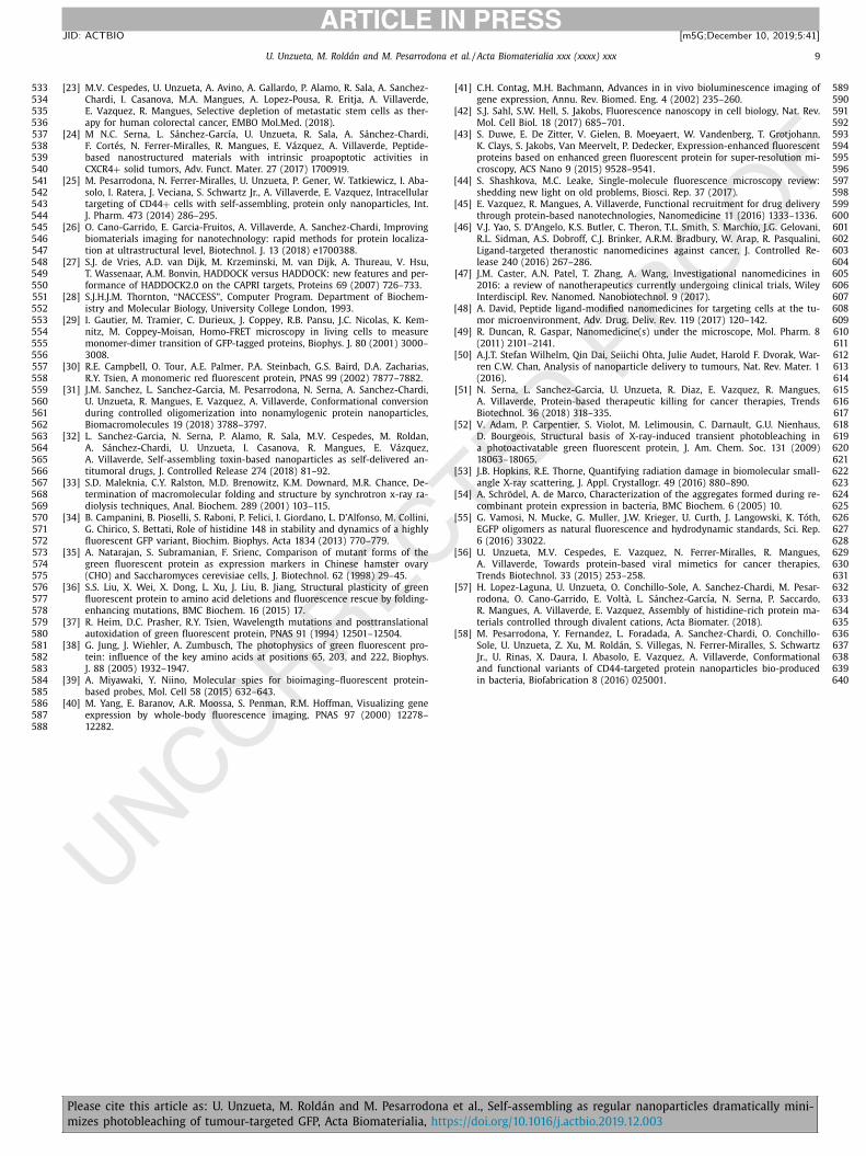
Fig. 4. Structural stability of protein nanoparticles. Radiation damage over (A) control GFP-H6 protein or over (B) T22-GFP-H6 and A5-GFP-H6 protein nanoparticles (NPs) and

building blocks (BBs) measured by progressive changes (indicated between red dashed line) observed over Small Angle X-ray scattering curves upon exposition at 12.4 keV

light source during subsequent frames. (For interpretation of the references to colour in this figure legend, the reader is referred to the web version of this article.)

Table 1

Comparative photostability (shown as enhancement factor) of engineered GFP pro-

teins.

GFP-H6 BB

BB a NP BB NP

T22-GFP-H6 1.06 7.72 – 5.42

A5-GFP-H6 1.08 2.54 – 2.34

R9-GFP-H6 2.85 5.49 – 1.93

a Fold reduction of 50 % photobleaching in NPs compared to either the parental

GFP-H6 (GFP-H6 column) or to the BB version of each protein (BB column) upon

SDS-mediated disassembly.

the one better describing the fluorescence extinction process 263

( Fig. 3 A). At this point, and following the selected decay model, 264

pixelwise analysis of Full Duration at Half Maximum (FDHM) for 265

all GFP oligomers and SDS-generated BBs completely supported 266

previously observed significant differences. Solutions of GFP NPs 267

were significantly more photostable than those of parental GFP-H6, 268

which showed similar fluorescence decay rate as in SDS-generated 269

BBs. An exception was R9-GFP-H6 BBs, which being still partially 270

organized as 9 nm supramolecular structures did not completely 271

reached the low photostability of the parental GFP-H6 ( Fig. 3 B). 272

In a recent study [31] , biophysical analyses of related pro- 273

tein NPs (in particular T22-DITOX-H6 and T22-GFP-H6, [32] ) were 274

suggestive of an enhanced structural stability of the oligomeric 275

versions of such proteins, measured through resistance to ther- 276

mal stress. We wondered if such robustness, presumably acquired 277

through self-assembling, might be related to the photostability de- 278

scribed in the present study. To address this question, we compar- 279

atively analyzed the X-ray radiation damage in SAXS on NP and BB 280

forms of T22-GFP-H6 and A5-GFP-H6, the two proteins in which 281

the disassembling protocol efficiently resulted in BBs. As observed 282

( Fig. 4 ), the assembled forms of both proteins were less vulnera- 283

ble to damage produced by radical modification upon X-ray radia- 284

tion over time than their unassembled counterparts. The detectable 285

alteration of scattering intensities at low angles suggested the 286

propensity of BBs to aggregate upon radiation damage, a common 287

Please cite this article as: U. Unzueta, M. Roldán and M. Pesarrodona et al., Self-assembling as regular nanoparticles dramatically mini-

mizes photobleaching of tumour-targeted GFP, Acta Biomaterialia, https://doi.org/10.1016/j.actbio.2019.12.003

U. Unzueta, M. Roldán and M. Pesarrodona et al. / Acta Biomaterialia xxx (xxxx) xxx 7

ARTICLE IN PRESS

JID: ACTBIO [m5G; December 10, 2019;5:41 ]

Fig. 5. BB interfaces in T22-GFP-H6 NPs. Two contiguous T22-GFP-H6 monomers of

T22-GFP-H6 NPs are depicted in transparent surface representation of an in silico

modelled NP. Interface residues (those supporting cross-molecular BB contacts) are

presented as sticks, and residues known to affect the fluorescence of the protein

are shown as spheres. Note the interaction and global proximity between them. In

the inset, an overview of the full NP.

structural alteration catalyzed by amino acid modification though 288

OH radicals [33] . Under the same radiation dose, the nanostruc- 289

tured GFP did not undergo observable macromolecular aggrega- 290

tion. In this sense, in our nanoparticle model, regions susceptible 291

to radiation damage (with high content on Cys, Met, Phe, Tyr, Trp, 292

Pro, His and Leu) are protected from solvent upon oligomeriza- 293

tion (Supplementary Table 1). These data confirm that oligomeric 294

forms of the protein are less sensitive to radiation stresses than the 295

unassembled versions probably due to a reduced solvent exposure 296

of oxygen-reactive amino acids. Also in this context, the analysis 297

of T22-GFP-H6 model ( Fig. 5 ) shows that the region of interaction 298

between the monomers is very close and contain several amino 299

acids described to be relevant in the generation of fluorescence. It 300

is known that mutations in His 148, Met154, Val164, Ile167, Ser202, 301

Thr 203 and Glu222 (marked as spheres in Fig. 5 ) induce changes 302

in GFP fluorescence [34–38] . Therefore, this fact points out that 303

NP formation modifies the local environment of these amino acids 304

that are important for the GFP chromophore formation, and there- 305

fore, self-assembling impacts on its stability under intense light ir- 306

radiation. 307

4. Discussion 308

Fluorescent proteins are exploited as molecular probes in 309

bioimaging [1 , 39] to report gene expression [40 , 41] , to dissect 310

intracellular structures [42] and to determine intracellular lo- 311

calization and interactions at single molecule resolution [43 , 44] . 312

They are also promising to develop protein-based NPs for cell- 313

targeted drug delivery in cancer, as they can be incorporated 314

in the BBs for theranostic purposes [45] . In this context, NPs 315

are usually functionalized with specific ligands of cell surface 316

receptors for cell-targeted drug delivery [46–48] . However, the 317

applicability of nanostructured vehicles as drug carriers has been 318

so far compromised by an only limited success in reaching a good 319

biodistribution of the drug. In most cases, only around 1 % of the 320

administered material accumulates in the target site, while the 321

rest distributes among healthy tissues [49 , 50] . This landscape is 322

not sufficient to enhance the therapeutic index of a free cytotoxic 323

compound, such as those used in oncotherapy [51] , and it is also 324

insufficient to fulfil the requirements of a precise theranostic 325

agent. In recent studies we have engineered a peptide-tagged 326

recombinant GFP, that self-assembles as NPs of 12 nm [20 , 22] , 327

to successfully deliver small molecular weight drugs [23] and 328

human pro-apoptotic factors [24] to metastatic cancer stem cells, 329

in colorectal cancer animal models. The biodistribution of this 330

material, as determined by fluorescence detection, is very selective 331

and it fulfil s the high standards required for both, drug delivery 332

and imaging [10] . The protein, in an assembled and fluorescent 333

form, is architectonically stable in circulation (Supplementary 334

Figure 4) [11] and is only found inside target cells, either in the 335

main tumour or in metastatic foci [10] , but not in normal organs. 336

Therefore, these nanostructured GFP show enormous potential 337

in preclinical drug trials and in diagnostic formulations, both in 338

cell culture and in vivo since targeted fluorescent NPs can be 339

used to specifically deliver fluorescent proteins and conjugated 340

drugs into target cells for precision imaging and theranosis. This 341

fact prompted us to investigate the photostability of the protein 342

fluorophore when GFP is organized as regular oligomeric NPs. 343

A self-assembling GFP, forming NPs of 12 nm in size, has been 344

engineered to target CXCR4 + tumour cells by the fusion to a 345

CXCR4-binding peptide, namely T22. This nanostructured complex, 346

biodistributes with exquisite precision upon intravenous injection 347

in colorectal cancer mouse models, in which more than 85% of 348

total detected fluorescence is accumulated specifically in tumoral 349

tissues (including small metastatic foci) [10 , 11] . This construct and 350

derivatives are, therefore, extremely efficient for theranosis [23 , 24] . 351

Then, we wanted to determine their photostability compared to 352

regular GFP versions to evaluate if the controlled oligomerization 353

might have a positive effect in the robustness of fluorescence emis- 354

sion. We initially anticipated a decrease in the specific fluores- 355

cence, since in the NPs, the close GFP-GFP contacts [20] could 356

cause substantial self-quenching. This has been recently demon- 357

strated in GFP-loaded HBV-like NPs, where the distance between 358

the internally localized GFP molecules strongly influenced their flu- 359

orescence signal [5] . However, in the current platform, the GFP flu- 360

orescence was not affected by self-assembling, as the engineered 361

GFP variants were equally emitting as NPs or BBs ( Fig. 1 A). The fu- 362

sion of a cationic peptide at the amino terminus of GFP had only a 363

moderate impact on fluorescence, which was reduced up to about 364

70 –90 % of the parental GFP-H6 ( Fig. 1 A). In addition, the pH of 365

the physiological media had no significant influence over both, the 366

self-assembling and specific fluorescence of the protein samples, 367

and they were also architectonically stable in human serum. This 368

is important when protein NPs are designed for in-vivo imaging 369

(Supplementary Figs. 1 and 4). Importantly, photobleaching was 370

dramatically reduced in oligomerized GFP compared to BB ver- 371

sions ( Figs. 2 and 3 ), stressing the unanticipated positive impact 372

of self-assembling in keeping the stability of the emitted fluores- 373

cence. Data also suggest that the observed fluorophore stability un- 374

der laser irradiation is related to a globally enhanced protein struc- 375

tural stability, since the radiation damage over NP forms of GFP is 376

clearly reduced over the parental unassembled GFP-H6 or the BB 377

versions ( Fig. 4 ). In this context, both synchrotron X-ray beams and 378

laser light induce photodamage to structurally similar fluorescent 379

proteins resulting in loss of absorbance and fluorescence, a fact 380

known to be influenced by the chromophore’s local environment 381

[52] . As an additional observation, the molecular weight of the tar- 382

get protein or protein complex influences the radiation damage in 383

small-angle X-ray scattering with a protective effect [53] . In this 384

context, recombinant GFP shows a tendency to uncontrolled multi- 385

merization and aggregation [54] and some oligomerizing GFP vari- 386

ants have shown affected fluorescence, photobleaching and blink- 387

ing, depending on oligomer size [55] . It is well known that several 388

residues involved in the chromophoric properties of GFP are close 389

to the GFP dimer interface, which has been postulated as one of 390

the contact sites between GFP BBs to form toroid NPs as those de- 391

scribed here [20] . These observations and concepts would account 392

for the particular differences observed in NPs performance and 393

stability of fluorescent NPs versus GFP monomers or dimeric BBs 394

Please cite this article as: U. Unzueta, M. Roldán and M. Pesarrodona et al., Self-assembling as regular nanoparticles dramatically mini-

mizes photobleaching of tumour-targeted GFP, Acta Biomaterialia, https://doi.org/10.1016/j.actbio.2019.12.003

8 U. Unzueta, M. Roldán and M. Pesarrodona et al. / Acta Biomaterialia xxx (xxxx) xxx

ARTICLE IN PRESS

JID: ACTBIO [m5G; December 10, 2019;5:41 ]

under electromagnetic radiation. Importantly, in the in silico 395

models of T22-GFP-H6 NPs, that perfectly fit the morphometric 396

nanoscale evaluation of the material [20 , 22] , the GFP residues in- 397

volved in fluorescence participate or are in close proximity to the 398

BB interfaces ( Fig. 5 ). This observation accounts for the enhanced 399

stability of GFP promoted by its controlled oligomerization in form 400

of NPs. 401

5. Conclusions 402

The administration of oligomeric GFP as cell-targeted NPs in 403

cancer imaging, therapy and theranostic contexts, benefits from 404

a multiplexed presentation of the tumoral cell ligand (here mod- 405

elled by T22, A5 or R9), what favours cell internalization [56] . 406

The nanoscale architecture of GFP also results in a highly selec- 407

tive biodistribution upon systemic administration, as it allows es- 408

caping from renal filtration [11] and the identification of small 409

metastatic foci because of the longer circulation time [10] . We have 410

here proved that in addition, the nanostructure of GFP NPs, easily 411

regulatable by the manipulation of divalent cations [57] , enhances 412

the structural stability and photostability of the system. Therefore, 413

being photobleaching a major obstacle in the application of GFP 414

and other fluorescent proteins in imaging and theranosis, the con- 415

trolled oligomerization of fluorescent proteins appears as a power- 416

ful method to enhance their photostability for their use in preci- 417

sion imaging and therapy. 418

Declaration of Competing Interest 419

The authors declare that they have no known competing finan- 420

cial interests or personal relationships that could have appeared to 421

influence the work reported in this paper. 422

CRediT authorship contribution statement 423

Ugutz Unzueta : Writing - original draft, Writing - review & 424

editing. Mònica Roldán : Writing - original draft, Writing - review 425

& editing. Mireia Pesarrodona : Writing - original draft, Writing 426

- review & editing. Raul Benitez : Writing - original draft, Writ- 427

ing - review & editing. Alejandro Sánchez-Chardi : Writing - orig- 428

inal draft, Writing - review & editing. Oscar Conchillo-Solé: Writ- 429

ing - original draft, Writing - review & editing. Ramón Mangues : 430

Writing - original draft, Writing - review & editing. Antonio 431

Villaverde : Writing - original draft, Writing - review & editing. 432

Esther Vázquez : Writing - original draft, Writing - review & edit- 433

ing. 434

Acknowledgments 435

This study has been funded by the Agencia Estatal de Investi- 436

gación (AEI) and Fondo Europeo de Desarrollo Regional (FEDER) 437

(grant BIO2016-76063-R , AEI/FEDER, UE ), AGAUR ( 2017SGR- 438

229 ) and CIBER-BBN (project VENOM4CANCER) granted to 439

AV, ISCIII ( PI15/00272 co-founding FEDER) granted to EV and 440

CIBER-BBN (project NANOSCAPE) granted to UUE. Protein pro- 441

duction and DLS measurements have been partially performed 442

by the ICTS “NANBIOSIS”, more specifically protein production 443

by the Protein Production Platform of CIBER-BBN/IBB ( http: 4 4 4

//www.nanbiosis.es/unit/u1- protein- production- platform- ppp/ ) 445

and nanoparticle size analysis by the Biomaterial Processing 446

and Nanostructuring Unit ( http://www.nanbiosis.es/portfolio/ 447

u6- biomaterial- processing- and- nanostructuring- unit/ ). We are 448

indebted to the Servei de Microscòpia of the UAB for excellent 449

technical service. UU is supported by PERIS program from the 450

health department of la Generalitat de Cataluña. R.B. was sup- 451

ported by research grant SAF2017-8819-C3 from the Spanish 452

Ministry of Science Innovation and Universities. AV received an

Q3 453

ICREA ACADEMIA award. EV, RM and AV are co-founders of NANO- 454

LIGENT, devoted to develop antitumoral drugs based on proteins. 455

Supplementary materials 456

Supplementary material associated with this article can be 457

found, in the online version, at doi: 10.1016/j.actbio.2019.12.003 . 458

References 459

[1] E.A. Rodriguez, R.E. Campbell, J.Y. Lin, M.Z. Lin, A. Miyawaki, A.E. Palmer, 460 X. Shu, J. Zhang, R.Y. Tsien, The growing and glowing toolbox of fluorescent 461 and photoactive proteins, Trends Biochem. Sci. 42 (2017) 111–129. 462

[2] R.M. Hoffman, Application of GFP imaging in cancer, Lab. Invest. 95 (2015) 463 432–452. 464

[3] V. Ntziachristos, J.S. Yoo, G.M. van Dam, Current concepts and future perspec- 465 tives on surgical optical imaging in cancer, J. Biomed. Opt. 15 (2010) 066024. 466

[4] R.M. Hoffman, The multiple uses of fluorescent proteins to visualize cancer in 467 vivo, Nat. Rev. Cancer 5 (2005) 796–806. 468

[5] S.E. Kim, S.D. Jo, K.C. Kwon, Y.Y. Won, J. Lee, Genetic assembly of double- 469 layered fluorescent protein nanoparticles for cancer targeting and imaging, 470 Adv. Sci. 4 (2017) 1600471. 471

[6] S. Yano, K. Takehara, S. Miwa, H. Kishimoto, H. Tazawa, Y. Urata, S. Ka- 472 gawa, M. Bouvet, T. Fujiwara, R. Hoffman, Fluorescence-guided surgery of a 473 highly-metastatic variant of human triple-negative breast cancer targeted with 474 a cancer-specific GFP adenovirus prevents recurrence, Oncotarget 7 (2016) 475 75635–75647. 476

[7] C.A. Metildi, S. Kaushal, C.S. Snyder, R.M. Hoffman, M. Bouvet, Fluorescence- 477 guided surgery of human colon cancer increases complete resection resulting 478 in cures in an orthotopic nude mouse model, J. Surg. Res. 179 (2013) 87–93. 479

[8] S. Miyamoto, S. Sperry, T. Yamashita, N.P. Reddy, B.W. O’Malley Jr., D. Li, Molec- 480 ular imaging assisted surgery improves survival in a murine head and neck 481 cancer model, Int. J. Cancer 131 (2012) 1235–1242. 482

[9] J. Lippincott-Schwartz, G.H. Patterson, Development and use of fluorescent 483 protein markers in living cells, Science 300 (2003) 87–91. 484

[10] U. Unzueta, M.V. Cespedes, N. Ferrer-Miralles, I. Casanova, J. Cedano, 485 J.L. Corchero, J. Domingo-Espín, A. Villaverde, R. Mangues, E. Vazquez, Intracel- 486 lular CXCR4( + ) cell targeting with T22-empowered protein-only nanoparticles, 487 Int. J. Nanomed. 7 (2012) 4533–4544. 488

[11] M.V. Cespedes, U. Unzueta, W. Tatkiewicz, A. Sanchez-Chardi, O. Conchillo-Sole, 489 P. Alamo, Z. Xu, I. Casanova, J.L. Corchero, M. Pesarrodona, J. Cedano, X. Daura, 490 I. Ratera, J. Veciana, N. Ferrer-Miralles, E. Vazquez, A. Villaverde, R. Mangues, 491 In vivo architectonic stability of fully de novo designed protein-only nanopar- 492 ticles, ACS Nano 8 (2014) 4166–4176. 493

[12] M.V. Cespedes, U. Unzueta, P. Alamo, A. Gallardo, R. Sala, I. Casanova, 494 M.A. Pavon, M.A. Mangues, M. Trias, A. Lopez-Pousa, A. Villaverde, E. Vazquez, 495 R. Mangues, Cancer-specific uptake of a liganded protein nanocarrier targeting 496 aggressive CXCR4 + colorectal cancer models, Nanomedicine 12 (2016) 1987– 497 1996. 498

[13] A .V. Mamontova, A .P. Grigoryev, A .S. Tsarkova, K.A .L.A .M. Bogdanov, Strug- 499 gle for Photostability: Bleaching Mechanisms of Fluorescent Proteins, Russ. J. 500 Bioorg. Chem + 43 (2017) 9. 501

[14] C. Zhang, J.B. Konopka, A photostable green fluorescent protein variant for 502 analysis of protein localization in Candida albicans, Eukaryot. Cell. 9 (2010) 503 224–226. 504

[15] B.T. Bajar, E.S. Wang, A.J. Lam, B.B. Kim, C.L. Jacobs, E.S. Howe, M.W. Davidson, 505 M.Z. Lin, J. Chu, Improving brightness and photostability of green and red flu- 506 orescent proteins for live cell imaging and FRET reporting, Sci. Rep. 6 (2016) 507 20889. 508

[16] A .M. Bogdanov, E.A . Bogdanova, D.M. Chudakov, T.V. Gorodnicheva, 509 S. Lukyanov, K.A. Lukyanov, Cell culture medium affects GFP photostabil- 510 ity: a solution, Nat. Methods 6 (2009) 859–860. 511

[17] C. Kiss, J. Temirov, L. Chasteen, G.S. Waldo, A.R. Bradbury, Directed evolution of 512 an extremely stable fluorescent protein, Protein Eng. Design Select 22 (2009) 513 313–323. 514

[18] Y. Fu, J. Zhang, J.R. Lakowicz, Metal-enhanced fluorescence of single green flu- 515 orescent protein (GFP), Biochem. Biophys. Res. Commun. 376 (2008) 712–717. 516

[19] A .V. Mamontova, A .M. Bogdanov, K.A . Lukyanov, Influence of cell growth con- 517 ditions and medium composition on EGFP photostability in live cells, BioTech- 518 niques 58 (2015) 258–261. 519

[20] F. Rueda, M.V. Cespedes, O. Conchillo-Sole, A. Sanchez-Chardi, J. Seras- 520 Franzoso, R. Cubarsi, A. Gallardo, M. Pesarrodona, N. Ferrer-Miralles, X. Daura, 521 E. Vazquez, E. Garcia-Fruitos, R. Mangues, U. Unzueta, A. Villaverde, Bottom- 522 Up instructive quality control in the biofabrication of smart protein materials, 523 Adv. Mater. 27 (2015) 7816–7822. 524

[21] M.T. de Pinho Favaro, N. Serna, L. Sanchez-Garcia, R. Cubarsi, M. Roldan, 525 A. Sanchez-Chardi, U. Unzueta, R. Mangues, N. Ferrer-Miralles, A.R. Azzoni, 526 E. Vazquez, A. Villaverde, Switching cell penetrating and CXCR4-binding ac- 527 tivities of nanoscale-organized arginine-rich peptides, Nanomedicine (2018). 528

[22] M. Pesarrodona, E. Crosas, R. Cubarsi, A. Sanchez-Chardi, P. Saccardo, U. Un- 529 zueta, F. Rueda, L. Sanchez-Garcia, N. Serna, R. Mangues, N. Ferrer-Miralles, 530 E. Vazquez, A. Villaverde, Intrinsic functional and architectonic heterogeneity 531 of tumor-targeted protein nanoparticles, Nanoscale 9 (2017) 6427–6435. 532

Please cite this article as: U. Unzueta, M. Roldán and M. Pesarrodona et al., Self-assembling as regular nanoparticles dramatically mini-

mizes photobleaching of tumour-targeted GFP, Acta Biomaterialia, https://doi.org/10.1016/j.actbio.2019.12.003

U. Unzueta, M. Roldán and M. Pesarrodona et al. / Acta Biomaterialia xxx (xxxx) xxx 9

ARTICLE IN PRESS

JID: ACTBIO [m5G; December 10, 2019;5:41 ]

[23] M.V. Cespedes, U. Unzueta, A. Avino, A. Gallardo, P. Alamo, R. Sala, A. Sanchez- 533 Chardi, I. Casanova, M.A. Mangues, A. Lopez-Pousa, R. Eritja, A. Villaverde, 534 E. Vazquez, R. Mangues, Selective depletion of metastatic stem cells as ther- 535 apy for human colorectal cancer, EMBO Mol.Med. (2018). 536

[24] M N.C. Serna, L. Sánchez-García, U. Unzueta, R. Sala, A. Sánchez-Chardi, 537 F. Cortés, N. Ferrer-Miralles, R. Mangues, E. Vázquez, A. Villaverde, Peptide- 538 based nanostructured materials with intrinsic proapoptotic activities in 539 CXCR4 + solid tumors, Adv. Funct. Mater. 27 (2017) 1700919. 540

[25] M. Pesarrodona, N. Ferrer-Miralles, U. Unzueta, P. Gener, W. Tatkiewicz, I. Aba- 541 solo, I. Ratera, J. Veciana, S. Schwartz Jr., A. Villaverde, E. Vazquez, Intracellular 542 targeting of CD44 + cells with self-assembling, protein only nanoparticles, Int. 543 J. Pharm. 473 (2014) 286–295. 544

[26] O. Cano-Garrido, E. Garcia-Fruitos, A. Villaverde, A. Sanchez-Chardi, Improving 545 biomaterials imaging for nanotechnology: rapid methods for protein localiza- 546 tion at ultrastructural level, Biotechnol. J. 13 (2018) e1700388. 547

[27] S.J. de Vries, A.D. van Dijk, M. Krzeminski, M. van Dijk, A. Thureau, V. Hsu, 548 T. Wassenaar, A.M. Bonvin, HADDOCK versus HADDOCK: new features and per- 549 formance of HADDOCK2.0 on the CAPRI targets, Proteins 69 (2007) 726–733. 550

[28] S.J.H.J.M. Thornton, “NACCESS”, Computer Program. Department of Biochem- 551 istry and Molecular Biology, University College London, 1993. 552

[29] I. Gautier, M. Tramier, C. Durieux, J. Coppey, R.B. Pansu, J.C. Nicolas, K. Kem- 553 nitz, M. Coppey-Moisan, Homo-FRET microscopy in living cells to measure 554 monomer-dimer transition of GFP-tagged proteins, Biophys. J. 80 (2001) 3000–555 3008. 556

[30] R.E. Campbell, O. Tour, A.E. Palmer, P.A. Steinbach, G.S. Baird, D.A. Zacharias, 557 R.Y. Tsien, A monomeric red fluorescent protein, PNAS 99 (2002) 7877–7882. 558

[31] J.M. Sanchez, L. Sanchez-Garcia, M. Pesarrodona, N. Serna, A. Sanchez-Chardi, 559 U. Unzueta, R. Mangues, E. Vazquez, A. Villaverde, Conformational conversion 560 during controlled oligomerization into nonamylogenic protein nanoparticles, 561 Biomacromolecules 19 (2018) 3788–3797. 562

[32] L. Sanchez-Garcia, N. Serna, P. Alamo, R. Sala, M.V. Cespedes, M. Roldan, 563 A. Sánchez-Chardi, U. Unzueta, I. Casanova, R. Mangues, E. Vázquez, 564 A. Villaverde, Self-assembling toxin-based nanoparticles as self-delivered an- 565 titumoral drugs, J. Controlled Release 274 (2018) 81–92. 566

[33] S.D. Maleknia, C.Y. Ralston, M.D. Brenowitz, K.M. Downard, M.R. Chance, De- 567 termination of macromolecular folding and structure by synchrotron x-ray ra- 568 diolysis techniques, Anal. Biochem. 289 (2001) 103–115. 569

[34] B. Campanini, B. Pioselli, S. Raboni, P. Felici, I. Giordano, L. D’Alfonso, M. Collini, 570 G. Chirico, S. Bettati, Role of histidine 148 in stability and dynamics of a highly 571 fluorescent GFP variant, Biochim. Biophys. Acta 1834 (2013) 770–779. 572

[35] A. Natarajan, S. Subramanian, F. Srienc, Comparison of mutant forms of the 573 green fluorescent protein as expression markers in Chinese hamster ovary 574 (CHO) and Saccharomyces cerevisiae cells, J. Biotechnol. 62 (1998) 29–45. 575

[36] S.S. Liu, X. Wei, X. Dong, L. Xu, J. Liu, B. Jiang, Structural plasticity of green 576 fluorescent protein to amino acid deletions and fluorescence rescue by folding- 577 enhancing mutations, BMC Biochem. 16 (2015) 17. 578

[37] R. Heim, D.C. Prasher, R.Y. Tsien, Wavelength mutations and posttranslational 579 autoxidation of green fluorescent protein, PNAS 91 (1994) 12501–12504. 580

[38] G. Jung, J. Wiehler, A. Zumbusch, The photophysics of green fluorescent pro- 581 tein: influence of the key amino acids at positions 65, 203, and 222, Biophys. 582 J. 88 (2005) 1932–1947. 583

[39] A. Miyawaki, Y. Niino, Molecular spies for bioimaging–fluorescent protein- 584 based probes, Mol. Cell 58 (2015) 632–643. 585

[40] M. Yang, E. Baranov, A.R. Moossa, S. Penman, R.M. Hoffman, Visualizing gene 586 expression by whole-body fluorescence imaging, PNAS 97 (20 0 0) 12278–587 12282. 588

[41] C.H. Contag, M.H. Bachmann, Advances in in vivo bioluminescence imaging of 589 gene expression, Annu. Rev. Biomed. Eng. 4 (2002) 235–260. 590

[42] S.J. Sahl, S.W. Hell, S. Jakobs, Fluorescence nanoscopy in cell biology, Nat. Rev. 591 Mol. Cell Biol. 18 (2017) 685–701. 592

[43] S. Duwe, E. De Zitter, V. Gielen, B. Moeyaert, W. Vandenberg, T. Grotjohann, 593 K. Clays, S. Jakobs, Van Meervelt, P. Dedecker, Expression-enhanced fluorescent 594 proteins based on enhanced green fluorescent protein for super-resolution mi- 595 croscopy, ACS Nano 9 (2015) 9528–9541. 596

[44] S. Shashkova, M.C. Leake, Single-molecule fluorescence microscopy review: 597 shedding new light on old problems, Biosci. Rep. 37 (2017). 598

[45] E. Vazquez, R. Mangues, A. Villaverde, Functional recruitment for drug delivery 599 through protein-based nanotechnologies, Nanomedicine 11 (2016) 1333–1336. 600

[46] V.J. Yao, S. D’Angelo, K.S. Butler, C. Theron, T.L. Smith, S. Marchio, J.G. Gelovani, 601 R.L. Sidman, A.S. Dobroff, C.J. Brinker, A.R.M. Bradbury, W. Arap, R. Pasqualini, 602 Ligand-targeted theranostic nanomedicines against cancer, J. Controlled Re- 603 lease 240 (2016) 267–286. 604

[47] J.M. Caster, A.N. Patel, T. Zhang, A. Wang, Investigational nanomedicines in 605 2016: a review of nanotherapeutics currently undergoing clinical trials, Wiley 606 Interdiscipl. Rev. Nanomed. Nanobiotechnol. 9 (2017). 607

[48] A. David, Peptide ligand-modified nanomedicines for targeting cells at the tu- 608 mor microenvironment, Adv. Drug. Deliv. Rev. 119 (2017) 120–142. 609

[49] R. Duncan, R. Gaspar, Nanomedicine(s) under the microscope, Mol. Pharm. 8 610 (2011) 2101–2141. 611

[50] A.J.T. Stefan Wilhelm, Qin Dai, Seiichi Ohta, Julie Audet, Harold F. Dvorak, War- 612 ren C.W. Chan, Analysis of nanoparticle delivery to tumours, Nat. Rev. Mater. 1 613 (2016). 614

[51] N. Serna, L. Sanchez-Garcia, U. Unzueta, R. Diaz, E. Vazquez, R. Mangues, 615 A. Villaverde, Protein-based therapeutic killing for cancer therapies, Trends 616 Biotechnol. 36 (2018) 318–335. 617

[52] V. Adam, P. Carpentier, S. Violot, M. Lelimousin, C. Darnault, G.U. Nienhaus, 618 D. Bourgeois, Structural basis of X-ray-induced transient photobleaching in 619 a photoactivatable green fluorescent protein, J. Am. Chem. Soc. 131 (2009) 620 18063–18065. 621

[53] J.B. Hopkins, R.E. Thorne, Quantifying radiation damage in biomolecular small- 622 angle X-ray scattering, J. Appl. Crystallogr. 49 (2016) 880–890. 623

[54] A . Schrödel, A . de Marco, Characterization of the aggregates formed during re- 624 combinant protein expression in bacteria, BMC Biochem. 6 (2005) 10. 625

[55] G. Vamosi, N. Mucke, G. Muller, J.W. Krieger, U. Curth, J. Langowski, K. Tóth, 626 EGFP oligomers as natural fluorescence and hydrodynamic standards, Sci. Rep. 627 6 (2016) 33022. 628

[56] U. Unzueta, M.V. Cespedes, E. Vazquez, N. Ferrer-Miralles, R. Mangues, 629 A. Villaverde, Towards protein-based viral mimetics for cancer therapies, 630 Trends Biotechnol. 33 (2015) 253–258. 631

[57] H. Lopez-Laguna, U. Unzueta, O. Conchillo-Sole, A. Sanchez-Chardi, M. Pesar- 632 rodona, O. Cano-Garrido, E. Voltà, L. Sánchez-García, N. Serna, P. Saccardo, 633 R. Mangues, A. Villaverde, E. Vazquez, Assembly of histidine-rich protein ma- 634 terials controlled through divalent cations, Acta Biomater. (2018). 635

[58] M. Pesarrodona, Y. Fernandez, L. Foradada, A. Sanchez-Chardi, O. Conchillo- 636 Sole, U. Unzueta, Z. Xu, M. Roldán, S. Villegas, N. Ferrer-Miralles, S. Schwartz 637 Jr., U. Rinas, X. Daura, I. Abasolo, E. Vazquez, A. Villaverde, Conformational 638 and functional variants of CD44-targeted protein nanoparticles bio-produced 639 in bacteria, Biofabrication 8 (2016) 025001. 640

Please cite this article as: U. Unzueta, M. Roldán and M. Pesarrodona et al., Self-assembling as regular nanoparticles dramatically mini-

mizes photobleaching of tumour-targeted GFP, Acta Biomaterialia, https://doi.org/10.1016/j.actbio.2019.12.003


Recommended