Decarboxylative alkylation for site-selectivebioconjugation of native proteins via oxidationpotentialsSteven Bloom1†, Chun Liu1†, Dominik K. Kölmel1, Jennifer X. Qiao1,2, Yong Zhang1,2, Michael A. Poss1,2,William R. Ewing1,2 and David W. C. MacMillan1*
The advent of antibody–drug conjugates as pharmaceuticals has fuelled a need for reliable methods of site-selectiveprotein modification that furnish homogeneous adducts. Although bioorthogonal methods that use engineered amino acidsoften provide an elegant solution to the question of selective functionalization, achieving homogeneity using native aminoacids remains a challenge. Here, we explore visible-light-mediated single-electron transfer as a mechanism towardsenabling site- and chemoselective bioconjugation. Specifically, we demonstrate the use of photoredox catalysis as aplatform to selectivity wherein the discrepancy in oxidation potentials between internal versus C-terminal carboxylates canbe exploited towards obtaining C-terminal functionalization exclusively. This oxidation potential-gated technology isamenable to endogenous peptides and has been successfully demonstrated on the protein insulin. As a fundamentally newapproach to bioconjugation this methodology provides a blueprint toward the development of photoredox catalysis as ageneric platform to target other redox-active side chains for native conjugation.
The last two decades have witnessed tremendous growth inresearch to address the challenge of site- and chemoselectiveprotein modification1. Propelled by a high demand for tech-
nologies that furnish homogeneously modified protein adducts,chemical biologists have successfully delivered a number of robustmethods that achieve site-selective protein functionalization viaprotein engineering and the incorporation of non-natural, bio-orthogonal amino acids2. Chief among these methods are ‘click’and Staudinger ligation strategies, where highly uniform productscan be obtained by genetically encoding an azide reporter in asite- and number-specific fashion3,4.
Pre-engineering of the protein scaffold (mutant proteins) hasproven to be an indispensable technology for selective protein modi-fication, but the bioconjugation strategies that harness the aminoacids of wild-type proteins remain elusive, despite their greatappeal. Traditionally, these native-modification methods mainlymake use of 2 of the 20 canonical amino-acid residues—cysteine5,6
and lysine—which incorporate heteroatom lone-pair nucleophiles.However, obtaining homogeneous products in which only a singleresidue at a single site has undergone reaction has proven challen-ging7. For example, selective modification of a specific lysineresidue is difficult given their high abundance on protein surfaces8.One solution has been to target proteins where one lysine residue ismore solvent-accessible to biotinylation than other Lys groups, asdemonstrated by Chen and co-authors9. In a similar vein, thisapproach has been applied by Bader and colleagues for the selectivelipidation of C-terminal cysteines10. More recently, elegant workfrom several groups has addressed this problem by extending thescope of natural amino acids employed in bioconjugation to Ntermini11, tyrosine, tryptophan and methionine residues12–19.These methods take advantage of the inherently low abundance ofthese residues on protein surfaces, thereby achieving a higherdegree of site selectivity.
Over the past several years, our laboratory has employed photo-redox catalysis as a platform for activating native functional groupstowards C–C and C–X bond formation. One such activation modehas focused on the use of naturally abundant carboxylic acids aslatent carbon-centred radicals20. These transient intermediates, gen-erated through single-electron transfer (SET) and subsequent CO2
extrusion, have been shown to undergo successful coupling with awide range of electrophilic partners including Michael acceptors21,vinyl sulfones22 and nickel complexes23. Recently, we questionedwhether this technology could be applied to more complex architec-tures that incorporate multiple carboxylic acids, such as endogenousproteins (Fig. 1). Indeed, carboxylic acids are naturally present inproteins due to their incorporation in aspartate, glutamate andC-terminal residues. Despite the abundance of these carboxylic-acid-bearing residues, we reasoned that the innate difference inoxidation potentials between side-chain alkyl carboxylates (that is,aspartate and glutamate)24 and C-terminal α-amino carboxylates20
should permit a high degree of site selectivity, with decarboxylation–functionalization occurring at the more readily oxidized C terminus.It is also important to note that traditional methods for carboxylicacid bioconjugation typically fall within the realm of amide bondcouplings and esterification with diazo compounds, two technologiesthat often suffer from indiscriminate regioselectivity25–29. In contrast,we reasoned that photoredox C-terminal decarboxylative functionali-zation might present a general strategy to target proteins in a site-selective manner, regardless of their intrinsic topological features.Moreover, we hypothesized that the presence of only one C-terminusposition on most protein structures should effectively enable singlesite modification using only canonical amino-acid residues.
Based on previous studies conducted in our laboratory, werecognized that the stability of the resulting radical intermediatefollowing decarboxylation is inherently linked to the carboxylate’sground-state oxidation potential30. Thus, we would expect internal
1Merck Center for Catalysis at Princeton University, Washington Road, Princeton, New Jersey 08544, USA. 2Bristol-Myers Squibb, Route 206 and ProvinceLine Road, Princeton, New Jersey 08543, USA. †These authors contributed equally to this work. *e-mail: [email protected]
ARTICLESPUBLISHED ONLINE: 4 DECEMBER 2017 | DOI: 10.1038/NCHEM.2888
Asp and Glu residues to have higher barriers to oxidation relativeto the C-terminal carboxylic acid due to formation of an unstabil-ized carbon-centred primary radical versus a heteroatom-stabilizedα-amino radical at the protein terminus. In addition to these selec-tivity considerations, we recognized the need to develop reactionconditions compatible with biological substrates, namely usingaqueous buffer, high dilution and mild temperatures. By virtueof operating through one-electron redox processes, photoredoxcatalysis presents a potentially valuable platform for selectivereaction design under aqueous conditions. More specifically,one-electron transfer mechanisms are less susceptible to many ofthe challenges faced when translating cationic or anionic mechan-isms to an aqueous environment (due to competitive trapping orattenuated nucleophilicities). As such, we recognized that a key
element for the successful execution of these ideals would be thedesign of a water-compatible photocatalyst. Furthermore, weelected to use α,β-unsaturated carbonyls as the electrophilecomponent, given their known ability to readily engage withcarbon-centred radicals under photoredox conditions in thepresence of water31. Although the cysteine thiol group is knownto react with highly electrophilic Michael acceptors, we rationalizedthat the judicious selection of a less reactive α,β-unsaturatedcarbonyl would allow for selective entrapment of the relativelyhigh-energy C-terminal radical. Finally, to be competitive withexisting methodologies that achieve single-site modification withnaturally occurring residues (for example, Tyr, Trp and Ntermini)13,14,16,17,32, we recognized that synthetic efficiencies in therange of 25% conversion or greater would render this system auseful bioconjugation technology.
Results and discussionAs a model system, we selected diethyl ethylidenemalonate 5 as theMichael acceptor and the N-terminal acetylated tetramer Ac-AGFP-OH as a representative short peptide sequence21. Initial evaluationsusing Ir[dF(CF3)ppy]2(dtbbpy)
+ (dF(CF3)ppy = 2-(2,4-difluoro-phenyl)-5-(trifluoromethyl)pyridine, dtbbpy = 4,4′-di-tert-butyl-2,2′-bipyridine) as the photocatalyst with 10 mM pH 7 buffer and 5%vol/vol glycerol (1 mM peptide) proved to be less than fruitful.Moreover, all attempts to use common photocatalysts that arewater-soluble (ruthenium-based catalysts, organic dyes) likewiseproved ineffective (<20% yield; see Supplementary Table 1, page5). We next began to consider strategies that employ biocatalyticcofactors that are known to readily operate in water. More specifi-cally, given that flavins have been shown to mediate acetate decar-boxylation (albeit by a two-electron pathway), we hypothesizedthat these cofactors might function as suitable photocatalysts forSET in aqueous bioconjugation processes33. Gratifyingly, a surveyof catalytic flavins (Supplementary Table 2, page 6) revealed that30 mol% of riboflavin tetrabutyrate (photocatalyst 1a, Fig. 2) wascapable of producing the decarboxylative conjugate additionadduct in 79% yield (Table 1, entry 3). It should be noted that spar-ging of the reaction mixtures with nitrogen before irradiation wasessential for the success of the reaction (reactions that were notsparged generally gave diminished yields).
Mechanistically, we propose that the flavin photocatalyst 1 isinitially promoted to its singlet excited state by excitation with a34 W blue light and undergoes subsequent intersystem crossing(quantum yield of ΦISC = 0.38 for riboflavin in water at pH 7)34.The resulting triplet-excited photocatalyst 2 is a strong single-electron oxidant (E1/2
red = 1.5 V versus saturated calomel electrode(SCE) in water)35 and should undergo facile SET with C-terminalcarboxylate 3 (E1/2
red = 1.3 V versus SCE for Ac-AGFP-OH inwater) (see Supplementary Section D for experimental details).Subsequent loss of CO2 from 3 furnishes α-amino radical 4, aspecies that is stabilized by the adjacent nitrogen. This transientintermediate then undergoes open-shell addition into Michaelacceptor 5 to provide an α-acyl radical 6, which, upon reductionby the photocatalyst, would generate the corresponding enolate36.Subsequent protonation would provide the bioconjugationproduct 7, while the α-acyl radical reduction step would regeneratethe ground state of photocatalyst 1 to complete the catalytic cycle.
With these SET bioconjugative decarboxylative conditions inhand, we next evaluated the functional group tolerance of this tech-nology with various peptides that incorporate the amino-acid resi-dues most commonly found on the surface of proteins (that is, weselected residues that are found with ≥2% surface abundance,based on crystallographic data)8. Using solid-phase peptide syn-thesis, we systematically altered the N-terminal residue of theparent Ac-XGFP-OH template to afford 14 peptides that incorpor-ate these most abundant amino acids. To our delight, a significant
HN
CO2CO2
HN
O
O
Endogenouspeptide or protein
Photocatalyst
C-terminal conjugation
C-terminal
Readily oxidized
carboxylate
Aspartic or glutamic
High abundance
acid carboxylates
Difficult to oxidize
CO2Et
CO2Et
Single site
Water-compatiblephotocatalyst
CO2Et
CO2Et
NIr
N
N
Critical design elements
Photocatalytic reactionat high dilution
>25% conversion tosingle-site alkylation product
E1/2red ~0.95 V (vs SCE)
E1/2red ~1.25 V (vs SCE)
a
b
Figure 1 | Photoredox-catalysed decarboxylative functionalization as anovel electron transfer mechanism towards site- and chemoselectivebioconjugation. a, Current methods targeting natural amino-acid motifs relyon the intrinsic availability of targeted residues on the protein surface toachieve homogeneously modified products. We postulated that photoredox-catalysed decarboxylation (a transformation that traditionally uses iridium-based photocatalysts) could capitalize on the inherent differences inoxidation potentials between the more abundant aspartic and glutamic acidresidues versus the lone C-terminal carboxylate to obtain an exclusiveC-terminal-modified product in which the net transformation involves adecarboxylative-alkylation at the more readily oxidized C terminus. b, Forgeneral applications of this technology, the ability to perform the reactionunder biocompatible conditions (in water at high dilution) and obtain yieldsgreater than 25% for a single modified product were defined as criticaldesign elements (oxidation potentials are reported in acetonitrile as asolvent)20,24. E1/2
red, half-wave reduction potential; SCE, standard calomelelectrode; V, volt.
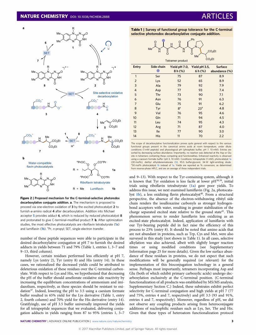
number of these peptide sequences were able to participate in thedesired decarboxylative conjugation at pH 7 to furnish the desiredadducts in yields between 71 and 79% (Table 1, entries 1, 3–7 and9–13, third column).
However, certain residues performed less efficiently at pH 7,namely Lys (entry 2), Tyr (entry 8) and His (entry 14). In thesecases, we rationalized the decreased yields could be attributed todeleterious oxidation of these residues over the C-terminal carbox-ylate. With respect to Lys and His, we hypothesized that decreasingthe pH of the buffer should ameliorate oxidative side reactivity byincreasing the equilibrium concentrations of ammonium and imi-dazolium, respectively, as these species should be resistant to oxi-dation37. Indeed, lowering the pH to 3.5 using a caesium formatebuffer resulted in 65% yield for the Lys derivative (Table 1, entry2, fourth column) and 70% yield for the His derivative (entry 14).Gratifyingly, use of pH 3.5 buffer universally improved the yieldsfor all tetrapeptide sequences that we evaluated, delivering conju-gation adducts in yields ranging from 87 to 95% (entries 1, 3–7
and 9–13). With respect to the Tyr-containing system, although itis known that Tyr oxidation is less facile at lower pH38,39, initialtrials using riboflavin tetrabutyrate (1a) gave poor yields. Toaddress this issue, we next examined lumiflavin (Fig. 2a, photocata-lyst 1b), a less oxidizing flavin photocatalyst40. From a structuralperspective, the absence of the electron-withdrawing ribityl sidechain renders the isoalloxazine carbonyls as stronger hydrogen-bond acceptors with water, resulting in greater stabilization of thecharge separated excited state relative to the ground state41. Thisphenomenon serves to render lumiflavin less oxidizing as anexcited-state photocatalyst. Indeed, application of lumiflavin withour Tyr-bearing peptide did in fact raise the efficiency of thisprocess to 23% (entry 8). It should be noted that amino acids thatare not abundant in proteins, such as Trp, Cys and Met, were alsosurveyed in this study (not shown in Table 1). In all cases, selectivealkylation was also achieved, albeit with slightly longer reactiontimes or using modified conditions (see SupplementaryInformation page 25 for more details). Given the low surface abun-dance of these residues in proteins, we do not expect that suchmodifications will be generally required (or relevant) for theimplementation of this bioconjugation technology in a generalsense. Perhaps most importantly, tetramers incorporating Asp andGlu (both of which exhibit primary carboxylic acids) undergo dec-arboxylation exclusively at the C-terminal position. (C-terminalfunctionalization of all products was established by MS/MS analysis,Supplementary Section C.) Indeed, these substrates exhibit perfectselectivity for C-terminal conjugation and high yields at pH 7 (77and 75%, entries 4 and 7, respectively) and pH 3.5 (93 and 91%,entries 4 and 7, respectively). Moreover, regardless of pH, we didnot observe any coupling products arising from heteroconjugateadditions of nucleophilic residues such as Lys, Ser, Thr and His.Given that these types of heteroatom functionalization protocol
b
CO2Et
CO2Et
1 8
Me
H+
5
N
N
NH
NMe
MeO
O
R
OCOnPr
OCOnPr
OnPrOC
OnPrOC
R = Me Lumiflavin 1b
Riboflavin tetrabutyrate
1
R =
CO2HN
O
O
N
N
NH
NMe
MeO
O
R
CO2HN
CO2HN
MeCO2Et
CO2Et
CO2HN
MeCO2Et
CO2Et
*
8
43
67
6
7
Site-selective oxidativedecarboxylation
N
N
NH
NMe
MeO
O
R1a
Water-compatibleflavin photocatalysts
N
N
NH
NMe
MeO
O
R
SET
2
SET
SET
a
Figure 2 | Proposed mechanism for the C-terminal-selective photoredoxdecarboxylative conjugate addition. a, The mechanism is proposed toproceed via one-electron oxidation of 3 by the excited photocatalyst 2 tofurnish α-amino radical 4 after decarboxylation. Addition into Michaelacceptor 5 provides adduct 6, which is reduced by reduced photocatalyst 8and protonated to give C-terminal-modified product 7. b, After optimizationstudies, the most effective photocatalysts are riboflavin tetrabutyrate (1a)and lumiflavin (1b). nPr, n-propyl; SET, single-electron transfer.
Table 1 | Survey of functional group tolerance for the C-terminalselective photoredox decarboxylative conjugate addition.
Tetramer product
Me NH
OHN
O
NH
O
N
O
Ph
Me
CO2Et
CO2Et
Yield pH 3.5,
92
95
95
90
87
90
23a
87
70
65
91
94
93
91
6 h (%)
79
76
74
77
75
73
8a
71
11
52
76
71
77
75
Yield pH 7.0,8 h (%)
Side chain
Ala
Val
Leu
Ile
Ser
Thr
Tyr
Lys
His
Arg
Asn
Gln
Asp
Glu
1
10
65
12
14
32
13
8
11
9
4
7
Entry Surfaceabundance (%)
7.9
4.6
4.3
3.0
8.9
7.1
4.8
4.0
2.2
8.9
6.3
4.5
7.4
6.2
The scope of decarboxylative functionalization proves quite general with respect to the variousfunctional groups present in the canonical amino acids at room temperature, under diluteconditions (1 mM peptide) and physiological pH (phosphate buffer, pH 7, 10 mM). Entries aresorted by decreasing surface abundance. Importantly, no reaction was detected at the Asp or Glusite in tetramers containing these competing acid functionalities. Enhanced reactivity is observedusing a caesium formate buffer (pH 3, 10 mM). Conditions: tetrapeptide (1 mM), photocatalyst 1a(30 mol%), diethyl ethylidenemalonate (5), 95:5 buffer:glycerol, 34 W light-emitting diode.a30 mol% photocatalyst 1b instead of 1a. Yields are reported as % conversion, as determinedfrom reverse-phase HPLC, and are an average of three independent trials.
represent a common strategy in bioconjugation chemistry, we feelthat this outcome reveals both the kinetic and thermodynamicbenefits of employing electron-transfer and open-shell mechanismsin lieu of closed-shell, nucleophile trapping protocols42.
Having assessed the functional group tolerance of our reactionon a large, representative group of tetrapeptides, we next decidedto examine the applicability of this photocatalytic methodologyfor endogenous peptides, including several with biological activity.
Me
CO2Et
CO2Et
O
NH
O
HN
O
NH
O
HN
O
NH
O
HN
O
NH
O
HN
O
NH
O
OH
Me
CO2Et
CO2Et
+ Photocat. 1a
95:5 pH 3.5 buffer:glycerol (1 mM)
Me
CO2Et
CO2Et
10 equiv.
+
30 mol% 34 W blue LED
+
Me
CO2Et
CO2Et
H2N
D R V Y I H P
40%a
F
Angiotensin II
Me
CO2Et
CO2Et
H2N
R P P G F S P
53%
F
Bradykinin
R
H2N
F R G D S P A S
Fibronectin binding inhibitor
S K P
Me
CO2Et
CO2Et
H2N
F F K N I V T P
Myelin basic (87–99)
R T PV H
Me
CO2Et
CO2Et
H2N
I A G F K G E Q
Type II collagen fragment
G P KP GG E
Bivalirudin
O
NH
O
HN
O
NH
O
HN
O
NH
O
HN
O
NH
O
HN
O
NH
a
15
16
66%
55%
41%
17
18
19
33%a,d20
or 1b
Me
CO2Et
CO2Et
H2N
Pro-adrenomedullin(153–185)b
33-mer peptide
52%
21
A G P G R TP ES L KS SL V
G A P A P PA HP Q PS AS G
LH F
15
33
30
ZHER2 Affibody
F N K E Q QN KV D F YN A
E Q R N A FN EN L S LI Q
20
58
40
31%c
22
E
K
H LI L
P SD D
P
Q
L A E A K KN LS A D AL N Q KA P
G G G G N G D F E E IR PF P P E E Y L
5
NH
OiPr
EtO2C
EtO2C
Me
NH
O O
NH2
b
c d
Nine internal carboxylate residues
High-affinity binder of HER2
58-mer peptide
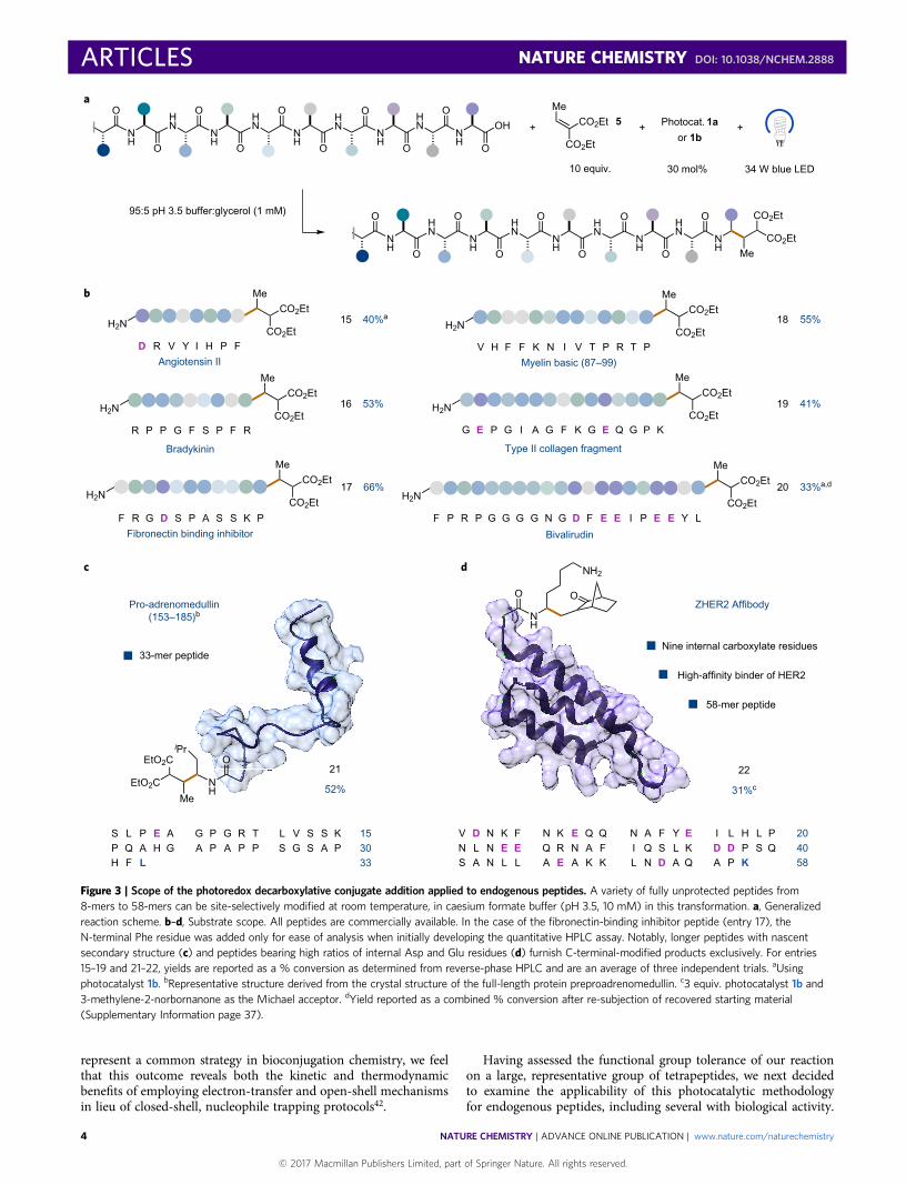
Figure 3 | Scope of the photoredox decarboxylative conjugate addition applied to endogenous peptides. A variety of fully unprotected peptides from8-mers to 58-mers can be site-selectively modified at room temperature, in caesium formate buffer (pH 3.5, 10 mM) in this transformation. a, Generalizedreaction scheme. b–d, Substrate scope. All peptides are commercially available. In the case of the fibronectin-binding inhibitor peptide (entry 17), theN-terminal Phe residue was added only for ease of analysis when initially developing the quantitative HPLC assay. Notably, longer peptides with nascentsecondary structure (c) and peptides bearing high ratios of internal Asp and Glu residues (d) furnish C-terminal-modified products exclusively. For entries15–19 and 21–22, yields are reported as a % conversion as determined from reverse-phase HPLC and are an average of three independent trials. aUsingphotocatalyst 1b. bRepresentative structure derived from the crystal structure of the full-length protein preproadrenomedullin. c3 equiv. photocatalyst 1b and3-methylene-2-norbornanone as the Michael acceptor. dYield reported as a combined % conversion after re-subjection of recovered starting material(Supplementary Information page 37).
At the outset, we selected commercial peptides ranging in size from8 to 10 amino acids. Among those examined were a cardiovascularregulator, angiotensin II, and an inflammation inhibitor, bradyki-nin. These peptides underwent C-terminal decarboxylative alky-lation with the desired monofunctionalization selectivity in yieldsof 40 and 53%, respectively (Fig. 3b, entries 15 and 16). Movingforward, we next examined mid-range, endogenous peptides con-sisting of 11–15 amino-acid residues. As highlighted in Fig. 3b,myelin basic protein (87–99), type II collagen and fibronectin-bindinginhibitor peptide (modified only by appending an N-terminal Phefor ease of spectroscopic analysis while developing a quantitativeHPLC assay) were all found to give good yields under our optimizedreaction conditions (41–66%, Fig. 3b, entries 17–19). It should benoted that in some cases, reactions of macropeptides affordedother unidentifiable side products, albeit in unappreciable quan-tities. As a testament to the levels of site selectivity of our new bio-conjugation protocol, we also demonstrated a successful alkylationof bivalirudin (Angiomax), an antithrombotic icosapeptide.Despite an initially lower yield of 28%, we observed exclusiveC-terminal functionalization in the presence of five additionalcarboxylic acids. HPLC analysis of the crude reaction mixturerevealed the remaining mass balance to be unreacted startingmaterial. As such, a second round of photocatalytic activationfollowing re-isolation of the unmodified peptide increased theoverall yield to 33% (Fig. 3b, entry 20). We hypothesize that thereaction halts at 28% conversion (with no further loss of startingmaterial) due to photocatalyst deactivation as evidenced by UV–vistime-course studies (Supplementary Section E).
Having demonstrated the selectivity of our reaction for the term-inal alkylation of linear peptides (4–20 amino acids), we next decidedto examine sequences with known secondary structure. For thispurpose, we selected commercially available pro-adrenomedullin(fragment 153–185) and the ZHER2 affibody derived fromimmunoglobulin-binding protein A. To our delight, pro-adrenome-dullin selectively delivered the C-terminal modified product in 52%yield (Fig. 3c, entry 21) using our standard conditions. To oursurprise, with the ZHER2 affibody, use of the diethyl ethylidenema-lonate Michael acceptor resulted in the formation of the desiredadduct along with side-chain conjugate addition adducts via acompeting Lys-addition pathway. At this stage, we rationalizedthat use of α,β-unsaturated carbonyls that are less susceptible to2e− nucleophile pathways than diethyl ethylidenemalonate, yet arestill able to participate in radical additions, might overcome thisissue. Indeed, the implementation of the strained, monocarbonyl-containing compound, 3-methylene-2-norbornanone, an estab-lished radicalphile under photoredox conditions43, eliminated anyundesired side reactivity, furnishing the modified product in 31%yield with intact α-helical structure (Fig. 3d, entry 22; seeSupplementary Section F for circular dichroism studies). In thiscase, recovered starting material accounted for the remainingmass balance.
A true test of any bioconjugation technology lies in its capacity toperform site-selective modification of peptides that exhibit tertiarystructure. However, the structural linkages that confer thesehigher-order architectures (for example, disulfide bridges) canthemselves be susceptible to chemical modification44, therebyoften changing the structure–function relationship of any givenhigher-order proteins. To test the viability of this new photoredoxprotocol in this context, we selected insulin as a suitable molecularplatform to examine the chemical selectivity of electron transfer andopen-shell mechanisms with a molecule that contains a variety offunctional groups (Fig. 4). Structurally, insulin is composed oftwo parent chains linked by two disulfide bridges, with a third dis-ulfide bond in the A-chain backbone. Insulin also contains fourtyrosines, residues that proved particularly challenging during ourassessment of tetrameric peptides. Furthermore, and unlike the
peptide substrates described thus far, insulin bears two C-terminalcarboxylic acids in addition to having two Glu residues on each ofthe A and B chains. In the event, subjecting native insulin to ouroptimized reaction conditions (with 3-methylene-2-norbornanoneas the electrophile) resulted in formation of a monoalkylatedadduct in 44% isolated yield wherein modification occurred exclu-sively at the A-chain C terminus. A minor adduct (less than 5%)was detected in which both the A- and B-chain C termini were func-tionalized (see Supplementary Information pages 43 and 143 fordetails). Importantly, product analysis revealed that all three disul-fide linkages remained intact and no side-chain decarboxylative orheteroatom conjugate addition was detected. The selectivity forA-chain monoalkylation is particularly noteworthy as currenttechnologies generally offer selective functionalization of insulin’sB chain through covalent modification at His10 (ref. 45), Tyr26(ref. 46) and Lys29 (ref. 47). This photoredox methodology there-fore offers a new technology that not only selectively targets aspecific C-terminal carboxylic acid, but also modifies the lightchain, a structural component that heretofore has not been suscep-tible to bioconjugation. We speculate that the observed selectivityfor A-chain modification could arise from either (1) inherent differ-ences in oxidation potentials between the C termini whereinA-chain oxidation is favoured or (2) adsorption of the photocatalystto a lipophilic region on the surface of the protein proximate to theA-chain C terminus. Moreover, expanding the scope of Michaelacceptors to norbornanones that bear bioorthogonal tags (that is,
flav.
O
+
Human insulin
F V N Q H L C G S H L V E A L Y L V C G E R G F F Y T P K T
G I V E Q C C T S I C S L Y Q L E N Y C N
B chain
A chain
49%
Selective A chainfunctionalization
Novel, site-selectivealkylation
HN
O
O
NH2
O
9:1 A chain monoalkylation:A/B chain dialkylation
MeMe
O
NH 7
No reduction of disulfide bonds No functionalization of internal Glu residues
ONH
O
41%Monoalkylkation
a
Ob
Figure 4 | Photoredox-mediated decarboxylative functionalization ofhuman insulin. a, Human insulin was functionalized at room temperatureusing our decarboxylative photoredox methodology to furnish highlyselective mono-alkylation at the C terminus of the A chain, exclusively.b, Functionalization of human insulin with a Michael acceptor incorporatinga bio-ambient alkyne. Reaction conditions: 1 equiv. insulin (500 nmol),10 equiv. of the respective Michael acceptor, 3 equiv. photocatalyst 1b,95:5 pH 3.5 caesium formate buffer:glycerol (1 mM), 34 W blue light-emitting diode, 8 h. Yield is reported as % conversion as determined fromreverse-phase HPLC. Current work exploring azide- and biotin-bearingMichael acceptors is ongoing.
an alkyne) has also demonstrated promising results (SupplementaryInformation pages 44 and 144). We fully expect this Michaelacceptor will be able to incorporate other bioorthogonal handles(biotin and azides), and work is ongoing in this area.
In conclusion, we present a photoredox bioconjugation strategythat selectively targets C-terminal carboxylic acids in lieu of otherfunctional groups found in protein structures. To our knowledge,this transformation represents an unprecedented approach to site-selective bioconjugation that can provide facile access to homo-geneous alkylation adducts by virtue of the inherent presence ofonly one C-terminal carboxylate in most peptide and protein struc-tures. Work is ongoing to apply this decarboxylative bioconjugationstrategy to the selective functionalization of a wide range of proteins,enzymes and antibodies with biologically active conjugates.Moreover, the development of photoredox catalysis as a genericplatform for site-selective bioconjugation will be investigated inthe context of targeting other redox-active side chains.
Data availability.All relevant data are provided in the SupplementaryInformation or are available from the authors upon request.
Received 15 June 2017; accepted 4 October 2017;published online 4 December 2017
References1. Boutureira, O. & Bernardes, G. J. L. Advances in chemical protein modification.
Chem. Rev. 115, 2174–2195 (2015).2. Krall, N., da Cruz, F. P., Boutureira, O. & Bernardes, G. J. L. Site-selective
protein-modification chemistry for basic biology and drug development. Nat.Chem. 8, 103–113 (2016).
3. Sletten, E. M. & Bertozzi, C. R. Bioorthogonal chemistry: fishing for selectivity ina sea of functionality. Angew. Chem. Int. Ed. 48, 6974–6998 (2009).
4. Saxon, E. & Bertozzi, C. R. Cell surface engineering by a modified Staudingerreaction. Science 287, 2007–2010 (2000).
5. Junutula, J. R. et al. Site-specific conjugation of a cytotoxic drug to an antibodyimproves the therapeutic index. Nat. Biotechnol. 26, 925–932 (2008).
6. Lyon, R. P., Meyer, D. L., Setter, J. R. & Senter, P. D. Conjugation of anticancerdrugs through endogenous monoclonal antibody cysteine resides. MethodsEnzymol. 502, 123–138 (2012).
7. Baslé, E., Joubert, N. & Pucheault, M. Protein chemical modification onendogenous amino acids. Chem. Biol. 17, 213–227 (2010).
8. Miller, S., Janin, J., Lesk, A. M. & Chothia, C. Interior and surface of monomericproteins. J. Mol. Biol. 196, 641–656 (1987).
9. Chen, X., Muthoosamy, K., Pfisterer, A., Neumann, B. & Weil, T. Site-selectivelysine modification of native proteins and peptides via kinetically controlledlabelling. Bioconjugate Chem. 23, 500–508 (2012).
10. Bader, B. et al. Bioorganic synthesis of lipid-modified proteins for the study ofsignal transduction. Nature 403, 223–226 (2000).
11. Rosen, C. B. & Francis, M. B. Targeting the N terminus for site-selective proteinmodification. Nat. Chem. Biol. 13, 697–705 (2017).
12. Romanini, D. W. & Francis, M. B. Attachment of peptide building blocks toproteins through tyrosine bioconjugation. Bioconjugate Chem. 19,153–157 (2008).
13. Tilley, S. D. & Francis, M. B. Tyrosine-selective protein alkylation usingπ-allylpalladium complexes. J. Am. Chem. Soc. 128, 1080–1081 (2006).
14. Joshi, N. S., Whitaker, L. R. & Francis, M. B. A three-component Mannich-typereaction for selective tyrosine bioconjugation. J. Am. Chem. Soc. 126,15942–15943 (2004).
15. Ban, H. et al. Facile and stable linkages through tyrosine: bioconjugationstrategies with the tyrosine-click reaction. Bioconjugate Chem. 24,520–532 (2013).
16. Antos, J. M., McFarland, J. M., Iavarone, A. T. & Francis, M. B. Chemoselectivetryptophan labeling with rhodium carbenoids at mild pH. J. Am. Chem. Soc.131, 6301–6308 (2009).
17. Antos, J. M. & Francis, M. B. Selective tryptophan modification withrhodium carbenoids in aqueous solution. J. Am. Chem. Soc. 126,10256–10257 (2004).
18. Seki, Y. et al. Transition metal-free tryptophan-selective bioconjugation ofproteins. J. Am. Chem. Soc. 138, 10798–10801 (2016).
19. Lin, S. et al. Redox-based reagents for chemoselective methioninebioconjugation. Science 355, 597–602 (2017).
20. Zuo, Z. & MacMillan, D. W. C. Decarboxylative arylation of α-amino acids viaphotoredox catalysis: a one-step conversion of biomass to drug pharmacophore.J. Am. Chem. Soc. 136, 5257–5260 (2014).
21. Chu, L., Ohta, C., Zuo, Z. & MacMillan, D. W. C. Carboxylic acids as a tracelessactivation group for conjugate additions: a three-step synthesis of (±)-pregabalin.J. Am. Chem. Soc. 136, 10886–10889 (2014).
22. Noble, A. & MacMillan, D. W. C. Photoredox-mediated α-vinylation ofα-amino acids and N-aryl amines. J. Am. Chem. Soc. 136,11602–11605 (2014).
23. Zuo, Z. et al. Merging photoredox with nickel catalysis: coupling of α-carboxylsp3-carbons with aryl halides. Science 345, 437–440 (2014).
24. Galicia, M. & González, F. J. Electrochemical oxidation of tetrabutylammoniumsalts of aliphatic carboxylic acids in acetonitrile. J. Electrochem. Soc. 149,D46–D50 (2002).
25. Hu, Q.-Y., Berti, F. & Adamo, R. Towards the next generation of biomedicines bysite-selective conjugation. Chem. Soc. Rev. 45, 1691–1719 (2016).
26. McGrath, N. A., Andersen, K. A., Davis, A. K. F., Lomax, J. E. & Raines, R. T.Diazo compounds for the bioreversible esterification of proteins. Chem. Sci. 6,752–755 (2015).
27. Rajagopalan, T. G., Stein, W. H. & Moore, S. The inactivation of pepsin bydiazoacetylnorleucine methyl ester. J. Biol. Chem. 241, 4295–4297 (1966).
28. Delpierre, G. R. & Fruton, J. S. Specific inactivation of pepsin by a diazo ketone.Proc. Natl Acad. Sci. USA 56, 1817–1822 (1966).
29. Totaro, K. A. et al. Systematic investigation of EDC/sNHS-mediatedbioconjugation reactions for carboxylated peptide substrates. Bioconj. Chem. 27,994–1004 (2016).
30. Noble, A., McCarver, S. J. & MacMillan, D. W. C. Merging photoredox andnickel catalysis: decarboxylative cross-coupling of carboxylic acids with vinylhalides. J. Am. Chem. Soc. 137, 624–627 (2015).
31. Slutskyy, Y. & Overman, L. E. Generation of the methoxycarbonyl radical byvisible-light photoredox catalysis and its conjugate addition with electron-deficient olefins. Org. Lett. 18, 2564–2567 (2016).
32. MacDonald, J. I., Munch, H. K., Moore, T. & Francis, M. B. One-step site-specificmodification of native proteins with 2-pyridinecarboxyaldehydes. Nat. Chem.Biol. 11, 326–331 (2015).
33. Novak, M., Miller, A., Bruice, T. C. & Tollin, G. The mechanism of flavin 4asubstitution which accompanies photolytic decarboxylation of α-substitutedacetic acids. Carbanion vs. radical intermediates. J. Am. Chem. Soc. 102,1465–1467 (1980).
34. Islam, S. D. M., Penzkofer, A. & Hegemann, P. Quantum yield of tripletformation of riboflavin in aqueous solution and of flavin mononucleotide boundto the LOV1 domain of Phot1 from Chlamydomonas reinhardtii. Chem. Phys.291, 97–114 (2003).
35. Lu, C. et al. Riboflavin (VB2) photosensitized oxidation of 2′-deoxyguanosine-5′-monophosphate (dGMP) in aqueous solution: a transient intermediatesstudy. Phys. Chem. Chem. Phys. 2, 329–334 (2000).
36. Bortolamei, N., Isse, A. A. & Gennaro, A. Estimation of standard reductionpotentials of alkyl radicals involved in atom transfer radical polymerization.Electrochim. Acta 55, 8312–8318 (2010).
37. Huvaere, K. & Skibsted, L. H. Light-induced oxidation of tryptophan andhistidine. Reactivity of aromatic N-heterocycles toward triplet-excited flavins.J. Am. Chem. Soc. 131, 8049–8060 (2009).
38. Harriman, A. Further comments on the redox potentials of tryptophan andtyrosine. J. Phys. Chem. 91, 6102–6104 (1987).
39. Stubbe, J. & van der Donk, W. A. Protein radicals in enzyme catalysis. Chem.Rev. 98, 705–762 (1998).
40. Sikorska, E. et al. Spectroscopy and photophysics of lumiflavins andlumichromes. J. Phys. Chem. A 108, 1501–1508 (2004).
41. Koziol, J. Studies on flavins in organic solvents – I. Spectral characteristics ofriboflavin, riboflavin tetrabutyrate, and lumichrome. Photochem. Photobiol. 5,41–54 (1966).
42. Garbaccio, R. M. in Comprehensive Organic Synthesis II 2nd edn, Vol. 1 (edsKnochell, P. & Molander, G. A.) Ch. 9, 438–462 (Elsevier, 2014).
43. Ravelli, D., Zema, M., Mella, M., Fagnoni, M. & Albini, A. Benzoyl radicalsfrom (hetero)aromatic aldehydes. Decatungstate photocatalyzedsynthesis of substituted aromatic ketones. Org. Biomol. Chem. 8,4158–4164 (2010).
44. Wagner, A. & Koniev, O. Developments and recent advancements in the field ofendogenous amino acid selective bond forming reactions for bioconjugation.Chem. Soc. Rev. 44, 5495–5551 (2015).
45. Uchida, K. & Stadtman, E. R. Modification of histidine residues in proteinsby reaction with 4-hydroxynonenal. Proc. Natl Acad. Sci. USA 89,4544–4548 (1992).
46. Wang, Y., Luo, Y. & Zhang, R. Investigation on insulin tyrosine modificationmediated by peroxynitrite. In Proc. IEEE/ICME Int. Conf. Complex Chem.Engineering Beijing, Beijing, 1813–1816 (IEEE, 2007).
47. Lindsay, D. G. & Shall, S. The acetylation of insulin. Biochem. J. 121,737–745 (1971).
AcknowledgementsThe authors acknowledge financial support provided by the NIHGMS (R01 01GM093213-04) and gifts from Merck and BMS. D.K.K. acknowledges the Deutsche
Forschungsgemeinschaft (DFG) for a postdoctoral fellowship (KO 4867/2-1). The authorsthank T. Muir, Z. Brown, R. Thompson and members of the Muir Laboratory for theiradvice and analytical support. The authors also thank I. Pelczer and K. Conover forassistance with NMR spectroscopy.
Author contributionsS.B., C.L. and D.K.K. performed and analysed the experiments. S.B., C.L., D.K.K. andD.W.C.M. designed the experiments and prepared this manuscript. J.X.Q., Y.Z., M.A.P.and W.R.E. provided discussions. J.X.Q. assisted with peptide synthesis.
Additional informationSupplementary information and chemical compound information are available in theonline version of the paper. Reprints and permissions information is available online atwww.nature.com/reprints. Publisher’s note: Springer Nature remains neutral with regard tojurisdictional claims in published maps and institutional affiliations. Correspondence andrequests for materials should be addressed to D.W.C.M.
Competing financial interestsThe authors declare no competing financial interests.