Deep-Inspiration Breath-Hold PET/CT: Clinical Findings with a New Technique for Detection and Characterization of Thoracic Lesions Gustavo S.P. Meirelles 1 , Yusuf Emre Erdi 2 , Sadek A. Nehmeh 2 , Olivia D. Squire 1 , Steven M. Larson 1 , John L. Humm 2 , and Heiko Sch¨ oder 1 1 Department of Radiology, Nuclear Medicine Service, Memorial Sloan-Kettering Cancer Center, New York, New York; and 2 Department of Medical Physics, Memorial Sloan-Kettering Cancer Center, New York, New York Respiratory motion during PET/CT acquisition can cause mis- registration and inaccuracies in calculation of standardized up- take values (SUVs). Our aim was to compare the detection and characterization of thoracic lesions on PET/CT with and without a deep-inspiration protocol. Methods: We studied 15 patients with suspected pulmonary lesions who underwent clinical PET/ CT, followed by deep-inspiration breath-hold (BH) PET/CT. In BH CT, the whole chest of the patient was scanned in 15 s at the end of deep inspiration. For BH PET, patients were asked to hold their breath 9 times for 20-s intervals. One radiologist reviewed images, aiming to detect and characterize pulmonary, nodal, and skeletal abnormalities. Clinical CT and BH CT were compared for number, size, and location of lesions. Lesion SUVs were compared between clinical PET and BH PET. Images were also visually assessed for accuracy of fusion and registra- tion. Results: All patients had lesions on clinical CT and BH CT. Pulmonary BH CT detected more lesions than clinical CT in 13 of 15 patients (86.7%). The total number of lung lesions detected increased from 53 with clinical CT to 82 with BH CT (P , 0.001). Eleven patients showed a total of 31 lesions with ab- normal 18 F-FDG uptake. BH PET/CT had the advantage of re- ducing misregistration and permitted a better localization of sites with 18 F-FDG uptake. A higher SUV was noted in 22 of 31 lesions on BH PET compared with clinical PET, with an average increase in SUV of 14%. Conclusion: BH PET/CT enabled an in- creased detection and better characterization of thoracic lesions compared with a standard PET/CT protocol, in addition to more precise localization and quantification of the findings. The tech- nique is easy to implement in clinical practice and requires only a minor increase in the examination time. Key Words: PET/CT; breath-hold; imaging; 18 F-FDG; cancer J Nucl Med 2007; 48:712–719 DOI: 10.2967/jnumed.106.038034 The recent introduction in clinical routine of combined PET/CT scanners, which provide inline fusion of anatomic and functional images, has enabled a more precise local- ization characterization of sites with radiotracer uptake. This has led to an increased acceptance of the method as a clinical tool for the diagnosis, staging, restaging, and mon- itoring of patients with a wide variety of cancers (1–6). Although the optimal protocol for PET/CT studies re- mains a subject of discussion, most centers perform the CT component of the examination in shallow breathing or at the end of expiration (7,8). The PET component, because of its longer acquisition time, is performed during free tidal breathing. Therefore, even with an inline scanner, the core- gistration of PET and CT can be imprecise, leading to misalignment of structures and lesions, repeated scans over certain regions, and omission of anatomic levels. Misalign- ment between PET and CT can reduce the diagnostic accu- racy of PET/CT for lesion detection. The accuracy of PET/CT for treatment monitoring could also be limited by respiratory motion: Because the CT images are used for attenuation correction of PET emission data, misalignment between PET and CT lesions will lead to overestimation or underestimation of standardized uptake values (SUVs). Furthermore, respiratory motion causes a spread of tracer activity within a given lesion over a larger area and, thus, underestimation of the true activity concentration (9–11). Although experienced PET/CT readers are familiar with these limitations and consider them in their study interpre- tation (12), it would be desirable to eliminate these artifacts completely and develop an imaging technique that allows accurate PET/CT alignment in the thorax. Indeed, efforts in this regard have led to the development of four-dimensional (4D) PET/CT, whereby respiratory gating of PET and CT can reduce or even eliminate these artifacts (11). However, this technique has not gained wide acceptance in diagnostic imaging practice because of the long acquisition time for 4D PET/CT, time-consuming postprocessing of raw data, and a higher radiation dose delivered to the patient (11). Received Nov. 8, 2006; revision accepted Jan. 2, 2007. For correspondence or reprints contact: Heiko Sch ¨ oder, MD, Department of Radiology/Nuclear Medicine, Memorial Sloan-Kettering Cancer Center, 1275 York Ave., Box 77, New York, NY 10021. E-mail: [email protected]712 THE JOURNAL OF NUCLEAR MEDICINE • Vol. 48 • No. 5 • May 2007 by on July 28, 2018. For personal use only. jnm.snmjournals.org Downloaded from
Transcript
Deep-Inspiration Breath-Hold PET/CT: ClinicalFindings with a New Technique for Detection andCharacterization of Thoracic Lesions
Gustavo S.P. Meirelles1, Yusuf Emre Erdi2, Sadek A. Nehmeh2, Olivia D. Squire1, Steven M. Larson1,John L. Humm2, and Heiko Schoder1
1Department of Radiology, Nuclear Medicine Service, Memorial Sloan-Kettering Cancer Center, New York, New York; and2Department of Medical Physics, Memorial Sloan-Kettering Cancer Center, New York, New York
Respiratory motion during PET/CT acquisition can cause mis-registration and inaccuracies in calculation of standardized up-take values (SUVs). Our aim was to compare the detection andcharacterization of thoracic lesions on PET/CT with and withouta deep-inspiration protocol. Methods: We studied 15 patientswith suspected pulmonary lesions who underwent clinical PET/CT, followed by deep-inspiration breath-hold (BH) PET/CT. InBH CT, the whole chest of the patient was scanned in 15 s atthe end of deep inspiration. For BH PET, patients were askedto hold their breath 9 times for 20-s intervals. One radiologistreviewed images, aiming to detect and characterize pulmonary,nodal, and skeletal abnormalities. Clinical CT and BH CT werecompared for number, size, and location of lesions. LesionSUVs were compared between clinical PET and BH PET. Imageswere also visually assessed for accuracy of fusion and registra-tion. Results: All patients had lesions on clinical CT and BHCT. Pulmonary BH CT detected more lesions than clinical CT in13 of 15 patients (86.7%). The total number of lung lesionsdetected increased from 53 with clinical CT to 82 with BH CT(P , 0.001). Eleven patients showed a total of 31 lesions with ab-normal 18F-FDG uptake. BH PET/CT had the advantage of re-ducing misregistration and permitted a better localization ofsites with 18F-FDG uptake. A higher SUV was noted in 22 of 31lesions on BH PET compared with clinical PET, with an averageincrease in SUV of 14%. Conclusion: BH PET/CT enabled an in-creased detection and better characterization of thoracic lesionscompared with a standard PET/CT protocol, in addition to moreprecise localization and quantification of the findings. The tech-nique is easy to implement in clinical practice and requires onlya minor increase in the examination time.
Key Words: PET/CT; breath-hold; imaging; 18F-FDG; cancer
J Nucl Med 2007; 48:712–719DOI: 10.2967/jnumed.106.038034
The recent introduction in clinical routine of combinedPET/CT scanners, which provide inline fusion of anatomicand functional images, has enabled a more precise local-ization characterization of sites with radiotracer uptake.This has led to an increased acceptance of the method as aclinical tool for the diagnosis, staging, restaging, and mon-itoring of patients with a wide variety of cancers (1–6).
Although the optimal protocol for PET/CT studies re-mains a subject of discussion, most centers perform the CTcomponent of the examination in shallow breathing or atthe end of expiration (7,8). The PET component, because ofits longer acquisition time, is performed during free tidalbreathing. Therefore, even with an inline scanner, the core-gistration of PET and CT can be imprecise, leading tomisalignment of structures and lesions, repeated scans overcertain regions, and omission of anatomic levels. Misalign-ment between PET and CT can reduce the diagnostic accu-racy of PET/CT for lesion detection. The accuracy ofPET/CT for treatment monitoring could also be limitedby respiratory motion: Because the CT images are used forattenuation correction of PET emission data, misalignmentbetween PET and CT lesions will lead to overestimation orunderestimation of standardized uptake values (SUVs).Furthermore, respiratory motion causes a spread of traceractivity within a given lesion over a larger area and, thus,underestimation of the true activity concentration (9–11).Although experienced PET/CT readers are familiar withthese limitations and consider them in their study interpre-tation (12), it would be desirable to eliminate these artifactscompletely and develop an imaging technique that allowsaccurate PET/CT alignment in the thorax. Indeed, efforts inthis regard have led to the development of four-dimensional(4D) PET/CT, whereby respiratory gating of PET and CTcan reduce or even eliminate these artifacts (11). However,this technique has not gained wide acceptance in diagnosticimaging practice because of the long acquisition time for4D PET/CT, time-consuming postprocessing of raw data,and a higher radiation dose delivered to the patient (11).
Received Nov. 8, 2006; revision accepted Jan. 2, 2007.For correspondence or reprints contact: Heiko Schoder, MD, Department
of Radiology/Nuclear Medicine, Memorial Sloan-Kettering Cancer Center,1275 York Ave., Box 77, New York, NY 10021.
Previous studies have shown the capability of acquiringPET/CT images at any phase within the respiratory cycle—in a short period of time—equivalent to the one adoptedand used in clinical practice with shallow breathing or atthe end of expiration (9). The aim of this study is tocompare the detection and characterization of thoraciclesions on regular clinical PET/CT with those on deep-inspiration breath-hold (BH) PET/CT, acquired at a singlebreathing phase at end inspiration.
MATERIALS AND METHODS
PatientsFifteen patients (8 male, 7 female; average age, 57.3 y; age range,
43–73 y) with a biopsy-proven diagnosis of cancer, confirmed bystaff pathologists at our institution, were included in this prospec-tive pilot study. Patients were selected for this study on the basis ofthe clinical referral, requesting evaluation of a documented orsuspected primary or secondary malignancy in the lungs or medias-tinum. Nine patients presented with newly diagnosed or recurrent/metastatic lung cancer, 1 with newly diagnosed esophageal cancer,3 with proven or suspected lung metastases from prior cervical orcolorectal cancer, 1 with prostate-specific antigen (PSA) relapseand nodules in lungs and mediastinum that were later proven to besarcoidosis, and 1 with rising thyroglobulin level after resection ofthyroid cancer.
In addition to the clinical PET/CT examination, in which PETand CT images are acquired during shallow breathing, all patientsunderwent a deep-inspiration BH PET/CT scan. All patients gavewritten informed consent. The study protocol was approved by theinstitutional review board.
Patients fasted for at least 6 h before injection of 514–611 MBqof 18F-FDG. During the uptake phase of approximately 60 min,patients remained in a quiet position and were trained to fol-low verbal breathing instructions (‘‘breathe in, hold, relax’’), whichaimed to ensure that they could hold their breath for at least 20 srepeatedly.
PET/CT Acquisition and Image ReconstructionAll PET/CT studies were acquired on a Discovery LS PET/CT
system (GE Healthcare). A scout image with settings of 30 mA and120 kV was first acquired to determine the scanning field of thepatient. This was followed by helical CT using the followingscanning parameters: 140 kV, 80 mA, a 5-mm-slice thickness in4.25-mm intervals, and gantry rotation period of 0.8 s. This scan wasthen followed by a whole-body PET scan that covered the same areawith 3 min per bed position. The effective axial field of view (FOV)was 14.8 cm for each bed position, as there was an overlapping of 5slices between each axial FOV. These CT and PET scans wereacquired under free shallow breathing, with no voice instructions.
After clinical PET/CT, patients remained in the same position(supine, arms above the head), and a plastic box with 2 infraredreflective markers was placed on the abdomen for respiratorymotion tracking. The Real-Time Position Management (RPM)Respiratory Gating System (Varian Medical Systems) was used tomonitor and track the patient’s respiratory motion, using theamplitude-gating mode. A second chest CT, encompassing theentire lungs, was then obtained with identical parameters as inthe first one, except for a voice command asking the patients to holdtheir breath in end-inspiration for the duration of the examination,which was approximately 15 s.
PET data were then obtained for one 15-cm bed position in 9independent frames of 20 s each (total, 3 min). During these 20-sacquisitions the patients were asked to hold their breath in end-inspiration. Between each of the 9 BH sessions, patients weregiven an approximately 20-s period for rest and relaxation. Thus,the entire BH PET acquisition required on average 6 min perpatient (9 · 20 s for acquisition 1 8 · 20 s for relaxation). In thispilot study, PET images were acquired for just one FOV to obtainproof-of-principle data and to limit patient discomfort. To ensurethat PET images spatially match to CT images, the amplitude ofthe breathing signal displayed on the RPM monitor during PETwas set to the same level as the amplitude of the BH CT.
At the end of the study, all of the 9 · 20-s sinogram frameswere added to generate the final 3-min dataset, thus matching theclinical PET acquisition time. These data were then corrected forattenuation using the BH CT images and reconstructed with theclinical ordered-subsets expectation maximization (OSEM) re-construction algorithm and parameters. The same reconstructionparameters were used for clinical PET/CT and BH PET/CT scans.The details of this new method were described recently (13).
Image AnalysisClinical PET/CT and BH chest PET/CT studies were examined
in separate instances by the same thoracic radiologist, who wasunaware of the clinical indication for the study or other clinicaldata. The aim was to detect and characterize pulmonary, nodal,and skeletal abnormalities on clinical PET/CT and BH PET/CTstudies.
Readings were performed on a dedicated workstation (Xeleris;GE Healthcare), which can display 3 orthogonal planes for CT,PET, and PET/CT fused images (sagittal, coronal, and transaxial)and a maximum-intensity-projection image.
The clinical CT was read first, followed by the BH CT of thesame patient. The number, location, and characteristics of lesionswere assessed on both CT examinations. The following CT win-dows were used: lung (window, 1,500 Hounsfield units [HU];level, 2800 HU), soft tissue (window, 450 HU; level, 50 HU), andbone (window, 2,000 HU; level, 200 HU).
Pulmonary nodules were defined as rounded opacities sur-rounded by lung parenchyma, measuring up to 3 cm; they wereconsidered a mass if they were .3 cm. Consolidations andground-glass opacities (GGOs) were defined as areas of increasedlung attenuation that did or did not obscure the adjacent lungvessels, respectively. Cavities were defined as areas of reducedlung attenuation enclosed by walls (14).
Nodes were regarded as diseased when measuring .1.0 cm inthe short axis, except for some sites—such as the internal mam-mary chain or the diaphragmatic region, where any visualizednode was considered pathologic. The lesion location was classifiedaccording to the mediastinal nodal station system (15).
After the interpretation of clinical CT and BH CT examinations,the same radiologist proceeded to analyze the clinical PET/CT andBH PET/CT. As BH PET images had been acquired for a singleFOV, the analysis of clinical PET and clinical PET/CT scans wasrestricted to this one FOV of the BH PET.
The presence of 18F-FDG uptake in lesions detected on CT wasassessed with the correlation of CT, PET (corrected and uncor-rected), and PET/CT fusion images. 18F-FDG uptake was consid-ered positive if it was greater than that in the surrounding normaltissue. A circular region of interest (ROI) was drawn on eachlesion with 18F-FDG uptake, on transaxial images, encompassing
BREATH-HOLD PET/CT FOR CHEST LESIONS • Meirelles et al. 713
by on July 28, 2018. For personal use only. jnm.snmjournals.org Downloaded from
the entire lesion. The maximum SUV (SUVmax), normalized tobody weight, was recorded for each finding.
A comparison was performed between the clinical CT and theBH CT for the number, size, and location of lesions. The 18F-FDGPET SUVmax values were compared between clinical PET andBH PET. Images were also visually assessed on clinical PET/CTand BH PET/CT for accuracy of fusion and alignment. Thenumber of lesions per patient on clinical CT versus BH CT andthe SUVs on clinical PET versus BH PET were compared using apaired t test. A P value , 0.05 was considered significant.
RESULTS
CT Findings
All patients had thoracic lesions (pulmonary, nodal, orskeletal) on clinical CT and BH CT (Table 1). The mostfrequent were pulmonary, followed by nodal and skeletallesions. Compared with clinical CT, the BH CT detectedmore pulmonary lesions in 13 of the 15 patients (87%). Noadditional lesions were seen with BH CT in lymph nodes orin the skeleton.
BH CT detected 29 additional lung lesions in 13 patients,especially nodules smaller than 0.5 cm (P , 0.001; Fig. 1).Nodules and GGOs were most frequently depicted with theBH CT. Tables 2 and 3 show the number, characteristics,and distribution of the pulmonary lesions detected by bothCT modalities.
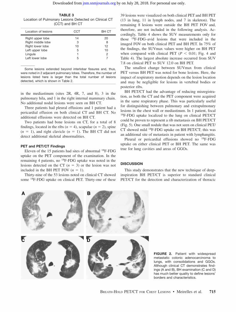
The BH CT allowed more precise localization andcharacterization of pulmonary findings than clinical CT inall cases. Nodules, GGOs, and consolidations were betterdefined and showed sharper contours. In 1 patient with dif-fuse disease, use of the BH technique permitted an improveddistinction between normal and diseased areas and en-hanced the definition of disease boundaries (Fig. 2). In 1case of lung cancer, use of the BH technique was partic-ularly useful because of the detection of signs of pulmonarycarcinomatous lymphangitis on the BH CT in a patientwithout evidence of distant disease on clinical PET/CT.These findings were characterized by irregular septal thick-ening, GGOs, and nodules, which were not seen on theclinical CT (Fig. 3). This patient also had foci of centri-lobular emphysema on the upper lobes, detected only onthe BH CT.
Twenty-seven lung lesions were seen on clinical CT andBH CT and were measured on both modalities. The size of
all lesions detected on clinical CT ranged from 0.3 to 9.8 cm(average 6 SD, 1.5 6 2.2 cm) and on BH CT ranged from0.2 to 9.6 cm (average 6 SD, 1.0 6 1.7 cm). With specificregard to lung nodules, the average size for nodules detectedon clinical CT was 0.9 cm and on BH CT it was 0.7 cm. Nopatient had lesions detected exclusively with clinical CT.
Four patients had a total of 12 enlarged lymph nodes onclinical CT and BH CT. Eight of these nodes were located
TABLE 1Pulmonary, Nodal, and Skeletal Chest Lesions Detected
on Clinical CT (CCT) and BH CT
Location
of lesions
No. of
patients
with lesions
on CCT
No. of
lesions
on CCT
No. of
patients
with lesions
on BH CT
No. of
lesions on
BH CT
Lung 15 33 15 62
Lymph nodes 4 12 4 12
Bones 2 8 2 8
Total 53 82
FIGURE 1. (A) Patient with lung cancer with 2 additionalnodules on left lung on BH CT (arrows). (B) Clinical CT does notshow lesions.
TABLE 2Pulmonary Lesions Detected on Clinical CT (CCT)
and BH CT
Type of lesion CCT BH CT
Nodules 22 49
Masses 4 4
Consolidations 4 4GGOs 2 4
Cavities 1 1
Total 33 62
714 THE JOURNAL OF NUCLEAR MEDICINE • Vol. 48 • No. 5 • May 2007
by on July 28, 2018. For personal use only. jnm.snmjournals.org Downloaded from
in the mediastinum (sites 2R, 4R, 7, and 8), 3 in thepulmonary hila, and 1 in the right internal mammary chain.No additional nodal lesions were seen on BH CT.
Three patients had pleural effusions and 1 patient had apericardial effusion on both clinical CT and BH CT. Noadditional effusions were detected on BH CT.
Two patients had bone lesions on CT, for a total of 8findings, located in the ribs (n 5 4), scapulae (n 5 2), spine(n 5 1), and right clavicle (n 5 1). The BH CT did notdetect additional skeletal abnormalities.
PET and PET/CT Findings
Eleven of the 15 patients had sites of abnormal 18F-FDGuptake on the PET component of the examination. In theremaining 4 patients, no 18F-FDG uptake was noted in thelesions detected on the CT (n 5 3) or the lesion was notincluded in the BH PET FOV (n 5 1).
Thirty-nine of the 53 lesions noted on clinical CT showedsome 18F-FDG uptake on clinical PET. Thirty-one of these
39 lesions were visualized on both clinical PET and BH PET(13 in lung, 11 in lymph nodes, and 7 in skeleton). Theremaining 8 lesions were outside the BH PET FOV and,therefore, are not included in the following analysis. Ac-cordingly, Table 4 shows the SUV measurements only forthose 18F-FDG–avid lesions that were included in theimaged FOV on both clinical PET and BH PET. In 75% ofthe findings, the SUVmax values were higher on BH PETwhen compared with clinical PET (P , 0.01; Fig. 4 andTable 4). The largest absolute increase occurred from SUV7.8 on clinical PET to SUV 12.0 on BH PET.
The smallest change between SUVmax from clinicalPET versus BH PET was noted for bone lesions. Here, theimpact of respiratory motion depends on the lesion locationand may be negligible for lesions in vertebral bodies orposterior ribs.
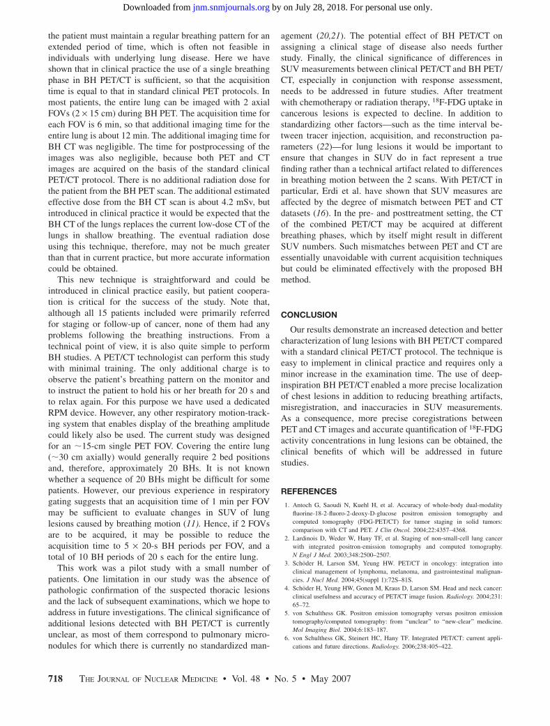
BH PET/CT had the advantage of reducing misregistra-tion, as both the CT and the PET component were acquiredin the same respiratory phase. This was particularly usefulfor distinguishing between pulmonary and extrapulmonarylesions in the chest wall or mediastinum. In 1 patient, focal18F-FDG uptake localized to the lung on clinical PET/CTcould be proven to represent a rib metastasis on BH PET/CT(Fig. 5). One small nodule that was not seen on clinical PET/CT showed mild 18F-FDG uptake on BH PET/CT; this wasan additional site of metastasis in patient with lymphangitis.
Pleural or pericardial effusions showed no 18F-FDGuptake on either clinical PET or BH PET. The same wastrue for lung cavities and areas of GGOs.
DISCUSSION
This study demonstrates that the new technique of deep-inspiration BH PET/CT is superior to standard clinicalPET/CT for the detection and characterization of thoracic
TABLE 3Location of Pulmonary Lesions Detected on Clinical CT
(CCT) and BH CT
Location of lesions CCT BH CT
Right upper lobe 14 20Right middle lobe 3 8
Right lower lobe 10 12
Left upper lobe 5 10Lingula 1 2
Left lower lobe 5 7
Some lesions extended beyond interlobar fissures and, thus,
were noted in 2 adjacent pulmonary lobes. Therefore, the number of
lesions listed here is larger than the total number of lesions
detected, which is shown in Table 2.
FIGURE 2. Patient with widespreadmetastatic colonic adenocarcinoma tolungs, with consolidations and GGOs.Although clinical CT demonstrates find-ings (A and B), BH examination (C and D)has much better quality to define lesions’borders and characteristics.
BREATH-HOLD PET/CT FOR CHEST LESIONS • Meirelles et al. 715
by on July 28, 2018. For personal use only. jnm.snmjournals.org Downloaded from
lesions. When compared with clinical scans, the BHmethod permitted detection of more pulmonary lesions onCT in 87% of patients. It also reduced breathing-inducedartifacts, which would otherwise lead to misregistration ofimages on the fused dataset, inaccuracies in SUV measure-ments, and erroneous localization of lung or chest walllesions.
The use of BH PET/CT enabled a better localization anddefinition of the lesions, especially in the lungs, whencompared with the regular clinical PET/CT protocol. In1 case, the clinical stage of a patient with lung cancer waschanged on BH PET/CT, because of a clearer visualizationon CT of findings compatible with carcinomatous lymph-angitis in the lung.
A major advantage of the BH PET/CT technique is theprecise registration of fused PET/CT images. Although theexperienced PET reader may often account for misregis-tration artifacts, in some cases this misalignment can pose aserious diagnostic problem (12,16,17). This is exemplified
in 1 of our patients with a chest lesion that was incorrectlylocalized to the right lung on clinical images but wasproven to be located in a rib using the BH technique.
Previously, shallow expiration has been recommended asa method to minimize motion artifacts. However, althoughshallow breathing can minimize the lung motion, it doesnot eliminate it completely. This can be achieved only in aclinically practical manner with our proposed BH PET/CTtechnique, which eliminates all motion artifacts.
In cancer patients, PET and PET/CT are increasingly usedin evaluation of the response to therapy. For this intent, mostphysicians rely on changes in SUV measurements from se-rial PET studies over time (18,19). However, as imagesacquired during free breathing are prone to respiratory arti-facts, with blurring of lesions and mismatch between PETand CT data, inaccuracies in PET measures of SUV maypropagate into undesirable overestimation or underestima-tion of the real treatment response (9,16). Adoption of theBH technique enables a more precise measurement of
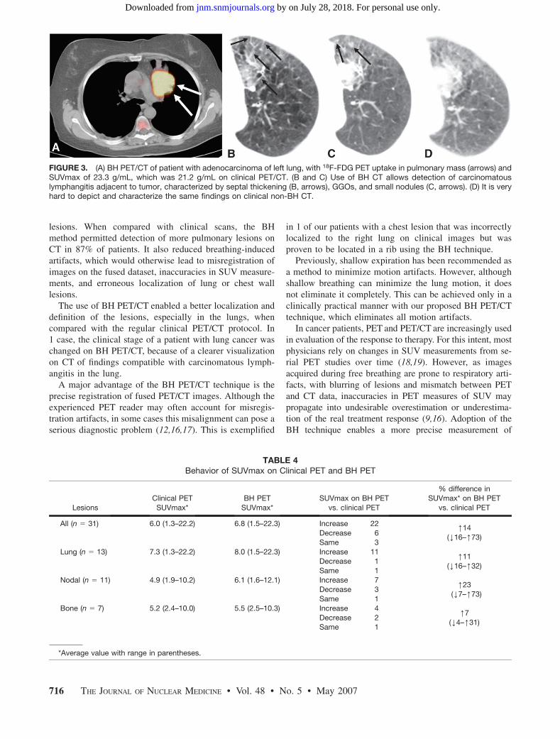
FIGURE 3. (A) BH PET/CT of patient with adenocarcinoma of left lung, with 18F-FDG PET uptake in pulmonary mass (arrows) andSUVmax of 23.3 g/mL, which was 21.2 g/mL on clinical PET/CT. (B and C) Use of BH CT allows detection of carcinomatouslymphangitis adjacent to tumor, characterized by septal thickening (B, arrows), GGOs, and small nodules (C, arrows). (D) It is veryhard to depict and characterize the same findings on clinical non-BH CT.
TABLE 4Behavior of SUVmax on Clinical PET and BH PET
Lesions
Clinical PET
SUVmax*
BH PET
SUVmax*
SUVmax on BH PET
vs. clinical PET
% difference in
SUVmax* on BH PET
vs. clinical PET
All (n 5 31) 6.0 (1.3–22.2) 6.8 (1.5–22.3) Increase 22
SUVs, and SUV measures obtained from BH images of lunglesions are expected to be higher than those from non-BHimages (9,11). In our study, SUV numbers calculated on BHPET were up to 73% higher than those on clinical PET. Suchan increase in SUV is more pronounced in lesions suscep-tible to breathing motion, including small lung nodules orlymph nodes in mobile sites, such as the pulmonary hila.Indeed, the vast majority of lung and hilar lesions showed anincrease in SUVmax on the BH PET images. On the otherhand, in some lesions, the SUVon the BH images was lowerthan that on clinical PET. This included one small lunglesion with SUVmax of 1.6 compared with 1.9. However, itshould be noted that SUV numbers below 2.5 are moreprone to inaccuracies, as the lesion contrast is low and imagenoise increases the uncertainty in SUV calculation. Inanother 5 lesions, the SUV numbers on BH PET were alsoslightly lower than those on the clinical study, but thesedifferences were between 4% and 7% and, thus, within thelimits of reproducibility for SUV measurements in general(18). However, more commonly, increases in SUV wereobserved on BH PET images, exceeding 10% in 15 of the
total of 31 lesions in our study. Indeed, increases in SUV arethe expected consequence of BH imaging in lesions withsignificant motion artifacts. In this pilot study, we did notobserve any SUV change that would have resulted in desig-nating a given lesion malignant instead of benign (or viceversa) if certain cut-offs, such as SUV 2.5 or 3.0, were to beused in characterizing lung nodules. This question should beaddressed in future investigations. Lesions in vertebral bod-ies and large lesions in the lung parenchyma or large lymphnodes may show less movement with respiration and,accordingly, changes in the SUV may not be so remarkable.
We did not observe any obvious differences in clinicalstage between clinical PET and BH PET. However, we wereable to characterize the pattern of carcinomatous lymph-angitis in one case on BH CT, which would not have beenpossible on clinical CT.
In contrast to 4D PET/CT, where data are acquired inseveral breathing phases (11), deep-inspiration BH PET/CTuses data from only a single respiratory phase, whichconsiderably shortens the acquisition time from about 10to 3 min per bed position. In addition, during 4D PET/CT,
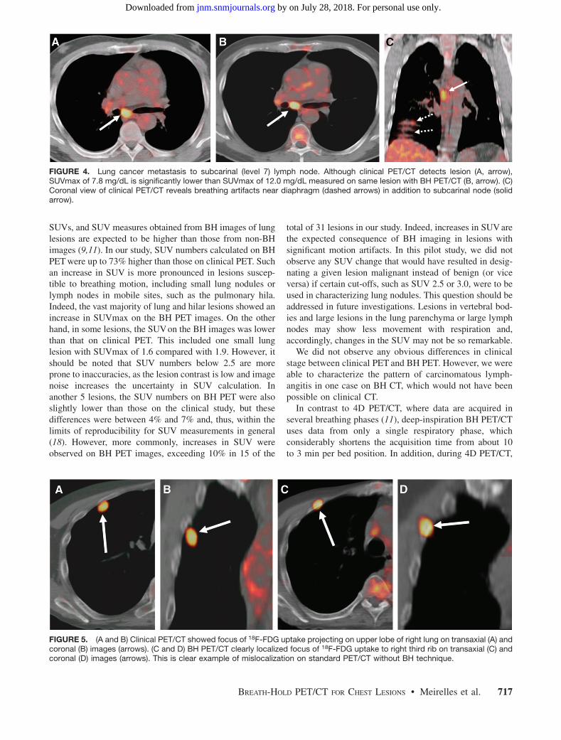
FIGURE 4. Lung cancer metastasis to subcarinal (level 7) lymph node. Although clinical PET/CT detects lesion (A, arrow),SUVmax of 7.8 mg/dL is significantly lower than SUVmax of 12.0 mg/dL measured on same lesion with BH PET/CT (B, arrow). (C)Coronal view of clinical PET/CT reveals breathing artifacts near diaphragm (dashed arrows) in addition to subcarinal node (solidarrow).
FIGURE 5. (A and B) Clinical PET/CT showed focus of 18F-FDG uptake projecting on upper lobe of right lung on transaxial (A) andcoronal (B) images (arrows). (C and D) BH PET/CT clearly localized focus of 18F-FDG uptake to right third rib on transaxial (C) andcoronal (D) images (arrows). This is clear example of mislocalization on standard PET/CT without BH technique.
BREATH-HOLD PET/CT FOR CHEST LESIONS • Meirelles et al. 717
by on July 28, 2018. For personal use only. jnm.snmjournals.org Downloaded from
the patient must maintain a regular breathing pattern for anextended period of time, which is often not feasible inindividuals with underlying lung disease. Here we haveshown that in clinical practice the use of a single breathingphase in BH PET/CT is sufficient, so that the acquisitiontime is equal to that in standard clinical PET protocols. Inmost patients, the entire lung can be imaged with 2 axialFOVs (2 · 15 cm) during BH PET. The acquisition time foreach FOV is 6 min, so that additional imaging time for theentire lung is about 12 min. The additional imaging time forBH CT was negligible. The time for postprocessing of theimages was also negligible, because both PET and CTimages are acquired on the basis of the standard clinicalPET/CT protocol. There is no additional radiation dose forthe patient from the BH PET scan. The additional estimatedeffective dose from the BH CT scan is about 4.2 mSv, butintroduced in clinical practice it would be expected that theBH CT of the lungs replaces the current low-dose CT of thelungs in shallow breathing. The eventual radiation doseusing this technique, therefore, may not be much greaterthan that in current practice, but more accurate informationcould be obtained.
This new technique is straightforward and could beintroduced in clinical practice easily, but patient coopera-tion is critical for the success of the study. Note that,although all 15 patients included were primarily referredfor staging or follow-up of cancer, none of them had anyproblems following the breathing instructions. From atechnical point of view, it is also quite simple to performBH studies. A PET/CT technologist can perform this studywith minimal training. The only additional charge is toobserve the patient’s breathing pattern on the monitor andto instruct the patient to hold his or her breath for 20 s andto relax again. For this purpose we have used a dedicatedRPM device. However, any other respiratory motion-track-ing system that enables display of the breathing amplitudecould likely also be used. The current study was designedfor an ;15-cm single PET FOV. Covering the entire lung(;30 cm axially) would generally require 2 bed positionsand, therefore, approximately 20 BHs. It is not knownwhether a sequence of 20 BHs might be difficult for somepatients. However, our previous experience in respiratorygating suggests that an acquisition time of 1 min per FOVmay be sufficient to evaluate changes in SUV of lunglesions caused by breathing motion (11). Hence, if 2 FOVsare to be acquired, it may be possible to reduce theacquisition time to 5 · 20-s BH periods per FOV, and atotal of 10 BH periods of 20 s each for the entire lung.
This work was a pilot study with a small number ofpatients. One limitation in our study was the absence ofpathologic confirmation of the suspected thoracic lesionsand the lack of subsequent examinations, which we hope toaddress in future investigations. The clinical significance ofadditional lesions detected with BH PET/CT is currentlyunclear, as most of them correspond to pulmonary micro-nodules for which there is currently no standardized man-
agement (20,21). The potential effect of BH PET/CT onassigning a clinical stage of disease also needs furtherstudy. Finally, the clinical significance of differences inSUV measurements between clinical PET/CT and BH PET/CT, especially in conjunction with response assessment,needs to be addressed in future studies. After treatmentwith chemotherapy or radiation therapy, 18F-FDG uptake incancerous lesions is expected to decline. In addition tostandardizing other factors—such as the time interval be-tween tracer injection, acquisition, and reconstruction pa-rameters (22)—for lung lesions it would be important toensure that changes in SUV do in fact represent a truefinding rather than a technical artifact related to differencesin breathing motion between the 2 scans. With PET/CT inparticular, Erdi et al. have shown that SUV measures areaffected by the degree of mismatch between PET and CTdatasets (16). In the pre- and posttreatment setting, the CTof the combined PET/CT may be acquired at differentbreathing phases, which by itself might result in differentSUV numbers. Such mismatches between PET and CT areessentially unavoidable with current acquisition techniquesbut could be eliminated effectively with the proposed BHmethod.
CONCLUSION
Our results demonstrate an increased detection and bettercharacterization of lung lesions with BH PET/CT comparedwith a standard clinical PET/CT protocol. The technique iseasy to implement in clinical practice and requires only aminor increase in the examination time. The use of deep-inspiration BH PET/CT enabled a more precise localizationof chest lesions in addition to reducing breathing artifacts,misregistration, and inaccuracies in SUV measurements.As a consequence, more precise coregistrations betweenPET and CT images and accurate quantification of 18F-FDGactivity concentrations in lung lesions can be obtained, theclinical benefits of which will be addressed in futurestudies.
REFERENCES
1. Antoch G, Saoudi N, Kuehl H, et al. Accuracy of whole-body dual-modality
fluorine-18-2-fluoro-2-deoxy-D-glucose positron emission tomography and
computed tomography (FDG-PET/CT) for tumor staging in solid tumors:
comparison with CT and PET. J Clin Oncol. 2004;22:4357–4368.
2. Lardinois D, Weder W, Hany TF, et al. Staging of non-small-cell lung cancer
with integrated positron-emission tomography and computed tomography.
N Engl J Med. 2003;348:2500–2507.
3. Schoder H, Larson SM, Yeung HW. PET/CT in oncology: integration into
clinical management of lymphoma, melanoma, and gastrointestinal malignan-
cies. J Nucl Med. 2004;45(suppl 1):72S–81S.
4. Schoder H, Yeung HW, Gonen M, Kraus D, Larson SM. Head and neck cancer:
clinical usefulness and accuracy of PET/CT image fusion. Radiology. 2004;231:
65–72.
5. von Schulthess GK. Positron emission tomography versus positron emission
tomography/computed tomography: from ‘‘unclear’’ to ‘‘new-clear’’ medicine.
Mol Imaging Biol. 2004;6:183–187.
6. von Schulthess GK, Steinert HC, Hany TF. Integrated PET/CT: current appli-
cations and future directions. Radiology. 2006;238:405–422.
718 THE JOURNAL OF NUCLEAR MEDICINE • Vol. 48 • No. 5 • May 2007
by on July 28, 2018. For personal use only. jnm.snmjournals.org Downloaded from
Heiko SchöderGustavo S.P. Meirelles, Yusuf Emre Erdi, Sadek A. Nehmeh, Olivia D. Squire, Steven M. Larson, John L. Humm and Detection and Characterization of Thoracic LesionsDeep-Inspiration Breath-Hold PET/CT: Clinical Findings with a New Technique for
http://jnm.snmjournals.org/content/48/5/712This article and updated information are available at:
Information about subscriptions to JNM can be found at:
http://jnm.snmjournals.org/site/misc/permission.xhtmlInformation about reproducing figures, tables, or other portions of this article can be found online at:
(Print ISSN: 0161-5505, Online ISSN: 2159-662X)1850 Samuel Morse Drive, Reston, VA 20190.SNMMI | Society of Nuclear Medicine and Molecular Imaging
is published monthly.The Journal of Nuclear Medicine