Deep Learning in Radiology: Recent Advances, Challenges and Future Trends Sarfaraz Hussein*, Harish Raviprakash* Uygar Teomete ⱡ , Ulas Bagci* * UCF Center for Research in Computer Vision ⱡ University of Miami Hospital
Transcript
Deep Learning in Radiology: Recent Advances, Challenges and Future
* UCF Center for Research in Computer Vision ⱡ University of Miami Hospital
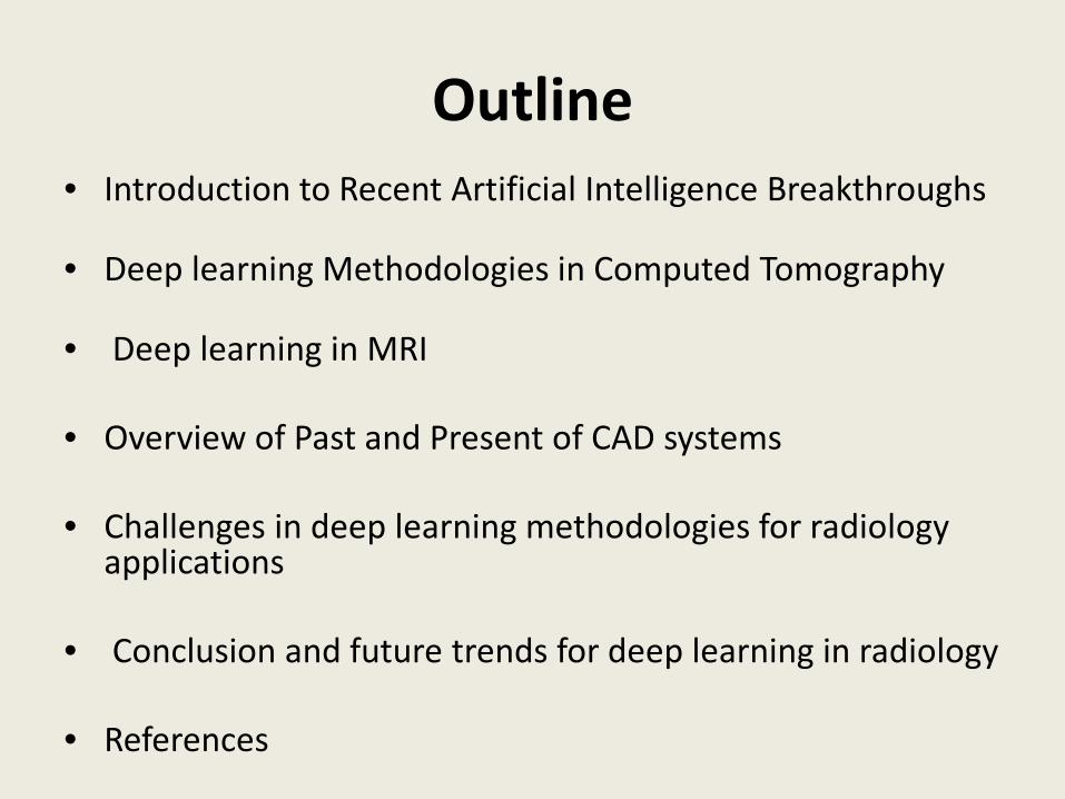
Outline • Introduction to Recent Artificial Intelligence Breakthroughs
• Deep learning Methodologies in Computed Tomography
• Deep learning in MRI
• Overview of Past and Present of CAD systems
• Challenges in deep learning methodologies for radiology
applications
• Conclusion and future trends for deep learning in radiology
• References
Recent AI Breakthroughs: Deep learning
• Deep learning algorithms can facilitate clinicians and radiologists in diagnosis and treatment planning.
• Following are some popular categories in DL: – Deep Belief Networks – Convolutional Neural Networks (CNN)
• 2D CNN • 3D CNN
– Auto-encoders – Recurrent Neural Networks
• Long Short Term Memory
Deep learning (DL) is a computer technology inspired by the functioning of brain. Artificial neural networks
automatically discover patterns in humongous amount of data. Data can be text, images, videos or any of your
choice
Images Credits: Nature, Science,
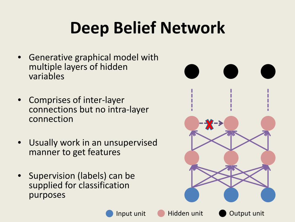
Deep Belief Network • Generative graphical model with
multiple layers of hidden variables
• Comprises of inter-layer
connections but no intra-layer connection
• Usually work in an unsupervised
manner to get features • Supervision (labels) can be
supplied for classification purposes
Input unit Hidden unit Output unit
Deep Belief Network- Applications
Face samples generated using DBN [1]
Images of digits generated using DBN[4]
Generated axial slices of brain [2]
Samples generated using Convolutional DBN [3]
Convolutional Neural Networks • In case of images there can be
thousands/millions of neurons (units)
• Use local connectivity of neurons to address over-parameterization
• The higher level features are found to be useful for image recognition
• Comprises of the following 4 stages: – Convolution – Non-linearity – Sub-sampling – Classification
A and B are connected to only a group of units rather than all of them [5]
Convolutional Neural Networks (2D CNN)
CNN architecture showing convolutional, max pooling, fully-connected and softmax (classification) layers [6] Visualizing learnt features from different layers of CNN [7,8]
Convolutional Neural Networks (3D CNN)
3D Convolutional Neural Network for Human Action Recognition [9]
Difference between 2D convolution when applied on (a) an image (b) video (volume) and (c) 3D convolution when applied on a volume [10]
Auto-encoders
• An unsupervised neural network
• Weights in the network are learnt so as to make the target values equal to the input values
• Comprises of two stages: – Encoding: maps input to a hidden representation – Decoding: the hidden representation is mapped back so as to be as close
to the input as possible
• In a denoising auto-encoder, the network is trained to reconstruct input from its corrupted version.
Auto-encoders [11]
• Application of deep auto-encoder to learn the hidden units that can reconstruct the image of a dress.
• The original image of a dress is encoded into a compressed form and then decoded to generate the reconstructed image of the same dress.
• During the stages of encoding and decoding, the network learns the parameters (weights) [11]
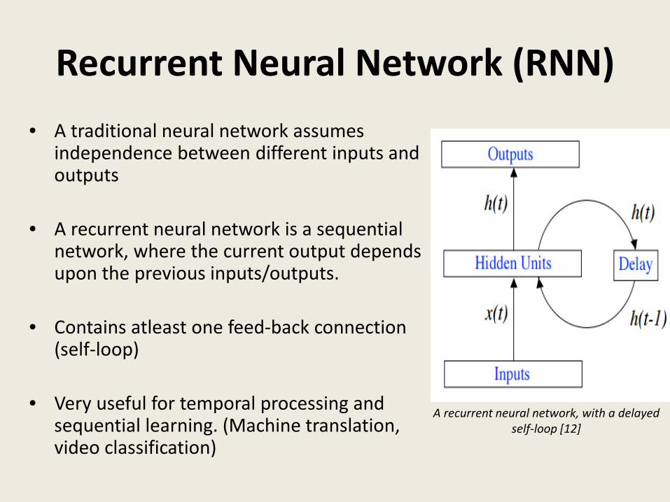
Recurrent Neural Network (RNN) • A traditional neural network assumes
independence between different inputs and outputs
• A recurrent neural network is a sequential
network, where the current output depends upon the previous inputs/outputs.
• Contains atleast one feed-back connection (self-loop)
• Very useful for temporal processing and sequential learning. (Machine translation, video classification)
A recurrent neural network, with a delayed self-loop [12]
Long Short Term Memory (LSTM) • RNNs struggle to model the long-term
dependencies between different inputs
• As the time different between input increases, the gradients of error used to train RNN start vanishing.
• LSTMs are designed to solve the long-term dependency problem of RNN
• An LSTM memory unit comprises of 3 gates: – Input gate: Controls the input signal to pass or
block – Forget gate: Decides whether to remember or
forget the previous state – Output gate: Allows/disallows the output of
the memory cell to pass to the next neurons
An LSTM memory cell with input gate, forget gate, output gate and a self-recurrent
connection [13]
Deep learning Methodologies in Computed Tomography
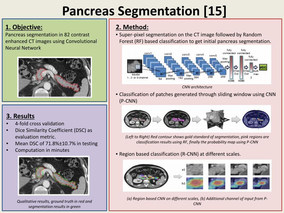
• Deep learning has been applied to a range of problems in Computed Tomography including: – Anatomy recognition [14] – Organ segmentation
evaluation metric. • Mean DSC of 71.8%±10.7% in testing • Computation in minutes
CNN architecture
(Left to Right) Red contour shows gold standard of segmentation, pink regions are classification results using RF, finally the probability map using P-CNN
(a) Region based CNN on different scales, (b) Additional channel of input from P-CNN Qualitative results, ground truth in red and
segmentation results in green
2D/3D Registration [17] 1. Objective: To perform realtime 2D/3D registration using CNN based regression
2. Method: • The goal is to train a CNN regressor to map from 2D/3D image to their
transformation parameter difference • Local image residual features are extracted representing difference
between rendered image and the X-ray image in local patches. • Regression problem is simplified by partitioning the parameter space
into 3 groups based on their difficulty • The CNN architecture comprises of 2 convolutional, 2 maxpooling and
1 fully connected layers.
3. Results • Evaluation on knee-prosthesis, virtual implant system and X-
ray echo fusion datasets • Evaluation metric: Mean target registration error in the
projection direction • Significant improvement in performance as well as time.
2D X-Ray image and a 3D model of the target object
Local Image Residuals
CNN Regression Model
Examples of Region of Interest and Local Image Residuals from the 3 datasets
Lung Texture Classification/Airway Detection [18] 1. Objective: Lung texture classification and airway detection using Convolutional classification Restricted Boltzmann Machine (RBM)
2. Method: • A classification RBM is constructed by having a extra layer of labeled
nodes to the visible layer. • As in convolutional neural network, a convolutional RBM use the
same weight sharing approach. • A convolutional classification RBM (CC-RBM) have all visible, hidden
and label layers • A CC-RBM can be trained as a discriminative model and be tested to
perform classification
3. Results • Lung tissue classification on 73 scans, 40 scans for airway
detection. • A combination of generative and discriminative learning gives
better classification accuracy than either of them alone.
Airway dataset ; airway centerline (green) and non-airway (red)
Convolutional RBM A classification RBM
Features learnt from Lung texture (left) and Airway (right) datasets.
Convolutional Classification RBM
Lymph Node Detection [19] 1. Objective: Lymph node (LN) detection in 176 CT scans using 2.5D Convolutional Neural Network.
2. Method: • The 3 views of CT are considered as different channels (RGB) of an
image.
• Data augmentation is performed using random translation and rotation
• 2 convolution, a max pooling, 2 locally fully connected and one drop-connect layers.
3. Results • 3-fold cross validation
• 388 mediastinal and 595 abdominal
lymph nodes
• Classification sensitivity of 70% for mediastinum and 83% for abdominal lymph node with 3 False Positives per patient
Lymph node in an axial CT slice marked in green
Data augmentation by random translation and rotation
CNN Network and the learnt features from the first layer
Lung Nodule Detection [20] 1. Objective: Detection of lung nodules in low dose CT images with CNN based False Positive rejection
2. Method: • Generating a substantial number of nodule candidates using 3
different methods. • Separate candidate generation methods for solid, sub-solid and large
solid nodules • A 50x50mm patch is generated around each nodule candidate which
serves as an input to 2D Convolutional Network • For each of the candidate nodules, 9 different views are considered
for classification • Different strategies are employed to fuse the outputs corresponding
to the 9 views
3. Results • Evaluations on 3 Low Dose CT datasets
with 1018, 55 and 612 scans • Sensitivity of 90.1% with 4 False
Positives per scan
Pulmonary Nodules across different views
A figurative overview of the method
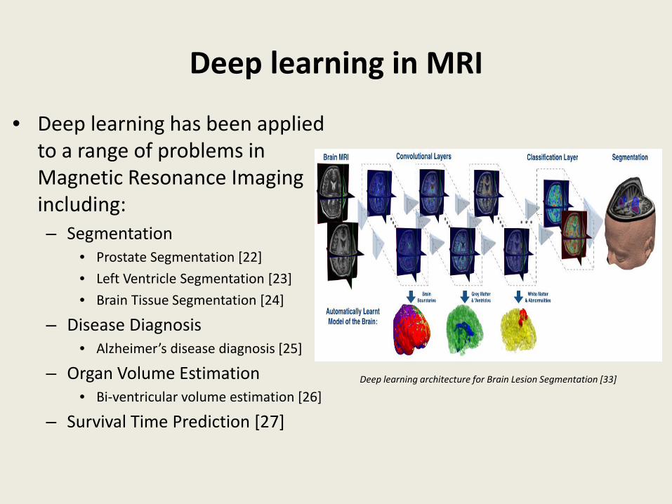
Deep learning in MRI
• Deep learning has been applied to a range of problems in Magnetic Resonance Imaging including: – Segmentation
– Organ Volume Estimation • Bi-ventricular volume estimation [26]
– Survival Time Prediction [27]
Deep learning architecture for Brain Lesion Segmentation [33]
Prostate Segmentation[22] 1. Objective: Prostate segmentation in 66 T2-weighted MR images using Stacked Sparse Auto Encoder and Sparse Patch Matching.
2. Method: • High level feature representation of the image patch.
• Infer likelihood map of prostate gland by using sparse patch matching with all atlases.
• Use the likelihood map to identify initialization region for deformable segmentation as well as an appearance force to drive the segmentation.
3. Results The proposed method reports Dice ratio, precision, Hausdorff distance, and average surface distance of 87.1±4.2, 87.1±7.3, 8.12±2.89 and 1.66±0.49 respectively.
Supervised SSAE architecture
Sparse Patch Mapping schematic
Qualitative results: ground truth in red and segmentation results in yellow. 3D visualization: Ground truth in gray, segmentation result in red
Left Ventricle Segmentation[23] 1. Objective: Left Ventricle segmentation 45 Cardiac MR images using Convolutional Neural Networks, Stacked Auto Encoder and Deformable Segmentation.
2. Method: • Detect LV and compute ROI around it by training the CNN.
• Stacked Encoders are used to infer the shape of LV from the ROI image.
• Use the inferred shape as initialization for deformable segmentation
of the LV.
3. Results The proposed method reports percentage of good contours, Dice metric, average perpendicular distance and conformity, were computed as 96.69%, 0.94, 1.81 mm and 0.86 respectively.
CNN Architecture
Stacked Auto Encoders
Qualitative results: ground truth in red and segmentation results green at the apex and mid
LV
Brain Tissue Segmentation[24] 1. Objective: Brain Tissue segmentation of developing neonates as well as young adults and ageing adults with T2-weighted or T-1 weighted MR images using Convolutional Neural Networks.
2. Method: • Multi-scale patches are fed into the CNN for voxel-wise classification
into different tissue classes.
3. Results The method reported for preterm infants 30weeks PMA, 40weeks PMA, ageing adults and young adults Dice coefficients across all tissue classes as 0.87,0.82,0.86 and 0.91 respectively.
CNN Architecture
Qualitative results: T2-weighted Image, ground truth and automatic segmentation results
30 weeks PMA (a) for the lateral sulcus using (from left to right), manual segmentation (b), only a patch of 25 25 voxels (c), only a patch of 51 51 voxels (d), only a patch of 75 75 voxels (e), and these 3 patch sizes combined (f). The tissues are labelled as follows: uWM in blue, cGM in yellow, and eCSF in red
Alzheimer’s Disease Diagnosis[25] 1. Objective:
AD, MCI, NC classification in 210 subjects using Convolutional Neural Networks.
2. Method: • Feature extraction based on reconstructing the input
• Multi-scale patches are fed into the CNN for voxel-wise classification into different tissue classes.
3. Results
• Accuracy as evaluation metric • five classification tasks: AD vs. NC,
AD+MCI vs. NC, AD vs. MCI, MCI vs NC and AD vs. MCI vs. NC.
Direct estimation of cardiac ventricular volumes using Recurrent Boltzmann Machines and Regression Forests.
2. Method: • Extract from multi-scale deep networks feature vector. • Regression Forest to predict if feature vector is biventricular cavity.
CNN Architecture
Multi-Scale Deep Network Architecture
Feature Learning and RF Regression
3. Results • Ejection Fraction (EF) is used to
estimate results. • Correlation of EF with Ground-truth
results in coefficient of 0.921 and .908 for LV and RV respectively.
EF: Manual(blue) vs Estimated (red)
Survival Time Prediction[27] 1. Objective:
Predicting survival time for 69 patients high-grade glioma using T1-MRI,DTI,fMRI and Convolutional Neural Networks, Principal Component Analysis (PCA) and Sparse Representation (SR).
2. Method: • Extract feature vector for fMRI and DTI from multi-channel CNN.
• Extract feature vector for T1-MRI from single channel CNN • Fuse the feature vectors and do PCA and SR to reduce dimensionality. • SVM to predict.
mCNN Architecture
Multi-modal Deep Network Architecture
3. Results Accuracy 89%
Prediction accuracy
Overview of Past and Present of CAD Systems
• Common Algorithms used • Basic CAD System • The popular applications
of CAD systems include: – Breast cancer detection – Lung cancer detection – EEG Signals
CAD for Breast Lesion and Lung Nodules using stacked denoising autoencoder [32]
Common Algorithms in CAD [28] • K-nearest neighbors
– Based on the closest training cases in feature space
• Decision Trees – Branching out from a node; similar to tree branches to reach the leaf node
• Fuzzy Logic – Fuzzification of input, followed by inference and then de-fuzzification to output.
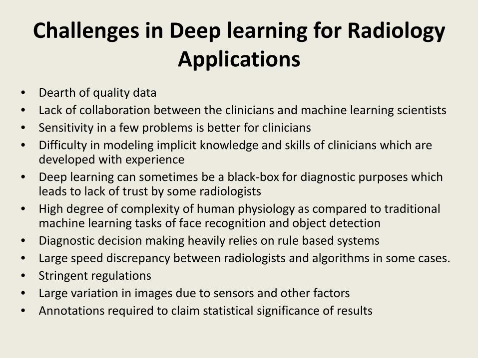
Challenges in Deep learning for Radiology Applications
• Dearth of quality data • Lack of collaboration between the clinicians and machine learning scientists • Sensitivity in a few problems is better for clinicians • Difficulty in modeling implicit knowledge and skills of clinicians which are
developed with experience • Deep learning can sometimes be a black-box for diagnostic purposes which
leads to lack of trust by some radiologists • High degree of complexity of human physiology as compared to traditional
machine learning tasks of face recognition and object detection • Diagnostic decision making heavily relies on rule based systems • Large speed discrepancy between radiologists and algorithms in some cases. • Stringent regulations • Large variation in images due to sensors and other factors • Annotations required to claim statistical significance of results
Potential Solutions: Data Dearth [30] • Transfer learning
– Transfer of knowledge from a non-radiological task but with a lot of annotated data (camera images, videos) to a radiological task
• Since the annotated medical data is limited, an alternative would be to just fine-tune a CNN model rather than train it from scratch.
• Fine-tuning requires a lot less data and outperforms (or performs as well) as network learnt from scratch.
• Fine tuning of CNN also increases the robustness to the size of training data as compared to the scratch trained CNN
• Experiments performed for 4 different clinical tasks with varying imaging modalities: – Polyp detection in colonoscopy videos – Pulmonary Embolism detection in CT pulmonary angiography – Colonoscopy frame classification – Intima-Media Boundary Segmentation in Carotid intima-media thickness
(CIMT) images.
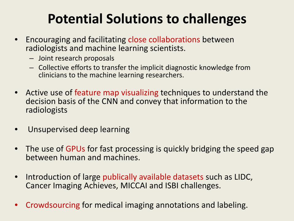
Potential Solutions to challenges • Encouraging and facilitating close collaborations between
radiologists and machine learning scientists. – Joint research proposals – Collective efforts to transfer the implicit diagnostic knowledge from
clinicians to the machine learning researchers.
• Active use of feature map visualizing techniques to understand the decision basis of the CNN and convey that information to the radiologists
• Unsupervised deep learning
• The use of GPUs for fast processing is quickly bridging the speed gap between human and machines.
• Introduction of large publically available datasets such as LIDC, Cancer Imaging Achieves, MICCAI and ISBI challenges.
• Crowdsourcing for medical imaging annotations and labeling.
Conclusion and Future Trends • There is a lot of untapped potential regarding the use
of deep learning for radiology • Healthcare will be most effected by the advancements
in AI than other industries. [31] • Transfer learning can help address the data dearth in
short term • Close collaboration between radiologists and ML
scientists can further advance this field.
References [1] Susskind, Joshua, Volodymyr Mnih, and Geoffrey Hinton. "On deep generative models with applications to recognition." Computer Vision and Pattern Recognition (CVPR), 2011 IEEE Conference on. IEEE, 2011. [2] Brosch, Tom, Roger Tam, and Alzheimer’s Disease Neuroimaging Initiative. "Manifold learning of brain MRIs by deep learning." International Conference on Medical Image Computing and Computer-Assisted Intervention. Springer Berlin Heidelberg, 2013. [3] Wu, Zhirong, et al. "3d shapenets: A deep representation for volumetric shapes." Proceedings of the IEEE Conference on Computer Vision and Pattern Recognition. 2015. [4] Calandra, Roberto, et al. "Learning deep belief networks from non-stationary streams." International Conference on Artificial Neural Networks. Springer Berlin Heidelberg, 2012. [5] Conv Nets: A Modular Perspective ( http://colah.github.io/posts/2014-07-Conv-Nets-Modular/ ) [6] A BRIEF REPORT OF THE HEURITECH DEEP LEARNING MEETUP #5 (https://blog.heuritech.com/2016/02/29/a-brief-report-of-the-heuritech-deep-learning-meetup-5/) [7] Deep Learning Tutorial ICML, Atlanta 2013, Yann LeCun and Marc'Aurelio Ranzato [8] Zeiler, Matthew D., and Rob Fergus. "Visualizing and understanding convolutional networks." European Conference on Computer Vision. Springer International Publishing, 2014. [9] Ji, Shuiwang, et al. "3D convolutional neural networks for human action recognition." IEEE transactions on pattern analysis and machine intelligence35.1 (2013): 221-231. [10] Tran, Du, et al. "Learning spatiotemporal features with 3d convolutional networks." 2015 IEEE International Conference on Computer Vision (ICCV). IEEE, 2015. [11] Deep Style: Inferring the Unknown to Predict the Future of Fashion, TJ TORRES, http://multithreaded.stitchfix.com/blog/2015/09/17/deep-style/ [12] Recurrent Neural Networks Neural Computation : Lecture 12, John A. Bullinaria, 2015 http://www.cs.bham.ac.uk/~jxb/INC/l12.pdf [13] LSTM Networks for Sentiment Analysis, http://deeplearning.net/tutorial/lstm.html [14] Yan, Zhennan, et al. "Multi-Instance Deep Learning: Discover Discriminative Local Anatomies for Bodypart Recognition." IEEE transactions on medical imaging 35.5 (2016): 1332-1343. [15] Roth, Holger R., et al. "Deeporgan: Multi-level deep convolutional networks for automated pancreas segmentation." International Conference on Medical Image Computing and Computer-Assisted Intervention. Springer International Publishing, 2015. [16] Cha, Kenny H., et al. "Urinary bladder segmentation in CT urography using deep-learning convolutional neural network and level sets." Medical physics43.4 (2016): 1882-1896. [17] Miao, Shun, Z. Jane Wang, and Rui Liao. "A CNN Regression Approach for Real-Time 2D/3D Registration." IEEE transactions on medical imaging 35.5 (2016): 1352-1363.
References [18] van Tulder, Gijs, and Marleen de Bruijne. "Combining Generative and Discriminative Representation Learning for Lung CT Analysis With Convolutional Restricted Boltzmann Machines." IEEE transactions on medical imaging 35.5 (2016): 1262-1272. [19] Roth, Holger R., et al. "A new 2.5 D representation for lymph node detection using random sets of deep convolutional neural network observations."International Conference on Medical Image Computing and Computer-Assisted Intervention. Springer International Publishing, 2014. [20] Setio, Arnaud Arindra Adiyoso, et al. "Pulmonary nodule detection in CT images: false positive reduction using multi-view convolutional networks."IEEE transactions on medical imaging 35.5 (2016): 1160-1169. [21] Li, Wen, Fucang Jia, and Qingmao Hu. "Automatic Segmentation of Liver Tumor in CT Images with Deep Convolutional Neural Networks." Journal of Computer and Communications 3.11 (2015): 146. [22] Guo, Yanrong, Yaozong Gao, and Dinggang Shen. "Deformable MR prostate segmentation via deep feature learning and sparse patch matching." IEEE transactions on medical imaging 35.4 (2016): 1077-1089. [23] Avendi, M. R., Arash Kheradvar, and Hamid Jafarkhani. "A combined deep-learning and deformable-model approach to fully automatic segmentation of the left ventricle in cardiac MRI." Medical image analysis 30 (2016): 108-119. [24] Moeskops, Pim, et al. "Automatic segmentation of MR brain images with a convolutional neural network." IEEE transactions on medical imaging 35.5 (2016): 1252-1261. [25] Hosseini-Asl, Ehsan, Robert Keynton, and Ayman El-Baz. "Alzheimer's disease diagnostics by adaptation of 3D convolutional network." Image Processing (ICIP), 2016 IEEE International Conference on. IEEE, 2016. [26] Zhen, Xiantong, et al. "Multi-scale deep networks and regression forests for direct bi-ventricular volume estimation." Medical image analysis 30 (2016): 120-129. [27] Nie, Dong, et al. "3D Deep Learning for Multi-modal Imaging-Guided Survival Time Prediction of Brain Tumor Patients." International Conference on Medical Image Computing and Computer-Assisted Intervention. Springer International Publishing, 2016. [28] Vasilakos, Athanasios V., Yu Tang, and Yuanzhe Yao. "Neural networks for computer-aided diagnosis in medicine: A review." Neurocomputing (2016). [29] https://www.researchgate.net/profile/Yingju_Chen/publication/233806620/figure/fig4/AS:202659295436813@1425329149496/General-steps-involving-in-computer-aided-diagnosis-CAD-system-where-gray-boxes-may-be.png [30] Tajbakhsh, Nima, et al. "Convolutional Neural Networks for Medical Image Analysis: Full Training or Fine Tuning?." IEEE transactions on medical imaging 35.5 (2016): 1299-1312. [31] 5 Industries Being Most Affected By Artificial Intelligence https://www.fowcommunity.com/blog/future-work/5-industries-being-most-affected-artificial-intelligence [32] Cheng, Jie-Zhi, et al. "Computer-Aided Diagnosis with Deep Learning Architecture: Applications to Breast Lesions in US Images and Pulmonary Nodules in CT Scans." Scientific reports 6 (2016). [33] Artificial Intelligence & Machine Learning for Semantic Imaging, Imperial College London http://wp.doc.ic.ac.uk/bglocker/project/semantic-imaging/