65
1 MELBOURNE | Oct.4-5, 2016 Le Lu, PhD, NIH-CC, Oct. 26 th , 2016 (GTC DC Talk DCS16103) DEEP NEURAL NETWORKS IN RADIOLOGY: PREVENTATIVE AND PRECISION MEDICINE PERSPECTIVES
| Date post: | 14-Feb-2017 |
| Category: |
Documents |
| Upload: | phungxuyen |
| View: | 217 times |
| Download: | 0 times |
1
MELBOURNE | Oct.4-5, 2016
Le Lu, PhD, NIH-CC, Oct. 26th, 2016 (GTC DC Talk DCS16103)
DEEP NEURAL NETWORKS IN RADIOLOGY: PREVENTATIVE AND PRECISION MEDICINE PERSPECTIVES
2
OUTLINES
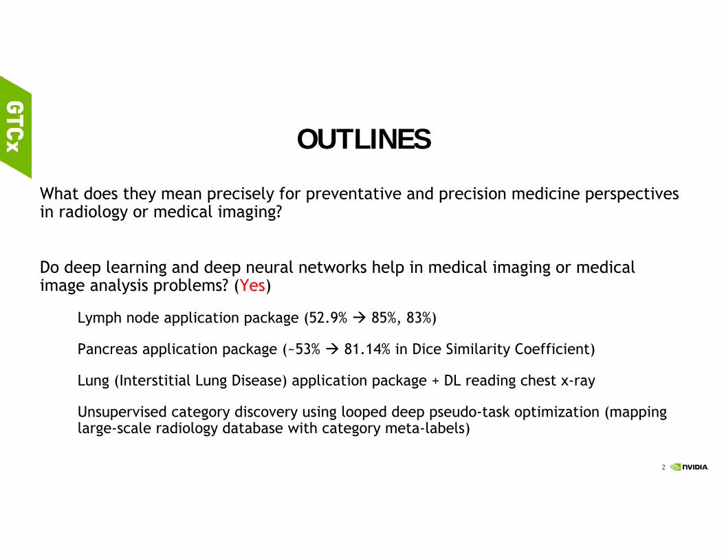
What does they mean precisely for preventative and precision medicine perspectives in radiology or medical imaging?
Do deep learning and deep neural networks help in medical imaging or medical image analysis problems? (Yes)
Lymph node application package (52.9% 85%, 83%)
Pancreas application package (~53% 81.14% in Dice Similarity Coefficient)
Lung (Interstitial Lung Disease) application package + DL reading chest x-ray
Unsupervised category discovery using looped deep pseudo-task optimization (mapping large-scale radiology database with category meta-labels)

3
COMPLEXITY & COMPOSABILITY

4
http://www.slate.com/articles/technology/future_tense/2016/06/microsoft_ceo_satya_nadella_humans_and_a_i_can_work_together_to_solve_society.html
5
PERSPECTIVES
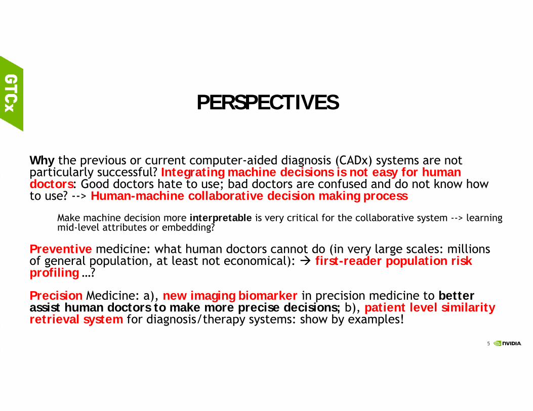
Why the previous or current computer-aided diagnosis (CADx) systems are not particularly successful? Integrating machine decisions is not easy for human doctors: Good doctors hate to use; bad doctors are confused and do not know how to use? --> Human-machine collaborative decision making process
Make machine decision more interpretable is very critical for the collaborative system --> learning mid-level attributes or embedding?
Preventive medicine: what human doctors cannot do (in very large scales: millions of general population, at least not economical): first-reader population risk profiling …?
Precision Medicine: a), new imaging biomarker in precision medicine to better assist human doctors to make more precise decisions; b), patient level similarity retrieval system for diagnosis/therapy systems: show by examples!

6
APPLICATION FOCUS: CANCER IMAGING
Cancer Facts and Figures 2016. Atlanta, Ga: American Cancer Society, 2016. Last accessed February 1, 2016.http://www.cancer.gov/types/common-cancers
Cancer Type Lung (Bronchus)
Colorectal Pancreatic Breast (Female-Male)
Prostate
Estimated New Cases
224,390 134,490 53,070 246,660 –2,600
180,890
Estimated Deaths
158,080 49,190 41,780 40,450 –440
26,120
7
OVERVIEW: THREE CATEGORIES OF KEY PROBLEMS (I)
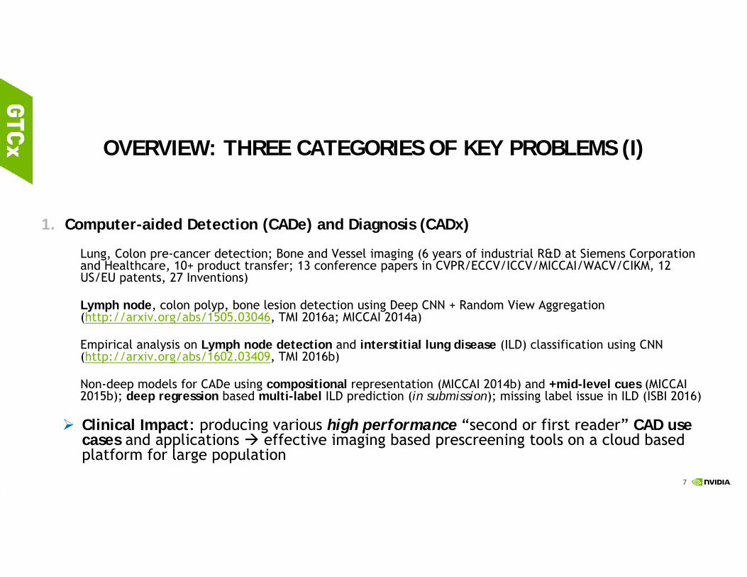
1. Computer-aided Detection (CADe) and Diagnosis (CADx)
Lung, Colon pre-cancer detection; Bone and Vessel imaging (6 years of industrial R&D at Siemens Corporation and Healthcare, 10+ product transfer; 13 conference papers in CVPR/ECCV/ICCV/MICCAI/WACV/CIKM, 12 US/EU patents, 27 Inventions)
Lymph node, colon polyp, bone lesion detection using Deep CNN + Random View Aggregation (http://arxiv.org/abs/1505.03046, TMI 2016a; MICCAI 2014a)
Empirical analysis on Lymph node detection and interstitial lung disease (ILD) classification using CNN (http://arxiv.org/abs/1602.03409, TMI 2016b)
Non-deep models for CADe using compositional representation (MICCAI 2014b) and +mid-level cues (MICCAI 2015b); deep regression based multi-label ILD prediction (in submission); missing label issue in ILD (ISBI 2016)
Clinical Impact: producing various high performance “second or first reader” CAD use cases and applications effective imaging based prescreening tools on a cloud based platform for large population

8
OVERVIEW: THREE CATEGORIES OF KEY PROBLEMS (II)
2. Semantic Segmentation in Medical Image Analysis
“DeepOrgan” for pancreas segmentation (MICCAI 2015a) via scanning superpixels using multi-scale deep features (“Zoom-out”) and probability map embedding http://arxiv.org/abs/1506.06448
Deep segmentation on pancreas and lymph node clusters with HED (Holistically-nested neural networks, Xie & Tu, 2015) as building blocks to learn unary (segmentation mask) and pairwise (labeling segmentation boundary) CRF terms + spatial aggregation or + structured optimization (The focus of three MICCAI 2016 papers since this is a much needed task Small datasets; (de-)compositional representation is still the key.)
CRF: conditional random fields
Clinical Impact: semantic segmentation can help compute clinically more accurate and desirable imaging bio-markers or precision measurement!

9
OVERVIEW: THREE CATEGORIES OF KEY PROBLEMS (III)
3. Interleaved or Joint Text/Image Deep Mining on a Large-Scale Radiology Image Database “large” datasets; no labels (~216K 2D key images/slices extracted from >60K unique patients)
Interleaved Text/Image Deep Mining on a Large-Scale Radiology Image Database (CVPR 2015, a proof of concept study)
Interleaved Text/Image Deep Mining on a Large-Scale Radiology Image Database for Automated Image Interpretation (its extension, JMLR, 17(107):1−31, 2016) http://arxiv.org/abs/1505.00670
Learning to Read Chest X-Rays: Recurrent Neural Cascade Model for Automated Image Annotation, (CVPR 2016) http://arxiv.org/abs/1603.08486
Unsupervised Category Discovery via Looped Deep Pseudo-Task Optimization Using a Large Scale Radiology Image Database, http://arxiv.org/abs/1603.07965
Clinical Impact: eventually to build an automated programmable mechanism to parse and learn from hospital scale PACS-RIS databases to derive semantics and knowledge … has to be deep learning based since effective image features are very hard to be hand-crafted cross different diseases, imaging protocols and modalities.

10
(A.0) Automated Lymph Node Detection
• Difficult due to large variations in appearance, location and pose.
• Plus low contrast against surrounding tissues.
Abdominal lymph node in CTMediastinal lymph node in CT
11
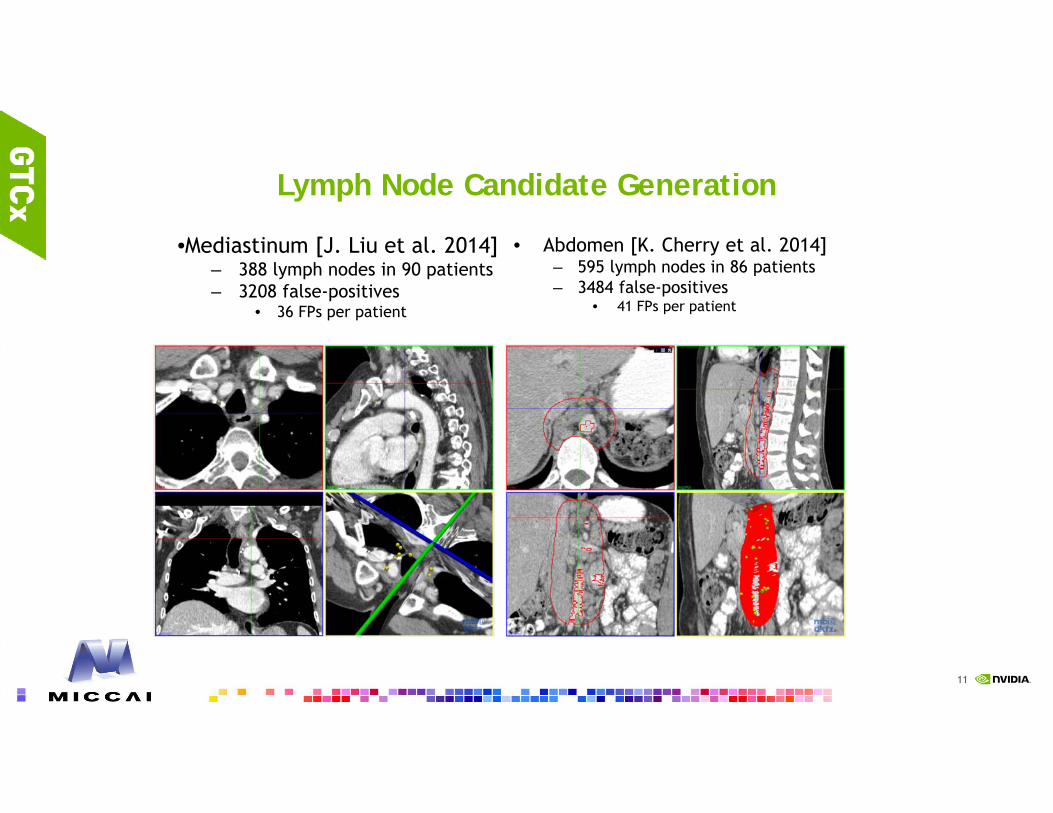
Lymph Node Candidate Generation
•Mediastinum [J. Liu et al. 2014]– 388 lymph nodes in 90 patients– 3208 false-positives
• 36 FPs per patient
• Abdomen [K. Cherry et al. 2014]– 595 lymph nodes in 86 patients– 3484 false-positives
• 41 FPs per patient
•Deep Detection Proposal Generation as future work

12
Shallow Models: 2D View Aggregation Using a Two-Level Hierarchy of Linear Classifiers [Seff et al. MICCAI 2014]
2D slice gallery for a LN candidate VOI (45 x 45 × 45 voxels).
Axial
Coronal
Sagittal
• VOI candidates generated via a random forest classifier using voxel‐level features (not the primary focus of this work), for high sensitivity but also high false positive rates.
• 2.5D: 3 sequences of orthogonal 2D slices then extracted from each candidate VOI (9 x 3 = 27 views).
NDSEG Fellowship
13
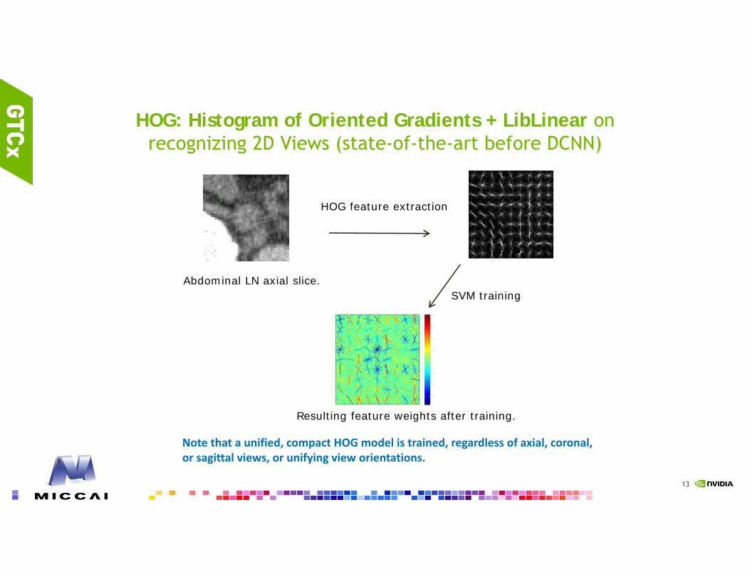
HOG: Histogram of Oriented Gradients + LibLinear on recognizing 2D Views (state-of-the-art before DCNN)
HOG feature extraction
Resulting feature weights after training.
Abdominal LN axial slice.SVM training
Note that a unified, compact HOG model is trained, regardless of axial, coronal, or sagittal views, or unifying view orientations.

14
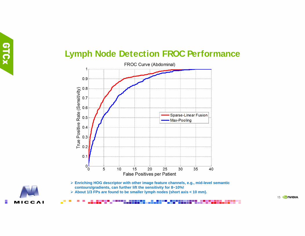
Lymph Node Detection FROC Performance
15
Lymph Node Detection FROC Performance
Enriching HOG descriptor with other image feature channels, e.g., mid-level semantic contours/gradients, can further lift the sensitivity for 8~10%!
About 1/3 FPs are found to be smaller lymph nodes (short axis < 10 mm).

16
MAKE SHALLOW TO GO DEEPER VIA MID-LEVEL CUES? [SEFF ET AL. MICCAI 2015]
We explore a learned transformation scheme for producing enhanced semantic input for HOG, based on LN-selective visual responses.
Mid-level semantic boundary cues learned from segmentation.
All LNs in both target regions are manually segmented by radiologists.
Target region # Patients # LNs
Mediastinal 90 389
Abdominal 86 595
17
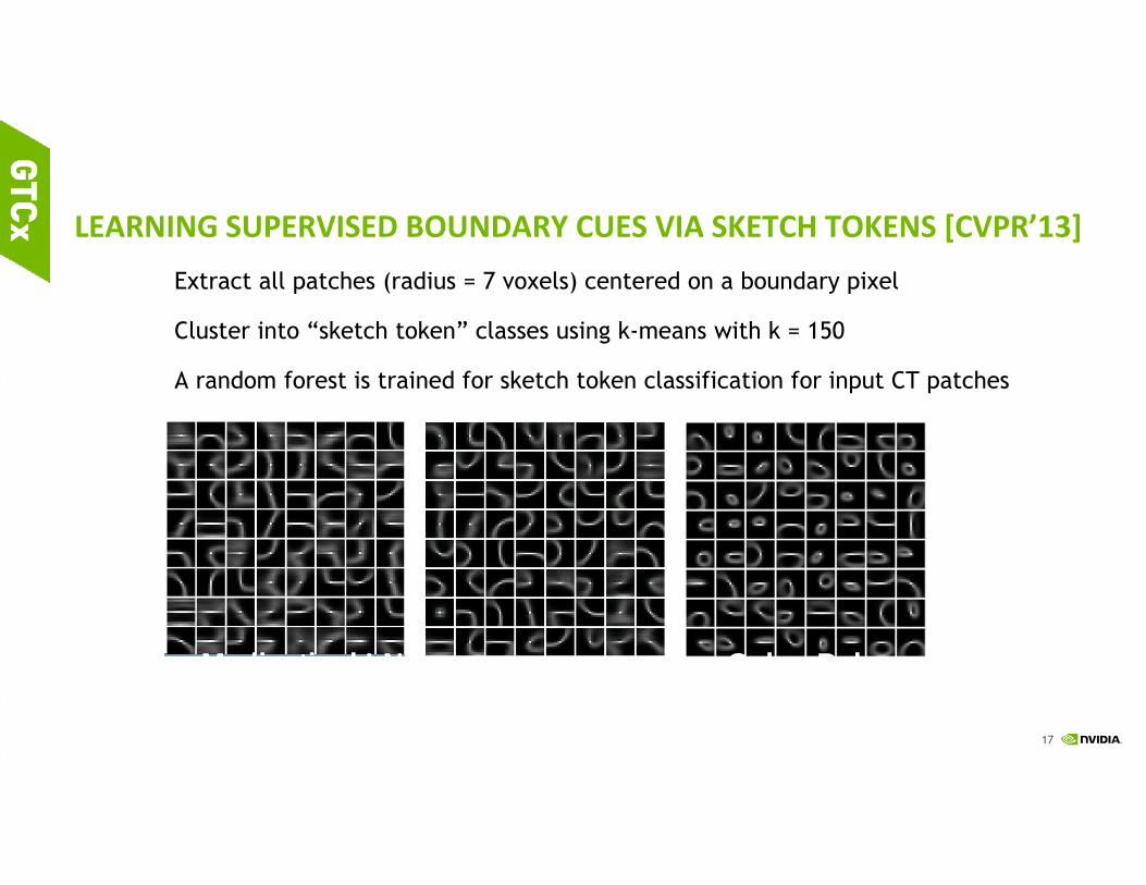
LEARNING SUPERVISED BOUNDARY CUES VIA SKETCH TOKENS [CVPR’13]Extract all patches (radius = 7 voxels) centered on a boundary pixel
Cluster into “sketch token” classes using k-means with k = 150
A random forest is trained for sketch token classification for input CT patches
Abdominal LNMediastinal LN Colon Polyps

18
MULTI-CHANNEL HOG FEATURE MAP CONSTRUCTION
An enhanced, 3-channel feature map:
19
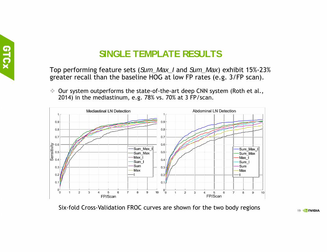
SINGLE TEMPLATE RESULTS
Top performing feature sets (Sum_Max_I and Sum_Max) exhibit 15%-23% greater recall than the baseline HOG at low FP rates (e.g. 3/FP scan).
Our system outperforms the state-of-the-art deep CNN system (Roth et al., 2014) in the mediastinum, e.g. 78% vs. 70% at 3 FP/scan.
Six-fold Cross-Validation FROC curves are shown for the two body regions
20
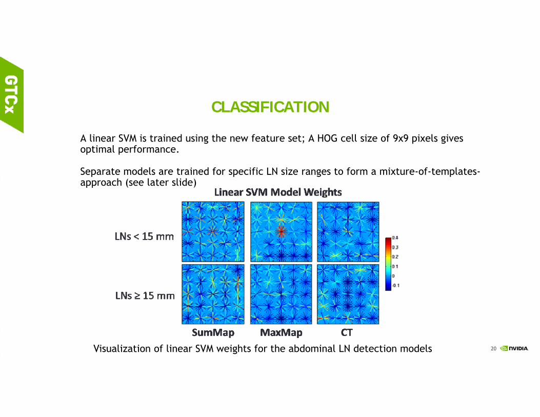
CLASSIFICATION
A linear SVM is trained using the new feature set; A HOG cell size of 9x9 pixels gives optimal performance.
Separate models are trained for specific LN size ranges to form a mixture-of-templates-approach (see later slide)
Visualization of linear SVM weights for the abdominal LN detection models

21
Wide distribution of LN sizes invites the application of size-specific models trained separately.
LNs > 20 mm are especially clinically relevant
Mixture Model Detection Results
Single template and mixture model performance for abdominal models

22
(A.1) Deep models: Random Sets of Convolutional Neural Network Predictions via Compositional Representation [Roth et al. MICCAI 2014, Shin et al. TMI 2016; Roth et al. TMI 2016]
Application to appearance modeling and detecting lymph node
Random translations, rotations and scale
Asst. Prof. Nagoya University (now)

23
Convolutional Neural Network Architecture

24
Experimental Results (~100% sensitivity but ~40 FPs/patient at candidate generation step; then 3-fold CV with data augmentation)
Mediastinum71% @ 3 FPs (was 55%)
Abdomen83% @ 3 FPs (was 30%)

25
Experimental Results (cont.)
Mediastinum82% @ 3 FPs
Abdomen80% @ 3 FPs
Training mediastinum and abdomen Jointly!
26
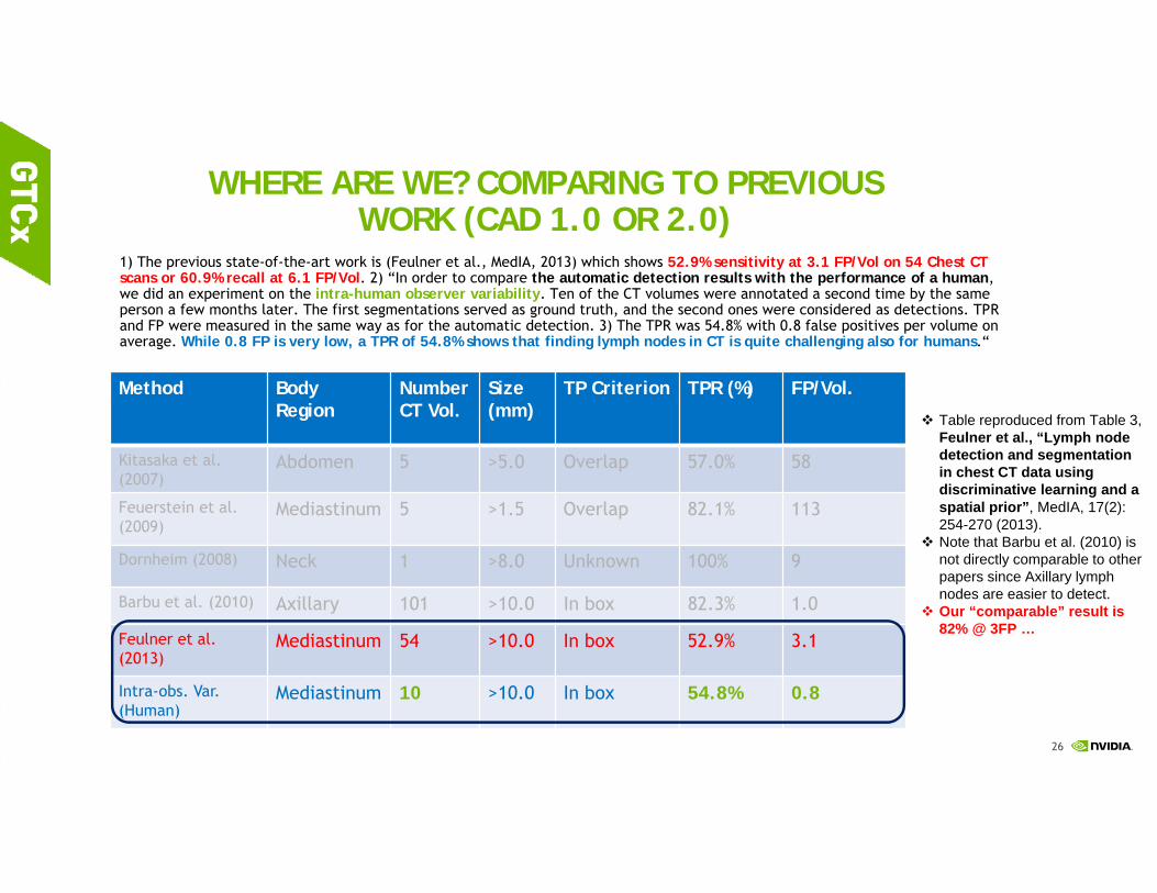
WHERE ARE WE? COMPARING TO PREVIOUS WORK (CAD 1.0 OR 2.0)
1) The previous state-of-the-art work is (Feulner et al., MedIA, 2013) which shows 52.9% sensitivity at 3.1 FP/Vol on 54 Chest CT scans or 60.9% recall at 6.1 FP/Vol. 2) “In order to compare the automatic detection results with the performance of a human, we did an experiment on the intra-human observer variability. Ten of the CT volumes were annotated a second time by the same person a few months later. The first segmentations served as ground truth, and the second ones were considered as detections. TPR and FP were measured in the same way as for the automatic detection. 3) The TPR was 54.8% with 0.8 false positives per volume on average. While 0.8 FP is very low, a TPR of 54.8% shows that finding lymph nodes in CT is quite challenging also for humans.“
Table reproduced from Table 3, Feulner et al., “Lymph node detection and segmentation in chest CT data using discriminative learning and a spatial prior”, MedIA, 17(2): 254-270 (2013).
Note that Barbu et al. (2010) is not directly comparable to other papers since Axillary lymph nodes are easier to detect.
Our “comparable” result is 82% @ 3FP …
Method Body Region
Number CT Vol.
Size (mm)
TP Criterion TPR (%) FP/Vol.
Kitasaka et al. (2007)
Abdomen 5 >5.0 Overlap 57.0% 58
Feuerstein et al. (2009)
Mediastinum 5 >1.5 Overlap 82.1% 113
Dornheim (2008) Neck 1 >8.0 Unknown 100% 9
Barbu et al. (2010) Axillary 101 >10.0 In box 82.3% 1.0
Feulner et al. (2013)
Mediastinum 54 >10.0 In box 52.9% 3.1
Intra-obs. Var. (Human)
Mediastinum 10 >10.0 In box 54.8% 0.8

27
VISUALIZATION ON TRANSFER LEARNING (LEARNED FROM THORACO-ABDOMINAL LNS) [SHIN ET AL., TMI 2016]
KRIBB Fellowship

28
BETTER LOCALIZATION AFTER FINE-TUNING?
Mediastinum LN via Transfer Learning from ImageNet: 85% @ 3 FPswhile Abdomen LN via transfer learning does not help (domain bias).
29
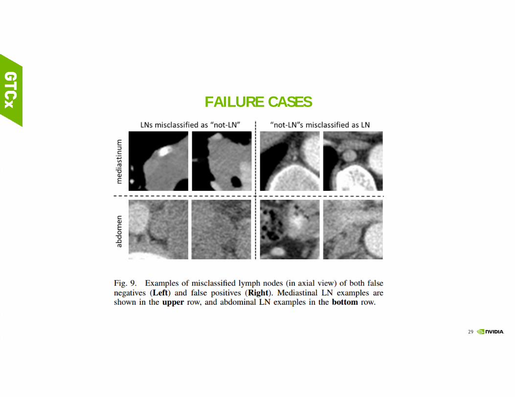
FAILURE CASES

30
Generalizable? Colon CADe Results using a deeper CNN on 1186 patients (or 2372 CTC volumes) via fine-tuning AlexNet
[Roth et al., TMI 2016]
[SVM baseline] Summers, et a., Computed tomographic virtual colonoscopy computer-aided polyp detection in a screening population, Gastroenterology, vol. 129, no. 6, pp.1832–1844, 2005. 1,186 patients with prone and supine CTC images (394/792 patients; 79/173 polyps training/testing split)

31
References:1. "A New 2.5D Representation for Lymph Node Detection using Random Sets of Deep Convolutional Neural Network
Observations", MICCAI 2014
2. "2D View Aggregation for Lymph Node Detection using a Shallow Hierarchy of Linear Classifiers", MICCAI 2014
3. “Detection of Sclerotic Spine Metastases via Random Aggregation of Deep Convolutional Neural Network Classifications", (Oral), MICCAI Spine Imaging Workshop 2014
4. "Leveraging Mid-Level Semantic Boundary Cues for Computer-Aided Lymph Node Detection", MICCAI 2015
5. "An Analysis of Robust Cost Functions for Deep CNN in Computer-aided Diagnosis", MICCAI DLMIA workshop 2015
6. "Anatomy-specific Classification of Medical Images using Deep Convolutional Nets", IEEE ISBI 2015
7. "Improving Computer-aided Detection using Convolutional Neural Networks and Random View Aggregation", IEEE Trans. on Medical Imaging, 2016
8. "Deep Convolutional Neural Networks for Computer-Aided Detection: CNN Architectures, Dataset Characteristics and Transfer Learning", IEEE Trans. on Medical Imaging, 2016
12/29/2016

32
SEMANTIC SEGMENTATION: TOP-DOWN OR BOTTOM-UP
PARADIGMS?
12/29/2016
33
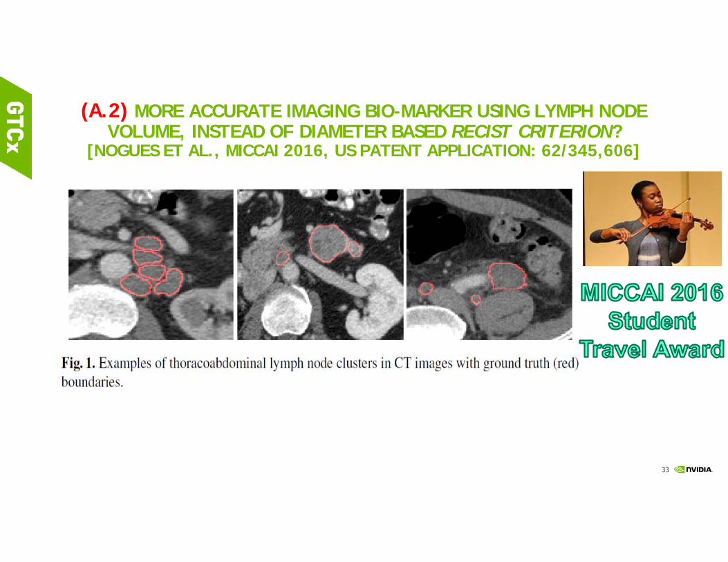
(A.2) MORE ACCURATE IMAGING BIO-MARKER USING LYMPH NODE VOLUME, INSTEAD OF DIAMETER BASED RECIST CRITERION?
[NOGUES ET AL., MICCAI 2016, US PATENT APPLICATION: 62/345,606]

34
Computing much moreprecise imaging based biomarkers, meta-information for precision medicine decision making, beyond the current Clinical Protocol … (Oncology Example) Mining reliable, high accuracy, clinically-relevant meta-measurements from unstructured, noisy low-level imaging data

35
AUTOMATIC LYMPH NODE CLUSTER SEGMENTATION USING HOLISTICALLY-NESTED NEURAL NETWORKS AND STRUCTURED OPTIMIZATION IN CT IMAGES
https://wiki.cancerimagingarchive.net/display/Public/CT+Lymph+Nodes annotated datasets are publicly available)

36
(A.2) A ROADMAP OF BOTTOM-UP DEEP PANCREAS SEGMENTATION: PATCH, REGION, HOLISTIC CNNS [FARAG ET AL., IEEE TIP 2017]
Ground truth Random Forest 2.5D Patch ConvNet prob.
P-ConvNet

37
MULTI-SCALE “ZOOM-OUT” R-CONVNET
Zoom-out in dual spaces

38
DEEPORGAN: R2-CONVNET VIA TWO-CHANNEL ENCODING[ROTH ET AL., MICCAI 2015]
~27%Dice score
~57%Dice score
~68%Dice score

39
4-FOLD CV PERFORMANCE ON 82 CT SCANS
Averaged surface-surface distances: 0.94+/-0.6mm (p<0.01) with R2-ConvNet from 1.46+/-1.5mm if just P-ConvNet is applied.
Previous state-of-the-art: [46.6% to 69.1%] DSC, all under LOO (Leave-one-patient-out).
40
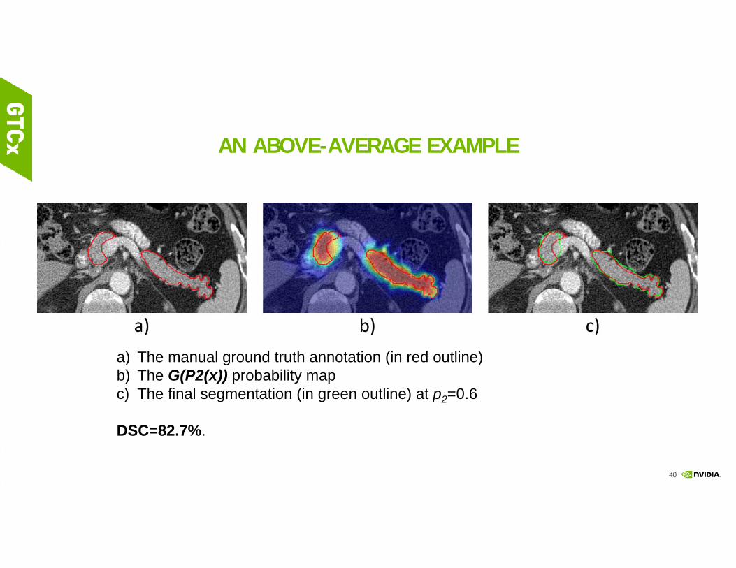
AN ABOVE-AVERAGE EXAMPLE
a) The manual ground truth annotation (in red outline)b) The G(P2(x)) probability mapc) The final segmentation (in green outline) at p2=0.6
DSC=82.7%.

41
SPATIAL AGGREGATION OF HOLISTICALLY-NESTED NETWORKS FOR AUTOMATED PANCREAS SEGMENTATION
[ROTH, ET AL., MICCAI 2016, US PATENT APPLICATION: 62/345,606]
Saining Xie and Zhuowen Tu, "Holistically-Nested Edge Detection", ICCV 2015 (Marr Prize Honorable Mention). Chen-Yu Lee, Saining Xie, Patrick Gallagher, Zhengyou Zhang, and Zhuowen Tu, "Deeply-Supervised Nets", AISTATS 2015.
Better CNN Architectures and pixelwise loss function greatly help for semantic segmentation problems under deep learning principles! stronger, more regularized gradient flow via SGD!!

42

43
MORE ACCURATE AND FASTER? [ROTH, ET AL. MICCAI 2016]
https://wiki.cancerimagingarchive.net/display/Public/Pancreas-CT annotated datasets are publicly available) https://www.synapse.org/#!Synapse:syn3193805/wiki/217789
Our newest results are (81.4% +/- 7.3%) in Dice and ~0.43 mm mean surface-to-surface distance with a stacked implementation of HNNs for both pancreas localization & segmentation.
Localize & Zoom better to see better or segment more accurately) proper Zooming is related to scale & attention models

44

45
References:1. "DeepOrgan: Multi-level Deep Convolutional Networks for Automated Pancreas Segmentation", MICCAI 2015
2. “A Bottom-up Approach for Automatic Pancreas Segmentation Abdominal CT Scans", Oral, MICCAI Abdominal Imaging Workshop 2014
3. "Spatial Aggregation of Holistically-Nested Networks for Automated Pancreas Segmentation", MICCAI 2016
4. Automatic Lymph Node Cluster Segmentation using Holistically-Nested Networks and Structured Optimization", MICCAI 2016
5. Pancreas Segmentation in MRI using Graph-based Decision Fusion on Convolutional Neural Networks", MICCAI 2016
6. "A Bottom-up Approach for Pancreas Segmentation Using Cascaded Superpixels and (Deep) Image Patch Labeling", to appear, IEEE Trans. Image Processing, 2016, arXiv:1505.06236, 2015
7. “Spatial Aggregation of Holistically-Nested Convolutional Neural Networks for Automated Pancreas Localization and Segmentation”, in preparation, 2016
12/29/2016

46
(A3) HOLISTIC ILD (INTERSTITIAL LUNG DISEASE) PREDICTION VIA MULTI-LABEL DEEP LEARNING (LOOKING FOR A CLINICALLY MORE
DESIRABLE PROTOCOL TO ASSIST DECISION MAKING)
ISTP Fellow

47

48
LABEL ANYTHING (THAT MATTERS) FROM EVERYWHERE?
Gao et al., “SEGMENTATION LABEL PROPAGATION USING DEEP CONVOLUTIONAL NEURAL NETWORKS AND DENSE CONDITIONAL RANDOM FIELD”, IEEE ISBI, 2016
49
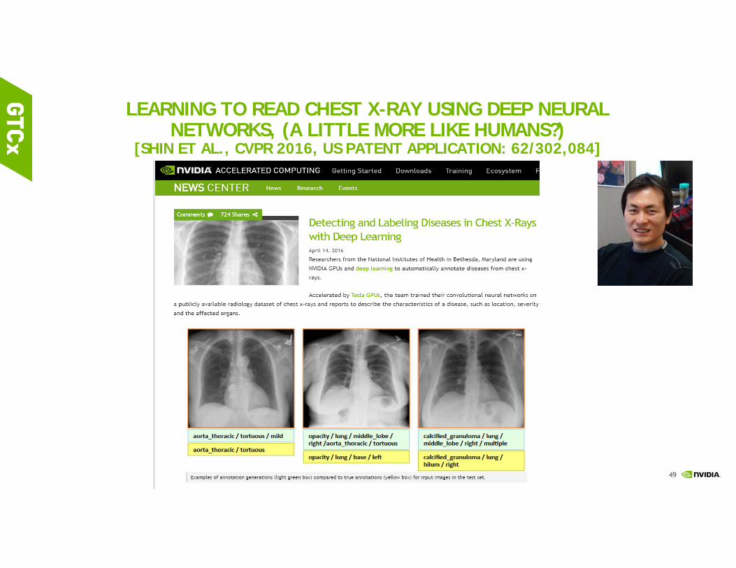
LEARNING TO READ CHEST X-RAY USING DEEP NEURAL NETWORKS, (A LITTLE MORE LIKE HUMANS?)
[SHIN ET AL., CVPR 2016, US PATENT APPLICATION: 62/302,084]

50
INTERLEAVED TEXT/IMAGE DEEP MINING ON A LARGE-SCALE RADIOLOGY DATABASE D
isease Ontology (O
D) is analogical to
WordN
et to ImageN
et
http://arxiv.org/abs/1603.08486

51
References:1. "Holistic Classification of CT Attenuation Patterns for Interstitial Lung Diseases via Deep CNNs", MICCAI DLMIA
workshop 2015
2. "SEGMENTATION LABEL PROPAGATION USING DEEP CONVOLUTIONAL NEURAL NETWORKS AND DENSE CONDITIONAL RANDOM FIELD", IEEE ISBI, 2016
3. "Deep Convolutional Neural Networks for Computer-Aided Detection: CNN Architectures, Dataset Characteristics and Transfer Learning", IEEE Trans. on Medical Imaging, 2016
4. Multi-label Deep Regression and Unordered Pooling for Holistic Interstitial Lung Disease Detection", MICCAI-MLMI 2016.
5. "Learning to Read Chest X-Rays: Recurrent Neural Feedback Model for Automated Image Annotation", IEEE CVPR, 2016
6. “Holistic Interstitial Lung Disease Detection using Deep Convolutional Neural Networks: Multi-label Learning and Unordered Pooling”, in preparation, 2016
12/29/2016
52
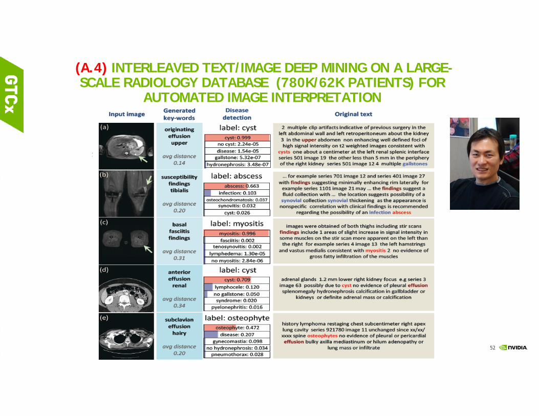
(A.4) INTERLEAVED TEXT/IMAGE DEEP MINING ON A LARGE-SCALE RADIOLOGY DATABASE (780K/62K PATIENTS) FOR
AUTOMATED IMAGE INTERPRETATION
Hoo-Chang Shin, Le Lu, Lauren Kim, Ari Seff, Jianhua Yao, Ronald M. Summers, IEEE Conf. CVPR 2015, to appear; JMLR on large scale health informatics issue (in submission)
53
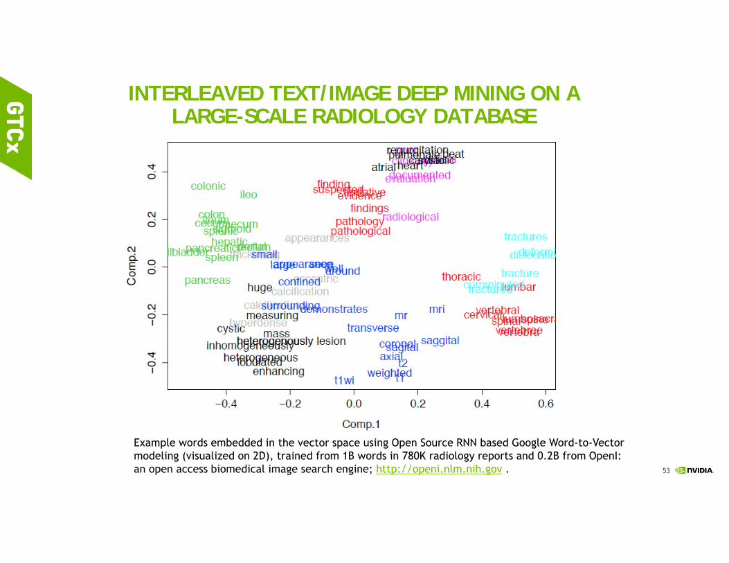
INTERLEAVED TEXT/IMAGE DEEP MINING ON A LARGE-SCALE RADIOLOGY DATABASE
Example words embedded in the vector space using Open Source RNN based Google Word-to-Vector modeling (visualized on 2D), trained from 1B words in 780K radiology reports and 0.2B from OpenI: an open access biomedical image search engine; http://openi.nlm.nih.gov .

54
55
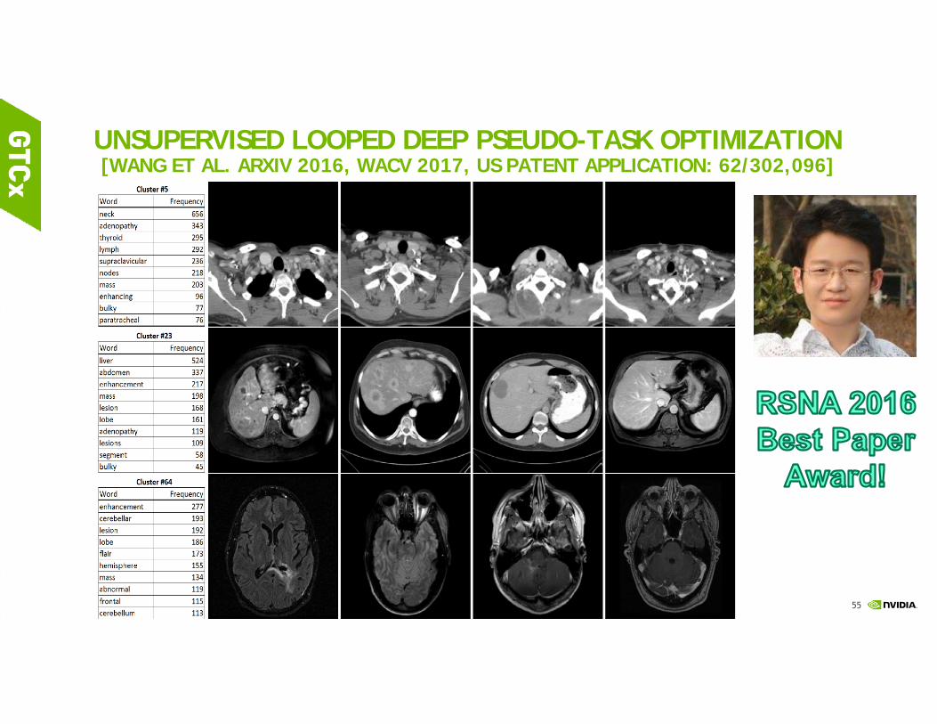
UNSUPERVISED LOOPED DEEP PSEUDO-TASK OPTIMIZATION [WANG ET AL. ARXIV 2016, WACV 2017, US PATENT APPLICATION: 62/302,096]
56
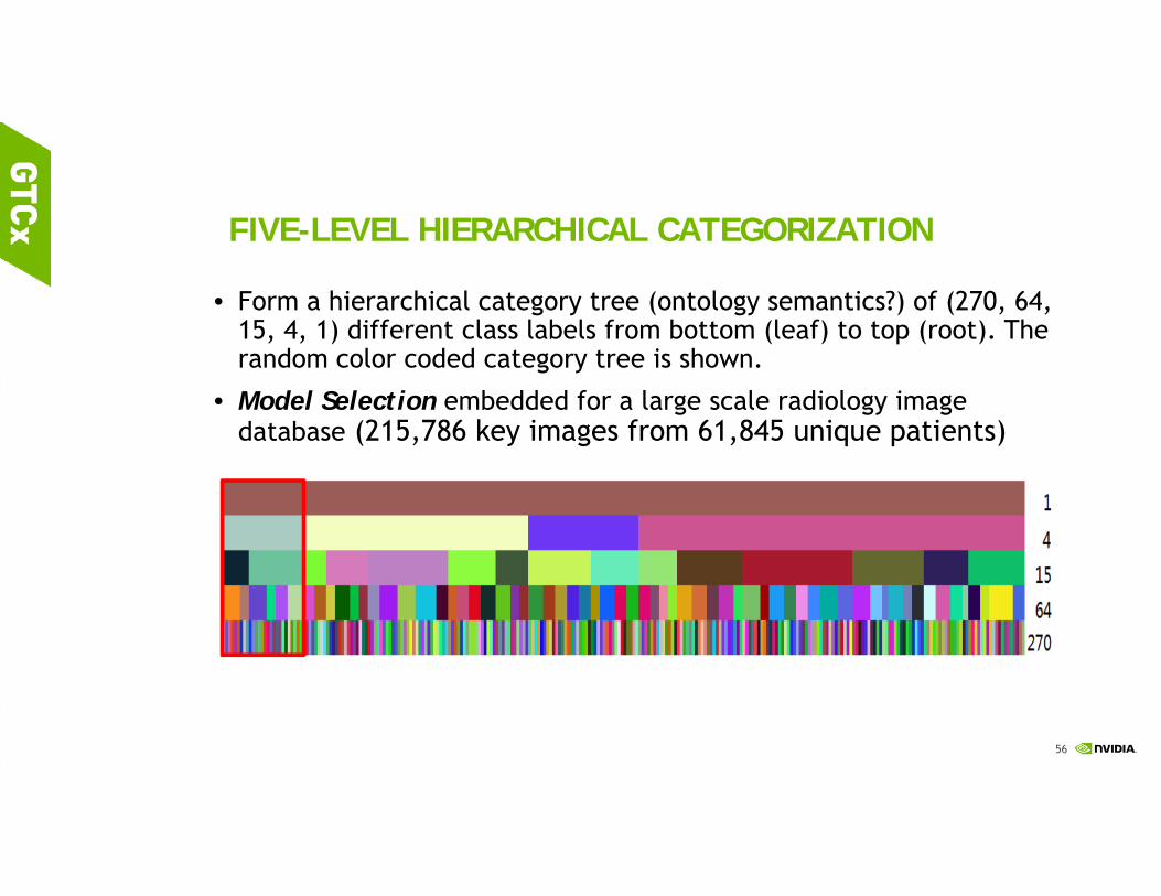
FIVE-LEVEL HIERARCHICAL CATEGORIZATION
• Form a hierarchical category tree (ontology semantics?) of (270, 64, 15, 4, 1) different class labels from bottom (leaf) to top (root). The random color coded category tree is shown.
• Model Selection embedded for a large scale radiology image database (215,786 key images from 61,845 unique patients)
57
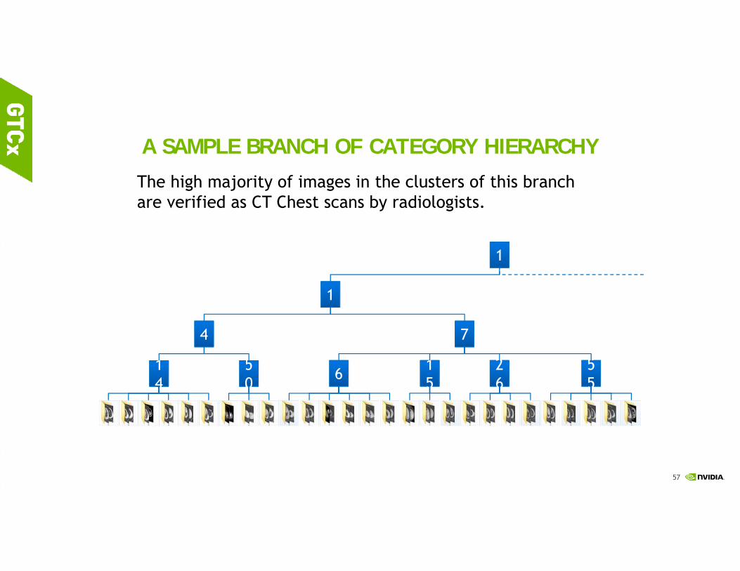
A SAMPLE BRANCH OF CATEGORY HIERARCHY
1414
5050 6 1
515
2626
5555
4 7
1
1
22 25 60 64 141 174 40 129 195 26 72 200 205 230 253 23 75 233 41 104 166 246 81 84 179 224 259
The high majority of images in the clusters of this branch are verified as CT Chest scans by radiologists.

58
*With “Radiologist-in-the-loop” Protocol to build an annotated Large-scale Radiology Image Database Flickr 30K, MS COCO …?
** Significantly better quantitative classification performance than [Shin et al., CVPR 2015; Shin et al. JMLR 2016], in recognizing learned categories!!
[Shin et al. CVPR 2015] may be the first work demonstrating the benefits of transfer learning from ImageNet to ~0.22M radiology key image database.

59
FRAMEWORK OF LDPO-PM: [WANG ET AL. ARXIV:1603.07965]
Fine‐tuned CNN model (with topic labels) or generic Imagenet CNN
model
Randomly Shuffled
Images for Each IterationTrain 70% Val 10% Test 20%
CNN features extraction and clustering
Clustering PM‐encode images (k‐means or RIM)arXiv:1603.07965 Fine-tuning the
CNN (Using renewed
cluster labels)
NLP on text reports for
each Cluster
Image Clusters with semantic
text labelsYes
NoConverged ?
PM based image encoding [1]:
A, Pattern mining inside each clusterB, Merge patterns from different clusters togetherC, Encode the images using merged patterns
[1] Mid-level Deep Pattern Mining. CVPR 2015; arxiv:1506.06343

60
Scene Recognition Dataset
MIT Indoor‐67(indoor scenes, 67 categories,
15620 images)
Building‐25(Architecture Style, 25
categories, 4794 images)
Scene‐15(Both indoor and outdoor, 15 categories, 4485 images)
Airport American Craftsman Bedroom

61
Results- Learned Features for Supervised classification
Method Accuracy (%) Comment
D-patch [1] 38.10 2012
D-parts [2] 51.40 2013
DMS [3] 64.03 2013
MDPM-Alex [4] 64.12 2015
MDPM-VGG [4] 76.95 2015
MetaObject [5] 78.90 2015
LDPO-PM-Alex* 63.68 Our unsupervised method
LDPO-PM-VGG* 72.52 Our unsupervised method
FC (CaffeRef) [4] 57.74 CNN FC feature
FC (VGG) [4] 68.87 CNN FC feature
CONV-FV (CaffeRef) [6] 69.70 Fisher Vector (supervised)
CONV-FV (VGG) [6] 81.00 Fisher Vector (supervised)
* Unsupervised feature representation learningon MIT Indoor‐67(67 categories, 15620 images)
A

62
References:1. "Interleaved Text/Image Deep Mining on a Large-Scale Radiology Image Database", IEEE CVPR 2015
2. "Interleaved Text/Image Deep Mining on a Large-Scale Radiology Image Database for Automated Image Interpretation", Journal of Machine Learning Research, arXiv:1505.00670, 2016
3. "Unsupervised Category Discovery via Looped Deep Pseudo-Task Optimization Using a Large Scale Radiology Image Database", arXiv:1603.07965, 2016
4. “Unsupervised Joint Mining of Deep Features and Image Labels for Large-scale Radiology Image Categorization and Scene Recognition”, …, 2016
12/29/2016
63
TAKE-HOME-MESSAGES
There exists the exact mapping of semantic object detection, object segmentation & parsing, and image-text captioning problems towards corresponding medical imaging tasks
Preventative and precision medicine in radiology are feasible means to advance healthcare through improved quantitative performance on hard and important clinical problems.
It is time to empower deep learning or deep neural networks under novel visual representations to solve previously poorly performed yet critical issues (lymph node, pancreas, chest X-ray, unconstrained ILD prediction, etc.) from doctors’ wish-list; and work with them to make new diagnosis protocols!
Key Technical Elements: Compositional & Hierarchical Visual Representations, Structured Prediction & Optimization, Heterogeneous Visual Cues (Boundary, etc.) integration, CNN Architectures & Loss Functions, Sequential vs. End-to-end Training …
64

65
Thank you & our amazing trainees,
collaborators!
Radiology and Imaging SciencesNational Institutes of Health Clinical Center
[email protected]; [email protected]
Thanks NIH Intramural Research Program (NIH-IRP) for support and NVidia for donating Tesla K40 and Titan X GPUs! NIH FARE awards (2014,2015, 2016), KRIBB Fellowship, NDSEG Fellowship, MICCAI student travel award 2016, RSNA trainee research prize 2016, …


