Deep Sea Visual Deep Sea Visual Adaptations in Adaptations in Teleost Fish Teleost Fish w of: Retinal specializations in the blue ma of: Retinal specializations in the blue ma s designed for sensitivity to low light leve s designed for sensitivity to low light leve Kerstin A. Fritsches, N. Justin Marshall and Eric J. Warrant Kerstin A. Fritsches, N. Justin Marshall and Eric J. Warrant Kenneth D. Hoadley Bio 602: BioChem Adaptations April 5 th , 2010
Transcript
Deep Sea Visual Adaptations in Deep Sea Visual Adaptations in Teleost FishTeleost Fish
Review of: Retinal specializations in the blue marlin: Review of: Retinal specializations in the blue marlin: eyes designed for sensitivity to low light levels.eyes designed for sensitivity to low light levels.
Kerstin A. Fritsches, N. Justin Marshall and Eric J. WarrantKerstin A. Fritsches, N. Justin Marshall and Eric J. Warrant
•At 600-700m: Day time light is similar to starlight conditions (clarke and Denton, 1962)
•Below 1000m, little to no down-welling light is available (Denton, 1990)
Spectral Changes:
•Light becomes monochromatic with peak around (480nm) as we get deeper
Jerlov, (1996)
Visual adaptations differ among ocean zones
Mesopelagic: •Sufficient light to produce an extended visual scene•Adaptations exist to increase optical sensitivity
Bathypelagic:•Insufficient light for extended visual scene. •Adaptations designed to maximize point source illumination (such as from bioluminescence.
Images.com
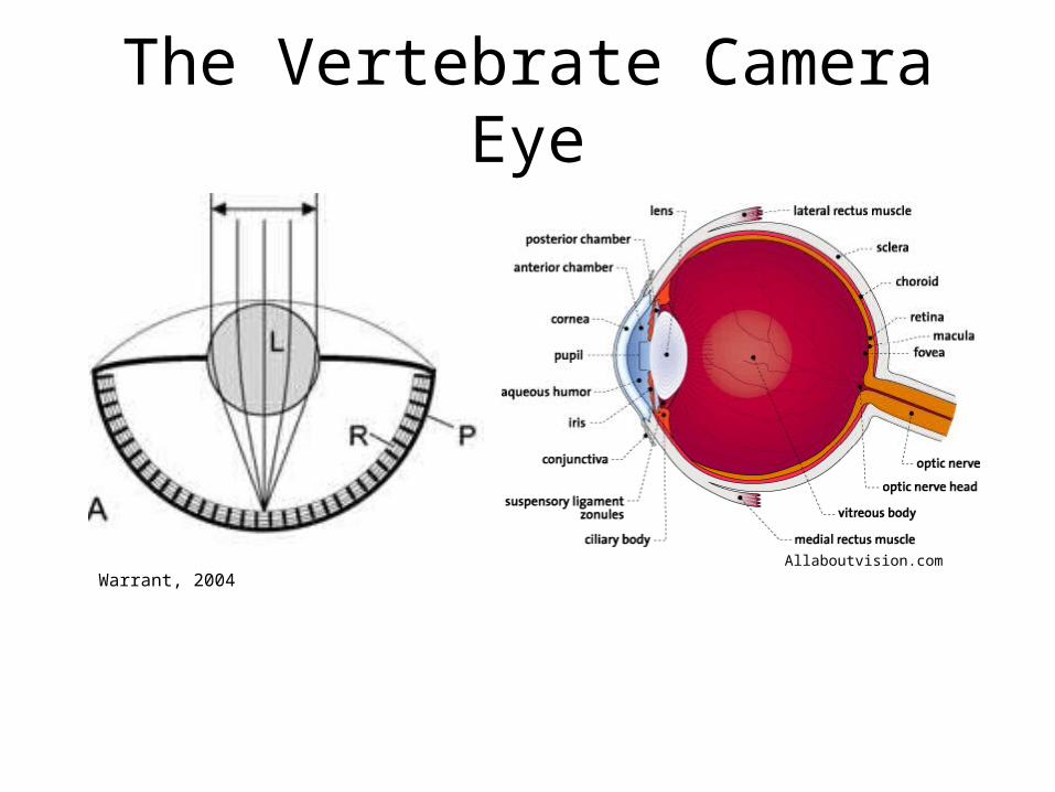
The Vertebrate Camera Eye
Allaboutvision.comWarrant, 2004
Photoreceptors
Rod Cell: wikipedia.com
Neuronal Cells capable of phototransduction
Cones: •Poor light sensitivity•High visual acuity•May have more than one photopigment (color vision)
Rods:•Very light sensitive•Typically only have one photopigment (achromatic vision)
Wikipedia.com
Visual Adaptations for Increasing Optical Sensitivity
•S= optical sensitivity•A= diameter of pupil•F=focal length of eye•D= diameter of photoreceptor•L= Length of photoreceptor•K= absorption coefficient of photoreceptor
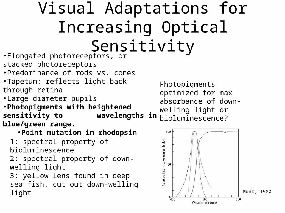
Visual Adaptations for Increasing Optical Sensitivity
•Elongated photoreceptors, or stacked photoreceptors•Predominance of rods vs. cones•Tapetum: reflects light back through retina•Large diameter pupils•Photopigments with heightened sensitivity to wavelengths in blue/green range.
Fritsches, Marshall and Warrant, 2003
Visual Adaptations for Increasing Optical Sensitivity
•Elongated photoreceptors, or stacked photoreceptors•Predominance of rods vs. cones•Tapetum: reflects light back through retina•Large diameter pupils•Photopigments with heightened sensitivity to wavelengths in blue/green range.
•Point mutation in rhodopsin
Photopigments optimized for max absorbance of down-welling light or bioluminescence?
1: spectral property of bioluminescence2: spectral property of down-welling light3: yellow lens found in deep sea fish, cut out down-welling light
How does pressure effect peak absorption of photopigments?
Munk, 1980
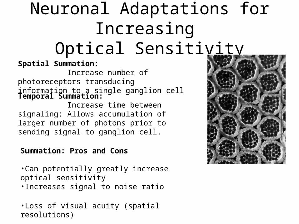
Neuronal Adaptations for Increasing Optical Sensitivity
Spatial Summation: Increase number of photoreceptors transducing information to a single ganglion cell
Temporal Summation: Increase time between signaling: Allows accumulation of larger number of photons prior to sending signal to ganglion cell.
Summation: Pros and Cons
•Can potentially greatly increase optical sensitivity•Increases signal to noise ratio
•Loss of visual acuity (spatial resolutions)
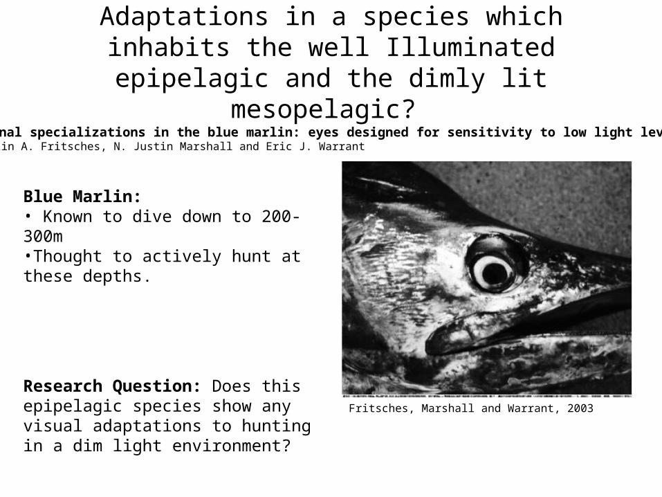
Adaptations in a species which inhabits the well Illuminated epipelagic and the dimly lit mesopelagic?
Retinal specializations in the blue marlin: eyes designed for sensitivity to low light levels.Kerstin A. Fritsches, N. Justin Marshall and Eric J. Warrant
Blue Marlin: • Known to dive down to 200-300m•Thought to actively hunt at these depths.
Research Question: Does this epipelagic species show any visual adaptations to hunting in a dim light environment?
Fritsches, Marshall and Warrant, 2003
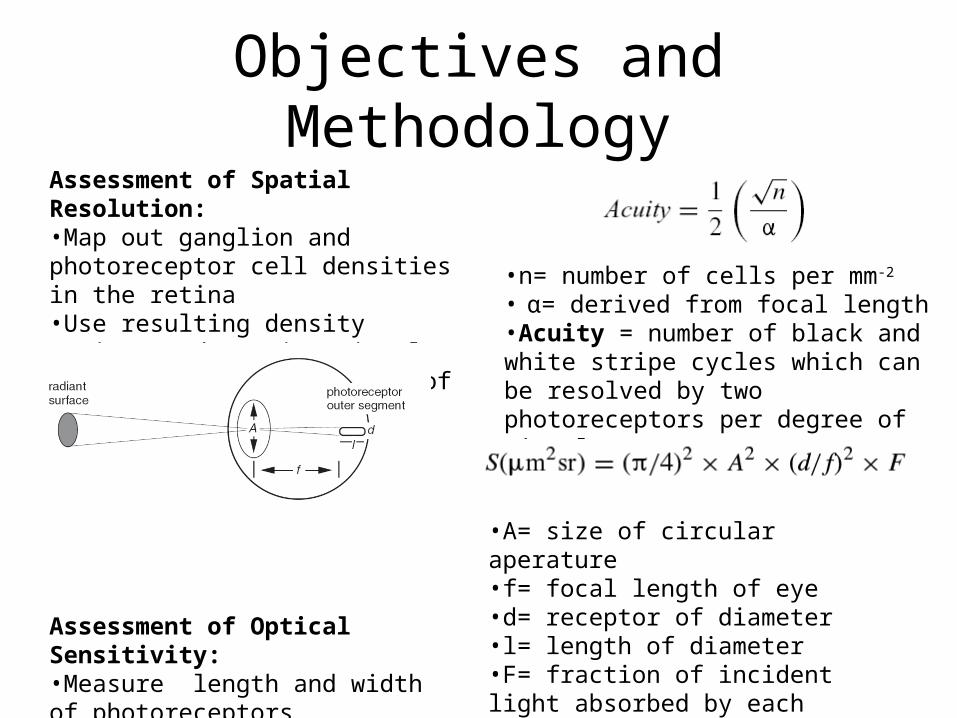
Objectives and MethodologyAssessment of Spatial Resolution:•Map out ganglion and photoreceptor cell densities in the retina•Use resulting density ratios to determine visual acuity in different parts of retina.
Assessment of Optical Sensitivity:•Measure length and width of photoreceptors •Determine optical sensitivity (S) using measurments and optical sensitivity equation as described by (Land, 1981).
•n= number of cells per mm-2
• α= derived from focal length•Acuity = number of black and white stripe cycles which can be resolved by two photoreceptors per degree of visual space
•A= size of circular aperature•f= focal length of eye•d= receptor of diameter•l= length of diameter•F= fraction of incident light absorbed by each photoreceptor
Cell Density Maps and Visual Acuity
Temporal Area Centralis: Peak density = 1600 ganglion cells/mm-2 . Max Acuity = 8.5 cycles / degree
Density increases from dorsal to ventral side for both ganglion and photoreceptor cells
Photoreceptors: predominantly double cones.
Ganglion: photoreceptor ratios:Central Retina: 100:1 Temporal area centralis: 40:1
Fritsches, Marshall and Warrant, 2003
Optical Sensitivity
•Dorsal Retina has wider photoreceptors•Dorsal Retina shows greater sensitivity compared with other regions
Dorsal Width: 3.7+/-0.6um
Ventral Width: 2.5+/-0.6um
Fritsches, Marshall and Warrant, 2003
Optical Sensitivity Comparison with Shallow Water Relative
Increased photoreceptor length, lens diameter and focal length contribute to greater optical sensitivity as compared with shallow water relative.
Fritsches, Marshall and Warrant, 2003
Conclusions and Critique Photoreceptors in the Ventral Retina show adaptations for increased optical sensitivity, necessary for hunting in mesopelagic.
Dorsal Retina: looks down (towards dark)
Temporal area centralis: Looks straight ahead
Ventral Retina: Looks up, towards well illuminated waters
Fritsches, Marshall and Warrant, 2003
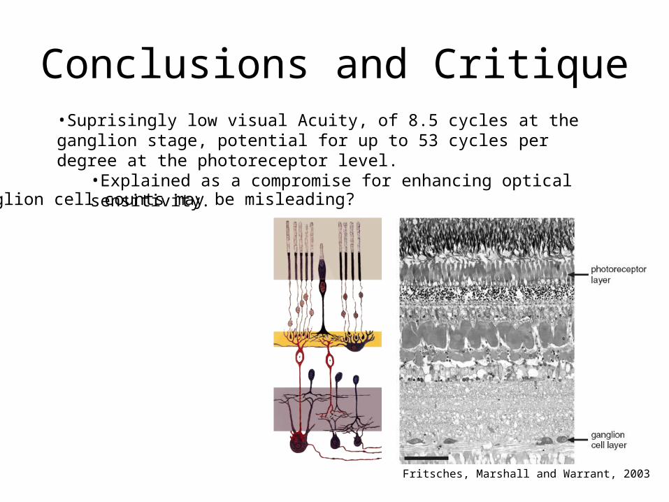
Conclusions and Critique•Suprisingly low visual Acuity, of 8.5 cycles at the ganglion stage, potential for up to 53 cycles per degree at the photoreceptor level.
•Explained as a compromise for enhancing optical sensitivity.
Ganglion cell counts may be misleading?
Fritsches, Marshall and Warrant, 2003
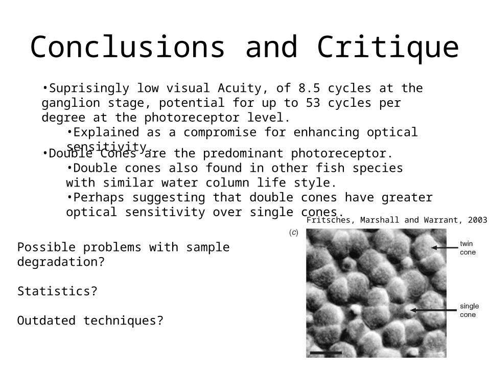
Conclusions and Critique•Suprisingly low visual Acuity, of 8.5 cycles at the ganglion stage, potential for up to 53 cycles per degree at the photoreceptor level.
•Explained as a compromise for enhancing optical sensitivity.
Possible problems with sample degradation?
Statistics?
Outdated techniques?
•Double Cones are the predominant photoreceptor.•Double cones also found in other fish species with similar water column life style. •Perhaps suggesting that double cones have greater optical sensitivity over single cones.
Fritsches, Marshall and Warrant, 2003
References• Kandel, E. R.; Schwartz, J.H.; Jessell, T.M. (2000). Principles of Neural Science (4th ed.). New York: McGraw-
Hill. pp. 507–513.• DENTON, E. J. (1990). Light and vision at depths greater than 200 metres. In Light and Life in the Sea (eds. P. J. Herring, A. K.
Campbell, M. Whitfield and L. Maddock), pp. 127–148. Cambridge University Press, Cambridge.• JERLOV, N. G. (1976). Marine Optics. Elsevier Scientific Publishing Company, Amsterdam.• LAND, M. F. (1981a). Optics and vision in invertebrates. In Handbook of Sensory Physiology, Vol. VII/6B (ed. H. Autrum), pp.
471–592. Springer, Berlin, Heidelberg, New York.• LAND,M. F. (1981b). Optics of the eyes of Phronima and other deep sea amphipods. Journal of Comparative Physiology A
145, 209–226.• KIRSCHFELD, K. (1974). The absolute sensitivity of lens and compound eyes. Zeitschrift fu¨r Naturforschung 29C, 592–596.• KIRSCHFELD, K. (1976). The resolution of lens and compound eyes. In Neural Principles in Vision (eds. F. Zettler and R.
Weiler), pp. 354–370. Springer, Berlin, Heidelberg, New York.• Warrant EJ, Locket NA (2004) Vision in the deep sea. Biol Rev 79:671–712