Deferoxamine preconditioning to restore impaired HIF-1a-mediated angiogenicmechanisms in adipose-derived stem cells from STZ-induced type 1 diabetic ratsM. Mehrabani*, M. Najafi†, T. Kamarul‡, K. Mansouri§, M. Iranpour¶, M. H. Nematollahi**, M. Ghazi-Khansari††and A. M. Sharifi*,‡‡
*Razi Drug Research Center, Department of pharmacology, Iran University of Medical Sciences, Tehran, Iran, †Department of Biochemistry, IranUniversity of Medical Sciences, Tehran, Iran, ‡Tissue Engineering Group (TEG) & Research, National Orthopedic Centre of Excellence inResearch & Learning (NOCERAL), Department of Orthopedics, Faculty of Medicine, University of Malaya, Kuala Lumpur, Malaysia, §MedicalBiology Research Center, Kermanshah University of Medical Sciences, Kermanshah, Iran, ¶Department of Pathology, Kerman University ofMedical Sciences, Kerman, Iran, **Department of Biochemistry, Kerman University of Medical Sciences, Kerman, Iran, ††Department ofPharmacology, Tehran University of Medical Sciences, Tehran, Iran and ‡‡Department of Tissue Engineering and regenerative Medicine, Schoolof Advanced Technologies in Medicine, Iran University of Medical Sciences, Tehran, Iran
Received 23 February 2015; revision accepted 22 June 2015
AbstractObjectives: Both excessive and insufficient angio-genesis are associated with progression of diabeticcomplications, of which poor angiogenesis is animportant feature. Currently, adipose-derived stemcells (ADSCs) are considered to be a promisingsource to aid therapeutic neovascularization. How-ever, functionality of these cells is impaired by dia-betes which can result from a defect in hypoxia-inducible factor-1 (HIF-1), a key mediator involvedin neovascularization. In the current study, wesought to explore effectiveness of pharmacologicalpriming with deferoxamine (DFO) as a hypoxiamimetic agent, to restore the compromised angio-genic pathway, with the aid of ADSCs derivedfrom streptozotocin (STZ)-induced type 1 diabeticrats (‘diabetic ADSCs’).Materials and methods: Diabetic ADSCs were trea-ted with DFO and compared to normal and non-treated diabetic ADSCs for expression of HIF-1a,VEGF, FGF-2 and SDF-1, at mRNA and proteinlevels, using qRT-PCR, western blotting andELISA assay. Activity of matrix metalloproteinases-2 and -9 were measured using a gelatin zymogra-phy assay. Angiogenic potential of conditionedmedia derived from normal, DFO-treated and non-treated diabetic ADSCs were determined by
in vitro (in HUVECs) and in vivo experimentsincluding scratch assay, three-dimensional tube for-mation testing and surgical wound healing models.Results: DFO remarkably enhanced expression ofnoted genes by mRNA and protein levels andrestored activity of matrix metalloproteinases -2and -9. Compromised angiogenic potential of con-ditioned medium derived from diabetic ADSCs wasrestored by DFO both in vitro and in vivo experi-ments.Conclusion: DFO preconditioning restored neovas-cularization potential of ADSCs derived from dia-betic rats by affecting the HIF-1a pathway.
Introduction
Impaired angiogenesis plays a pivotal role in the patho-genesis of a variety of disorders such as diabetes melli-tus (DM) (1,2). Inadequate neovascularization of theskin, nerves and myocardium clinically translates intothe major manifestations of diabetes, including impairedwound healing, neuropathy and poor prognosis aftermyocardial infarction (3,4). In this setting, therapeuticangiogenesis is an ideal option towards restoring normalcirculation in many human diseases, particularly DM.
Recently, adult stem cell-based medicine has beenshown to be a promising therapeutic strategy, facilitatingrepair of diabetic ischaemic tissues (5). Some criteriaamong others, make ADSCs attractive candidates foruse in cell therapies. ADSCs are multipotent mesenchy-mal stromal cells (MSCs), which are easily accessible,easy to culture to high numbers and able to self-renew
Correspondence: A. M. Sharifi, Department of pharmacology,School of Medicine, Iran University of Medical Sciences, Tehran,Iran. Tel.: +98 021 88622523; Fax: +98 021 88622523; E-mails:[email protected], [email protected] and [email protected]
and differentiate into various cell types, in particularendothelial cells and vascular myocytes (6,7). Moreover,autologous ADSCs, due to their secretion of manypotent angiogenic factors and not being subject toimmune rejection, have been considered to be excellentchoice for curative neovascularization treatment (8–10).However, previous reports have indicated that ADSCsfron a diabetic source, compared to normal ADSCs,exhibit poor and insufficient angiogenic induction(11,12). Clinically, such an obstacle may prevent appli-cation of autologous diabetic ADSCs. There is, thus, aneed to identify and revive intrinsic impaired mecha-nisms before cell implantation.
It has also been demonstrated that hypoxia-induciblefactor-1 (HIF-1) transcriptional complex regulates awide range of hypoxia-induced cell processes includingangiogenic factor production in ADSCs (13,14). TheHIF-1 transcriptional complex plays a central role in cellresponses when oxygen availability changes; it is com-posed of a hypoxia-regulated alpha subunit and a stablebeta subunit (15). Under normoxic conditions, the HIF-1a subunit rapidly becomes hydroxylated by HIF prolyl-hydroxylases (PHDs), followed by ubiquitination andproteasomal degradation. PHDs enzymes belong to theoxygenase superfamily, which needs iron and 2-oxoglu-tarate as cofactors (16–18). In normoxia, use of an iron-chelating agent causes enzyme inhibition, leading toHIF-1a stabilization. HIF-1, through binding to hypoxiaresponse elements (HREs), can up-regulate expressionof several angiogenic genes such as vascular endothelialgrowth factor (VEGF), stromal cell-derived factor 1(SDF-1), fibroblast growth factor-2 (FGF-2) and matrixmetalloproteinases (MMPs)-2 and -9 (19,20). MMPs areextracellular endoproteinases secreted by MSCs, andplay important roles during angiogenesis. They facilitatemigration of endothelial cells by disrupting extracellularmatrix (ECM), and can also release angiogenic factorsincluding VEGF and FGF-2 from the ECM. SeveralMMPs particularly, MMP-2 and -9, have been recog-nized as being more involved in proceeding to angio-genesis (21,22).
To date, different strategies have been proposed topromote functional capacity of impaired stem cells byaffecting the HIF-1a pathway, including preconditioningof stem cells with hypoxic shock (23) and pharmacologi-cal agents that mimic hypoxia (24,25). DFO is an iron-chelating agent used clinically in cases of iron overload,such as thalassaemia (26). It inhibits the HIF prolyl-hydroxylases and can stabilize HIF-1a through its iron-chelating activity. Thus, DFO acts as a hypoxia mimeticagent in normoxia (27). In this respect, accumulating evi-dence has shown that DFO can increase VEGF secretionfrom ADSCs from non-diabetic sources and bone marrow
mesenchymal stem cells (BMSCs) from non-diabeticsources, via the HIF-1a pathway (13,28). We, thus,sought to evaluate effects of DFO preconditioning onrestoring angiogenic potential of ADSCs derived fromSTZ-induced diabetic rats to enhance efficacy of cell ther-apy in DM, both in vitro and in vivo.
Material and methods
Antibodies and reagents
Polyclonal antibodies against FGF-2 and SDF-1 werefrom Abcam (Cambridge, UK). Fluorescein isothio-cyanate (FITC)-conjugated anti-rat antibodies againstCD45, CD90, FITC-conjugated mouse IgG isotype con-trol, purified mouse IgG isotype control and purifiedanti-rat antibody to CD73 were purchased from BD Bio-science and eBioscience (San Diego, CA, USA). FITC-conjugated CD11b antibody was obtained from Gene-Tex, Inc. (United States) and FITC-conjugated anti-ratCD44 and CD31 were purchased from Serotec (Oxford,UK). HIF inhibitor was from Santa Cruz Biotechnology(San Jose, CA, USA) and rat VEGF Quantikineenzyme-linked immunosorbent assay (ELISA) kit wasfrom R&D Systems. Monoclonal antibodies againstHIF-1a, DFO, gelatin A and B, streptozotocin, proteaseand phosphatase inhibitor cocktails, collagenase type Iand MTT were purchased from Sigma (Sigma Aldrich,St Louis, MO, USA). Minimal essential medium a(aMEM), foetal bovine serum (FBS) and penicillin-streptomycin were from Gibco (Invitrogen, Carlsbad,CA, USA) and Trizol reagent was from Invitrogen(Merelbeke, Belgium). Oligo (dT) primer, moloney mur-ine leukaemia virus reverse transcriptase (M-MLV RT)and Maxima SYBR Green/ROX qPCR Master Mix (2X)were purchased through Fermentas (UK). Horseradishperoxidase-linked anti-rabbit secondary antibody andanti-glyceraldehyde-3-phosphate dehydrogenase antibody(GAPDH) were obtained from Cell Signaling (Danvers,MA, USA). Cytodex 3 microcarrier bead was fromAmersham Pharmacia Biotech (UK) and Amicon Ultra-15 Centrifugal Filter Unit with Ultracel-3 membranewas from Millipore.
Experimental animals
Male Wistar rats aged 8–10 weeks, weighing 200–250 gwere purchased from the animal facility of the PastureInstitute of Iran. They were maintained in standard con-ditions (individual plastic cages, temperature 20–26 °Cand humidity of 40–70%, with regular light cycles of12/12 h light/dark). The study was accepted by the Ethi-cal Committee of Tehran University of Medical Sciences
Deferoxamine restore Angiogenesis in stem cell 533
and was performed in accordance with its guideline baseon the National Institutes of Health Principles of Labo-ratory Animal Care (NIH publication no. 85-23, revised1985).
Induction of diabetes
Twenty rats were classified randomly into two groups:normal group (n = 10) and diabetic group (n = 10).Diabetes was induced in rats of the diabetic group by asingle intraperitoneal injection of streptozotocin (STZ)(55 mg/kg). One week after injection, blood glucoselevels were measured. Only animals with blood glucoselevels >300 mg/dl were included in the study. Over thenext 4 months, blood glucose was measured twice everymonth to ensure that its level was over 300 mg/dl.
ADSC isolation
All procedures were performed under sterile conditions.First, rats were euthanized using ketamine/xylazine over-dose. After shaving abdominal hair, skin was cleansedusing povidone-iodine solution. Epididymal fat-padswere excised from diabetic and non-diabetic (normal)animals and were soaked in phosphate-buffered saline(PBS) containing 5% penicillin/streptomycin andantimycotic agent. Debris was removed, fat tissues wereminced, and washed extensively in PBS to removehaematopoietic cells. For isolation of ADSCs, adiposetissues were exposed to 0.075% collagenase type I pre-pared in aMEM containing 2% penicillin/streptomycinunder gentle agitation for 30 min at 37 °C. Collagenasewas neutralized with growth medium (aMEM containing15% FBS and 1% penicillin/streptomycin) and the mix-ture was centrifuged at 2000 rpm for 6 min. After dis-carding supernatants, pellets were suspended in 1 mlice-cold lysis buffer for 10 min and subsequentlywashed in PBS. Growth medium was added to pelletsand filtered using a 100 lm nylon mesh. Filtered frac-tions were incubated in a humidified chamber with 5%CO2 atmosphere at 37 °C. Adherent cells at third pas-sages were used as ADSCs in the next steps (7).
Characterization of ADSCs
ADSC surface marker expressions were evaluated usingfluorescence-activated cell sorting (FACS) analysis with aFACS Calibur cytometer (Becton Dickinson, San Diego,CA, USA). Briefly, cultured ADSCs from three to fourpassages were pelleted and washed three times in PBS.Afterwards 2 9 105 cells were incubated in saturatedconcentration of FITC-conjugated anti-rat antibodiesagainst CD11b, CD31, CD44, CD45, CD90 and purified
CD73 for 40 min at 4 °C in the dark. FITC-unconjugatedantibody was incubated with secondary conjugated anti-body for 15 min. Mouse IgG isotype served as negativecontrol. Cells were centrifuged for 6 min and resuspendedin PBS. Finally, results were analysed using Cell Questsoftware and compared to isotype control.
Colony-forming unit (CFU) assay
CFU assay was used to compare size and number ofcolonies derived from normal and diabetic ADSCs.Appropriate numbers of cells at early passages wereseeded in six-well plates. Growth medium was changedevery 3–4 days and after 2 weeks, dishes were washedin 10–15 ml PBS. Then, colonies were fixed and stainedwith 5 ml 0.5% crystal violet solution in methanol for30 min. After rinsing in PBS, numbers and sizes ofcolonies were determined using NIH ImageJ software(http://rsbweb.nih.gov/ij). Colonies with a minimum of50 cells were included.
Quantification of intracellular iron by atomic absorptionspectroscopy
Cultured ADSCs (passage 3) were pelleted and washedthree times in cold PBS. Next, 1 9 106 cells were lysedwith 500 ll of 50 mM NaOH for 2 h on a shaker. Celllysates were used for quantification of total amount ofiron, using atomic absorption spectroscopy (VarianSpectrAA-600 Atomic Absorption Spectrometer withGTA 100). Intracellular iron concentration was reportedas iron (pg) per cell.
Preparation of different concentrations of DFO
DFO was dissolved in distilled water and kept at 4 °C.Required concentrations of DFO (75, 150, 300 lM) wereprepared from the stock solution. HIF-1 inhibitor wasused according to the manufacturer’s instructions.Briefly, 30 lM HIF-1 inhibitor was used 1 h before pre-treatment of ADSCs with 300 lM DFO.
MTT assay
The MTT assay is one of the most frequently used col-orimetric assays for determining cell viability. Activityof mitochondrial dehydrogenases in viable cells leads toreduction of tetrazolium salts to dark blue formazan.Briefly, normal ADSCs (nADSCs) and diabetic ADSCs(dADSCs) were seeded into 96-well microplates at10,000 cells per well. Final reaction products ofnADSCs and dADSCs were measured after 24, 48 and72 h. For evaluation of cytotoxic effects of DFO on
dADSCs, cells were seeded into 96-well microplates;after 24 h, they were exposed to different concentrationsof DFO (0, 75, 150, 300 lM). After 24, 48 and 72 h,MTT solution was added into each well and plates wereincubated for 4 h at 37 °C in the dark. Medium wasremoved and dimethylsulphoxide (DMSO) (100 ll)was added to each well. Absorbance of the solution wasmeasured at 570 nm using a microplate reader (Bio-TekELX800, Winooski, VT, USA).
Real-time PCR
Normal, non-treated and treated diabetic ADSCs afterexposure to DFO, were used for total RNA extraction, byutilization of Trizol reagent according to the manufac-turer’s protocols. Then, RNA quality and concentrationwere evaluated spectrophotometerically. ComplementaryDNA was synthesized by 1 ll M-MLV RT, 1 ll deoxyri-bonucleotide triphosphates (dNTPs), 0.5 ll RNase Inhibi-tor, 2 ll oligo (dT) primer and 3 lg RNA in final volumeof 20 ll. The mixture was incubated for 1 h at 42 °C, fol-lowed by incubation at 72 °C for 10 min. Quantitativereverse transcription-polymerase chain reaction (qRT-PCR) was performed using the Rotor-Gene 6000 System(Corbett Research). Reaction mixture contained cDNAtemplate, 15 ll the Maxima SYBR Green/ROX qPCRMaster Mix and specific primers (Table 1). GAPDH wasused as endogenous control gene. Thermal cycling profilewas as follows: Initial denaturation at 95 °C for 10 min,and 40 cycles of denaturing at 95 °C for 15 s, annealingat 60 °C for 30 s and extension for 30 s at 72 °C. Thresh-old cycle (Ct) was determined for GAPDH and targetgenes of each sample. DCt was calculated for each sampleand relative gene expressions were calculated.
Western blot analysis
Non-treated and treated cells were trypsinized, andwashed centrifugally twice, in cold PBS. Cells werelysed using ice-cold lysis buffer containing a proteaseand phosphatase inhibitor cocktail and centrifuged for30 min, 12,000 g at 4°C. Total protein concentrationwas determined using the Bradford method (29). Equal
concentration of samples (150 lg) was loaded andseparated on SDS-PAGE gel. Next, proteins were trans-ferred to nitrocellulose membranes followed by incuba-tion with primary antibodies overnight at 4 °C.Antibodies included mouse monoclonal anti-HIF-1a(1:500), rabbit polyclonal anti-FGF-2 (5:1000), rabbitanti-SDF-1 (2.5:1000) and rabbit anti-GAPDH (1:5000).After this step, membranes were incubated with sec-ondary antibody (anti-mouse or anti-rabbit conjugated tohorseradish peroxidase) for 1 h at 4 °C. An enhancedchemiluminescence kit was used to visualize proteinbands which were then quantified by densitometry,using Total Lab software (UK).
Preparation of conditioned media
After reaching 80% confluence, cultured ADSCs (in six-well plates) were rinsed in PBS and fed with 3 mlserum-free medium. After 24 h, supernatants were col-lected and frozen at �80 °C until the following step.For in vivo experiments, conditioned medium (CM) wasconcentrated (50-fold) by ultra-filtration centrifugal filterunits, with 3 kD cut-off, according to the manufacturer’sinstructions. For collecting CM from pre-treated ADSCs,cells were treated with DFO (150 and 300 lM) and/orHIF-1 inhibitor, for 24 h then washed in PBS severaltimes. Then they were incubated with serum-free med-ium and supernatant was collected after 24 h.
ELISA assay for release of VEGF
VEGF concentration was measured in 50 ll differentculture media (CM) from standards in 0 to 2000 pg/mlrange, using an ELISA kit, according to the manufac-turer’s instructions. VEGF expression was normalized toprotein concentration.
Gelatin zymography
A zymography assay was used to analyse effects ofDFO on activities of matrix metalloproteinases (MMPs)-2 and -9, in nADSCs and dADSCs. CM were preparedas described above and subjected to zymographic assay.
Table 1. Rat primers for qRT-PCR
Gene Sense strand Antisense strand Size of product
Deferoxamine restore Angiogenesis in stem cell 535
Briefly, supernatants were mixed with 3X loading buffer(125 mM Tris-HCl, 20% glycerol, 2% SDS and 0.002%bromophenol blue, pH 6.8). Mixtures were separatedelectrophoretically using 10% polyacrylamide gel, in thepresence of 0.1% SDS containing 0.1% gelatin A(MMP-2) or B (MMP-9). Then, gels were soaked in Tri-ton X-100 for 60 min and incubated in developing buf-fer (50 mM Tris-HCl, 200 mM NaCl, 5 mM CaCl2 and0.01% NaN3, pH 7.5) overnight at 37 °C. Subsequentlygels were stained with Coomassie Blue solution (0.1%Coomassie Brilliant Blue G-250, 40% methanol, 10%acetic acid and deionized water) and then destained with40% methanol, 10% acetic acid and deionized water.When enzyme-digested regions were visualized as whitebands against a blue background, gels were pho-tographed and bands were quantified using densitometricanalysis from Total Lab software.
In vitro scratch assay
The scratch assay is a straightforward method to evaluatecell migration in vitro. HUVECs were obtained from thePasteur Institute (Tehran, Iran). They were loaded intosix-well plates, all at the same density, and then 5 mlgrowth medium was added to each well. After reaching90% confluence, scratch wounds were created in the cellmonolayers by scraping them with a sterile 1000 ll pip-ette tip. After removal of detached cells by washing inPBS, cells were fed CM derived from treated and non-treated cells. The HUVECs were then incubated to allowcells to migrate into the scratch area. The next step wascapturing photographic images during cell migration(from five random, separate, microscopic fields) and ana-lysing them using NIH Image J software (30).
HUVEC capillary tube formation in three-dimensionalcollagen gel
The HUVECs were mixed with Cytodex 3 microcarrierbeads and placed in an incubator at 37 °C and themixed suspension was flicked every 20 min to allow foruniform distribution of cells on the microcarrier beads.Beads were seeded in 12-well plates and then incubatedovernight at 37 °C. On the following day, cell-coatedbeads were embedded in ice-cold collagen matrix andserum-free medium and the mixture was loaded into 96-well plates and allowed to solidify for 45 min at 37 °C.Next, serum-free medium, and CM derived from normal,DFO-treated and non-treated diabetic ADSCs wereadded to wells. Cells were photographed after 48 h.Sprout formation was analysed using NIH Image J soft-ware according to the standard method, and are pre-sented as percentages of control (31).
Wound healing model
Forty-eight male Wistar rats were randomly divided intofour groups as follows: treated with serum-free media,those treated with CM derived from diabetic rats, normal,and 300 lM DFO-treated ADSCs. Excisional wounds werecreated in the splinting model, as described previously (8).In brief, following administration of anaesthesia (intramus-cular injection of xylazine and ketamine), dorsal areas ofrats were shaved and one 8 mm full-thickness excisionalskin wound, per animal, was created in the midline of theneck. Wounds were injected with 100 ll concentratedmedium and concentrated CM derived from normal,300 lM DFO-treated and non-treated diabetic ADSCs(60 ll around the wound and 40 ll into the wound bed).The animals were housed individually for 2 weeks.
Wound area analysis
Digital photographs of wounds were captured after cre-ating during healing. Wound areas were blindly mea-sured in mm2 squares, using NIH Image J software.Then, wound closure rates were calculated using the fol-lowing formula (32):
Wound closure rate (%)
¼ ðWound area on day 0�Wound area on day xÞ=Wound area on day 0� 100
x ¼ 0; 3; 7; 10; 14
Histological examination
Rats were sacrificed on days 3, 7, 10 and 14 and skin sam-ples, including wounds plus 10 mm of surrounding skin,were collected for histological examination. Skin speci-mens were fixed in 10% formalin solution and embeddedin paraffin wax blocks. Afterwards, 3 lm sections werestained with Masson’s trichrome for collagen fibre stain-ing and haematoxylin and eosin (H&E). Angiogenesis,epithelialization, collagen levels and macrophage countswere evaluated by examining three fields per section ofthe wounds at 100x and 400x magnification. Each samplewas given a histological score using Abramov’s histologi-cal scoring system (33) as summarized in Table 2.
Statistical analysis
Statistical significance of mean differences between groupswas analysed by one-way or repeated ANOVA withTukey’s post-test, and two-way ANOVA with Duncan andBonferroni post-test, as appropriate. Unpaired Student’st-testing was performed for comparisons between twogroups (diabetic and normal ADSCs). Statistically,
differences were assumed significant at P < 0.05. Resultsare presented as mean � standard error of the mean(SEM).
Results
ADSC characterization and morphology of diabetic andnormal ADSCs
Flow cytometry indicated that ADSCs (passage 3–4)were positive for CD90, CD73 and CD44 and were neg-ative for haematopoietic and endothelial lineage markers
CD11b, CD31 and CD45 (Fig. 1a). The cells hadfibroblast-like morphology at this passage level. Mor-phologically, dADSCs had more flattened appearancescompared to nADSCs of the same passage (Fig. 1b).
CFU assay and iron content of diabetic and normalADSCs
CFU assays revealed that dADSCs formed significantlylower numbers of colonies (6.9 � 0.4%) than nADSCs(11.66 � 0.7%) (P < 0.01). Also, colonies derived fromdADSCs significantly were smaller (P < 0.001) thanthose derived from normal cells (Fig. 2a,b). Basal totaliron content was found to be 0.041 � 0.002 and0.046 � 0.001 pg iron per cell for nADSCs anddADSCs respectively (n = 3). There were no significantdifferences between means of iron content of diabeticADSCs and normal ADSCs (Fig. 2c).
DFO, by MTT assay, showed no cytotoxic effect after24 h
Cell viability was evaluated by MTT assay; nADSCsseemed to be more proliferative than dADSCs after 72 h
Table 2. Histological score based on Abramov’s histological scoringsystem (33)
Figure 1. Characterization and morpho-logical assessment of ADSCs. (a) Flowcytometry showed that the ADSCs were neg-ative for CD45, CD11b and CD31 but posi-tive for CD44, CD73 and CD90. Data areshown as a histogram plot, with black repre-senting isotype control and white demonstrat-ing experiments. (b) Assessment ofmorphology of ADSCs demonstrated that (I)diabetic ADSCs had more flattened androunder morphology than (II) normal ADSCs.Dashed lines indicate cells.
Deferoxamine restore Angiogenesis in stem cell 537
(P < 0.01; Fig. 3a). Pre-treatment of dADSCs withDFO (75, 150 and 300 lM) revealed no cytotoxic effectafter 24 h (Fig. 3). The results demonstrate that DFO(150 lM) seemed to increase proliferation of ADSCscompared to non-treated dADSCs after 24 h (P < 0.01;Fig. 3c). On the other hand, DFO significantly causedcytotoxic effects after 48 and 72 h (P < 0.05; Fig. 3).Hence, DFO was applied at concentrations of 150 and300 lM for 24 h in all other experiments of this study.
DFO preconditioning enhanced HIF-1a proteinexpression in diabetic ADSCs
Western blotting assay was used to compare HIF-1aprotein expression in nADSCs, DFO-treated and non-treated dADSCs. The results indicated that non-treateddADSCs had significantly lower HIF-1a protein expres-sion levels compared to nADSCs (P < 0.001). HIF-1expression was enhanced in dADSCs at 150 (P < 0.01)
and 300 lM (P < 0.001) concentration of DFO in aconcentration manner, so that non-significant changeswere detected between normal group and treated groups.This effect was remarkably reversed by pre-treatmentwith 30 lM of HIF-1 inhibitor 1 hour before exposureto 300 lM DFO (P < 0.001) (Fig. 4a). HIF-1a expres-sion was also assessed in dADSCs pre-treated with150 lM DFO over the appropriate time period (0, 6, 12,24 h). DFO significantly increased HIF-1a expressionafter 12 h (P < 0.05) and to a highest level after 24 h(P < 0.001), before showing cytotoxic effects (Fig. 4b).Therefore, DFO was applied for 24 h in all other experi-ments of this study.
DFO enhanced VEGF, FGF-2 and SDF-1 mRNAexpression levels
Quantitative real-time PCR was performed on nADSCs,DFO-treated and non-treated dADSCs and results indicated
(a)
I II
(b)
(c)
I II
Figure 2. Colony formation and ironassessment of ADSCs. (a) Representativeimages of colonies of diabetic ADSCs (I) andnormal ADSCs (II) by light microscopy. (b)Assessment of size (I) and number (II) ofcolonies derived from diabetic and normalADSCs, by NIH imageing. (c) Assessment ofiron content of normal and diabetic ADSCs.Data are represented as mean � SEM (n = 3,**P < 0.01, ***P < 0.001 versus normalcells). Statistical significance was measuredby unpaired Student’s t-test.
that for all genes, dADSCs had significant reduction inrelated mRNA levels, compared to nADSCs, so thatVEGF, SDF-1 and FGF-2 expression levels were0.13 � 0.01 (P < 0.001), 0.07 � 0.001 (P < 0.001) and0.01 � 0.008 (P < 0.01) times lower in dADSCs respec-tively. In treated subgroups with 150 and 300 lM concen-trations DFO, expression levels of VEGF were estimatedto be in the order of 0.42 � 0.01 times (P < 0.001) and1.36 � 0.1 times (P < 0.05) respectively, versus nADSCs.Furthermore, in treated cells with 150 and 300 lM concen-trations DFO, expression levels of SDF-1 were estimatedto be in the order of 0.32 � 0.01 times (P < 0.01) and0.68 � 0.9 times (P > 0.05) respectively versus nADSCs.FGF-2 expression was estimated in both 150 and 300 lM
concentrations of DFO-treated diabetic ADSCs to be in theorder of 0.47 � 0.08 times (P < 0.05) versus nADSCs.Notably, VEGF expression was higher at 300 lM concen-tration of DFO-treated dADSCs compared to normal cells.SDF-1 expression was enhanced at 300 lM concentrationof DFO so that non-significant change was detectedbetween normal ADSCs and 300 lM DFO-treated ADSCs.On the other hand, treatment with DFO did not promoteexpression of FGF-2 to reach the normal level (Fig. 5a).
Promotion of VEGF protein secretion by DFO
To investigate effects of DFO on VEGF protein release,ELISA was performed on CM derived from ADSCs.
(a) (b)
(c) (d)Figure 3. Effect of DFO on proliferation ofADSCs. (a) Viability of diabetic cells signifi-cantly was lower than viability of normalADSCs after 72 h measured by MTT assay.Diabetic ADSCs were treated with 75 lM (b),150 lM (c) and 300 lM (d) of DFO for 24,48 and 72 h and their viability was measuredby MTT assay. Data are represented as mean� SEM (n = 5; *P < 0.05, **P < 0.01,***P < 0.001 versus normal cells). Statisticalsignificance was measured by repeated mea-sures ANOVA.
(a)I
IIII
I(b)
Figure 4. Effect of DFO on HIF-1a expression at protein level. (a) Representative blot (I) and semi-quantitative data (II) of HIF-1a expressionat protein level in normal and diabetic ADSCs and treated dADSCs at different concentrations of DFO after 24 h. (b) Representative blot (I) andsemi-quantitative data (II) of HIF-1a expression at protein levels in treated dADSCs with DFO (150 lM) after different times. Data are representedas mean � SEM (n = 3; *P < 0.05, ***P < 0.001 versus normal cells and ##P < 0.01, ###P < 0.001 versus diabetic cells and xxxP < 0.001 versus300 lM DFO-treated dADSCs). Statistical significance measured by one-way ANOVA.
Deferoxamine restore Angiogenesis in stem cell 539
Secretion of VEGF in CM of dADSCs was significantlylower than that obtained from nADSCs (P < 0.001), andsecretion of VEGF in CM was significantly enhancedby 150 (P < 0.01) and 300 lM (P < 0.001) DFO com-pared to non-treated dADSCs, cells in a concentration-dependent manner. VEGF secretion was restored at300 lM concentration of DFO (P < 0.01). This effectwas reversed by the use of HIF-1 inhibitor (P < 0.001;Fig. 5b).
DFO promoted FGF-2 and SDF-1 protein expressionlevels
Translation of FGF-2 and SDF-1 genes to protein wasdetermined by western blot assay. In nADSCs, expres-sion of FGF-2 (P < 0.001) and SDF-1 (P < 0.001)genes was higher than dADSCs and level of SDF-1 pro-tein was enhanced at 300 lM concentration of DFO
(P < 0.001) compared to dADSCs. Treatment with DFO(150 and 300 lM) promoted expression of FGF-2 so thatnon-significant change was detected between nADSCsand treated dADSCs. The elevating effect of DFO onFGF-2 (P < 0.001) and SDF-1 (P < 0.001) proteinexpression was significantly reversed by use of HIF-1inhibitor (Fig. 5c).
DFO promoted MMP-2 and -9 activity in diabeticADSCs
MMP-2 and MMP-9 enzyme activities were assessed bygelatin zymography. CM collected from culturednADSCs had significantly higher levels of zymographicactivity than of dADSCs, for MMP-2 and for MMP-9(P < 0.001). DFO significantly increased enzyme activ-ity of MMP-2 and MMP-9 compared to dADSCs(150 lM; P < 0.001 and 300 lM; P < 0.001). Notably,
(a)
I
II
III
(b)
(c)
Figure 5. Effect of DFO on angiogenic gene expression. (a) Diabetic ADSCs were exposed to DFO (150 and 300 lM) for 24 h and mRNAexpression levels of VEGF (a), SDF-1(b) and FGF-2 (c) genes determined by qRT-PCR. (b) Secretion measurement of VEGF in CM of ADSCsafter treatment with DFO by ELISA assay. (c) Representative blots and semi-quantitative data of SDF-1 and FGF-2 expression at protein levels innon-treated and treated ADSCs with different concentrations of DFO after 24 h. Data are represented as mean � SEM (n = 3; *P < 0.05,**P < 0.01, ***P < 0.001 versus normal cells and #P < 0.05, ##P < 0.01, ###P < 0.001 versus diabetic cells and xxxP < 0.001 versus 300 lMDFO-treated dADSCs). Statistical significance measured by one-way ANOVA.
enzyme activity of MMP-2 and -9 of CM derived from300 lM concentration of DFO-treated dADSCs was sim-ilar to those derived from normal cells (Fig. 6).
Migration increased with conditioned medium derivedfrom DFO pre-treated cells
CM derived from normal (P < 0.001), 150 lM(P < 0.01) and 300 lM (P < 0.001) DFO-treateddADSCs remarkably increased migration of HUVECs incomparison with CM derived from non-treated dADSCs.Potentials of CM derived from 150 and 300 lM DFO-treated ADSCs to promote migration were similar tothose derived from normal cells. Non-significant changesin migration rate were detected between CM derivedfrom diabetic group and serum-free medium (Fig. 7a,b).
DFO enhanced tube formation in a three-dimensionalmodel of angiogenesis
CM derived from nADSCs (P < 0.001) and 300 lM(P < 0.01) DFO-treated ADSCs had significantly higherlevels of endothelial cell sprout formations compared toCM derived from dADSCs. The potential of CMderived from 300 lM DFO-treated ADSCs to promote
sprout formation was similar to that derived fromnormal cells. Hence, DFO was applied at concentrationsof 300 lM for evaluating effects of CM derived fromDFO-treated diabetic ADSCs on wound healing(Fig. 7c,d).
Effect of CM derived from DFO pre-treated diabeticADSCs on wound closure
On the first day, mean wound area was51.45 � 0.12 mm2. Analysis showed that there were noremarkable differences in primary wound area betweenthe four groups (P > 0.05). A two-way analysis of vari-ances showed that wounds injected with CM derivedfrom normal (normal ADSC group; P < 0.05) and300 lM DFO-treated ADSCs (DFO-treated ADSCgroup; P < 0.05) showed significantly higher levels ofwound closure rate compared to those injected with CMderived from dADSCs and FBS-negative medium(Fig. 8a,b). Analysis with Duncan and Bonferroni post-testing also showed that wound closure rate increased inall groups over 14 days, so that wound closure rate ingroups injected with CM derived from normal or300 lM DFO-treated ADSCs was significantly higherthan those injected with CM derived from dADSCgroups after 10 days (P < 0.05).
Effect of CM derived from DFO pre-treated diabeticADSCs on angiogenesis in wounds
Two-way ANOVA testing indicated that there was a sig-nificant effect of time (P < 0.001) and treatments(P < 0.001) on angiogenesis in wounds. It was shownthat interaction between time and groups was significantin all parameters (P < 0.001), so that groups injectedwith CM derived from normal and 300 lM DFO-treatedADSCs had significantly higher levels of angiogenesiscompared to those injected with CM derived fromdADSCs groups (P < 0.001). However, non-significantdifferences in angiogenesis were detected betweenwounds injected with CM from normal and DFO-treatedADSCs, or between those injected with CM derivedfrom dADSCs and FBS-free medium (P > 0.001). Anal-ysis with Duncan post-testing also showed that angio-genesis significantly increased over the first 3 days andreached its maximum level after 7 days. It thendecreased by days 10 and 14 in all groups. Analysis byBonferroni post-testing also showed that angiogenicactivity of CM derived from normal or 300 lM DFO-treated ADSCs was significantly (P < 0.001) higher thanCM derived from dADSCs on 3 and 7 days, and non-significant differences were observed between groupsregarding angiogenesis on days 10 and 14 (Fig. 9a,b).
Figure 6. Assessment of gelatinases by gelatin zymography inADSCs. Representative zymogram (a) and semi-quantitative data ofMMP-2 (b) and MMP-9 (c) activity in conditioned media derived fromnADSCs, DFO-treated and non-treated dADSCs. The areas of proteaseactivity appeared as white bands. Data are represented as mean �SEM. Data are represented as mean � SEM (n = 3; **P < 0.01,***P < 0.001 versus normal cells and ###P < 0.001 versus diabeticcells and xxxP < 0.001 versus 300 lM DFO-treated dADSCs). Statisti-cal significant differences by one-way ANOVA.
Deferoxamine restore Angiogenesis in stem cell 541
Effect of CM derived from DFO pre-treated diabeticADSCs on collagenization and epitheliarization
Two-way ANOVA testing indicated that there was a sig-nificant effect of time (P < 0.001) and treatments(P < 0.001) on collagenization and epitheliarization ofwounds in all groups. It was shown that groups whichwere injected with CM derived from normal and 300 lMDFO-treated ADSCs had significantly higher levels ofcollagenization and epitheliarization compared to thoseinjected with CM derived from dADSC groups(P < 0.001). Epitheliarization also was enhanced in allgroups over the 14 days experimentation (P < 0.001).Groups treated with CM derived from normal and DFO-treated cells compared to those injected with CM derivedfrom dADSCs had higher levels of epitheliarization, sothat differences between CM derived from normal or300 lM DFO-treated ADSCs and CM derived fromdADSCs were significant after 10 days (P < 0.001). Onday 14, wounds of all groups were fully epitheliarized but
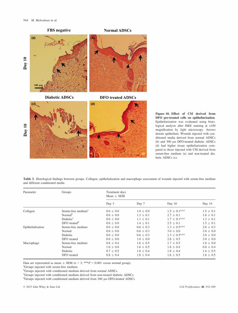
in groups injected with CM derived from dADSCs, scabscovered wounds until day 14; otherwise scabs were notseen in wounds injected with CM derived from treatedgroup (Fig. 10; Table 3). Collagen levels also increasedafter 3 days and reached to maximum levels by 10 daysthen reduced in all groups. Collagen levels were signifi-cantly (P < 0.001) higher in groups injected with CMderived from normal and DFO-treated ADSCs comparedto those treated with CM derived from dADSC groups(P < 0.001; Fig. 11; Table 3). There was no significantdifference in macrophage count between any groupsduring the study (P > 0.05; Table 3).
Discussion
Over the last decade, cell therapy based on ADSCs hasemerged as an appealing approach for promoting angio-genesis in ischaemic tissues (12). However, diseasesaffecting vasculature can inversely influence therapeutic
(a)
(b)
(c)
(d)
I II III IV V I II III
IV V
Figure 7. Assessment of angiogenic potential of conditioned media derived from ADSCs. (a) Representative image of migration of HUVECscultured in different media (I) medium without FBS, (II) CM derived from normal ADSCs, (III) diabetic ADSCs, (IV) 150 lM and (V) 300 lMDFO-treated ADSCs by inverted microscopy after 24 h. (b) Assessment of areas free of HUVECs by NIH image software, in five separated fields.(c) Sprout formation of HUVECs in (I) serum-free medium, (II) CM derived from nADSCs, (III) dADSCs, (IV) 150 lM and (V) 300 lM DFO-trea-ted dADSCs were monitored by inverted microscopy after 48 h. (d) Change in sprout formation of HUVECs determined by NIH image. Data repre-sented as mean � SEM (n = 3; **P < 0.01, ***P < 0.001 versus normal cells and ##P < 0.01, ###P < 0.001 versus diabetic cells). Statisticalsignificance measured by one-way ANOVA.
potential of autologous ADSCs utilized in cell therapy(34,35). Hence, for enhancing angiogenesis, pre-treat-ment strategies are required to revive normal function ofautologous ADSCs before transplantation. To date,hypoxia preconditioning has been considered a desirableapproach to enhance survival and therapeutic neovascu-larization capacity of implanted cells (23,36). Interest-ingly, pharmacological agents mimicking hypoxia have
been found to be used more easily and economically torestore impaired angiogenic mechanisms (24). In thecurrent study, for the first time, we employed a pharma-cological preconditioning approach based on targetingHIF-1a to restore hampered cell functions of ADSCs,under diabetic conditions. First, proliferative capacity aswell as angiogenic gene profiles of dADSCs were exam-ined compared to healthy controls. The current results
Figure 8. Assessment of rat wound closurerate after treatment with different condi-tioned media derived from ADSCs. Follow-ing creating 8 mm full-thickness excisionalskin wounds, wounds were injected with100 ll concentrated medium and concentratedCM derived from ADSCs. Yellow dash linesindicate margins of primary wounds. (a) Rep-resentative gross picture of healing process ofwounds injected with conditioned mediaderived from non-treated diabetic ADSCs(control) and 300 lM DFO-treated dADSCs(DFO-treated) after 0, 7, 10, 12 and 14 days.(b) Quantification of wound closure rate in allgroups using NIH ImageJ software. CMderived from normal and 300 lM DFO-treatedADSCs significantly increased wound healingrates compared to CM derived from dADSCgroups over 10 days. Data are represented asmean � SEM. (n = 3; *P < 0.05, **P < 0.01versus normal cells, #P < 0.05 versus diabeticcells). Statistical significance by two-wayANOVA.
(a)
I II III IV
V VI VII VIII
(b)
Figure 9. Assessment of rat wound angio-genesis after treatment with different con-ditioned media derived from ADSCs. (a)Haematoxylin and eosin-stained histologicalsections for assessment of angiogenesis inwounds treated with serum-free medium(I&V), CM derived from (II&VI) nADSCs,(III&VII) dADSCs and (IV&VIII) 300 lMDFO-treated dADSCs are shown after 3 and7 days respectively. Arrows denote bloodvessels. (b) Comparison between angiogenicscores in groups after 3 and 7 days. Data arerepresented as mean � SEM. (n = 3;***P < 0.001 versus normal cells). Statisticalsignificance measured by two-way ANOVA.
Deferoxamine restore Angiogenesis in stem cell 543
(a) (b)
(c) (d)
Figure 10. Effect of CM derived fromDFO pre-treated cells on epitheliarization.Epitheliarization was evaluated using histo-logical analysis after H&E staining at x100magnification by light microscopy. Arrowsdenote epithelium. Wounds injected with con-ditioned media derived from normal ADSCs(b) and 300 lM DFO-treated diabetic ADSCs(d) had higher tissue epitheliarization com-pared to those injected with CM derived fromserum-free medium (a) and non-treated dia-betic ADSCs (c).
Table 3. Histological findings between groups. Collagen, epithelialization and macrophage assessment of wounds injected with serum-free mediumand different conditioned media
Data are represented as mean � SEM (n = 3; ***P < 0.001 versus normal group).aGroups injected with serum-free medium.bGroups injected with conditioned medium derived from normal ADSCs.cGroups injected with conditioned medium derived from non-treated diabetic ADSCs.dGroups injected with conditioned medium derived from 300 lM DFO-treated ADSCs
seem to indicate that proliferative possibility of dADSCswas lower than normal cells, at similar passages. More-over, in the CFU test, colonies derived from dADSCswere remarkably smaller and fewer than from nADSCs.In agreement with us, other studies have reported signif-icantly lower proliferation and colony formation capacityof BMSCs obtained from diabetic rats compared to con-trols (37,38); furthermore, similar results have beenreported for epidermal stem cells (ESCs) derived fromdiabetic rats (39). In contrast, non-significant changes ingrowth rate have been detected in dADSCs compared tonADSCs of mice (40). This discrepancy could be relatedto differences in experimental conditions such as dura-tion of post-diabetes induction. Our current report hasalso shown diabetic cells to display a more flattenedappearance. This morphology could be due to prematureageing processes of dADSCs, which represent lowerproliferative capacity and also show changes at themolecular level compared to control cells (41).
In addition to macroscopic differences, expression ofHIF-1a and its downstream angiogenic genes (VEGF,FGF-2 and SDF-1) as well as activity of MMP-2 andMMP-9 were significantly lower in dADSCs comparedto nADSCs. Correspondingly, conditioned mediumderived from dADSCs showed a significantly lower
potential in scratch assays, three-dimensional tube for-mation and wound healing. Several lines of evidence arein agreement with this report indicating significantdefects in secretion of angiogenic cytokines. such asVEGF, from MSCs as well as impairment in expressionof other genes associated with cell survival (12,42). Incontrast, Gu and colleagues have reported non-signifi-cant differences between angiogenic capacity ofdADSCs and nADSCs after implantation into ischaemicskin (11). Following assessment of impaired mecha-nisms in dADSCs, this study has tried to establisheffects of DFO on dADSCs, and to evaluate the effec-tiveness of DFO to correct hampered mechanisms withthe goal of utilizing the cells in cell therapy for diabeticpatients. In the cell viability assay, DFO from 75 to300 lM did not show any cytotoxic effect after 24 h.Consistent with the current results, in a recent study per-formed on nADSCs, DFO from 50 to 500 lM for 24 hdid not show any cytotoxic effects (13). These findingssuggest that DFO pre-treatment may be a safe pharma-cological approach for promoting angiogenic activity ofADSCs. These results have also demonstrated that DFOpreconditioning significantly enhanced HIF-1a expres-sion at the protein level in a time- and concentration-dependent manner. In agreement with this report, one
(a) (b)
(c) (d)Figure 11. Effect of CM derived fromDFO pre-treated cells on collagenization.Collagenization was evaluated using Mas-son’s trichrome staining at x400 magnifica-tion, by light microscopy. Collagen fibres arestained blue and arrows denote collagenfibres. Wounds injected with conditionedmedia derived from normal ADSCs (b) and300 lM DFO-treated diabetic ADSCs (d) hadhigher tissue collagenization compared tothose injected with CM derived from non-treated diabetic ADSCs (c) and serum-freemedium (a).
Deferoxamine restore Angiogenesis in stem cell 545
pervious study has shown that DFO-induced expressionof HIF-1a protein in BMSCs, reached its maximumlevel after 24 h (43).
The present invetsigation has also elucidated thatDFO significantly increased expression of VEGF, SDF-1 and FGF-2 in mRNA and protein levels. Existence ofHREs at the promoter level of VEGF, SDF-1 and FGF-2 genes is the most important possible reason for con-cordant stabilization of HIF-1a and up-regulation of rel-evant genes here (20,44,45). These results are supportedby work performed by Hou et al., confirming that DFO,in a HIF-1a-dependent manner, significantly increasedexpression of VEGF and SDF-1 genes in an excisionaldiabetic wound model (27). Similarly, local injection ofDFO has prevented skin flap necrosis in mice by elevat-ing expression of HIF-1a, VEGF and endothelial pro-genitor cell (EPCs) mobilization (46). In a further studyperformed by Chekanov and colleagues, DFO enhancedexpression of FGF-2 in fibroblasts and smooth musclecells (47). In contrast, Potier et al. showed that FGF-2expression was non-significantly increased in DFO pre-treated BMSCs at mRNA and protein levels (28). Thisdiscrepancy might be due to differences in time andconcentration of DFO treatment. Moreover, these resultsindicated that high compared to low concentration ofDFO (300 and 150 lM) increased expression of FGF-2genes to a similar extent. Based on this finding, furtherexperiments are required to clarify whether there areother intervening pathways that prevent the promotingeffect of DFO on FGF-2 expression.
In addition to angiogenic factors, proteolytic enzymesalso contribute to angiogenic activity of ADSCs. MSCssecrete MMP-2 and-9, which are two pivotal members ofthe MMP enzyme family. These are strongly involved inphysiological processes, including endothelial invasionduring angiogenesis (48,49), and homing processes ofstem cells (50). MMPs have been shown to increase angio-genesis through several mechanisms including remod-elling components of the ECM, degradation of basementmembranes, cleaving of endothelial cell–cell adhesionsand pericyte detachment from vessels (49). In the currentstudy, we found that DFO pre-treatment significantly pro-moted activity of secreted MMP-2 and -9 via a HIF-1-de-pendent pathway. These results have also been reported inprevious investigations indicating that promoter activitiesof MMP-2 and -9 were HIF-1a-dependent (51,52). Najafiet al. demonstrated that DFO remarkably enhanced MMP-2 and -9 enzyme activity in BMSCs by a HIF-1a-mediatedmechanism (43). In contrast, one study on hepatoma cellshas shown that MMP expression is HIF-1a independent(53). Furthermore, it must be noted that activity of theseenzymes would be adversely increased in chronic ischae-mic areas such as diabetic wounds (54–56).
The current study has also shown that CM derivedfrom DFO-treated ADSCs significantly improved migra-tion and sprout formation capacity of endothelial cells.Along with this result, one further investigation has alsoshown that hypoxic preconditioning promotes the abilityof dADSCs and nADSCs to enhance angiogenesis byparacrine means (11,12). Conversely, Jiang and col-leagues reported that there was no remarkable differ-ences between numbers and lengths of tubules producedby HUVECs treated with CM from DFO-preconditionedand control ADSCs (13).
To investigate whether enhanced in vitro angiogeniccapacity of DFO pre-treated ADSCs contributed toimproved in vivo angiogenic paracrine potential ofADSCs in injured tissue, we established a surgicalwound model in rats. Histological examination showedthat CM derived from normal and treated ADSCs(300 lM DFO) significantly enhanced angiogenesis. Ithas previously been shown that transplantation of MSCspromoted angiogenesis in the wound area of diabeticmice. This effect was predominantly mediated by para-crine effects rather than by their direct differentiationand incorporation in vascular walls (8). MSCs-derivedCM contain high levels of important potent angiogenicgrowth factors and chemoattractants including VEGF,FGF-2 and SDF-1 (57,58). VEGF enhances migrationand proliferation of endothelial cells (59,60). FGF-2seems to be a crucial factor for proliferation of endothe-lial and smooth muscle cells and development of matureblood vessels (61). SDF-1 acts as a chemoattractant andaugments stem cell homing and vasculogenesis (62).Furthermore, SDF-1 regulates angiogenesis throughbinding to its receptors C-X-C chemokine receptor type4 (63) and it has been observed that its deficiency con-tributes to impaired wound healing in aged mice (64).
In addition, our current study has evaluated effects ofCM derived from ADSCs on epitheliarization, collage-nization and wound closure rate. Results have shown thatCM derived from DFO-treated cells restored these parame-ters by around day 10 and thus might be related to theirhigher angiogenic potential compared to CM derived fromnon-treated cells. It has also been reported that angiogene-sis (which plays a key role during wound healing pro-cesses) also influences other non-angiogenic parametersrelated to wound healing (65–67). Moreover, there is a reatdeal of evidence describing a role for VEGF in accelerat-ing wound healing through ameliorating wound closurerate, epitheliarization and collagenization (68). However,more experiments are required to elucidate exact mecha-nisms involved in improving these histological findings,by CM derived from normal or DFO-treated cells.
Moreover, current results have evaluated underlyingmechanisms involved in impairment of angiogenic
activity of ADSCs derived from type 1 diabetic rats, andmay not be similar to other types of diabetes, includingtype 2, which is commonly associated with obesity. Ithas been shown that adipose tissues form obese humansand mice are poorly oxygenated which leads to induc-tion of HIF-1a in tissues (69–71). Hence, in contrast toour results, HIF-1a and its downstream pathways mightbe activated in poorly oxygenated adipose tissues fromtype 2 diabetic patients. However, on the other hand, itmay not occur. Botusan and colleagues have demon-strated that hyperglycaemia can make HIF-1a more sen-sitive to degradation and reduces its stability in hypoxia(72). Thus, it is possible that hypoxia, presented in adi-pose tissues of type 2 diabetic patients, was not able toinduce HIF-1 expression. Hence, further research isneeded to clarify these mechanisms in type 2 diabetes.
In conclusion, these results may provide a reliableapproach for pharmacological preconditioning prior toclinical application of ADSCs derived from patients withtype 1 diabetes, to revive their normal therapeutic value.It could also be concluded that HIF-1a may act as animportant and key mediator in the effect of DFO to up-regulate pivotal angiogenic genes and therefore restoreimpaired mechanisms in ADSCs from rats with type 1diabetes.
Acknowledgements
This work was supported by a grant from Iran Univer-sity of Medical Sciences and University of Malaya.
Conflict of interest
The authors confirm that there are no conflicts of inter-est.
References
1 Thangarajah H, Vial IN, Grogan RH, Yao D, Shi Y, Januszyk Met al. (2010) HIF-1alpha dysfunction in diabetes. Cell Cycle 9, 75–79.
2 De Vriese AS, Verbeuren TJ, Van de Voorde J, Lameire NH, Van-houtte PM (2000) Endothelial dysfunction in diabetes. Br. J. Phar-macol. 130, 963–974.
3 Krock BL, Skuli N, Simon MC (2011) Hypoxia-induced angiogen-esis good and evil. Genes Cancer 2, 1117–1133.
4 Martin A, Komada MR, Sane DC (2003) Abnormal angiogenesisin diabetes mellitus. Med. Res. Rev. 23, 117–145.
5 Patel DM, Shah J, Srivastava AS (2013) Therapeutic potential ofmesenchymal stem cells in regenerative medicine. Stem Cells Int.2013, 496218. doi:10.1155/2013/496218.
6 Gimble JM, Katz AJ, Bunnell BA (2007) Adipose-derived stemcells for regenerative medicine. Circ. Res. 100, 1249–1260.
7 Bunnell BA, Flaat M, Gagliardi C, Patel B, Ripoll C (2008) Adi-pose-derived stem cells: isolation, expansion and differentiation.Methods 45, 115–120.
8 Wu Y, Chen L, Scott PG, Tredget EE (2007) Mesenchymal stemcells enhance wound healing through differentiation and angiogene-sis. Stem Cells 25, 2648–2659.
9 Murohara T, Shintani S, Kondo K (2009) Autologous adipose-derived regenerative cells for therapeutic angiogenesis. Curr.Pharm. Des. 15, 2784–2790.
10 Ankrum JA, Ong JF, Karp JM (2014) Mesenchymal stem cells:immune evasive, not immune privileged. Nat. Biotechnol. 32, 252–260.
11 Gu JH, Lee JS, Kim DW, Yoon ES, Dhong ES (2012) Neovascularpotential of adipose-derived stromal cells (ASCs) from diabeticpatients. Wound Repair Regen. 20, 243–252.
12 El-Ftesi S, Chang EI, Longaker MT, Gurtner GC (2009) Agingand diabetes impair the neovascular potential of adipose-derivedstromal cells. Plast. Reconstr. Surg. 123, 475–485.
13 Liu GS, Peshavariya HM, Higuchi M, Chan EC, Dusting GJ, JiangF (2013) Pharmacological priming of adipose-derived stem cellsfor paracrine VEGF production with deferoxamine. J. Tissue Eng.Regen. Med. In Press. doi:10.1002/term.1796.
14 Xu Y, Zuo Y, Zhang H, Kang X, Yue F, Yi Z et al. (2010) Induc-tion of SENP1 in endothelial cells contributes to hypoxia-drivenVEGF expression and angiogenesis. J. Biol. Chem. 285, 36682–36688.
15 Wang GL, Jiang B-H, Rue EA, Semenza GL (1995) Hypoxia-in-ducible factor 1 is a basic-helix-loop-helix-PAS heterodimer regu-lated by cellular O2 tension. Proc. Natl Acad. Sci. 92, 5510–5514.
16 Mole DR, Schlemminger I, McNeill LA, Hewitson KS, Pugh CW,Ratcliffe PJ et al. (2003) 2-oxoglutarate analogue inhibitors of HIFprolyl hydroxylase. Bioorg. Med. Chem. Lett. 13, 2677–2680.
18 Karuppagounder SS, Ratan RR (2012) Hypoxia-inducible factorprolyl hydroxylase inhibition: robust new target or another big bustfor stroke therapeutics? J. Cereb. Blood Flow Metab. 32, 1347–1361.
19 Pugh CW, Ratcliffe PJ (2003) Regulation of angiogenesis byhypoxia: role of the HIF system. Nat. Med. 9, 677–684.
20 Ceradini DJ, Kulkarni AR, Callaghan MJ, Tepper OM, Bastidas N,Kleinman ME et al. (2004) Progenitor cell trafficking is regulatedby hypoxic gradients through HIF-1 induction of SDF-1. Nat. Med.10, 858–864.
21 Van Hinsbergh VW, Koolwijk P (2008) Endothelial sprouting andangiogenesis: matrix metalloproteinases in the lead. Cardiovasc.Res. 78, 203–212.
22 Stetler-Stevenson WG (1999) Matrix metalloproteinases in angio-genesis: a moving target for therapeutic intervention. J. Clin.Invest. 103, 1237–1241.
23 Hu X, Yu SP, Fraser JL, Lu Z, Ogle ME, Wang J-A et al. (2008)Transplantation of hypoxia-preconditioned mesenchymal stem cellsimproves infarcted heart function via enhanced survival ofimplanted cells and angiogenesis. J. Thorac. Cardiovasc. Surg.135, 799–808.
24 Doorn J, Fernandes HA, Le BQ, van de Peppel J, van LeeuwenJP, De Vries MR et al. (2013) A small molecule approach to engi-neering vascularized tissue. Biomaterials 34, 3053–3063.
25 Mirzamohammadi S, Aali E, Najafi R, Kamarul T, Mehrabani M,Aminzadeh A et al. (2014) Effect of 17b-estradiol on mediatorsinvolved in mesenchymal stromal cell trafficking in cell therapy ofdiabetes. Cytotherapy 17, 46–57.
26 Brittenham GM, Griffith PM, Nienhuis AW, McLaren CE, YoungNS, Tucker EE et al. (1994) Efficacy of deferoxamine in prevent-ing complications of iron overload in patients with thalassemiamajor. N. Engl. J. Med. 331, 567–573.
27 Hou Z, Nie C, Si Z, Ma Y (2013) Deferoxamine enhances neovas-cularization and accelerates wound healing in diabetic rats via theaccumulation of hypoxia-inducible factor-1a. Diabetes Res. Clin.Pract. 101, 62–71.
28 Potier E, Ferreira E, Dennler S, Mauviel A, Oudina K, Logeart-Avramoglou D et al. (2008) Desferrioxamine-driven upregulationof angiogenic factor expression by human bone marrow stromalcells. J. Tissue Eng. Regen. Med. 2, 272–278.
29 Bradford MM (1976) A rapid and sensitive method for the quanti-tation of microgram quantities of protein utilizing the principle ofprotein-dye binding. Anal. Biochem. 72, 248–254.
30 Liang C-C, Park AY, Guan J-L (2007) In vitro scratch assay: aconvenient and inexpensive method for analysis of cell migrationin vitro. Nat. Protoc. 2, 329–333.
31 Griffith CK, Miller C, Sainson RC, Calvert JW, Jeon NL, HughesCC et al. (2005) Diffusion limits of an in vitro thick prevascular-ized tissue. Tissue Eng. 11, 257–266.
32 Kumar MS, Sripriya R, Raghavan HV, Sehgal PK (2006) Woundhealing potential of Cassia fistula on infected albino rat model. J.Surg. Res. 131, 283–289.
33 Abramov Y, Golden B, Sullivan M, Botros SM, Miller JJR, Al-shahrour A et al. (2007) Histologic characterization of vaginal vs.abdominal surgical wound healing in a rabbit model. WoundRepair Regen. 15, 80–86.
34 Dzhoyashvili NA, Efimenko AY, Kochegura TN, Kalinina NI,Koptelova NV, Sukhareva OY et al. (2014) Disturbed angiogenicactivity of adipose-derived stromal cells obtained from patientswith coronary artery disease and diabetes mellitus type 2. J. Transl.Med. 12, 337.
35 Shin L, Peterson DA (2012) Impaired therapeutic capacity of autol-ogous stem cells in a model of type 2 diabetes. Stem Cells Transl.Med. 1, 125–135.
36 Wei L, Fraser JL, Lu Z-Y, Hu X, Yu SP (2012) Transplantation ofhypoxia preconditioned bone marrow mesenchymal stem cellsenhances angiogenesis and neurogenesis after cerebral ischemia inrats. Neurobiol. Dis. 46, 635–645.
37 Jin P, Zhang X, Wu Y, Li L, Yin Q, Zheng L et al. (2010) Strep-tozotocin-induced diabetic rat–derived bone marrow mesenchymalstem cells have impaired abilities in proliferation, paracrine, anti-apoptosis, and myogenic differentiation. Transplant. Proc. 42,2745–2752.
38 Kim YS, Kwon JS, Hong MH, Kang WS, Jeong H-Y, Kang H-J et al. (2013) Restoration of angiogenic capacity of diabetes-in-sulted mesenchymal stem cells by oxytocin. BMC Cell Biol. 14,38.
39 Zhong Q-L, Liu F-R, Liu D-W, Peng Y, Zhang X-R (2011) Ex-pression of b-catenin and cyclin D1 in epidermal stem cells of dia-betic rats. Mol. Med. Rep. 4, 377–381.
40 Nambu M, Ishihara M, Kishimoto S, Yanagibayashi S, YamamotoN, Azuma R et al. (2011) Stimulatory effect of autologous adiposetissue-derived stromal cells in an atelocollagen matrix on woundhealing in diabetic db/db mice. J. Tissue Eng. 2, 158105.
41 Sethe S, Scutt A, Stolzing A (2006) Aging of mesenchymal stemcells. Ageing Res. Rev. 5, 91–116.
42 Khan M, Akhtar S, Mohsin S, N Khan S, Riazuddin S (2010)Growth factor preconditioning increases the function of diabetes-impaired mesenchymal stem cells. Stem Cells Dev. 20, 67–75.
43 Najafi R, Sharifi AM (2013) Deferoxamine preconditioning potenti-ates mesenchymal stem cell homing in vitro and in streptozotocin-diabetic rats. Expert Opin. Biol. Ther. 13, 959–972.
44 Black SM, DeVol JM, Wedgwood S (2008) Regulation of fibrob-last growth factor-2 expression in pulmonary arterial smooth mus-
cle cells involves increased reactive oxygen species generation.Am. J. Physiol. 294, C345–C354.
45 Shi X, Guo L, Seedial S, Si Y, Wang B, Takayama T et al. (2014)TGF-b/Smad3 inhibit vascular smooth muscle cell apoptosisthrough an autocrine signaling mechanism involving VEGF-A. CellDeath Dis. 5, e1317.
46 Wang C, Cai Y, Zhang Y, Xiong Z, Li G, Cui L (2014) Localinjection of deferoxamine improves neovascularization in ischemicdiabetic random flap by increasing HIF-1a and VEGF expression.PLoS ONE 9, e100818.
47 Chekanov VS, Nikolaychik V (2002) Iron contributes toendothelial dysfunction in acute ischemic syndromes. Circulation105, e35.
48 Arkell J, Jackson CJ (2003) Constitutive secretion of MMP9 byearly-passage cultured human endothelial cells. Cell Biochem.Funct. 21, 381–386.
49 Rundhaug JE (2005) Matrix metalloproteinases and angiogenesis.J. Cell Mol. Med. 9, 267–285.
50 De Becker A, Van Hummelen P, Bakkus M, Broek IV, De WeverJ, De Waele M et al. (2007) Migration of culture-expanded humanmesenchymal stem cells through bone marrow endothelium is regu-lated by matrix metalloproteinase-2 and tissue inhibitor of metallo-proteinase-3. Haematologica 92, 440–449.
51 Munoz-Najar U, Neurath K, Vumbaca F, Claffey K (2005) Hy-poxia stimulates breast carcinoma cell invasion through MT1-MMPand MMP-2 activation. Oncogene 25, 2379–2392.
52 Choi JY, Jang YS, Min SY, Song JY (2011) Overexpression ofMMP-9 and HIF-1a in breast cancer cells under hypoxic condi-tions. J. Breast Cancer 14, 88–95.
53 Miyoshi A, Kitajima Y, Ide T, Ohtaka K, Nagasawa H, Uto Yet al. (2006) Hypoxia accelerates cancer invasion of hepatoma cellsby upregulating MMP expression in an HIF-1a-independent man-ner. Int. J. Oncol. 29, 1533–1539.
54 Kang L, Chen Q, Wang L, Gao L, Meng K, Chen J et al. (2009)Decreased mobilization of endothelial progenitor cells contributesto impaired neovascularization in diabetes. Clin. Exp. Pharmacol.Physiol. 36, e47–e56.
55 Liu Y, Min D, Bolton T, Nub�e V, Twigg SM, Yue DK et al.(2009) Increased matrix metalloproteinase-9 predicts poor woundhealing in diabetic foot ulcers. Diabetes Care 32, 117–119.
56 Lobmann R, Ambrosch A, Schultz G, Waldmann K, Schiweck S,Lehnert H (2002) Expression of matrix-metalloproteinases and theirinhibitors in the wounds of diabetic and non-diabetic patients. Dia-betologia 45, 1011–1016.
57 Kwon HM, Hur SM, Park KY, Kim CK, Kim YM, Kim HS et al.(2014) Multiple paracrine factors secreted by mesenchymal stemcells contribute to angiogenesis. Vascul. Pharmacol. 63, 19–28.
58 Kinnaird T, Stabile E, Burnett M, Shou M, Lee C, Barr S et al.(2004) Local delivery of marrow-derived stromal cells augmentscollateral perfusion through paracrine mechanisms. Circulation109, 1543–1549.
59 Bao P, Kodra A, Tomic-Canic M, Golinko MS, Ehrlich HP, BremH (2009) The role of vascular endothelial growth factor in woundhealing. J. Surg. Res. 153, 347–358.
60 Cebe-Suarez S, Zehnder-Fj€allman A, Ballmer-Hofer K (2006) Therole of VEGF receptors in angiogenesis; complex partnerships.Cell. Mol. Life Sci. 63, 601–615.
61 Murakami M, Simons M (2008) Fibroblast growth factor regulationof neovascularization. Curr. Opin. Hematol. 15, 215–220.
62 Locatelli F, Bersano A, Ballabio E, Lanfranconi S, PapadimitriouD, Strazzer S et al. (2009) Stem cell therapy in stroke. Cell. Mol.Life Sci. 66, 757–777.
63 Kiefer F, Siekmann AF (2011) The role of chemokines and theirreceptors in angiogenesis. Cell. Mol. Life Sci. 68, 2811–2830.
64 Loh SA, Chang EI, Galvez MG, Thangarajah H, El-ftesi S, VialIN et al. (2009) SDF-1a expression during wound healing in theaged is HIF dependent. Plast. Reconstr. Surg. 123, 65S–75S.
65 Lugo LM, Lei P, Andreadis ST (2010) Vascularization of the der-mal support enhances wound re-epithelialization by in situ deliveryof epidermal keratinocytes. Tissue Engin. 17, 665–675.
66 Tonnesen MG, Feng X, Clark RAF (2000) Angiogenesis in woundhealing. in Journal of Investigative Dermatology Symposium Pro-ceedings. J. Investig. Dermatol. Symp. Proc. 5, 40–46.
67 Oryan A, Mohammadalipour A, Moshiri A, Tabandeh MR (2014)Topical application of aloe vera accelerated wound healing, model-ing, and remodeling. Ann. Plast. Surg. In Press. doi:10.1097/SAP.0000000000000239.
68 Brem H, Kodra A, Golinko MS, Entero H, Stojadinovic O, WangVM et al. (2009) Mechanism of sustained release of vascular
endothelial growth factor in accelerating experimental diabetic heal-ing. J. Invest. Dermatol. 129, 2275–2287.
69 Hosogai N, Fukuhara A, Oshima K, Miyata Y, Tanaka S, SegawaK et al. (2007) Adipose tissue hypoxia in obesity and its impacton adipocytokine dysregulation. Diabetes 56, 901–911.
70 Rausch ME, Weisberg S, Vardhana P, Tortoriello DV (2008) Obe-sity in C57BL/6J mice is characterized by adipose tissue hypoxiaand cytotoxic T-cell infiltration. Int. J. Obes. 32, 451–463.
71 Ye J, Gao Z, Yin J, He Q (2007) Hypoxia is a potential risk factorfor chronic inflammation and adiponectin reduction in adipose tis-sue of ob/ob and dietary obese mice. Am. J. Physiol. Endocrinol.Metab. 293, 1118–1128.
72 Botusan IR, Sunkari VG, Savu O, Catrina AI, Gr€unler J, LindbergS et al. (2008) Stabilization of HIF-1alpha is critical to improvewound healing in diabetic mice. Proc. Natl Acad. Sci. 105, 19426–19431.