Deficiency of C4 from Donor or Recipient MouseFails to Prevent Renal Allograft Rejection
Tao Lin,*† Wuding Zhou,* Conrad A. Farrar,*Roseanna E.G. Hargreaves,* Neil S. Sheerin,*and Steven H. Sacks*From the Department of Nephrology and Transplantation,* King’s
College London School of Medicine at Guy’s, King’s College, and
St. Thomas’ Hospitals, London, United Kingdom; and the
Department of Urology,† West China Hospital, Sichuan
University, Chengdu, People’s Republic of China
Complement effector products generated in the trans-planted kidney are known to mediate transplant re-jection, but which of the three main activation path-ways of complement trigger this response is unclear.Here we assessed the role of the classical and lectinpathways by studying the common component C4 inmouse kidney transplant rejection. We transplantedwild-type or C4-null H-2b donor kidneys into H-2k orH-2d recipients, or vice-versa, to assess the roles ofdonor kidney and recipient expression of comple-ment. Intragraft C4 gene expression rose substan-tially during rejection. However, we found no signif-icant association between graft acceptance and thepresence of C4 in either the donor kidney or recipientmouse. At the time of rejection, we found no signifi-cant differences in alloantibody response in the dif-ferent groups. Tubular deposition of C3 to C9 oc-curred regardless of the absence or presence of C4 ineither the donor or recipient mouse, indicating thatC4 was dispensable for complement activation at thissite. These data suggest that complement activationand renal allograft rejection are independent of theclassical and lectin pathways in these models, imply-ing that in the absence of these pathways the alterna-tive pathway is the main trigger for complement-mediated rejection. (Am J Pathol 2006, 168:1241–1248;DOI: 10.2353/ajpath.2006.050360)
Complement activation is a well recognized cause oftissue injury in inflammatory conditions including organtransplant rejection.1 The main complement effectorproducts are 1) the soluble fragments including C3a andC5a, which mediate inflammation; 2) the membrane at-
tack complex C5b-9, which causes membrane injuryleading to cell activation and cell death; and 3) the large,surface-bound fragments of C3 that mediate immune celladhesion.2 Formation of these products requires thecleavage of C3, which can be initiated by the classical,alternative, and lectin pathways.
The fourth component is an essential intermediary forthe classical and lectin pathways of complement activa-tion. The classical pathway is triggered by antibody andC1q binding to target structures, resulting in the cleavageof C4. In addition, C1q can bind directly to activatingsurfaces, such as apoptotic blebs, nuclear material, andC-reactive protein, bypassing the need for antibody toinitiate the cleavage of C4.3,4 In contrast, the lectin path-way uses mannose-binding lectin and associated serineproteases to initiate the cleavage of C4.5 Once cleavedby the classical or lectin pathways, C4 attaches to acti-vator surface, forms a complex with C2 that activates C3,and subsequently leads to membrane insertion of com-plement. In addition, bound C4 acts as a ligand for com-plement receptor CD35 expressed on leukocytes.6 Thesubsequent degradation product C4d has no known bi-ological function but serves as a marker of humoral al-lorejection.7,8 The soluble fragment C4a has only weakproinflammatory properties. Because each bound mole-cule of C4 can result in up to 10 molecules of attachedC3, C4 constitutes an important amplification and regu-latory step in the complement cascade.
Although most circulating complement is produced byhepatocytes, extrahepatic synthesis occurs at severaltissue locations. Important local sources of C3 and C4include resident tissue cells (eg, epithelial cells, endothe-lial cells, and fibroblasts) and migratory leukocytes (eg,neutrophils and macrophage/monocytes).9,10 Recentwork in a renal allograft model has shown that intrarenalproduction of C3 was essential for graft rejection.11
Transplants from C3-deficient mouse donors had lessinflammation and generated a weaker recipient immune
Supported by grants from the Medical Research Council, UK, and Well-come Trust, UK.
Accepted for publication December 29, 2005.
Address reprint requests to Steven H. Sacks, Department of Nephrol-ogy and Transplantation, 5th Floor, Thomas Guy House, Guy’s Hospital,St. Thomas St., London SE1 9RT, UK. E-mail: [email protected].
American Journal of Pathology, Vol. 168, No. 4, April 2006
response than transplants from wild-type donors. Be-cause C4 is needed for two of the three major pathwaysleading to C3 activation, we aimed to define the contri-bution of C4 in the mechanism of allograft rejection. Thisis important because selective inhibition might allow lim-ited therapeutic blockade with fewer unwanted effects.
We tested the hypothesis that C4 was a determinant ofacute allograft rejection. Our prediction was that deficientlocal or systemic production of C4 would lead to in-creased graft acceptance. For this purpose, we used aknown complement-sensitive model of renal allograft re-jection (C57BL/6 donor to B10.Br recipient) and in addi-tion investigated two other donor-recipient straincombinations.
Materials and Methods
Mice
Male C57BL/6 (B6; H-2b), B10.Br (H-2k), BLAB/c (H-2d),and C3H/HeN (H-2k) mice were obtained from HarlanOlac (Bicester, UK). The C4�/� mice were derived byhomologous recombination in embryonic stem cells12
and backcrossed onto the C57BL/6 parental strain for 11generations. C4 mRNA and protein is undetectable inthese mice by real-time polymerase chain reaction (PCR)or Western blotting. All procedures were conducted inaccordance with the Home Office Animal (Scientific Pro-cedures) Act of 1986.
Renal Transplantation
Donor mice, 6 to 8 weeks of age, were anesthetized andthe abdomen was opened through a midline incision. Theleft kidney was excised and preserved in cold saline.Recipient mice were then anesthetized and the right na-tive kidney was excised. Renal transplantation was per-formed with end-to-side anastomoses of the donor renalvein to the inferior vena cava and the donor aortic cuff tothe aorta.13 Urinary tract reconstruction was accom-plished using ureter-to-bladder anastomosis.14 No immu-nosuppressive therapy was administrated at any timeduring the experiment. The left native kidney was re-moved at 1 week after transplantation. We placed thegraft in the right flank because left nephrectomy is tech-nically easier to perform. After that, renal function wasdetermined by measuring blood urea nitrogen at days 8,10, 12, 14, and 16 and then once a week. The end-pointof graft survival was taken as the time to blood ureanitrogen �50 mmol/L or death, depending on which wasfirst.
RNA Isolation and Real-Time ReverseTranscriptase (RT)-PCR
Total RNA was isolated using the RNeasy mini kit (QiagenLtd., West Sussex, UK). Five �g of total RNA was reverse-transcribed into cDNA using the M-MLV reverse tran-scriptase. Quantification of gene expression was per-
formed using DNA Engine Opticon System (MJResearch, Cambridge, MA). C4 primer was designedusing the computer program Oligo 4 (National Bio-sciences Inc., Plymouth, MN), and the sequence is for-ward 5�-GGATCCAGCAGTTTCGAAAG-3�, reverse 5�-ACTGGACATGGG TCGTGGAA-3�. Melting curves wereused to detect primer-dimer conformation and nonspe-cific amplification. Threshold cycle (Ct) numbers weredetermined and transformed using the �Ct and ��Ctmethods as described by the manufacturer, usingGAPDH as the internal control.
In Situ Hybridization
Mouse C4 mRNA was performed on frozen sections us-ing the HybriProbe in situ hybridization assay kit (Biog-nostik, Gottingen, Germany). The mouse C4-specific oli-gonucleotide was derived from the published sequence(accession number: NM�009780). Briefly, 4-�m frozensections were fixed in 4% paraformaldehyde on silanizedslides (DAKO, High Wycombe, UK). Sections were pre-hybridized for 4 hours, hybridized for 16 hours with fluo-rescein isothiocyanate (FITC)-labeled probe for C4mRNA, and then washed with standard saline citrate.Bound probe was detected by immunochemical stainingusing an alkaline phosphatase-conjugated F(ab�) anti-body fragment to FITC (DAKO) followed by the substratemixture BCIP/NBT (DAKO). This resulted in purple prod-uct at the site of hybridization. Slides were then counter-stained with hematoxylin for 10 seconds.
Histological Examination of Renal Tissue
Renal tissue was excised and fixed in 4% formalin andembedded in paraffin. For histological analysis, 4-�msections were mounted on slides and stained with peri-odic acid-Schiff (PAS). The sections were reviewed in ablinded manner by two experienced persons for evalua-tion of rejection.
Immunofluorescence
Four-�m frozen sections of the renal allografts werestained for C4, C3d, and C9 using an indirect immuno-fluorescence method. The slides were fixed in acetoneand then blocked in normal goat serum. The primaryantibodies for C3 and C9 staining were rabbit anti-humanC3d (dilution 1:200; Dako Ltd., Cambridgeshire, UK) andrabbit anti-rat C9 (dilution 1:100; generously provided byDr. B. Paul Morgan, University of Wales). The secondaryantibody was a FITC-conjugated polyclonal goat anti-rabbit IgG (dilution 1:200; Jackson ImmunoResearch,West Grove, PA). For C4 staining, rat monoclonal anti-body to C4 (dilution 1:50; Abcam Ltd., Cambridge, UK)and FITC-conjugated goat anti-rat IgG (dilution 1:200;ICN Biomedicals Inc, Aurora, OH) were used. C3- andC9-positive staining was quantified using Lucia imageanalysis software (Jencons-PLS, Forest Row, UK). At amagnification of �400, for each animal, six fields fromtwo stained kidney sections were photographed. Areas of
1242 Lin et alAJP April 2006, Vol. 168, No. 4
positive staining in each image were outlined, high-lighted, and values computed.
Flow Cytometry
The presence of circulating donor-specific IgM and IgGantibodies were evaluated in the recipient serum by flowcytometry (FACScan; BD Biosciences, Cowley, UK). Na-ıve donor-strain mouse splenocytes were isolated andresuspended in 50 �l of phosphate-buffered saline � 2%bovine serum albumin at a concentration of 1 � 107/ml.The splenocytes were stained with a phycoerythrin-conjugated antibody specific for CD3 (BD Bio-sciences), followed by incubation with the sera fromrecipients. Then the cells were incubated with FITC-conjugated goat antibody specific for mouse IgM orIgG (Sigma, Dorset, UK). Each incubation was per-formed at 4°C for 20 minutes. The cells were washed,fixed, and analyzed by two-color flow cytometry gatedon T cells. The mean fluorescence intensity of each testsample was compared with that obtained from serumof a naıve mouse and the relative mean fluorescencewas determined. Specificity of alloantibody bindingwas assessed by testing the serum against recipientstrain splenocytes. Preliminary studies using a titrationof serum against donor splenocytes found the concen-tration of 1:25 had a linear relationship with meanfluorescence intensity, which was used in all subse-quent experiments.
Statistical Analysis
Survival analysis between groups was calculated usingthe log-rank method. Complement C3 and C9 depositionwere analyzed using one-way analysis of variance. Anti-body production was assessed by the Mann-Whitneytest. All results were generated using GraphPad Prismsoftware (GraphPad, San Diego, CA). Statistical signifi-cance was considered by a P value �0.05.
Results
Intragraft Expression of C4 Is Increased inRenal Allograft Rejection
Wild-type B6 (H-2b) donor kidneys transplanted intoB10.Br (H-2k) recipients were rejected acutely, with graftloss occurring by day 9 after transplantation in all cases.Real-time PCR analysis of day 8 renal transplants isshown in Figure 1. The results indicate that the amount ofC4 transcript in rejecting grafts was increased by 2 to 3logs. Localization studies with C4 mRNA probe are illus-trated in Figure 2, A and B. The renal tubule is seen to bethe main site of C4 expression in normal C57BL/6 kidney.Immunofluorescent staining for tissue C4 is presented inFigure 2, C and D. It shows evidence of C4 deposition inthe peritubular capillary walls of rejecting grafts.
C4-Null Kidney Transplants Are RejectedAcutely
To assess the influence of intragraft complement geneexpression on acute rejection, we determined graft sur-vival times with C4�/� B6 donor kidney transplanted intowild-type B10.Br recipients. The results are illustrated inFigure 3. Acute rejection occurred in 80% of C4�/� donorkidneys, compared with 100% for C4�/� (wild-type) do-nor grafts. This difference is not significant. Graft histol-ogy at rejection is shown in Figure 4, A and B. The extent
Figure 1. Complement C4 mRNA expression in normal B6 kidneys andrejecting B6 grafts. Real-time RT-PCR of C4 transcripts in rejecting B6 grafts.C4 mRNA levels are expressed as fold increase over normal B6 kidneys. Eachanalysis was performed on RNA isolated from three individual kidneys, andassays were run in duplicate.
Figure 2. Localization of C4 expression in wild-type donor kidney. A and B:In the wild-type normal B6 kidney, in situ hybridization of C4 mRNA showedpositive staining in the cytoplasm of tubule cells. There was no stainingfound in C4�/� kidney tissue. C: Immunofluorescence staining for C4 inallografts showed staining predominantly of peritubular capillary walls. D:Background C4 staining in normal B6 kidney tissue. Original magnifications,�250.
C4 Deficiency in Kidney Allograft Rejection 1243AJP April 2006, Vol. 168, No. 4
of tubulointerstitial infiltration in rejected C4�/� donorkidneys was not obviously different from that in rejectedC4�/� kidneys. C4 staining was still clearly detected inthe peritubular capillary walls of C4�/� grafts (Figure 4C),indicating that recipient protein was the source of thecapillary C4 staining. Residual C4 mRNA was detected inrejecting C4�/� grafts (Figure 5); this could have beenthe result of expression by infiltrating leukocytes15 or
contaminating blood cells. As for the surviving C4�/�
grafts, except for infiltration around the major arteries thehistology was no different from normal tissue (data notshown). In addition, the renal function of long-time sur-viving recipients, as measured by blood urea nitrogen,was normal.
C4-Null Recipients Reject Wild-Type Grafts
We next examined the effect of circulating C4, by trans-planting C4�/� B6 recipients with donor kidneys fromB10.Br mice. Graft survival data are presented in Figure6. With C4�/� recipients the rate of acute rejection was90%, compared to 80% for wild-type recipients. Thus, thehypothesis that deficient recipient C4 would prevent graftrejection was unsupported. Histological analysis at rejec-
Figure 3. Effect of donor C4 status on graft survival: B10.Br recipients ofwild-type kidney reject their transplants within 9 days. Eight of ten B10.Brrecipients of C4�/� grafts rejected their transplants in 9 days; the other tworecipients did not acutely reject but survived until killing at day 72 or day 110.P 0.42 comparing C4�/� and wild-type grafts.
Figure 4. Histology and C4 staining in the rejecting B6 grafts. A: Typicalrejecting kidney allograft (B6 to B10.Br) at day 8 showing heavy interstitialinfiltration, venulitis, and tubulitis (3) (PAS stain). B: Typical rejectingC4�/� kidney graft at day 8 showing a similar pattern to B6 wild-type graft(PAS stain). C: Diffuse C4 deposition was detected in the peritubular capillarywalls of C4�/� grafts, which was not present in normal nontransplantedC4�/� kidney (D). a, Arteriole; v, vein; t, tubule. Original magnifications,�250.
Figure 5. Real-time RT-PCR of C4 transcripts in rejecting grafts. C4 mRNAlevels are expressed as fold increase over normal B6 kidneys. Three sampleswere used in each analysis, and assays were run in duplicate.
Figure 6. Effect of recipient C4 on graft survival. In B10.Br to B6 group, tworecipients were sacrificed at days 56 and 82; the others rejected their grafts atday 8 or day 9. C4�/� recipients rejected 9 of 10 B10.Br grafts before day 9.Another recipient was killed at day 34.
1244 Lin et alAJP April 2006, Vol. 168, No. 4
tion showed no difference in the extent of tubular damageand leukocyte infiltration (Figure 7, A and B). There wasno deposition of C4 in the peritubular capillaries (Figure7C), which again proved that systemic C4 was the sourceof staining in the wild-type recipient experiments. Thesurviving C4�/� grafts displayed normal features, exceptthat infiltrating cells were present around some of thelarger arteries.
Different Donor-Recipient Strain CombinationsShow No Effect of C4 from Donor or Recipient
The B6 to B10.Br transplant model described above ex-hibited marked cell-mediated rejection, supporting thesuggestion that B10.Br mice have a predominant Th1response. We next transplanted C4�/� or C4�/� B6 do-nor kidneys into BALB/c (H-2d) mice. These are reportedto have a Th2 phenotype that favors an antibody-medi-ated response.16,17 The graft survival data are shown inFigure 8. There was no clear difference in graft survivalbetween the two sets of donor kidneys. Histological datafrom rejecting grafts is illustrated in Figure 9, A and B.This indicates features of cell-mediated rejection in bothgroups of transplants.
We also examined strain combinations involving C3H/HeN (H-2k) mice. C3H reportedly give weaker transplantrejection, which might allow detection of any subtle ef-fects of C4.18 The outcome of these transplant experi-ments is summarized in Table 1. When either the trans-plant donor or recipient was C4�/�, acute rejectionoccurred in five of six recipients. This was not signifi-
cantly different compared to the respective wild-type do-nor or recipient controls.
Alloantibody Response Is Unaffected byC4 at Rejection
Previous studies have shown that complement-deficientmice have impaired antibody responses.19 To investigateif this is the case in our model, the donor-specific IgG andIgM alloantibody responses were determined by flowcytometry. Because the majority of recipients failed tosurvive more than 9 days after transplantation, serum
Figure 7. Histology and C4 staining in the rejecting B10.Br grafts. A and B:B10.Br allografts in wild-type and C4�/� B6 recipients at day 8 showingsimilar pattern of interstitial infiltration, venulitis, and tubulitis (3) (PASstain). C: Immunofluorescence staining for C4 in B10.Br to C4�/� allograftsshowed background staining. Original magnifications, �250.
Figure 8. Kidney transplant rejection in BALB/c recipients. Seven of eightBALB/c recipients of C4�/� grafts rejected their transplants within 9 days.The other one was killed at day 43. In B6 to BALB/c group, acute rejectionoccurred in every case.
Figure 9. Histology of rejecting B6 graft. A: Rejecting kidney allograft (B6into BALB/c) at day 8 typically showing a heavy interstitial infiltration,venulitis, and tubulitis (3) (PAS stain). B: Typical rejecting C4�/� kidneygraft at day 8 showing a similar pattern to B6 wild-type graft (PAS stain).Original magnifications, �250.
Table 1. Survival of Allografts in C57BL/6 and C3H/HeNStrain Combination
* P 0.44 versus C4�/� grafts.† P 0.71 versus C4�/� recipients.‡ Sacrifice at the end of experiment.
C4 Deficiency in Kidney Allograft Rejection 1245AJP April 2006, Vol. 168, No. 4
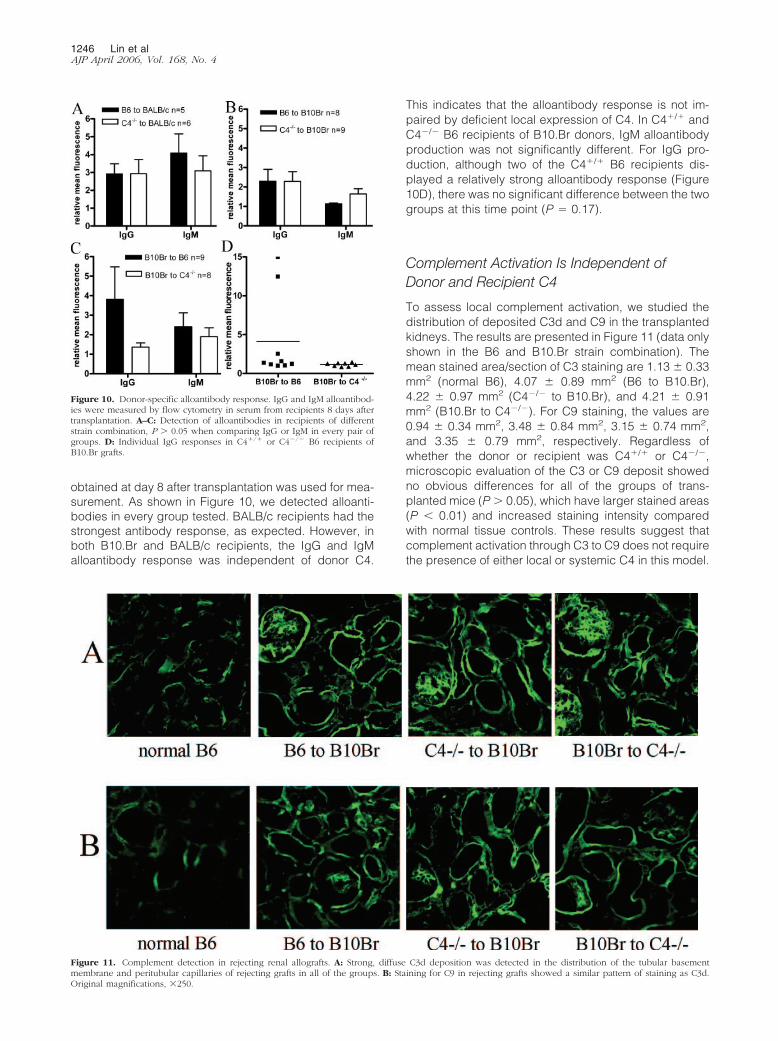
obtained at day 8 after transplantation was used for mea-surement. As shown in Figure 10, we detected alloanti-bodies in every group tested. BALB/c recipients had thestrongest antibody response, as expected. However, inboth B10.Br and BALB/c recipients, the IgG and IgMalloantibody response was independent of donor C4.
This indicates that the alloantibody response is not im-paired by deficient local expression of C4. In C4�/� andC4�/� B6 recipients of B10.Br donors, IgM alloantibodyproduction was not significantly different. For IgG pro-duction, although two of the C4�/� B6 recipients dis-played a relatively strong alloantibody response (Figure10D), there was no significant difference between the twogroups at this time point (P 0.17).
Complement Activation Is Independent ofDonor and Recipient C4
To assess local complement activation, we studied thedistribution of deposited C3d and C9 in the transplantedkidneys. The results are presented in Figure 11 (data onlyshown in the B6 and B10.Br strain combination). Themean stained area/section of C3 staining are 1.13 0.33mm2 (normal B6), 4.07 0.89 mm2 (B6 to B10.Br),4.22 0.97 mm2 (C4�/� to B10.Br), and 4.21 0.91mm2 (B10.Br to C4�/�). For C9 staining, the values are0.94 0.34 mm2, 3.48 0.84 mm2, 3.15 0.74 mm2,and 3.35 0.79 mm2, respectively. Regardless ofwhether the donor or recipient was C4�/� or C4�/�,microscopic evaluation of the C3 or C9 deposit showedno obvious differences for all of the groups of trans-planted mice (P � 0.05), which have larger stained areas(P � 0.01) and increased staining intensity comparedwith normal tissue controls. These results suggest thatcomplement activation through C3 to C9 does not requirethe presence of either local or systemic C4 in this model.
Figure 10. Donor-specific alloantibody response. IgG and IgM alloantibod-ies were measured by flow cytometry in serum from recipients 8 days aftertransplantation. A–C: Detection of alloantibodies in recipients of differentstrain combination, P � 0.05 when comparing IgG or IgM in every pair ofgroups. D: Individual IgG responses in C4�/� or C4�/� B6 recipients ofB10.Br grafts.
Figure 11. Complement detection in rejecting renal allografts. A: Strong, diffuse C3d deposition was detected in the distribution of the tubular basementmembrane and peritubular capillaries of rejecting grafts in all of the groups. B: Staining for C9 in rejecting grafts showed a similar pattern of staining as C3d.Original magnifications, �250.
1246 Lin et alAJP April 2006, Vol. 168, No. 4
Discussion
In a total of 48 transplants in which the donor kidney wasfrom a C4-positive or -negative mouse, our results showthat the graft survival rate for the C4-negative transplantswas not increased. Furthermore, in another 32 transplantprocedures, when the recipient was either C4-positive or-negative, the negative recipients showed no graft sur-vival advantage. Thus C4, whether expressed in the do-nor kidney or recipient, was dispensable in this model ofacute rejection.
All graft tissues contained a deposit of C3d and C9 inthe region of the tubular basement membrane regardlessof strain combination, indicating that complement activa-tion and subsequent membrane insertion did not requirethe presence of C4. Because the classical and lectinpathways depend on C4 for the formation enzyme com-plexes capable of cleaving C3, our data suggest nomajor role for the classical and lectin pathways leading toC3 cleavage and complement insertion. Instead, our dataimply that the alternative pathway was responsible for C3cleavage, in the absence of the classical and lectin path-ways. Once cleaved, C3 leads to the formation of C3a,C3b, C5a, and C5b-9. Proinflammatory and immunoregu-latory functions downstream of C3 cleavage have re-cently been shown to mediate renal transplant rejection ina B6 to B10.Br allograft model.11 The data presentedhere infer a role for the alternative pathway in drivingthese functions of complement.
The effector function of C4 in acute antibody-mediatedrejection is well recognized.20 In the presence of boundantibody and C1, activation of C4 leads to the cleavageof C3 and consequently to the formation of effector prod-ucts that mediate endothelial injury at the site of Ig andC1 binding. Circulating C4 might be expected to exertgreater influence on vascular rejection than locally pro-duced C4, because of the abundance of C4 in the circu-lation and in view of the contact of blood with endothe-lium. However, mice with deficient circulating C4 did notexhibit lower rejection rates compared to wild-type mice.
In addition to its effector function, complement canalso regulate the B cell response.21 For example, Marshand colleagues20 grafted C3- or C4-deficient mice withallogeneic donor skin and found that complement defi-ciency impaired the conversion of IgM to IgG alloanti-body responses. In the present study, we were unable todetect a difference in the alloantibody response at day 8after transplantation, with the exception of two recipientwild-type mice. However, primary B-cell responses gen-erally require 14 days to achieve peak serum antibodylevel22 and the study by Marsh and colleagues20 onlydetected a difference between complement-deficientand wild-type animals at the peak of the immune re-sponse, which was 28 days after skin grafting. Thus, ourantibody data collected at 8 days after renal transplan-tation do not contradict the results for the skin-graftedmice20 but suggest that at the time of rejection C4-medi-ated regulation of the alloantibody response had no im-pact on our results. Despite the detection of alloantibod-ies in each strain combination, the dominant tissuepathology was cell-mediated injury. Taken together with
the lack of effect of circulating C4 on graft survival, thissuggests that the predominant mechanism of rejectionwas cell mediated and that this was independent of C4.
Spontaneous acceptance of grafted tissue in somemouse strain combinations has been recognized sincethe 1970s.23,24 The rate of spontaneous graft acceptancein mice is also donor-tissue related, given that kidneyallograft acceptance rates exceed those for cardiac andskin allografts.18,25 Recent research has shown thatmouse kidney allograft survival rates vary from 20 to 80%depending on the donor-recipient strain combina-tion.18,26 The mechanism behind such survival remainsunproven. Here, we observed it again in all of the threestrain combinations tested in our study. However, thespontaneous acceptance rate was no different when ei-ther the donor or recipient was C4-negative. Therefore, itis unlikely that C4 has any effect on spontaneousacceptance.
The apparent lack of C4 at the tubular basement mem-brane in the presence of bound C3 suggests that C3cleavage at this site occurred by a C4-independent path-way. Low production of C4 is unlikely to have been alimiting factor, because there was marked up-regulationof C4 mRNA localized to the tubular epithelium in reject-ing grafts, and proximal tubule cells are known to effi-ciently translate and secrete expressed C4.27,28 Althoughseveral mouse strains are reported to have low genera-tion of C4 as a result of posttranscriptional regulation,29
all our comparisons used a C4-high strain (B6) againstC4 knockout mice. Thus, comparison was between highand absent C4 in the donor or recipient arrangementsused. Furthermore, even when the recipient was B10.Br,which has been described as a low producer,30 the cir-culating protein was sufficient to allow C4 deposition onthe capillary wall. Therefore, although we cannot fullyexclude the possibility of low production causing the lackof tubular C4 protein staining, our immunohistologicaland survival data strongly suggest that C4 cleavage didnot participate in the activation of intrarenal C3. Antibodydetection of C4 in the peritubular capillary walls adjacentto negatively stained tubules provided an internal controlshowing that the marker antibody used was capable ofdetecting bound C4.
In conclusion the data presented here deny a signifi-cant role for C4 leading to complement activation andgraft rejection in a mouse renal allograft model. Comple-ment activation at the tubular basement membrane in theabsence of either locally or systemically derived C4 sug-gests an effect independent of the commonly describedclassical and lectin pathways, which both involve C4.Rather our data suggest that a C4-independent pathway,such as the alternative pathway, is the main driver ofcomplement-mediated rejection in our model.
Acknowledgment
We thank Dr. P. O’Donnell for kindly providing expertisein the histopathology analysis.
C4 Deficiency in Kidney Allograft Rejection 1247AJP April 2006, Vol. 168, No. 4
References
1. Sacks SH, Chowdhury P, Zhou W: Role of the complement system inrejection. Curr Opin Immunol 2003, 15:487–492
2. Baldwin III WM, Larsen CP, Fairchild RL: Innate immune responses totransplants: a significant variable with cadaver donors. Immunity2001, 14:369–376
3. Korb LC, Ahearn JM: C1q binds directly and specifically to surfaceblebs of apoptotic human keratinocytes: complement deficiency andsystemic lupus erythematosus revisited. J Immunol 1997,158:4525–4528
4. Gasque P: Complement: a unique innate immune sensor for dangersignals. Mol Immunol 2004, 41:1089–1098
5. Fujita T: Evolution of the lectin-complement pathway and its role ininnate immunity. Nat Rev Immunol 2002, 2:346–353
6. Kalli KR, Fearon DT: Binding of C3b and C4b by the CR1-like site inmurine CR1. J Immunol 1994, 152:2899–2903
8. Racusen LC, Halloran PF, Solez K: Banff 2003 meeting report: newdiagnostic insights and standards. Am J Transplant 2004,4:1562–1566
9. McPhaden AR, Whaley K: Complement biosynthesis by mononuclearphagocytes. Immunol Res 1993, 12:213–232
10. Morgan BP, Gasque P: Extrahepatic complement biosynthesis:where, when and why? Clin Exp Immunol 1997, 107:1–7
11. Pratt JR, Basheer SA, Sacks SH: Local synthesis of complementcomponent C3 regulates acute renal transplant rejection. Nat Med2002, 8:582–587
12. Fischer MB, Ma M, Goerg S, Zhou X, Xia J, Finco O, Han S, Kelsoe G,Howard RG, Rothstein TL, Kremmer E, Rosen FS, Carroll MC: Regu-lation of the B cell response to T-dependent antigens by classicalpathway complement. J Immunol 1996, 157:549–556
13. Zhang Z, Schlachta C, Duff J, Stiller C, Grant D, Zhong R: Improvedtechniques for kidney transplantation in mice. Microsurgery 1995,16:103–109
14. Han WR, Murray-Segal LJ, Mottram PL: Modified technique for kidneytransplantation in mice. Microsurgery 1999, 19:272–274
15. Pratt JR, Abe K, Miyazaki M, Zhou W, Sacks SH: In situ localization of
C3 synthesis in experimental acute renal allograft rejection. Am JPathol 2000, 157:825–831
16. Reiner SL, Locksley RM: The regulation of immunity to Leishmaniamajor. Annu Rev Immunol 1995, 13:151–177
17. Sher A, Coffman RL: Regulation of immunity to parasites by T cellsand T cell-derived cytokines. Annu Rev Immunol 1992, 10:385–409
18. Zhang Z, Zhu L, Quan D, Garcia B, Ozcay N, Duff J, Stiller C,Lazarovits A, Grant D, Zhong R: Pattern of liver, kidney, heart, andintestine allograft rejection in different mouse strain combinations.Transplantation 1996, 62:1267–1272
19. Fearon DT, Carroll MC: Regulation of B lymphocyte responses toforeign and self-antigens by the CD19/CD21 complex. Annu RevImmunol 2000, 18:393–422
20. Marsh JE, Farmer CK, Jurcevic S, Wang Y, Carroll MC, Sacks SH: Theallogeneic T and B cell response is strongly dependent on comple-ment components C3 and C4. Transplantation 2001, 72:1310–1318
21. Carroll MC: The complement system in regulation of adaptive immu-nity. Nat Immunol 2004, 5:981–986
22. Vongwiwatana A, Tasanarong A, Hidalgo LG, Halloran PF: The role ofB cells and alloantibody in the host response to human organ allo-grafts. Immunol Rev 2003, 196:197–218
23. Russell PS, Chase CM, Colvin RB, Plate JM: Induced immune de-struction of long-surviving, H-2 incompatible kidney transplants inmice. J Exp Med 1978, 147:1469–1486
24. Russell PS, Chase CM, Colvin RB, Plate JM: Kidney transplants inmice. An analysis of the immune status of mice bearing long-term,H-2 incompatible transplants. J Exp Med 1978, 147:1449–1468
25. Bickerstaff AA, Wang JJ, Pelletier RP, Orosz CG: The graft helps todefine the character of the alloimmune response. Transpl Immunol2002, 9:137–141
26. Bickerstaff AA, Wang JJ, Pelletier RP, Orosz CG: Murine renalallografts: spontaneous acceptance is associated with regulated Tcell-mediated immunity. J Immunol 2001, 167:4821–4827
27. Seelen MA, Brooimans RA, van der Woude FJ, van Es LA, Daha MR:IFN-gamma mediates stimulation of complement C4 biosynthesis inhuman proximal tubular epithelial cells. Kidney Int 1993, 44:50–57
28. Gerritsma JS, Gerritsen AF, De Ley M, van Es LA, Daha MR: Inter-feron-gamma induces biosynthesis of complement components C2,C4 and factor H by human proximal tubular epithelial cells. Cytokine1997, 9:276–283
29. Ogata RT, Zepf NE: C4 from C4-high and C4-low mouse strains haveidentical sequences in the region corresponding to the isotype-spe-cific segment of human C4. Eur J Immunol 1990, 20:1607–1610
30. Nakayama K, Pattanakitsakul S, Yokoyama S, Kimura H, Nonaka M,Takahashi M: Post-transcriptional regulation of complement C4 in lowC4-producing strain of mouse. Immunogenetics 1990, 31:361–367