Dynamic Article LinksC<Lab on a Chip
Cite this: Lab Chip, 2011, 11, 912
www.rsc.org/loc PAPER
Publ
ishe
d on
27
Janu
ary
2011
. Dow
nloa
ded
by Q
ueen
s U
nive
rsity
- K
ings
ton
on 2
1/05
/201
3 17
:42:
56.
View Article Online / Journal Homepage / Table of Contents for this issue
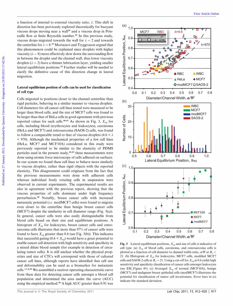
Deformability-based cell classification and enrichment using inertialmicrofluidics†
Soojung Claire Hur,ae Nicole K. Henderson-MacLennan,b Edward R. B. McCabec and Dino Di Carlo*de
Received 12th November 2010, Accepted 6th January 2011
DOI: 10.1039/c0lc00595a
The ability to detect and isolate rare target cells from heterogeneous samples is in high demand in cell
biology research, immunology, tissue engineering and medicine. Techniques allowing label-free cell
enrichment or detection are especially important to reduce the complexity and costs towards clinical
applications. Single-cell deformability has recently been recognized as a unique label-free biomarker for
cell phenotype with implications for assessment of cancer invasiveness. Using a unique combination of
fluid dynamic effects in a microfluidic system, we demonstrate high-throughput continuous label-free
cell classification and enrichment based on cell size and deformability. The system takes advantage of
a balance between deformability-induced and inertial lift forces as cells travel in a microchannel flow.
Particles and droplets with varied elasticity and viscosity were found to have separate lateral dynamic
equilibrium positions due to this balance of forces. We applied this system to successfully classify
various cell types using cell size and deformability as distinguishing markers. Furthermore, using
differences in dynamic equilibrium positions, we adapted the system to conduct passive, label-free and
continuous cell enrichment based on these markers, enabling off-chip sample collection without
significant gene expression changes. The presented method has practical potential for high-throughput
deformability measurements and cost-effective cell separation to obtain viable target cells of interest in
cancer research, immunology, and regenerative medicine.
Introduction
Alteration in the deformability (or mechanical strength) of single
cells has been identified to be a useful indicator of changes in
cellular phenotype of importance for biological research. Various
diseases are associated with cell deformability alterations
including cancer,1–4 blood diseases (sickle cell anemia, hereditary
spherocytosis, and immune haemolytic anaemia),5,6 and inflam-
mation.7,8 In particular, the stiffness of individual cancer cells has
been found to be drastically reduced when compared to normal
tissue of the same origin. Furthermore, decreasing single-cell
stiffness was correlated with increasing invasiveness or meta-
static potential. Biomechanical assays confirmed this correlation
both with in vitro human cancer cell lines as well as clinical
biopsies.1–4 These results are practically important considering
aDepartment of Mechanical and Aerospace Engineering, University ofCalifornia, Los Angeles, Los Angeles, CA, 90095, USAbDepartment of Pediatrics, University of California, Los Angeles, LosAngeles, CA, 90095, USAcLinda Crnic Institute for Down Syndrome and Department of Pediatrics,University of Colorado School of Medicine, Aurora, CO, 80045, USAdDepartment of Bioengineering, University of California, Los Angeles, LosAngeles, CA, 90095, USA. E-mail: [email protected]; Tel: +1 310 9833235eCalifornia NanoSystems Institute, Los Angeles, CA, 90095, USA
† Electronic supplementary information (ESI) available: ESI_Figure 1–7and ESI_Movie. See DOI: 10.1039/c0lc00595a
912 | Lab Chip, 2011, 11, 912–920
the simplicity and low potential cost for obtaining label-free
biophysical measurements. A label-free deformability biomarker
would likely have lower operating costs than current molecular-
based biomarkers that require pre-processing steps, dyes, and/or
costly antibodies. Furthermore, disease states of interest can be
expanded to those without predetermined immunological
markers as long as a correlation between deformability pheno-
type and clinical outcome is confirmed. Specifically, deform-
ability-based target cell classification/enrichment would be useful
for cancer research and diagnostics since it would enable
controlled experiments correlating cell mechanics of cancer cell
lines with known genetic defects as well as analysis of malignant
cells of unknown origin (e.g. circulating tumor cells (CTCs) in
peripheral blood or malignant cells in biopsy samples) for cancer
staging, relapse detection, molecular analysis of cancer drug
resistance, and potentially early detection.1,3,4,9
Current techniques developed for measuring deformability
and elastic properties of cells include micropipette aspiration,10–12
atomic force microscopy,4,13,14 optical deformability,3 magnetic
bead twisting assays15 and optical tweezers.2 Despite the success
in obtaining overall deformability measurements for cells of
interest, the low throughput (1 cell min�1 to 1 cell s�1) of current
cell deformability measurement techniques renders current
technologies ill-suited for statistical analysis of large heteroge-
neous biological samples or rare cell detection. For example,
current throughput does not allow routine screening of millions
This journal is ª The Royal Society of Chemistry 2011
Fig. 1 Classifying and separating cells and particles by deformability.
(a) The balance between two lateral forces, namely inertial lift force, FL,
and viscoelasticity induced force, Fv. leads to unique lateral inertial
focusing equilibrium positions, Xeq, for (b) deformable particles and (c)
rigid particles with various diameters, a. (d) The microfluidic device used
for cancer cell enrichment based on these parameters consists of an inlet
with a coarse filter, a straight focusing (40 � 93 mm) region, and a grad-
ually expanding separation region ending in 5 branched outlets with high
fluidic resistance. Outlets, denoted as B or C, represent the designated
collection outlets for blood cells and cancer cells, respectively in enrich-
ment experiments. All schematics represent the top view of the micro-
fluidic device.
Publ
ishe
d on
27
Janu
ary
2011
. Dow
nloa
ded
by Q
ueen
s U
nive
rsity
- K
ings
ton
on 2
1/05
/201
3 17
:42:
56.
View Article Online
of cells, which is often desired for statistically robust diagnostic
and research applications (e.g. detection/enumeration of cancer
cells in blood or biopsies). Moreover, post-measurement
enrichment of cell populations with uniform deformability has
not been demonstrated for current technologies although high-
purity isolation of viable cells with preserved gene expression
profiles would facilitate the comprehensive assessment of single-
cell mechanics correlated with unexplored genes responsible for
such changes in phenotype. Therefore, novel techniques allowing
deformability activated target cell enrichment and/or high-
throughput deformability measurement of individual cells would
expand the research use and clinical adoption of this biomarker.
In order to address the limited throughput and off-chip viable
sample collection of current techniques, we present a passive
label-free cell classification and enrichment technique utilizing
intrinsic properties of living cells (i.e. size and deformability).
The system employs inertial focusing in combination with
deformability-induced migration to identify and separate more
deformable cells of interest in a label-free, continuous flow, and
high-throughput manner. The microfluidic system operates by
yielding unique equilibrium positions in a channel cross-section
for flowing particles with heterogeneous size/deformability.
First, to systematically investigate the effect of particle deform-
ability on our measurements and to calibrate our system we
evaluated the unique focusing behavior for silicone oil droplets
over a range of viscosities. Then, we applied the system to assay
a large variety of blood cells and cultured cell lines. Furthermore,
as a proof-of-concept towards analysis of CTCs from blood,
cancer cells, spiked in a dilute blood sample prior to the injection,
were separated and enriched using our approach. Viability and
global gene expression of the collected cells were examined in
order to determine whether the proposed fluidic process
adversely affects processed cells. Taking advantage of microscale
physics, the presented method provides a simple target cell
classification and enrichment approach for a variety of applica-
tions where deformability and size are suitable biomarkers.
Theoretical background: Inertial focusing ofdeformable particles in poiseuille flow
Inertial effects in microfluidic systems have recently been gaining
much attention because of the ability to easily focus and order
particles and cells continuously without external forces.16 In
brief, an inertial lift force, FL, induces lateral migration of
particles in confined flow (see Fig. 1) and creates distinct
inertial lift focusing positions at finite particle Reynolds
number,17 RP ¼rexUmaxa2
mexDh
¼ Rc
�a
Dh
�2
. Here, rex, Umax, mex, Rc,
a are the density, the maximum velocity, the dynamic viscosity of
the continuous phase fluid, the channel Reynolds number, and
the particle diameter, respectively, while Dh is the hydraulic
diameter of the channel, defined as Dh ¼2WH
W þHwhere W and H
are the channel width and height, respectively. Particularly
relevant to this work, we have previously demonstrated that
particles/cells can be inertially focused at two distinct lateral
focusing positions and one uniform z-position using high aspect
ratio channels (2 < H/W).18 These channels reduced the proba-
bility of particle overlap and out-of-focus blur and provided
This journal is ª The Royal Society of Chemistry 2011
similar cell signature images, allowing accurate cell-type classi-
fication with extreme throughput. These high-aspect ratio
channel structures are used to focus cells to two lateral positions
in this work.
In addition to nonlinearity associated with the inertia of the
fluid, nonlinear lateral migration can occur when the particle
itself is deformable. Early theoretical investigations reported that
elastic solid particles experience a force away from the wall in
Poiseuille flow,19 such that particles laterally migrated to the
channel centerline, even in the zero-Reynolds-number limit.19,20
Following these works, several theoretical, numerical and a few
experimental studies expanded the investigation of this
phenomenon to consider the motion of bubbles, liquid drops,
vesicles and viscous capsules surrounded by elastic membranes,
flowing in wall-bounded flows.19–24 Lateral migration of
deformable particles was found to result from a nonlinearity
caused by matching of velocities and stresses at the particle/
droplet interface.24 That is, the magnitude of lateral drift velocity
and lift force is closely related to the deformed shape of the
object. For droplets with surface tension, s, the
Weber, We ¼ rexU2maxa
s, or capillary, Ca ¼ mexUmaxa
sWnumber
provides a dimensionless parameter (inertial stress vs. surface
tension or viscous stress vs. surface tension) that characterizes the
relative deformation expected for a droplet.25 The internal-to-
external viscosity ratio, l ¼ min/mex, is another significant
parameter characterizing droplet deformation and drift.21,22,24,25
The drift velocity (i.e., lift force) was found to increase with the
droplet deformability and the direction of migration is
predominantly toward the channel centerline for all deformable
Lab Chip, 2011, 11, 912–920 | 913
Publ
ishe
d on
27
Janu
ary
2011
. Dow
nloa
ded
by Q
ueen
s U
nive
rsity
- K
ings
ton
on 2
1/05
/201
3 17
:42:
56.
View Article Online
objects. A notable exception to this trend, and migration towards
the wall, was observed with viscous droplets when the viscosity
ratio ranged between 0.5 and 10.22,24,25
The fact that deformable particles experience an additional lift
force suggests the possibility of high throughput deformability-
induced particle classification and separation.26–28 Deformation-
induced lift forces will act in superposition with inertial lift forces
to create modified lateral equilibrium positions that are depen-
dent on particle deformability. Consequently, the lateral equi-
librium position can then be used as the measure of particle
deformability when the particle size is taken into account.
Furthermore, the differences in lateral equilibrium position
among cell types can be utilized for deformability-induced target
cell enrichment by directing entrained target cells to separate
designated outlets.
Methods
Cell preparation
Whole blood samples were drawn from healthy volunteers into
venous blood collection tubes (BD Vacutainer�) containing 0.4
mL of trisodium citrate (13.2 g L�1), citric acid (4.8 g L�1) and
dextrose (14.7 g L�1). Leukocytes were either purchased from the
Center for AIDS Research Virology Core/BSL3 Facility or
obtained by selectively lysing erythrocytes using RBC lysis buffer
(eBioscience). HeLa cells (a cervical carcinoma cell line), MCF7
cells (a breast carcinoma cell line) and SAOS-2 (an osteosarcoma
cell line) were cultured in the growth media suggested by
American Type Culture Collection (ATCC�) and incubated at
37 �C in 5% CO2 until near confluence. Breast cancer cells
(modMCF7), representing a highly invasive phenotype,29 were
obtained by chemically modifying MCF7 cells with 100 nM 12-
O-tetradecanoylphorbol-13-acetate (TPA) for 18–24 h.3 For
spiking experiments, blood and cancer cells were mixed to ach-
ieve a final ratio of 1 : 100 cancer cells to blood cells. Dilute cell
solution with relatively low total number concentration (202 000
cells mL�1) was used for the experiment to avoid particle–particle
interactions, leading to defocusing of particles at the expanding
outlet and lower purity of collected samples.30 Indeed, it was
observed that more blood cells were directed towards the cancer
outlet, contaminating the collected sample, when the cell solu-
tions with higher number concentration (100 million cells mL�1)
were introduced into the system (see ESI_Movie 1†).
Particle/capsule suspensions
Experiments with polydisperse elastic particles and viscous oil
droplets with a wide range of elastic moduli and kinematic
viscosity were conducted to isolate dominant contributions to
lateral migration of particles. Particles/droplets were created by
forming stable surfactant coated spheres in deionized water
with 3% w/v Tween 80 (Fisher Chemical) through vigorously
mixing two immiscible liquids (uncured polydimethylsiloxane
(PDMS) polymer or silicone oils).31 Interfacial tension between
the two immiscible phases was kept uniform and small by
controlling surfactant concentration (�0.5 mJ m�2), which is
the reported value for a stabilized emulsion.32 Moreover, the
interface of droplets was considered to be saturated with the
surfactant molecules since the molar concentration of Tween 80
914 | Lab Chip, 2011, 11, 912–920
in the solution (28 mM) was much higher than its critical
micelle concentration (13 mM at 25 �C). Saturation of the
surfactant molecules at the water/oil interface allows the no-slip
boundary condition at the interface to be valid.33 After curing,
PDMS solid particle emulsions were centrifuged to collect
microparticles whose diameters ranged between 2 and 30 mm.34
Solid elastic particles made of PDMS and viscous oil droplets
with a deformable interface are referred to as PDMS particles
and viscous droplets, respectively. The deformability of PDMS
particles was controlled by varying crosslinker density35 while
that of viscous oil capsules was controlled by choosing silicone
oil with various kinematic viscosities (1 cSt < n < 1000 cSt,
PDMS 200, Dow Corning). Young’s moduli of microparticles
were assumed to be identical to that of bulk PDMS specimens36
and were directly measured (for 10 : 1–65 : 1 base to crosslinker
ratios) with standard tensile tests (Instron 4111, elongation rate
at 0.5 mm min�1) while those of samples with lower crosslinker
density (74 : 1 and 80 : 1) were interpolated based on the
aforementioned measurements. Young’s moduli of PDMS
particles were found to range from 0.5 to 1000 kPa (see
ESI_Figure 2(a)†).
Inertial focusing of deformable particles/capsules and high speed
imaging
We assayed the lateral equilibrium positions of PDMS parti-
cles, viscous oil droplets, red blood cells, peripheral blood
mononuclear cells, and benign and metastatic cancer cells by
individually injecting solutions through a single straight
channel with high aspect ratio (i.e., W : H z 1 : 2). Micro-
particle/cell containing samples were injected into the device
with a syringe pump (Harvard Apparatus, PHD 20000)
equipped with a 10 mL glass syringe (Hamilton) to sustain an
overall flow rate, Q, ranging between 25 mL min�1 and 450 mL
min�1. The solution in a vertically oriented glass syringe was
continuously agitated during injection in order to maintain
a uniform concentration throughout the experiment. The
loaded syringe was connected to 1/32 � 0.020 0 PEEK tubing
(Upchurch Scientific) by a ½0 0 luer stub (Instech Solomon) and
tubing was secured in the punched inlet and outlet of the
microfluidic device. High-speed microscopic images of inertially
focused cells/particles were recorded downstream using
a Phantom v7.3 high speed camera (Vision Research Inc.) and
Phantom Camera Control software. All high speed images were
taken using 1 ms exposure time and image intervals were varied
according to the flow rate. The lateral equilibrium position
(Xeq) of individual particles/cells was determined by measuring
the distance between the particle center and the channel wall
(Xeq equal to 0 or 1 indicates that a particle was centered at the
channel wall or centerline, respectively) with the aid of high-
speed microscopy and image viewer software, Irfanview (see
ESI_Figure 1†). Particle/cell diameter, a, and lateral equilib-
rium position, Xeq, were determined with �0.56 mm accuracy
for each data point. More than 100 Xeq data points for each
individual particle/cell type were taken in order to obtain clear
statistical insight. The maximum and average standard error
for each averaged Xeq data point was found to be 0.07 and
0.01, respectively.
This journal is ª The Royal Society of Chemistry 2011
Publ
ishe
d on
27
Janu
ary
2011
. Dow
nloa
ded
by Q
ueen
s U
nive
rsity
- K
ings
ton
on 2
1/05
/201
3 17
:42:
56.
View Article Online
Device design and fabrication
For cell classification we implemented a straight high aspect ratio
channel (W ¼ 38 mm, H ¼ 85 mm and L ¼ 4.5 cm), consisting of
one inlet with coarse filters and one outlet, in which particle/cells
are inertially focused to two lateral focusing positions at
a uniform z-plane. For cell enrichment we designed a deform-
ability activated cell sorting (DACS) microfluidic device, which is
composed of one inlet with coarse filters, a straight focusing
channel (W ¼ 40 mm, H ¼ 93 mm and L ¼ 4.5 cm), a gradually
expanding region, and 5 branched outlets with fluidic resistors
(see Fig. 1(d)). The focusing channel length was designed to be
4.5 cm to ensure complete particle ordering as the critical channel
length, Lf, required to focus smaller red blood cells (6 mm < a < 8
mm), was found to range from 1.8 to 3.1 cm at Rc ¼ 21. Lf was
calculated by balancing the shear-gradient lift force with Stokes
drag as we previously reported.18 A gradually expanding region
was empirically evaluated to maintain focused cells in the
focusing streamline while enhancing the Xeq differences between
cell types when compared to straight angled expansions
(ESI_Figure 3(c) and (d)†). The expanding region was designed
by gradually increasing the angle between the channel wall and
the flow direction by 2� per 100 mm. Each outlet has a fluidic
resistor (a serpentine channel) attached in order to minimize the
flow ratio distortion due to any small variation in the fluidic
resistance at the outlet (e.g. small variation in tubing length or
small debris partially clogging one or more outlets).
The microfluidic devices were fabricated with conventional
soft lithography techniques using PDMS. In brief, the mold was
fabricated by spin-coating a negative photoresist (KMPR 1050,
Microchem) on a 400 silicon wafer to obtain desired height of
microfluidic channels (85 or 93 mm). PDMS (Sylgard 184, Dow
Corning) was cast on to the prepared mold and degassed. Inlet
and outlets of the devices were punched in the cured PDMS cast
with a pin vice (Pin vise set A, Technical Innovation, Inc.) and
bonded to slide glass using air plasma (Plasma Cleaner, Harrick
Plasma).
Enrichment of cancer cells spiked in peripheral blood
Blood cell suspensions spiked with cancer cells were injected into
the DACS in order to separate and enrich cancer cells. Two
different flow rates (corresponding to Rc ¼ 21 and 42) and two
fluidic resistances were tested in order to examine the effect of
flow rate and outlet fluidic resistance on enrichment. These
values for Reynolds number, Rc, were determined based on Xeq
and cancer cell yield data conducted in separate experiments (See
ESI_Figure 3(a) and (b)†). In the range tested the difference
between blood and cancer cell equilibrium positions are maxi-
mized, while maintaining high yield and throughput. A mixture
of cancer and blood cells were prepared by spiking modMCF7 or
SAOS-2 cells into dilute whole blood or mononuclear blood cells
at a ratio of 1 to 100. The fractions collected from blood (two
outer) and cancer (three inner) outlets were combined and used
for flow cytometry analysis (BD FACSCalibur, BD Bioscience),
performed for quantitative evaluation of DACS target cell
enrichment ability. Cancer cells were fluorescently labeled with 2
mM Calcein AM (Invitrogen�) prior to preparing the cell
mixture, whereas leukocytes were labeled with anti-CD45 PerCP
This journal is ª The Royal Society of Chemistry 2011
(BD Bioscience). The intensities of Calcein AM (green fluores-
cence) and PerCP were measured using the detector FL1 and
FL3, respectively, to distinguish cancer cells and leukocytes. The
gates were set for statistical analysis such that more than 96% of
the population of stained leukocytes (FL3) or cancer cells (FL1)
is counted in the gated regions as determined from the intensity
measurements of individual cell types. The enrichment
ratio, ER ¼ ðCancer=BloodÞoutlet
ðCancer=BloodÞinlet
, was determined by comparing
the ratios of blood and cancer cells at designated outlets,
obtained using FACS analysis. In addition, cancer and blood cell
yield was determined by flowing blood cells and cancer cells
separately through the device and counting the number of cells
collected at each outlet using a Coulter Counter (Z2TM Coulter
Counter�, Beckman Coulter).
Global gene expression and cell viability
We assessed whether shear stress acting on cells flowed through
the inertial microfluidic device would result in altered global gene
expression. First, MCF7 cells were harvested from the flask
grown under normal conditions following standard subculturing
protocols. Half of the cell population was saved in micro-
centrifuge tubes and labeled as control samples (C1–C4). The
control samples were kept at room temperature (not under
growth conditions) while the remaining MCF7 cells harvested
from the same flask were flowed through the DACS device at
Rc ¼ 21 for 3 h. The processed cells were collected in micro-
centrifuge tubes and labeled as flow samples (F1–F4). To assay
long-term changes in gene expression, a portion of the control
and flow samples were separately cultured under normal growth
conditions in 12 well plates, harvested after 24 h and labeled as 24
h control (24 h C1–C4) and flow (24 h F1–F4) samples, respec-
tively. To reduce false-positive results, four replicates were
arrayed from two independent cultures of MCF-7 cells per-
formed on separate days per each experimental condition
(e.g., control, flow, control 24 h and flow 24 h). Live/dead assay
using Calcein AM and Ethidium homodimer-2 (Invitrogen) was
performed for subpopulations of each experimental condition,
prior to the microarray analysis, in order to determine the
viability of processed cancer cells following established proto-
cols. The number ratio between live and dead cells was deter-
mined by counting stained cells in fluorescence images (N ¼ 10)
taken at random locations. Total RNA isolation was done using
TRIzol� Reagent (Invitrogen) followed by RNeasy MinElute
Cleanup (Qiagen). RNA from individual samples was prepared
for cDNA synthesis and hybridization to Affymetrix Gen-
eChip� Human Gene 1.0 ST arrays and gene expression of over
28 000 gene probe sets was measured. Eluted total RNAs were
quantified and adjusted to a final concentration of 100 ng. cDNA
was synthesized following the WT Expression Kit protocol
(Ambion). cDNA was fragmented and labeled using the Gen-
eChip WT Terminal Labeling and Hybridization Kit (Affyme-
trix). Finally 2.5 mg of cDNA was hybridized at 45 �C with
rotation for 18 h (Affymetrix GeneChip Hybridization Oven
640) to an Affymetrix GeneChip Human Gene 1.0 ST Array as
per the manufacturer’s instructions (Affymetrix, Inc.). The
GeneChip arrays were washed and then stained with streptavi-
din–phycoerythrin on an Affymetrix Fluidics Station 450,
Lab Chip, 2011, 11, 912–920 | 915
Publ
ishe
d on
27
Janu
ary
2011
. Dow
nloa
ded
by Q
ueen
s U
nive
rsity
- K
ings
ton
on 2
1/05
/201
3 17
:42:
56.
View Article Online
followed by scanning on an Affymetrix GeneChipScanner 3000
7G. Results (CEL files) were normalized with MAS5 in dChip
software37 with the average modeling method and 5th percentile
of region as background subtraction with median probe intensity
brightness and call percent as the baseline array (sample # F3).
The MAS5-normalized data were further analyzed with dChip.37
Annotations and sequences of probes on the Affymetrix� Gene
Chip� Human 1.0 ST Array were based on release version 30
(January 2010). The unsupervised learning method of average
linkage hierarchical clustering (HC) with the Euclidean distance
measure was used to cluster the microarray samples on the basis
of their gene expression profiles in dChip. The most highly
varying genes were selected by restricting the unsupervised
analysis to genes with coefficients of variance between 0.38 and
1000. This resulted in 364 Affymetrix-annotated genes (2035
probe sets), which will be referred to as the most varying genes.
Differentially expressed gene lists were generated by comparing
the experimental group (F or 24 h F) to the baseline (C or 24 h C)
using the following parameters: (i) two sample t-test with p <
0.05, (ii) absolute expression value of fold change between
samples greater than 2 with lower 90% confidence interval, and
(iii) absolute difference between samples greater than 100.
Furthermore, Ingenuity Pathways Analysis (Ingenuity�Systems) software using the Benjamani–Hochburg multiple
testing correction p-value was used to determine significant
KEGG canonical pathways, novel network pathways and func-
tional enrichment groups disrupted within the sets of differen-
tially expressed genes. The settings applied for pathway/function
analyses include (i) general settings: direct and indirect rela-
tionships; (ii) data sources: all; (iii) species: human only; (iv)
tissues and cell lines: breast cancer cell lines only; with (iii) and
(iv) set to filter molecules and relationships.
Fig. 2 Particle viscoelastic properties affect lateral equilibrium position.
High-speed microscopic images of (a) elastic solid particles (i.e., relatively
rigid) and (b) viscous oil droplets (i.e., highly deformable) of various sizes
are shown. Xeq for viscous oil droplets and elastic particles varies as
a function of (c) particle diameter to channel width, a/W, and (d) viscosity
ratio, l. Error bars in (c) indicate the standard error and l is the ratio
between the dynamic viscosity of oil and water at 25 �C. Bars in part (d)
represent the range of equilibrium position measurements for particles
and oil droplets whose diameters range from 6 mm (bottom) to 20 mm
(top). All data points were obtained by flowing particulates at Rc ¼ 21.
Results and discussion
Lateral equilibrium position is a function of particle viscoelastic
properties
The lateral equilibrium positions (Xeq) of particles and viscous
droplets strongly depend on particle size and viscosity (see
Fig. 2(a and b)). Compared to the baseline equilibrium positions
of rigid PDMS particles (elasticity over a range of 0.5–1000 kPa,
raw data can be found in ESI_Figure 2(a)†) deformable droplets
occupied equilibrium positions much closer to the channel
centerline (Fig. 2(c)). Additionally, the droplets were observed to
shift towards the channel-center as viscosity decreased (from
1000 to 5 cSt). For lower viscosities, droplets were observed to
adopt more deformed shapes at steady state (Fig. 2(d)) which is
associated with increased deformation-induced lift.19,21,23,25 These
data are consistent with a lateral force that is inversely related to
droplet viscosity ratio in general agreement with previous theo-
retical and computational studies.22,38,39 Moreover, in agreement
with previous computational studies, the shape of droplets with
lower viscosity (l < 10) exhibited a larger curvature near the
channel wall, suggesting that the surface of the droplet near the
wall experiences larger stresses.39 Note that the Xeq of oil droplets
with l ¼ 970 was found to be much closer to the channel
centerline than that of solid particles although the droplets
exhibited an undistorted, spherical shape similar to that of elastic
916 | Lab Chip, 2011, 11, 912–920
solid particles. This could be attributed to the fact that the
interface of oil droplets is a non-solid boundary, allowing addi-
tional dynamics that could contribute to differences in lateral
drift, such as internal fluid circulation and tank-treading motion
of the surfactant molecules at the interface, which are not present
for solid particles.
Interestingly, as the viscosity decreases below 5 cSt (l < 4.6),
the lateral equilibrium position again moved closer to the
channel wall (Fig. 2(d)) despite the similarity in the droplet
shape. This counter intuitive phenomenon may have its origins in
a shift in the direction of deformability-induced migration as
This journal is ª The Royal Society of Chemistry 2011
Publ
ishe
d on
27
Janu
ary
2011
. Dow
nloa
ded
by Q
ueen
s U
nive
rsity
- K
ings
ton
on 2
1/05
/201
3 17
:42:
56.
View Article Online
a function of internal to external viscosity ratio, l. This shift in
direction has been previously explored theoretically for buoyant
viscous drops moving near a wall39 and a viscous drop in Pois-
euille flow at finite Reynolds number.25 In this previous study,
viscous drops migrated towards the wall for l ¼ 2 and towards
the centerline for l¼ 8.25 Mortazavi and Tryggvason argued that
this phenomenon could be explained since droplets with higher
viscosity (l¼ 8) more effectively slow down the surrounding flow
in between the droplet and the channel wall, thus lower viscosity
droplets (l ¼ 2) have a thinner lubrication layer, yielding smaller
lateral equilibrium positions.25 Further studies will be needed to
clarify the definitive cause of this direction change in lateral
migration.
Fig. 3 Lateral equilibrium positions, Xeq and size of cells is indicative of
cell type. (a) Xeq of blood cells, carcinoma, and osteosarcoma cells is
plotted as a function of cell diameter to channel width ratio, a/W at Rc ¼21. (b) Histogram of Xeq for leukocytes, MCF7 cells, modified MCF7
cells and SAOS-2 cells at Rc¼ 21. Using a cut-off for Xeq at 0.6 yields high
sensitivity and specificity classification of cancer cells amongst leukocytes
(see ESI_Figure 4†). (c) Averaged Xeq of normal (MCF10A), benign
(MCF7) and malignant breast epithelial cells (modMCF7) illustrates the
potential for classification of tumor cell invasiveness. Error bars in (c)
indicate the standard deviation.
Lateral equilibrium position of cells can be used for classification
of cell type
Cells migrated to positions closer to the channel centerline than
rigid particles, behaving in a similar manner to viscous droplets.
Cell diameters for all cancer cell lines tested were measured to be
larger than blood cells, and the size of MCF7 cells was found to
be larger than that of HeLa cells in good agreement with previous
reported values for such cells.40,41 As shown in Fig. 3, Xeq for
cells, including blood (erythrocytes and leukocytes), carcinoma
(HeLa and MCF7) and osteosarcoma (SAOS-2) cells, was found
to follow a comparable trend to that of viscous droplets (4.6 < l
< 970). Although the mechanical properties of a few cell lines
(HeLa, MCF7 and MCF10A) considered in this study were
previously reported to be similar to the elasticity of PDMS
particles used in the present study,42,43 these measurements were
done using atomic force microscopy of cells adhered on surfaces.
In our system we found these cell lines to behave more similarly
to viscous droplets, rather than rigid objects with the reported
elasticity. This disagreement could originate from the fact that
the previous measurements were done with adherent cells
whereas individual freely rotating cells in suspension were
observed in current experiments. The experimental results are
also in agreement with the previous report, showing that the
viscous properties of cells dominate under high frequency
perturbation.44 Notably, breast cancer cells with increased
metastatic potential (i.e. modMCF7 cells) were found to migrate
even closer to the centerline than benign breast cancer cells
(MCF7) despite the similarity in cell diameter range (Fig. 3(a)).
In general, cancer cells were also easily distinguishable from
blood cells based on their size and equilibrium positions. A
histogram of Xeq for leukocytes, breast cancer cells and osteo-
sarcoma cells illustrates that more than 97% of cancer cells were
found to have Xeq greater than 0.6 (see Fig. 3(b)). This indicates
that successful gating (0.6 < Xeq) would have a great potential to
enable cancer cell detection with high sensitivity and specificity in
a mixed dilute blood sample (for example in detection of circu-
lating tumor cells). It is still unclear whether the physical prop-
erties and size of CTCs will correspond with those of cultured
cancer cell lines, although reports have identified that cell size
and deformability can be used as a biomarker for metastatic
cells.1,3,4,45 We assembled a receiver operating characteristic curve
from these data for detecting cancer cells amongst a blood cell
population and determined the area under the curve (AUC)
using the empirical method.46 A high AUC greater than 0.91 was
This journal is ª The Royal Society of Chemistry 2011 Lab Chip, 2011, 11, 912–920 | 917
Publ
ishe
d on
27
Janu
ary
2011
. Dow
nloa
ded
by Q
ueen
s U
nive
rsity
- K
ings
ton
on 2
1/05
/201
3 17
:42:
56.
View Article Online
attained for all cancer cell lines tested (see ESI_Figure 4†), sug-
gesting that the current technique would be useful in identifying
cancer cells in blood with high sensitivity and specificity.
Moreover, the lateral equilibrium position of malignant cancer
cells (modMCF7) was found to be distinctively different from
those of benign cancer cells (MCF7) and normal tissue cells
(MCF10A) from the same origin (see Fig. 3(c)), indicating the
potential to enable determination of cancer invasiveness or stage
from disaggregated biopsy samples. This is in good agreement
with previous reports showing that modMCF7 cells are much
more deformable than their benign or healthy counterparts.3
Additionally, the technique holds promise for other cases where
deformability changes accompany phenotypic changes, for
example in assaying the level of leukocyte activation7,8 or degree
of embryonic stem cell differentiation.47 A combination of cell
size and Xeq measurements may provide a unique cellular
signature, allowing automatic target cell enumeration through
image-based or other optical detection approaches. Further-
more, the simplicity of the device (a single rectangular channel) is
expected to allow the system to be easily parallelized in order to
further enhance the throughput.18
Fig. 4 Passive label-free deformability-activated cell enrichment. (a) An
image of our separation outlet is shown, constructed from sequences of
high-speed microscopy images with a time interval of 33 ms. Red and blue
arrows indicate the outlets for blood cells and enrichment of cancer cells,
respectively. Individual cancer cells separated from the mixture are
marked with blue and green circles. (b) Flow cytometry data show that
the cell populations in the initial sample and those collected from cancer
and blood outlets are different. The initial fraction of the cancer cells
(modMCF7 and SAOS-2) to leukocytes was 0.8% and 1.4%, respectively.
(c) A comparison of enrichment ratios for modMCF7 and SAOS-2 cells
at varied flow conditions is shown.
Passive label-free enrichment of spiked cancer cells in dilute
whole blood
Using the significant differences in lateral equilibrium position
between cancer and blood cells we conducted label-free enrich-
ment and collection of these cells (Fig. 4(a)). Fig. 4(b) shows the
cellular components at the inlet and the fractions from the blood
and cancer outlets. The maximum enrichment ratio for both
modMCF-7 and SAOS-2 cells was found at Rc ¼ 21 with
modified fluidic resistances at the blood outlets (0.1% reduction
in overall fluidic resistance). In agreement with Xeq measure-
ments (Fig. 3(a)), more deformable metastatic breast cancer cells
(modMCF7) were found to have a better enrichment ratio than
osteosarcoma cells (SAOS-2). Metastatic breast cancer cells were
enriched by a factor of 5.4 with 96% recovery of target cells at the
collection outlets while osoteosarcoma cells were enriched by
3.2� with 97% yield. Note that the reported values are the
conservative, lower limit of the enrichment ratio as the signal
from double stained cells exhibiting high intensity levels at both
FL1 and FL3 was excluded in the enrichment ratio determina-
tion. This signal from double stained cells could originate from
leukocytes bound to cancer cells deemed immunologically
foreign since the blood sample and cancer cells were obtained
from immunologically different subjects (e.g., T-cell mediated
immune response48).
The throughput of a single device is �22 000 cells min�1 when
the device is operated at Rc ¼ 42. Although the current
throughput is considerably slower than conventional rare cell
enrichment systems, the approach is label-free and does not
require complex, additional electrical/optical components as
other active, label-free target cell separation techniques do.49
Additionally, throughput can be further improved by parallel-
izing the device as previously shown by our research group.50
With parallelization of 45 single-devices, 1 mL of RBC-lysed
blood (�8 million WBCs) is expected to be processed within 8
min. Moreover, owing to the simplicity of the device (e.g.
a single-layer device with one input and no additional external
918 | Lab Chip, 2011, 11, 912–920 This journal is ª The Royal Society of Chemistry 2011
Publ
ishe
d on
27
Janu
ary
2011
. Dow
nloa
ded
by Q
ueen
s U
nive
rsity
- K
ings
ton
on 2
1/05
/201
3 17
:42:
56.
View Article Online
force requirements) and high yield, the system can be easily
cascaded in series in a similar way to the system recently reported
by Sim et al.51 in order to achieve higher levels of enrichment
without significant sample loss. We also note that the system is
very robust, operating stably for >3 h without clogging or
intervention due to our innovations in microfluidic design
(including low shear transitions that prevent cell rupture and
clogging and high-impedance outlets which maintain flow rate
uniform with small clogs). Immediate promising application
areas for the system include as a simple processing unit integrated
upfront to state-of-the-art image based target cell detection
systems9,45 or FACS in order to enhance the overall throughput
by reducing the RBC/WBC background.
Gene expression and cell viability is not significantly affected by
inertial separation
The overall gene expression profile of MCF7 cells before and
after flow through the system showed only minor changes and
the processed cells remained highly viable and proliferated for
over a week (ESI_Figure 5†). Unsupervised clustering using the
Fig. 5 Inertial focusing does not significantly affect cell viability and
gene expression. (a) Viability tests show that cells flowed through the
device remain highly viable similar to control cells not flowed through the
system. (b) Unsupervised hierarchical clustering of microarray data for
MCF7 cells for 8 control and 8 flow conditions (number of annotated
genes¼ 364) illustrates that the processed cells do not have distinct global
gene expression compared to control samples.
This journal is ª The Royal Society of Chemistry 2011
364 most varying annotated genes (2035 probe sets) was per-
formed to determine whether cells flowed through the device
have globally distinct gene expression profiles since (i) not all of
the 2035 probe sets have annotations and (ii) the same clustering
pattern seen in the 364 gene list is observed with the 2035 gene
list. Hierarchical clustering showed that control and flow samples
(C and F) and 24 h samples were clustered together (see
Fig. 5(b)), indicating that the gene expression profiles for pro-
cessed MCF7 cells were not globally distinct from the control
samples. Interestingly, but unrelated to processing in the device,
the 24 h C/24 h F samples clustered closely together, suggesting
an effect due to culture prior to analysis. Moreover, gene filtering
between control (C1–C4) and flow (F1–F4) samples collected at
the same day of processing using a t-test (p < 0.05) and the fold-
change criterion (2 fold-change) showed that no single gene was
differentially expressed between those samples. However, gene
filtering analysis (2 fold-change) of 24 h samples revealed one
probe set without an annotated gene name that was up-regulated
in 24 h flow samples compared to 24 h control samples.
Temporal differences revealed 46 and 69 probe sets corre-
sponding to 27 (gene list 1) and 25 (gene list 2) Affymetrix-
annotated genes, that were differentially expressed between C and
24 h C, and F and 24 h F, respectively. Of these, twenty-seven
probe sets/16 annotated genes (gene list 3) overlapped between C
versus 24 h C and F versus 24 h F. These three gene lists were used
for hierarchical clustering dendrograms (ESI_Figure 6†) and
imported into the Ingenuity program for pathway analysis.
Pathway analysis revealed that aminoacyl-tRNA biosynthesis,
glycine, threonine and serine metabolism, and alanine and
aspartate metabolism were significant canonical pathways in
both the C vs. 24 h C and F vs. 24 h F samples. Aryl hydrocarbon
receptor signaling was a unique canonical pathway in the C vs. 24
h C samples only (ESI_Figure 7†). No significant, non-canonical
network pathways or functional enrichment groups were revealed
in the C vs. 24 h C or F vs. 24 h F comparisons.
We conclude that there are no significant alterations caused by
initially flowing cells through the device as evidenced by no
statistical differences in gene expression at 2-fold change. Only
a limited number of genes in the C vs. 24 h C and F vs. 24 h F
samples have altered expression after 24 h of incubation. 27 total/
16 annotated genes were in common between C vs. 24 h C and F
vs. 24 h F samples, indicating that the major subset of the gene
expression differences are independent of flow. An important
assessment of the temporal effects of flow can be seen in pathway
alterations. There is only one pathway difference between C vs.
24 h C and F vs. 24 h F samples: aryl hydrocarbon receptor
signaling which is a type of toxic and carcinogenic exposure
response. In C samples, 2/3 genes in this pathway were down-
regulated. The remaining 3 pathways are the same for C vs. 24 h
C and F vs. 24 h F, further proof of minimal impact of the long-
term effects of flowing cells through the device.
Furthermore, the expression profiles of potential genes of
interest for development of targeted anticancer therapeutics
(MDR1, MRP, LRP, p53)52 or breast cancer prognostic and
clinical stage forecasting (errB-2)53,54 were not shown to be
differentially expressed between control and experimental
samples, which does not rule out the use of this approach to
isolate cells for assessment of anticancer drug efficacy and
identification of a personalized therapy.
Lab Chip, 2011, 11, 912–920 | 919
Publ
ishe
d on
27
Janu
ary
2011
. Dow
nloa
ded
by Q
ueen
s U
nive
rsity
- K
ings
ton
on 2
1/05
/201
3 17
:42:
56.
View Article Online
Conclusions
We have developed a microfluidic device for passive label-free
cell classification and enrichment that uniquely uses cell size and
deformability as distinguishing markers. It has been found that
suspended cells behave much like viscous droplets moving closer
to the channel centerline than rigid particles. Consequently, more
deformable and larger metastatic cancer cells were observed with
lateral equilibrium positions closer to the channel centerline than
blood cells, benign cancer and normal tissue cells from the same
origin. Given these results, clinical and research instruments are
now possible for high-throughput cell classification using cell
deformability as a biomarker. Moreover, using lateral equilib-
rium position differences we adapted the system to conduct label-
free cell enrichment based on cell size and deformability, which
may have immediate use in lowering the WBC background for
imaging-based cell detection. Further improvements in micro-
fluidic design and sequential processing leading to improved
enrichment ratios are expected to increase the application areas
for this approach. Importantly, little change in the global gene
expression profiles of processed cells indicates that the proposed
technique is appropriate for clinical and research applications in
which gene expression analysis, or establishment of in vitro culture
are desired. Taking advantage of cellular-scale hydrodynamics in
inertial flows, the presented technique has practical potential for
cost-effective cell separation and high-throughput deformability
measurements of clinical and biological importance.
Acknowledgements
The authors thank Jong-se Park PhD and Professor Thomas H.
Hahn PhD for the standard tensile tests, Dennis J. Yoon and
Professor Daniel T. Kamei PhD for instructing Coulter
measurements, Eric J. Tsang and Professor Patricia A. Zuk PhD
for providing osteosarcoma cells, and Karin Chen M.D., Jamie
Powers M.D. and UCLA CFAR virology laboratory for
providing de-identified blood samples. We also thank Lance
Hultin and Marianne Chow for assisting with FACS analysis.
Flow cytometry was performed in the UCLA Jonsson Compre-
hensive Cancer Center and Center for AIDS Research Flow
Cytometry Core Facility that is supported by the National
Institutes of Health Awards CA-16042 and AI-28697, by the
Jonsson Cancer Center, the UCLA AIDS Institute and the
UCLA school of Medicine. This work was partially supported by
the National Science Foundation under grant 0930501.
References
1 S. Suresh, Acta Biomater., 2007, 3, 413–438.2 S. Suresh, J. Spatz, J. Mills, A. Micoulet, M. Dao, C. Lim, M. Beil and
T. Seufferlein, Acta Biomater., 2005, 1, 15–30.3 J. Guck, S. Schinkinger, B. Lincoln, F. Wottawah, S. Ebert,
M. Romeyke, D. Lenz, H. M. Erickson, R. Ananthakrishnan,D. Mitchell, J. Kas, S. Ulvick and C. Bilby, Biophys. J., 2005, 88,3689–3698.
4 S. Cross, Y. Jin, J. Rao and J. Gimzewski, Nat. Nanotechnol., 2007, 2,780–783.
5 M. Brandao, A. Fontes, M. Barjas-Castro, L. Barbosa, F. Costa,C. Cesar and S. Saad, Eur. J. Haematol., 2003, 70, 207–211.
6 N. Mohandas and S. Shohet, Curr. Top. Hematol., 1978, 1, 71.7 E. Drost and W. MacNee, Eur. J. Immunol., 2002, 32, 393–403.8 A. Skoutelis, V. Kaleridis, C. Gogos, G. Athanassiou, Y. Missirlis
and H. Bassaris, Cytokine, 2000, 12, 1737–1740.
920 | Lab Chip, 2011, 11, 912–920
9 S. Nagrath, L. Sequist, S. Maheswaran, D. Bell, D. Irimia, L. Ulkus,M. Smith, E. Kwak, S. Digumarthy and A. Muzikansky, Nature,2007, 450, 1235–1239.
10 G. Lee and C. Lim, Trends Biotechnol., 2007, 25, 111–118.11 K. Ward, W. Li, S. Zimmer and T. Davis, Biorheology, 1991, 28, 301.12 R. Hochmuth, J. Biomech., 2000, 33, 15–22.13 M. Rosenbluth, W. Lam and D. Fletcher, Biophys. J., 2006, 90, 2994–3003.14 M. Lekka, P. Laidler, D. Gil, J. Lekki, Z. Stachura and
A. Hrynkiewicz, Eur. Biophys. J., 1999, 28, 312–316.15 N. Wang and D. Ingber, Biochem. Cell Biol., 1995, 73, 327–336.16 D. Di Carlo, Lab Chip, 2009, 9, 3038–3046.17 J. Matas, J. Morris and E. Guazzelli, J. Fluid Mech., 2004, 515, 171–
195.18 S. C. Hur, H. T. K. Tse and D. Di Carlo, Lab Chip, 2010, 10, 274–280.19 C. K. W. Tam and W. Hyman, J. Fluid Mech., 1973, 59, 177–185.20 L. Leal, Annu. Rev. Fluid Mech., 1980, 12, 435–476.21 S. K. Doddi and P. Bagchi, Int. J. Multiphase Flow, 2008, 34, 966–986.22 P. Chan and L. Leal, J. Fluid Mech., 1979, 92, 131–170.23 M. Abkarian and A. Viallat, Biophys. J., 2005, 89, 1055–1066.24 J. Magnaudet, S. Takagi and D. Legendre, J. Fluid Mech., 2003, 476,
115–157.25 S. Mortazavi and G. Tryggvason, J. Fluid Mech., 2000, 411, 325–350.26 M. Chabert and J. Viovy, Proc. Natl. Acad. Sci. U. S. A., 2008, 105, 3191.27 M. Faivre, M. Abkarian, K. Bickraj and H. Stone, Biorheology, 2006,
43, 147–159.28 H. W. H. Hou, A. A. S. B. Bhagat, A. G. L. Chong, P. M. Mao,
K. S. W. Tan, J. Han and C. T. Lim, Lab Chip, 2010, 10, 2605–2613.29 M. D. Johnson, J. A. Torri, M. E. Lippman and R. B. Dickson, Exp.
Cell Res., 1999, 247, 105–113.30 W. Lee, H. Amini, H. Stone and D. Di Carlo, Proc. Natl. Acad. Sci.
U. S. A., 2010, 107, 22413–22418, DOI: 10.1073/pnas.1010297107.31 D. Di Carlo, J. Edd, D. Irimia, R. Tompkins and M. Toner, Anal.
Chem., 2008, 80, 2204–2201.32 P. De Gennes, F. Brochard-Wyart and D. Qu�er�e, Capillarity and wetting
phenomena: drops, bubbles, pearls, waves, Springer Verlag, 2004.33 R. Dagastine, R. Manica, S. Carnie, D. Chan, G. Stevens and
F. Grieser, Science, 2006, 313, 210.34 T. Svedberg and H. Rinde, J. Am. Chem. Soc., 1924, 46, 2677–2693.35 M. Ochsner, M. R. Dusseiller, H. M. Grandin, S. Luna-Morris,
M. Textor, V. Vogel and M. L. Smith, Lab Chip, 2007, 7, 1074–1077.36 R. Dingreville, J. Qu and M. Cherkaoui, Effective elastic modulus of
nano-particles, Proceedings, 9th International Symposium onAdvanced Packaging Materials: Processes, Properties and Interfaces,IEEE-Wiley, 2004.
37 C. Li and W. Wong, The Analysis of Gene Expression Data, 2003, 120–141.38 A. J. Griggs, A. Z. Zinchenko and R. H. Davis, Int. J. Multiphase
Flow, 2007, 33, 182–206.39 W. Uijttewaal, E. Nijhof and R. Heethaar, Phys. Fluids A, 1993, 5, 819.40 G. Gobert and H. Schatten, J. Electron Microsc., 2000, 49, 539.41 K. Rafferty, Virchows Arch. B, 1986, 50, 167–180.42 S. Leporatti, D. Vergara, A. Zacheo, V. Vergaro, G. Maruccio,
R. Cingolani and R. Rinaldi, Nanotechnology, 2009, 20, 055103.43 Q. Li, G. Lee, C. Ong and C. Lim, Biochem. Biophys. Res. Commun.,
2008, 374, 609–613.44 B. D. Hoffman and J. C. Crocker, Annu. Rev. Biomed. Eng., 2009, 11,
259–288.45 D. Marrinucci, K. Bethel, M. Luttgen, R. Bruce, J. Nieva and
P. Kuhn, Archives of Pathology and Laboratory Medicine, 2009,133, 1468–1471.
46 T. Lasko, J. Bhagwat, K. Zou and L. Ohno-Machado, Journal ofBiomedical Informatics, 2005, 38, 404–415.
47 F. Chowdhury, S. Na, D. Li, Y. Poh, T. Tanaka, F. Wang andN. Wang, Nat. Mater., 2009, 9, 82–88.
48 M. Sayegh and L. Turka, N. Engl. J. Med., 1998, 338, 1813.49 D. Gossett, W. Weaver, A. Mach, S. Hur, H. Tse, W. Lee, H. Amini
and D. Di Carlo, Anal. Bioanal. Chem., 2010, 397, 3249–3267.50 A. J. Mach and D. Di Carlo, Biotechnol. Bioeng., 2010, 107, 302–311.51 T. Sim, K. Kwon, J. Park, J. Lee and H. Jung, Lab Chip, 2011, 11, 93–99.52 Z. Duan, D. Lamendola, Y. Duan, R. Yusuf and M. Seiden, Cancer
Chemother. Pharmacol., 2005, 55, 277–285.53 C. Fan, D. Oh, L. Wessels, B. Weigelt, D. Nuyten, A. Nobel, L. van’t
Veer and C. Perou, N. Engl. J. Med., 2006, 355, 560.54 M. West, C. Blanchette, H. Dressman, E. Huang, S. Ishida, R. Spang,
H. Zuzan, J. Olson, J. Marks and J. Nevins, Proc. Natl. Acad. Sci.U. S. A., 2001, 98, 11462.
This journal is ª The Royal Society of Chemistry 2011