37
Degenerative Diseases of Brain
| Date post: | 26-Dec-2015 |
| Category: |
Documents |
| Upload: | rudolf-russell |
| View: | 231 times |
| Download: | 0 times |

Degenerative Diseases of Brain

Degenerative Diseases
• Spontaneous, progressive degeneration of neurons
• Sporadic, Familial
• Overlapping features

Degenerative Diseases
• Spontaneous, progressive degeneration of neurons
• Sporadic, Familial
• Overlapping features– Alzheimer disease– Parkinsonism– Huntington Disease

Alzheimer Disease
• Dementia, memory impairment, recent memory loss
• Most common cause of dementia in elderly
• >50 years (Down syndrome at 40y)
• 10% familial

Alzheimer Disease
• Clinical– Progressive memory and cognitive loss– Over 5-15 years– Starts with recent memory loss, missing words– Progress to language difficulties and loss of
higher cortical function– Some: have parkinsonism like features– Death is due to pneumonia and other infections

Alzheimer Disease
• Pathogenesis:– Genetic factors
• Amyloid precursor protein (APP)– Ch 21
– Deposition of beta-amyloid (senile plaques)
– Mutation in APP
– Toxic to neurons
• Presenilin 1
• Presenilin 2

Alzheimer Disease
• Pathogenesis:– Hyperphosphorylation of tau protein
• Deposition of tau protein
– Expression of ApoProtein E• Binds to APP

Alzheimer Disease
• Morphology:– Brain atrophy
• Frontal, temporal, parietal lobes (small gyri, wide sulci)• Dilated ventricles
– Micro:• Neurofibrillary tangles
– Contain tau protein
• Senile plaques– Contains amyloid beta
• Amyloid angiopathy• Lewy bodies


Parkinsonism
• Degenerative disease
• Affect motor function
• Rigidity, mask face, gait disturbance, slow voluntary movement, tremor
• Progress over years (10y)
• Dementia
• Death from infections or trauma (fall)
Parkinsonism
• Pathogenesis– Disturbance of dopaminergic pathway in substantia
nigra in the basal ganglia
– Can be due to Parkinson disease or due to trauma, toxins, vascular insult, encephalitis.
– Parkinson disease• Genetic/environmental
• Mutation in alpha-synuclein
• Degeneration of substantia nigra and locus ceruleus
• Sixth decade of life

Parkinsonism
• Morphology:– Depigmentation of substantia nigra and locus
ceruleus– Micro:
• Gliosis
• Loss of neurons
• Lewy bodies (intracytoplasmic inclusions contain alpha-synuclein and ubiquitin)

Huntington Disease
• Hereditary progressive fatal disorder
• Involve extra pyramidal motor system
• Involuntary movements (chorea)
• Dementia
• Autosomal dominant
• Appear in adulthood

Huntington Disease
• Gene: Huntingtin protein (ch 4)– Unknown function
• Trinucleotide repeat mutation– Normal: CAG: 6-34 repeat– Huntington diseas: CAG: 40-55 repeat– Early onset disease: CAG: 70 repeat

Huntington Disease
• Morphology:– Brain atrophy <1100g– Atrophy of caudate nucleus, and putamen– In advanced cases atrophy of globus pallidus– Dilated ventricles– Micro:
• Loss of neurons
• gliosis

Huntington Disease
• Clinical– 4th-5th decade– Anticipation phenomenon– Chorea– Seizure– Rigidity– Depression, cognitive impairment– Death: suicide, infections

Primary Disease of Myelin

Multiple Sclerosis
• Young adults 18-40y• Attack of neurological abnormalities in different
regions of the body• Autoimmune disease
– T cell mediated injury– Antibody mediated injury– Dystruction of myelin and oligodendrocytes
• Genetic: HLA-DR2• Environmental: regional risk
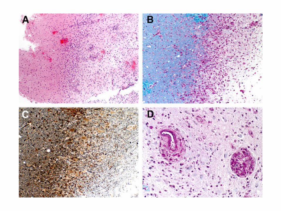
Multiple Sclerosis
• Morphology– Multiple plaques in brain, spinal cord– Common sites: peri-ventricular white matter,
optic nerve, white matter of spinal cord– Micro:
• Areas of demyelination • Lymphocytic infiltrate• Shadow plaques: axons with thin myelin (re
myelination)
Multiple Sclerosis
• Clinical– Visual disturbances, paresthesia, speech
disturbance, gait abnormalities– Cognitive impairment (not severe)– CSF: high protein, lymphocytes, gamma
globulin (oligoclonal band)– Some rapidly progress- death– Others have normal life span

Other Myelin diseases:
Acute disseminated encephalomyelitis
Central pontine myelinolysis
Leukodystrophies

Metabolic diseases
• Thiamine deficiency– Chronic alcoholics– Wernicke-Korsakoff syndrome– Peripheral neuropathy– Wernick encephalopathy: confusion, paralysis,
ataxia– Korsakoff psychosis: memory loss– Findings: Mamillary body hemorrhage

Metabolic diseases
• Hepatic encephalopathy– Confusion– Flabbing tremor– Large astrocytes (Alzheimer type II)

Peripheral Neuropathy

Peripheral Neuropathy
• Axonal Degeneration
• Segmental Demyelination

Peripheral Neuropathy• Axonal Degeneration
– Wallerian degeneration• Injury to proximal part with degeneration of the
distal segment• Followed by regeneration (incomplete) • eg. Localized cut of nerve, vasculitis
– Distal axonal degeneration• eg. Generalized injury of cell bodies, thiamine
deficiency, toxic: lead, arsenic• Degeneration distally and extends proximally in
continuous fashion• Regeneration (incomplete)

Peripheral Neurophathy
• Segmental Demyelination:– Injury to myelin sheath (segmental)
– Preservation of axons
– poor conduction
– New myelin sheath (remyelination)
– “onion bulb” formation
– Secondary axonal degeneration
– eg. Hereditary neuropathy, Guillain-Barre syndrome, Leukodystrophies

Peripheral Neurophathy
• Clinical– Defect in motor, sensory or autonomic nerve– “Glove-and-stocking” distribution seen in distal
axonal degeneration– Motor: muscle weakness, atrophy– Loss of deep tendon reflexes– Postural hypotension, constipation

Guillain-Barre Syndrome
• Etiology: unknown• Follow viral infection, Mycoplasma, allergies,
surgery• ? Immune defect• Rapid, progressive ascending motor weakness• Minimal sensory loss• Death from involvement of respiratory muscle• Path: segmental demyelination with inflammatory
infiltrate

Neoplasm of Peripheral Nerve
• Schwannoma
• Neurofibroma
• Malignant peripheral nerve sheath tumor

Schwannoma
• Well-circumscribed, encapsulated tumor
• At the edge of peripheral nerve
• Common in 8th cranial nerve (acoustic neuroma)
• Micro– Antoni A, Antoni B– Verocay bodies


Neurofibroma
• Solitary or multiple (neurofibromatosis)
• Mixture of schwann cells and fibroblasts
• Malignant peripheral nerve sheath tumor


