+ All Categories
Home > Documents > Degenerative Joint Disease Dr. Abdulrahman Algarni, MD, SSC (Ortho), ABOS Assist. Professor, King...

Degenerative Joint Disease Dr. Abdulrahman Algarni, MD, SSC (Ortho), ABOS Assist. Professor, King...

Date post: 17-Dec-2015
Category:
Upload: sherman-cobb
View: 224 times
Download: 4 times
Share this document with a friend
Popular Tags:
47
Degenerative Joint Disease Dr. Abdulrahman Algarni, MD, SSC (Ortho), ABOS Assist. Professor, King Saud University Consultant Orthopedic and Arthroplasty Surgeon King Khaled University Hospital, Dallah Hospital
Transcript

Degenerative Joint Disease

Dr. Abdulrahman Algarni, MD, SSC (Ortho), ABOSAssist. Professor, King Saud University

Consultant Orthopedic and Arthroplasty SurgeonKing Khaled University Hospital, Dallah Hospital
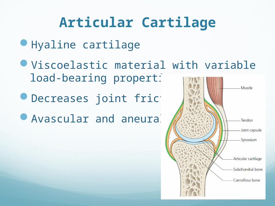
Articular Cartilage

Hyaline cartilage

Viscoelastic material with variable load-bearing properties

Decreases joint friction

Avascular and aneural

Cartilage Composition

Cartilage Composition

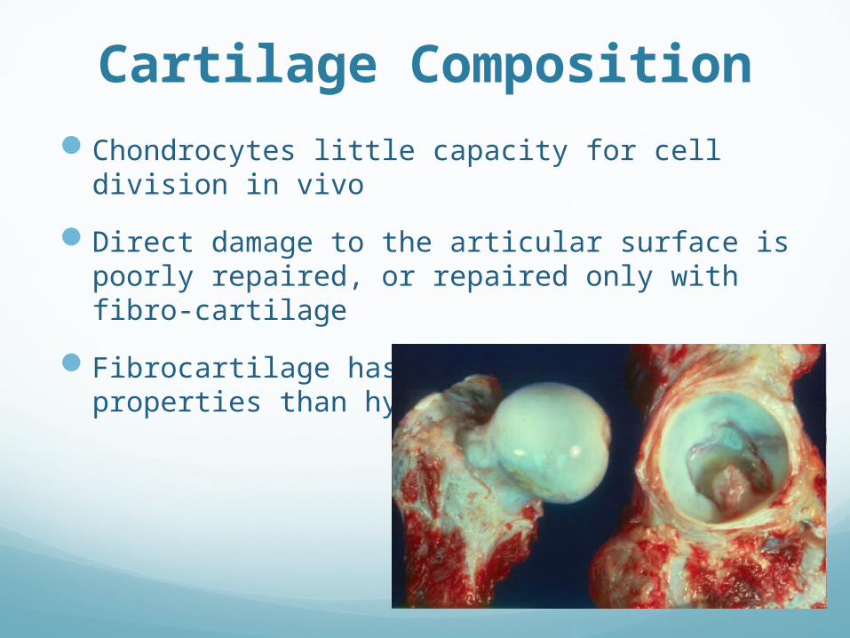
Chondrocytes little capacity for cell division in vivo

Direct damage to the articular surface is poorly repaired, or repaired only with fibro-cartilage

Fibrocartilage has inferior biomechanical properties than hyaline cartilage

Cartilage CompositionIf the collagen network is disrupted, the matrix becomes

waterlogged and soft

Followed by loss of proteoglycans, cellular damage and splitting (‘fibrillation’) of the articular cartilage.

Damaged chondrocytes begin to release matrix-degrading enzymes

Capsule and Ligaments

Fibrous structure with tough condensations on its surface (ligaments)

Together with the overlying muscles, help to provide stability.

Synovium and synovial fluid

Thin membrane

Richly supplied with blood vessels, lymphatics and nerves.

target tissue in joint infections and autoimmune disorders such as rheumatoid arthritis

Provides a nonadherent covering for

the articular surfaces

Produces synovial fluid

Synovium and synovial fluid

Synovial fluid nourishes the avascular articular cartilage

plays an important part in reducing friction during movement

has slight adhesive properties which assist in maintaining joint stability.

The volume remains fairly constant, regardless of movement.

When a joint is injured fluid increases ( joint effusion)

Degenerative Joint Disease

Primary’ (‘idiopathic’) osteoarthritis (OA)

Chronic disorder

Progressive softening and disintegration of articular cartilage

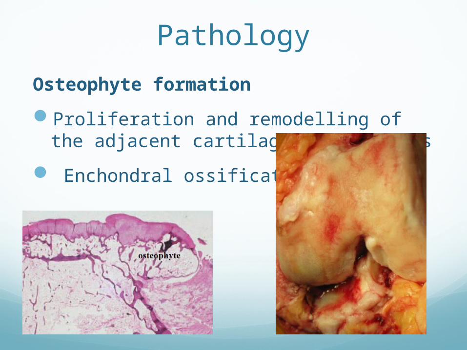
New growth of cartilage and bone at the joint margins (osteophytes)

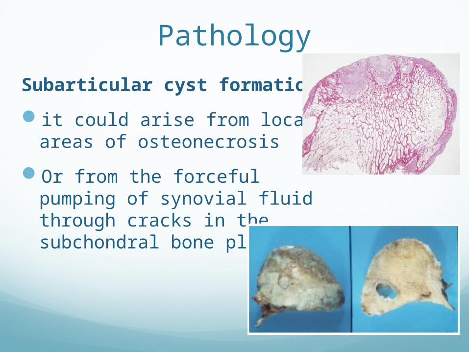
Subchondral bone sclerosis and cyst formation

Mild synovitis and capsular fibrosis.

Degenerative Joint Disease

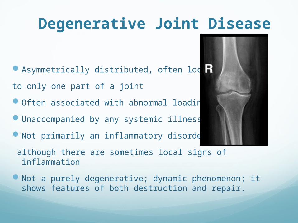
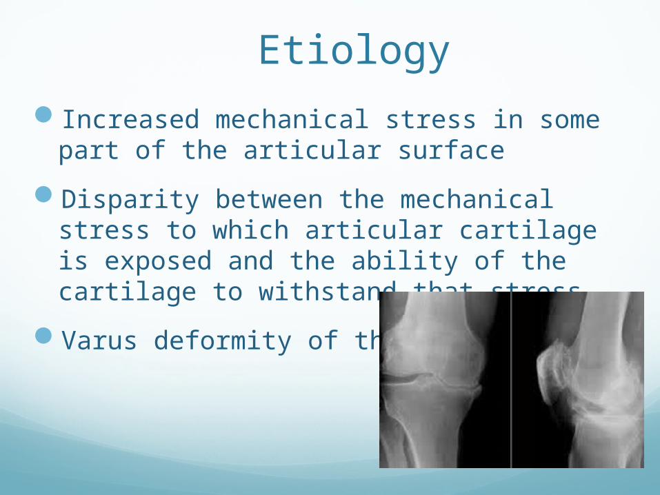
Asymmetrically distributed, often localized

to only one part of a joint

Often associated with abnormal loading

Unaccompanied by any systemic illness

Not primarily an inflammatory disorder

although there are sometimes local signs of inflammation

Not a purely degenerative; dynamic phenomenon; it shows features of both destruction and repair.

Secondary OAMetabolic: crystaline deposition disease(gout, CPPD),

Paget’s disease

Inflammatory: RA, SLE, Reiter’s syndrome

Neuropathic: DM, tabes dorsalis

Hematologic: SCD, hemophelia

Endocrine: DM, acromegaly

Secondary OA

Trauma: osteochondral, malunion, sport injury

Congenital/developmental: hip dyplasia, multiple epiphyseal dysplasia

Infection

Necrosis: Perthe’s disease, osteonecrosis, steroids

Etiology

Increased mechanical stress in some part of the articular surface

Disparity between the mechanical stress to which articular cartilage is exposed and the ability of the cartilage to withstand that stress.

Varus deformity of the knee

Etiology

More a process than a disease

Increases in frequency with age.

Obesity (hips and knees take 3-4 body weight with each step)

Family history

Prevalence

Osteoarthritis is the commonest of all joint diseases.

Osteoarthritis is much more common in some joints (hip, knee, spine and the fingers) than in others (the elbow, wrist and ankle).

More joints are affected in women than in men.

Prevalence

Common in our community especially knees

Much more in females

Presents earlier than in West

About 90% of those over 40 have asymptomatic degeneration of weight bearing joints

Commonest joints are knee, hip, Cervical spine & Lumbar Spine,1st Carpometacarpal,1st Metatarsophalangeal and Interphalangeal joints

Pathology

Cardinal features

Progressive cartilage destruction

Subarticular cyst formation

Sclerosis of the surrounding bone

Osteophyte formation

Capsular fibrosis.

Pathology

Progressive cartilage destruction

Increased water content: swelling and softening of cartilage

Depletion of Proteoglycans

Pathology

Progressive cartilage destruction

Chondrocyte damage and synovitis › proteolytic enzymes› collagen disruption

Fibrillation on weight bearing surfaces

Pathology

Subarticular cyst formation

it could arise from local areas of osteonecrosis

Or from the forceful pumping of synovial fluid through cracks in the subchondral bone plate

Pathology

Sclerosis of the surrounding bone

Bone becomes exposed

may be polished, or burnished, to ivory-like smoothness (eburnation)

Pathology

Osteophyte formation

Proliferation and remodelling of the adjacent cartilage at the edges

Enchondral ossification

Pathology

Marked vascularity and venous congestion of the subchondral bone

The capsule and synovium are often thickened but cellular activity is slight

Progressive bone erosion› BONE COLLAPSE

Fragmented osteophyte› LOOSE BODIES

Loss of height and ligamentous laxity› MALALIGNMENT

Clinical Features

Intermittent course, with periods of remission sometimes lasting for months.

One or two of the weight-bearing joints (hip or knee)

Symptoms

Pain

Stiffness

Loss of function

Symptoms

Pain

Localized or rarely referred to a distant site; e.g. pain in the knee from hip osteoarthritis.

Insidious

aggravated by exertion and relieved by rest

Advanced stage, night pain or at rest

Symptoms

Possible causes of pain

Bone pressure due to vascular congestion and intraosseous hypertension; most important

Mild synovial inflammation

Capsular fibrosis with pain on stretching the shrunken tissue

Muscular fatigue

Symptoms

Stiffness

Initially after periods of inactivity

Later, constant and progressive

Loss of function

Signs

Swelling

Intermittent (effusion)

continuous (large osteophytes)

Deformity; mal-alignment

Tenderness

Signs

Limited movement

Crepitus

Instability

Loss of cartilage and bone, asymmetrical capsular contracture and/or muscle weakness

Imaging

Asymmetrical loss of cartilage (narrowing of the ‘joint space’)

Subchondral bone sclerosis

Cysts close to the articular surface

Imaging

Osteophytes at the margins of the joint

Late features

Malalignment

joint subluxation

bone loss

Loose bodies

Imaging

Signs of other disorders

Symmetric narrowing in inflammatory OA e.g. RA

Complications

Capsular herniation: Knee OA; marked effusion and herniation of the posterior capsule (Baker’s cyst).

Complications

Loose bodies

Rotator cuff dysfunction: acromioclavicular (AC) joint OA

Complications

Spinal stenosis

Spondylolisthesis

severe segmental instability; at L4/5

Management

Joint (or joints) involved

Stage of the disorder

Severity of the symptoms

Age of the patient

Functional needs

Management

EARLY TREATMENT

Maintain movement and muscle strength

Protect the joint from ‘overload

Relieve pain

Modify daily activities

Conservative Treatment

Maintain movement and muscle strength

Physiotherapy (Physical therapy)

Pain relief: massage; application of warmth

Prevent contractures

Muscle strengthening

Range of motion

Conservative Treatment

Load reduction

Weight-reduction

Shock-absorbing shoes

Walking stick

Unloading brace

Conservative Treatment

Modify activity

avoiding activities like climbing stairs

Medications

Systemic: paracetamol, non-steroidal anti-inflammatory drugs (NSAIDs)

Local: not recommended

Surgical Treatment

Joint Debridement (Arthroscopy)

Corrective Osteotomy

Arthroplasty (Joint Replacement)

Arthrodesis (Fusion)

Surgical Treatment

Joint Debridement (Arthroscopy)

Removal of loose bodies

Removal of meniscal or labral tears

Surgical Treatment

Corrective Osteotomy

Realign axis and redistribute weight

Knee; hip

Young, active, mild OA

Surgical Treatment

Corrective Osteotomy

Pain relief

Vascular decompression of the subchondral bone

Redistribution of loading forces towards less damaged parts of the joint

Surgical Treatment

Arthrodesis

Transfer painful stiff into painless stiff joint

Small joints; hand, foot and spine

Surgical TreatmentArthroplasty (Joint Replacement)

Nowadays the procedure of choice for advanced OA

Total Joint Replacement

Knee, hip, shoulder, ankle and elbow

Painful, deformed stiff joint, old patient

Surgical TreatmentArthroplasty (Joint Replacement)

Partial Joint Replacement

Same patient as for osteotomy

Knee


Recommended