early onset AD (EOAD)—control cohort, and examined the effect on transcript level with comprehensive third-generation long-read sequencing. We characterized the ABCA7 coding sequence with next-generation sequencing in 928 EOAD patients and 980 matched control individu-als. With MetaSKAT rare variant association analysis, we observed a fivefold enrichment (p = 0.0004) of PTC muta-tions in EOAD patients (3%) versus controls (0.6%). Ten novel PTC mutations were only observed in patients, and PTC mutation carriers in general had an increased famil-ial AD load. In addition, we observed nominal risk reduc-ing trends for three common coding variants. Seven PTC mutations were further analyzed using targeted long-read cDNA sequencing on an Oxford Nanopore MinION plat-form. PTC-containing transcripts for each investigated PTC mutation were observed at varying proportion (5–41% of
Abstract Premature termination codon (PTC) mutations in the ATP-Binding Cassette, Sub-Family A, Member 7 gene (ABCA7) have recently been identified as intermedi-ate-to-high penetrant risk factor for late-onset Alzheimer’s disease (LOAD). High variability, however, is observed in downstream ABCA7 mRNA and protein expression, disease penetrance, and onset age, indicative of unknown modifying factors. Here, we investigated the prevalence and disease penetrance of ABCA7 PTC mutations in a large
European Early Onset Dementia (EU EOD) consortium side author list is given in acknowledgements.
Arne De Roeck and Tobi Van den Bossche contributed equally.
Electronic supplementary material The online version of this article (doi:10.1007/s00401-017-1714-x) contains supplementary material, which is available to authorized users.
1 Neurodegenerative Brain Diseases Group, VIB Center for Molecular Neurology, VIB, Antwerp, Belgium
2 Institute Born-Bunge, University of Antwerp, Antwerp, Belgium
3 Department of Neurology, Antwerp University Hospital, Edegem, Belgium
4 Department of Neurology and Memory Clinic, Hospital Network Antwerp (ZNA) Middelheim and Hoge Beuken, Antwerp, Belgium
5 Alzheimer’s Disease and Other Cognitive Disorders Unit, Neurology Department, Hospital Clínic, Institut d’Investigacions Biomediques August Pi i Sunyer (IDIBAPS), Barcelona, Spain
6 Department of Neuroscience, Psychology, Drug Research and Child Health (NEUROFARBA), University of Florence, Florence, Italy
7 IRCCS Don Gnocchi, Florence, Italy8 Neurological Tissue Bank of the Biobanc, Hospital Clinic,
Institut d’Investigacions Biomediques August Pi i Sunyer (IDIBAPS), Barcelona, Spain
9 Department of Neurology, Fundación Jiménez Díaz, Madrid, Spain
the total read count), implying incomplete nonsense-medi-ated mRNA decay (NMD). Furthermore, we distinguished and phased several previously unknown alternative splic-ing events (up to 30% of transcripts). In conjunction with PTC mutations, several of these novel ABCA7 isoforms have the potential to rescue deleterious PTC effects. In con-clusion, ABCA7 PTC mutations play a substantial role in EOAD, warranting genetic screening of ABCA7 in geneti-cally unexplained patients. Long-read cDNA sequencing revealed both varying degrees of NMD and transcript-mod-ifying events, which may influence ABCA7 dosage, disease severity, and may create opportunities for therapeutic inter-ventions in AD.
Keywords Early Onset Alzheimer’s disease · ATP-Binding Cassette · Sub-Family A · Member 7 (ABCA7) · Third-generation long-read sequencing · RNA sequencing · Loss-of-function · Modifier
Introduction
Alzheimer’s disease (AD, MIM: 104300) is the most common form of dementia. More than 20 genomic loci have been identified to contribute to AD risk [17, 18, 26, 27, 37, 46]. Among those, the gene encoding ATP-Bind-ing Cassette, Sub-Family A, Member 7 (ABCA7, MIM: 605414) is of particular interest, because both common variants and rare variants are reported to affect AD risk
[11, 13, 16, 27, 41, 44, 47, 49]. ABCA7 plays a role in lipid metabolism [20, 32, 43, 48] and microglial phago-cytosis [15, 21, 31], and was linked to altered amyloid β (Aβ) processing [23, 43, 45], the predominant hypothesis on AD pathogenesis.
Deleterious premature termination codon (PTC) muta-tions (nonsense, frameshift, and splice site mutations) in ABCA7 are observed at varying disease penetrance, with a 1.5–4× increased frequency in AD patients across populations [11, 16, 47, 49]. PTC mutation carriers appear more frequent among AD patients with a positive family history, though a wide range of disease onset age is observed [7, 11]. Two pedigrees have been reported in which a PTC mutation in ABCA7 (p.Arg578fs and p.Glu709fs) co-segregates with disease [10, 11]. Although the mode-of-action of ABCA7 PTC mutations in AD pathogenesis is unknown, a plausible mechanism is loss-of-function (LOF) due to nonsense-mediated mRNA decay (NMD). This is in line with mouse Abca7 knockout experiments leading to increased Aβ brain lev-els [15, 23, 43]. Single-epitope quantification of human brain mRNA and protein levels of ABCA7 in PTC muta-tion carriers, however, is conflicting. High variability is observed between individuals in general and between PTC mutation carriers [1, 11]. Furthermore, mRNA and protein levels do not seem to correlate [1], necessitat-ing analysis of ABCA7 expression in a broader context. In addition to PTC mutations, rare predicted deleteri-ous missense mutations and some common missense
10 Memory Unit, Department of Neurology, University Hospital Mútua de Terrassa, University of Barcelona School of Medicine, Terrassa, Barcelona, Spain
11 Centro de Investigación Biomédica en Red de Enfermedades Neurodegenerativas (CIBERNED), Instituto de Salud Carlos III, Madrid, Spain
12 Neuroimaging Laboratory, Division of Neurosciences, Center for Applied Medical Research (CIMA), University of Navarra, Pamplona, Spain
13 Department of Neurology, Clínica Universidad de Navarra, University of Navarra School of Medicine, Pamplona, Spain
14 Department of Neurobiology, Care Sciences and Society (NVS), Division of Neurogeriatrics, Center for Alzheimer Research, Karolinska Institutet, Huddinge, Stockholm, Sweden
15 Genetics Unit, Department of Geriatric Medicine, Karolinska University Hospital, Stockholm, Sweden
16 Molecular Markers Laboratory, Istituto di Ricovero e Cura a Carattere Scientifico (IRCCS), Istituto Centro San Giovanni di Dio-Fatebenefratelli, Brescia, Italy
17 MAC Memory Center, Istituto di Ricovero e Cura a Carattere Scientifico (IRCCS), Istituto Centro San Giovanni di Dio-Fatebenefratelli, Brescia, Italy
18 Faculty of Medicine, University of Lisbon, Lisbon, Portugal19 Neurology Unit, Department of Clinical and Experimental
Sciences, Centre for Neurodegenerative Disorders, University of Brescia, Brescia, Italy
20 Center for Neuroscience and Cell Biology, University of Coimbra, Coimbra, Portugal
21 Department of Psychiatry and Psychotherapy, Technische Universität München, Munich, Germany
22 Department of Neurology, IIB Sant Pau, Hospital de la Santa Creu i Sant Pau, Universitat Autònoma de Barcelona, Barcelona, Spain
23 3rd Department of Neurology, Medical School, Aristotle University of Thessaloniki, Thessaloniki, Greece
24 Laboratory of Biochemistry, Department of Chemistry, Aristotle University of Thessaloniki, Thessaloniki, Greece
25 Department of Pathology, First Medical Faculty, Charles University, Prague, Czech Republic
26 Department of Pathology and Molecular Medicine, Thomayer Hospital, Prague, Czech Republic
27 Institute of Pathology, Third Medical Faculty, Charles University, Prague, Czech Republic
477Acta Neuropathol (2017) 134:475–487
1 3
variants were, respectively, linked to risk increasing [16] and protective effects [11, 44], though requiring further confirmation.
The observation that ABCA7 PTC mutations exert a relatively strong effect on individual risk and familial occurrence of AD warrants further exploration of their potential in individualized genetic diagnosis and risk prediction [12]. To address the current complexity on a clinical and molecular level, we examined the prevalence and characteristics of ABCA7 coding mutations in a large European cohort of early onset AD patients (EOAD, onset age ≤65), a subgroup of AD patients that would strongly benefit from improved diagnosis, and genetic counseling. For a subset of PTC mutations, we performed targeted transcript analysis with third-generation (long-read) sequencing to gain further insight in the mode-of-action of these mutations and ABCA7 dosage modifying events.
Materials and methods
Study population
The EOAD patients and control individuals were recruited within the European Early Onset Demen-tia (EU EOD) consortium. Details about recruitment, in- and exclusion criteria, and demographic and patient characteristics were previously described [51]. In sum-mary, patients [n = 928, 60.2% (558/927) female, 51.2% (391/763) APOE ε4+ (MIM: 107741)] were diagnosed according to NINCDS-ADRDA [34] and/or NIA-AA [19, 35] diagnostic criteria, and had a mean onset age of 57.4 ± 5.6 years (30–65). In 47.7% (274/575) of patients, a positive familial history for dementia was reported. Patients were ascertained in neurological cent-ers with an expertise in memory disorders, and origi-nated from Spain (n = 403), Italy (n = 159), Sweden (n = 160), Germany (n = 83), Portugal (n = 66), Greece (n = 52), and the Czech Republic (n = 5) (Table S1). Seventeen EOAD patients carried a known pathogenic mutation in PSEN1, PSEN2, or APP (MIM: 104311, 600759, and 104760). Control individuals (n = 980, 64.9% (607/936) female) had a mean inclusion age of 63.9 ± 7.7 years (34-89), and were recruited in Spain (n = 223), Italy (n = 304), Sweden (n = 295), Portu-gal (n = 120), Greece (n = 35), and the Czech Republic (n = 3). The study was approved by the respective ethics committees, and all participants and/or their legal guard-ian provided written informed consent before inclusion.
ABCA7 sequencing
Genomic DNA (gDNA) was amplified with Illustra GenomiPhi v2 (Thermo Fisher, Waltham, MA, USA). Enrichment of 43 of the 47 canonical ABCA7 (RefSeq NM_019112.3) exons and splice sites was based on a cus-tom multiplex PCR assay (primer sequences available upon request) generated with mpcr software (Multiplicom, Niel, Belgium). Regions of interest were amplified using flank-ing primers with universal adapter sequences [5′-TCGTC GGCAGCGTCAGATGTGTATAAGAGACAG-(sequence-specific forward primer) and 5′-GTCTCGTGGGCTC GGAGATGTGTATAAGAGACAG-(sequence-specific reverse primer)]. Amplicons were PCR barcoded with Nex-tera XT sequences (Illumina, USA) targeting previously incorporated adapters. Samples were 2 × 300 bp paired-end sequenced with MiSeq Reagent Kit v3 (Illumina, San Diego, CA, USA). Adapter clipping of demultiplexed FASTQ output was performed with fastq-mcf [3], aligned with Burrows-Wheeler Aligner MEMv0.7.5a [30] and vari-ants were called with GATKv2.4 UnifiedGenotyper and GATKv3.5 HaplotypeCaller. Variants were annotated with GenomeComb [42] and SnpEff [9].
For four exons (12, 17, 19, and 23), no compatible prim-ers were found. Exons 12, 17, and 19 were analyzed with Sanger sequencing: exons were PCR amplified, subse-quently dideoxy-terminated with BigDye termination cycle sequencing kit v3.1 (Thermo Fisher), and sequenced with ABI3730 DNA Analyzer (Thermo Fisher). Sanger sequences were analyzed using Seqman (DNAstar, Madison, WI, USA) and NovoSNP software [52]. Exon 23 (73 bp + 4 bp splice sites) was not duly screened. Of note, this exon contains no known PTC mutations [Exome Aggregation Consortium (ExAC) (accessed April 2017)]. In addition, gaps are present (less than 50% of individuals sequenced at 20× coverage) in exon 9 (29 bp), 16 (23 bp), 18 (4 bp), and 21 (87 bp), as depicted in Fig. 1, mainly due to repetitive regions flanking ABCA7 exons, limiting efficient PCR primer design.
The sequencing read-depth statistics for all coding and splice sites regions are represented in Fig. 1 based on Rsam-tools [36] and Circos visualization [25]. All PTC variants and c.5570+5G>C were validated on gDNA with Sanger sequencing as described above. In addition, we validated rare [minor allele frequency (MAF) <0.01%] missense vari-ants with at least 20× read depth, a less than threefold dif-ference between reference and alternative allele counts for heterozygous variants, and a Phred-scaled CADD score [24] above 20, corresponding to the 1% most deleterious variants in the human genome. This cutoff was chosen given the rel-atively high mutational tolerance of ABCA7 and incomplete disease penetrance for very deleterious PTC mutations.
478 Acta Neuropathol (2017) 134:475–487
1 3
Association analysis
Rare variant (MAF < 0.01) association meta-analysis by ethnicity was performed separately for PTC and predicted deleterious missense mutations using heterogeneous genetic effect SKAT-O statistics within the MetaSKAT framework [29]. APOE dosage (number of ε4 alleles) was tested as a covariate. Summary odds ratios (ORMH) were calculated with a Cochran–Mantel–Haenszel test in
PLINK [40], stratified by country of origin, on individu-als without missing genotyping information.
For common variants (MAF ≥ 0.01), a fixed-effects (Cochran–Mantel–Haenszel) meta-analysis was used to calculate single variant allelic association. We evaluated all SNPs located within the CDS and 15 bp exon flank-ing regions passing Hardy–Weinberg equilibrium quality control (p > 0.001). Odds ratios are reported for the minor allele with 95% confidence intervals. Pairwise linkage dise-quilibrium (LD) was calculated in PLINK [40]. To account
Fig. 1 ABCA7 mutation screening in EOAD. From the outside to the inside: HGVS nomenclature is denoted for known (black) and novel (red) PTC mutations, suggestively associated protective common variants (green), intron retaining variant c.5570+5G>C (orange), and predicted deleterious missense variants (gray). The second rim cor-responds to the percentage of included individuals covered at more than 20× on the corresponding exonic position (0–50% in red and 50–100% in green). Exons highlighted in blue were screened with Sanger sequencing. The third layer consists of the UTR (narrow) and
CDS (broad) architecture of ABCA7 (NM_019112.3). Next, the corre-sponding predicted protein domains (UniProtKB entry: Q8IZY2) are shown (transmembrane domains in green, extracellular parts in blue and red corresponds to ABC domains). In the center, the number of carriers per PTC (filled bars) and deleterious missense (open bars) variant are shown. Patients are represented in red, while control indi-viduals are shown in blue. Linked variants (red, green, and gray line) segregated on the same haplotype
479Acta Neuropathol (2017) 134:475–487
1 3
for LD between variants, multiple testing thresholds were based on spectral decomposition [38], leading to a study-wide multiple testing p value cutoff of 0.0033. Carriers of a known pathogenic mutation in APP, PSEN1, or PSEN2 were excluded from association analyses.
Transcript analysis of PTC mutation carriers
Fresh frozen brain was available for patient EOD-P1 (c.67-1G > A mutation). RNA was extracted from anterior cingulate cortex tissue with Ambion RiboPure™ kit (Life Technologies, Carlsbad, CA, USA). Reagent volumes were adapted to support 25 mg of input brain material. In addition, RNA from blood was extracted with Tempus™ blood RNA tube (Thermo Fisher) for carriers EOD-P7 (p.Met370fs) and EOD-P19 (c.3577+1G>C). For a sub-set of ABCA7 PTC mutations, EBV-transformed lympho-cytes (p.Glu709fs, p.Trp1336*, and c.5570+5G>C) or fresh frozen brain tissue (p.Leu1403fs) from AD patients was available through the BELNEU consortium [11]. RNA was extracted with Ambion RiboPure™ kit (Life Technologies). All RNA was treated with Ambion TUR-BODNase kit (Life Technologies) to degrade potential gDNA contamination. First-strand cDNA was synthesized with SuperScript®III First-Strand Synthesis System (Life Technologies).
We performed cDNA sequencing on a MinION platform (Oxford Nanopore Technologies, Oxford, UK). Exonic primers were designed with Primer3Plus to generate ampli-cons containing exons of interest, flanked by at least one splicing event on each side (Table S2). All PCR amplifi-cations were performed with 35 cycles to have sufficient amplification of the lowly expressed ABCA7 in all tissues. Titanium Taq (Clontech Laboratories, Mountain View, CA, USA) or Platinum Taq (Thermo Fisher) enzymes were used with or without supplementation of betain, depend-ing on the GC content of the amplicon. In case of overlap-ping PCR amplicons, a 5′ barcoding adapter sequence was added to the primers and amplicons were barcoded with the PCR 96 barcoding genomic DNA (R9) kit (Oxford Nanopore Technologies) using 15 amplification cycles on a 1/125 diluted template. DNA was purified with Agen-court AMPure XP SPRI beads (Beckman Coulter, Brea, CA, USA) and concentrations were measured with Qubit (Thermo Fisher). Amplicons were then pooled equimolar to a total quantity of 0.2 pmol. The resulting PCR library was then further prepared according to the manufacturer’s protocol. Briefly, DNA was treated with NEBNext Ultra II End Repair/dA-Tailing Module (New England Bio-Labs, Ipswich, MA, USA). The product was then purified with AMPure XP beads, after which the ONT sequencing adapter and hairpin were ligated with the NEB Blunt/TA Ligase Master Mix (New England BioLabs). Next, biotin
containing hairpin tethers were added and washed MyOne Streptavidin C1 Dynabeads (Thermo Fisher) were used for pull-down. Finally, the sequencing library was eluted from the beads and supplemented with Running Buffer with Fuel Mix. SQK-MAP006/NSK007 chemistry and FLO-MAP103/MIN104 flow cells were used for sequencing. Base calling was done with the 2D plus barcoding proto-col (Metrichor, Oxford, UK) after which FASTQ sequences were extracted with poretools v0.6.0 [33] according to the “best” protocol. Sequencing reads were aligned to the human genome (hg19) with GMAP v2016-06-30 [53] to account for splicing events. Wild-type (WT), nonsense, and rescue alleles were quantified with Rsamtools v1.26.0 for aligned sequences [36], and with GMAP splicesites out-put for splicing events. The PTC expression fraction was calculated as (expressionNonsense/expressionNonsense+WT); 0% corresponds to complete degradation of PTC bearing transcripts and 50% to equal observance of PTC and WT transcripts. Results were validated with Sanger sequencing of cDNA amplicons as described above and compared with RNA sequencing (RNAseq) data from a Belgian unrelated AD-control transcriptome study [51]. Briefly, total RNA was isolated from Epstein–Barr virus immortalized lymph-oblasts derived from whole blood lymphocytes. RNA iso-lation of lymphoblast cells was performed with the RNe-asy mini kit (Qiagen Inc., Valencia, CA, USA) according to the manufacturer’s protocol. Sequence libraries were constructed using the Truseq stranded mRNA Library Prep Kit v2 (Illumina) using 1 mg total RNA for each sample. Sequencing of prepared libraries was performed using an Illumina HiSeq 2000 sequencer generating an average of 72 × 106 ± 6 × 10⁶ paired-end sequence reads/sample. Subsequent data processing consisted out of removal of read adapters and trimming of read ends with Trimmomatic [6]. Reads were then aligned to hg19 using the Bowtie short read aligner integrated in Tophat2 [22].
Results
PTC mutations
Sequencing of the ABCA7 CDS in 928 EOAD patients and 980 control individuals revealed 17 different PTC mutations (six frameshift indels, six nonsense muta-tions, and five PTC-introducing splice site muta-tions) (Table 1), which were more frequent in EOAD patients (3.02%; n = 28) than controls (0.61%; n = 6) [p value = 0.0004, ORMH = 5.01 (95% CI = 1.59–15.72)]. This association remained significant after correction for APOE (p = 0.001). Most PTC mutations (n = 10) were not reported before and—together with c.67-1G > A, c.3577 + 1G > C, and p.Arg1489*—were absent from
480 Acta Neuropathol (2017) 134:475–487
1 3
control individuals (Fig. 1; Table S3). Patient EOD-P5 carried two PTC mutations (p.Trp69*and c.579+1G>T) segregating on the same haplotype (Figure S1). Two previ-ously reported variants (p.Trp1336*, c.4416+2T>G) were only observed in control individuals. The two most fre-quent PTC mutations (p.Glu709fs and p.Leu1403fs) were observed more in patients than control individuals (Fig. 1; Table 1). All carriers of p.Glu709fs and p.Leu1403fs shared the same respective haplotype (Table S4). Car-riers of less frequent multiple occurring variants (c.67-1G>A, p.Thr849fs, c.4416+2T>G, and p.Arg1489) were
geographically confined (respectively, originating from Spain, Portugal, Italy, and the Iberian Peninsula), though no familial relatedness was known (Table S3).
Patients (n = 28, 75.0% female, 54.2% (13/24) APOE ε4 +) carrying PTC mutations had a mean onset age of 56.7 ± 5.5 years (42-65, upper limit determined by inclu-sion criteria for EOAD), and a mean disease duration of 8.6 ± 1.8 years (Table S3). In comparison, carriers of established pathogenic mutations in PSEN1 (n = 12), PSEN2 (n = 3), or APP (n = 2) had a lower mean onset age of, respectively, 46.0 ± 9.0, 53.0 ± 5.1, or 49.5 ± 1.5 years
Table 1 ABCA7 PTC mutations
An overview of all premature termination codon (PTC) inducing mutations observed in this study. Genomic coordinates are based on hg19. HGVS = mutation nomenclature according to the Human Genome Variation Society. dbSNP notations refer to Reference SNP IDs (rs) from dbSNP build 142. MAF minor allele frequency. The previous reports of the mutation in discovery populations of Cuyvers et al. (1), Steinberg et al. (2), Le Guennec et al. (3), or Nuytemans et al. (4) are denoted. Abundance of the PTC transcript is shown based on MinION cDNA sequencing (Figures S3–S5, S8, and S10). PTC expression fraction corresponds to the abundancy of sequencing reads containing the PTC muta-tion, in which case 0% is associated with complete degradation of PTC transcripts, and 50% with equal expression of PTC and WT alleles. Reading frame restorations are denoted when identified and correspond either to in-frame skipping of the exon harboring the mutation of inter-est, or alternative splicing which restores the reading frame without introduction of a PTC. The same cDNA PCR amplicon was used to measure exon skipping events for p.Trp1336* and p.Leu1403fs; as a result, both exons could be quantified for two different individuals (range)
(35-63, p = 0.0002) (Figure S2). Patient EOD-P6.1 (ABCA7 c.302 + 1G>C) also carried a pathogenic PSEN1 mutation (p.His163Arg) and had the earliest onset age (42 years) of all ABCA7 PTC carriers. Two affected rela-tives of EOD-P6.1 also carried PSEN1 p.His163Arg, but not ABCA7 c.302+1G>C, and had a slightly older onset age of 46 years.
A positive familial history for dementia was reported in 61.5% (16/26) of patient carriers. Of note, for patient EOD-P21.1 carrying p.Leu1403fs, DNA was available of two affected relatives with onset ages of 68 and 70 years, who both also carried the mutation. In addition, EOD-P7 (p.Met370fs) and EOD-P20 (p.Leu1403fs) had a negative familial history for dementia, but both had a first-degree relative with Parkinson’s disease. Information on clinical presentation was available for 23 patients, of whom 82.6% (19/23) had a predominant amnestic presentation. In two patients, the onset of memory dysfunction was accom-panied by language dysfunction. EOD-P6.1 (carrying PSEN1 p.His163Arg) had prominent behavioral symptoms (aggressiveness). Only one patient presented with a clear nonamnestic phenotype (logopenic progressive aphasia). Neuropathology was available for EOD-P1, confirming the clinical AD diagnosis (Fig. 2, Neuropathological descrip-tion S1).
Transcript analysis of PTC mutations
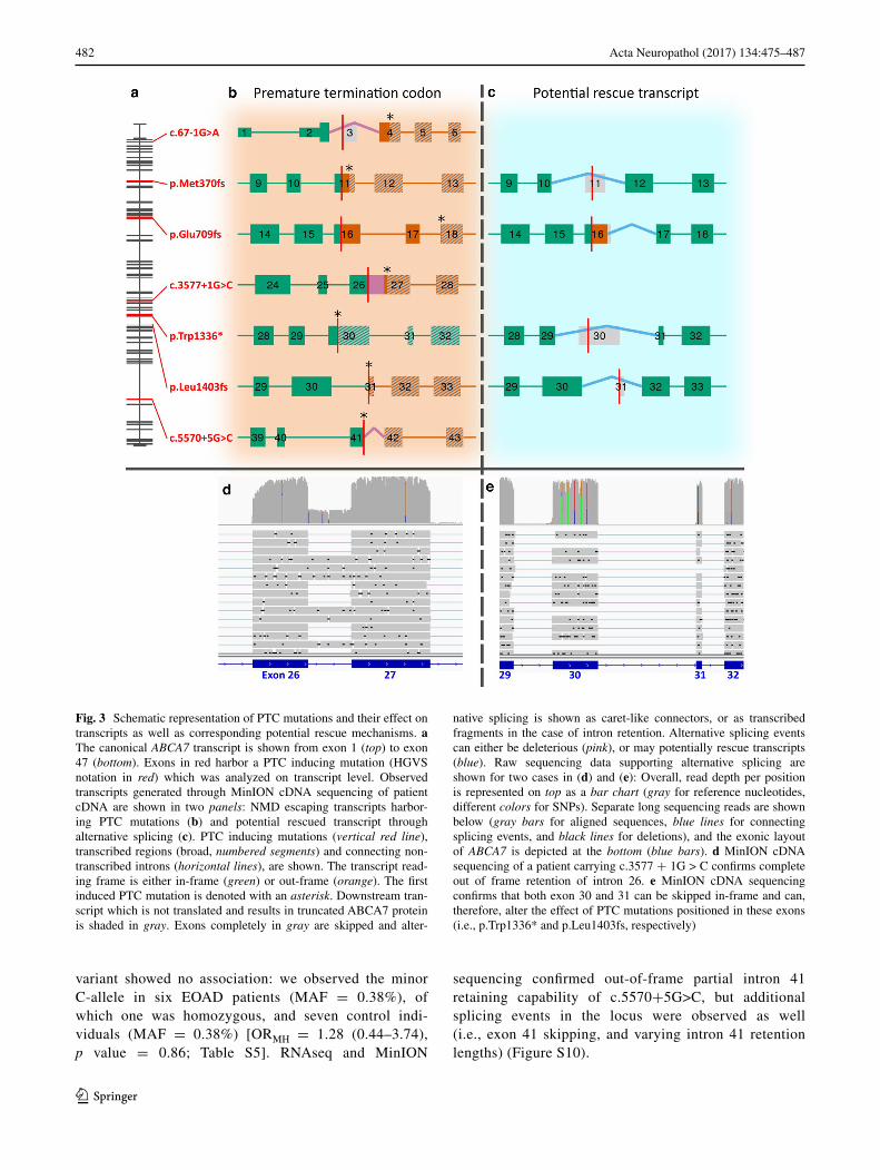
We examined ABCA7 expression with MinION sequenc-ing for three frameshift mutations (p.Met370fs, p.Glu709fs, and p.Leu1403fs), one nonsense (p.Trp1336*), one splice donor (c.3577 + 1G > C), and one splice acceptor mutation (c.67-1G>A). We observed varying degrees of PTC bearing transcripts in all cDNA libraries (Fig. 3; Table 1), indicative of incomplete
NMD. The most N-terminal mutation (c.67−1G>A) had the highest NMD efficiency with only 5% of sequencing reads showing out-of-frame exon 3 skipping. All other mutations presented higher PTC abundancy (21–41%), approaching expression equal to the WT allele (50%). On top of apparent NMD escape, we observed alterna-tive splicing events, absent from public databases (e.g., Ensembl, GENCODE, and UCSC genes), in mutated regions of interest. Some have the ability to restore reading frameshifts caused by PTC mutations (Fig. 3; Table 1). For three mutations (p.Met370fs, p.Trp1336*, and p.Leu1403fs), in-frame skipping of the respective PTC bearing exon [exon 11 (168 bp), exon 30 (255 bp), and exon 31 (33 bp)] was observed in patient carriers (Figures S3, S4, and S5). Overall, potential PTC res-cue transcripts had a modest abundance (2–8%); how-ever, in brain cDNA of one individual, we observed skipping of exon 30 in 30% of all sequencing reads, in this case unrelated to a PTC (Figure S5). These in-frame exon skipping transcripts were validated with RNAseq, confirming their presence in individuals not carrying an ABCA7 mutation (Figure S6 and S7). Fur-thermore, for p.Glu709fs (exon 16), we observed usage of a cryptic splice donor site (10%) in the same exon (Figure S8), which can negate the frameshift effect of p.Glu709fs. Validation of this splicing isoform with RNAseq is presented in Figure S9, along with confir-mation of its presence in a physiological context. West-ern blotting was performed on brain of c.67−1G>A and p.Leu1403fs (Method S1), confirming a reduc-tion of expression by PTC mutations (Figure S11), as well as an overall variability in expression. One splice mutation (c.5570+5G>C)—3 bp downstream of the canonical splice donor site—was previously reported to cause out-of-frame intron retention [47]. Here, this
Fig. 2 Neuropathological findings in patient EOD-P1. Abundant beta-A4 amyloid pathology in the form of diffuse and cored amyloid plaques (a) and amyloid angiopathy involving leptomeningeal vessels (b). Prominent phospho-tau pathology in the form of neurofibrillary
tangles, dystrophic neurites, and neuropil threads (c). Scale bars: a, c 50 μm, b 20 μm. An extended neuropathological description is avail-able in Neuropathological description S1
482 Acta Neuropathol (2017) 134:475–487
1 3
variant showed no association: we observed the minor C-allele in six EOAD patients (MAF = 0.38%), of which one was homozygous, and seven control indi-viduals (MAF = 0.38%) [ORMH = 1.28 (0.44–3.74), p value = 0.86; Table S5]. RNAseq and MinION
sequencing confirmed out-of-frame partial intron 41 retaining capability of c.5570+5G>C, but additional splicing events in the locus were observed as well (i.e., exon 41 skipping, and varying intron 41 retention lengths) (Figure S10).
Fig. 3 Schematic representation of PTC mutations and their effect on transcripts as well as corresponding potential rescue mechanisms. a The canonical ABCA7 transcript is shown from exon 1 (top) to exon 47 (bottom). Exons in red harbor a PTC inducing mutation (HGVS notation in red) which was analyzed on transcript level. Observed transcripts generated through MinION cDNA sequencing of patient cDNA are shown in two panels: NMD escaping transcripts harbor-ing PTC mutations (b) and potential rescued transcript through alternative splicing (c). PTC inducing mutations (vertical red line), transcribed regions (broad, numbered segments) and connecting non-transcribed introns (horizontal lines), are shown. The transcript read-ing frame is either in-frame (green) or out-frame (orange). The first induced PTC mutation is denoted with an asterisk. Downstream tran-script which is not translated and results in truncated ABCA7 protein is shaded in gray. Exons completely in gray are skipped and alter-
native splicing is shown as caret-like connectors, or as transcribed fragments in the case of intron retention. Alternative splicing events can either be deleterious (pink), or may potentially rescue transcripts (blue). Raw sequencing data supporting alternative splicing are shown for two cases in (d) and (e): Overall, read depth per position is represented on top as a bar chart (gray for reference nucleotides, different colors for SNPs). Separate long sequencing reads are shown below (gray bars for aligned sequences, blue lines for connecting splicing events, and black lines for deletions), and the exonic layout of ABCA7 is depicted at the bottom (blue bars). d MinION cDNA sequencing of a patient carrying c.3577 + 1G > C confirms complete out of frame retention of intron 26. e MinION cDNA sequencing confirms that both exon 30 and 31 can be skipped in-frame and can, therefore, alter the effect of PTC mutations positioned in these exons (i.e., p.Trp1336* and p.Leu1403fs, respectively)
483Acta Neuropathol (2017) 134:475–487
1 3
Other coding variants
We identified 19 predicted deleterious missense muta-tions (CADD score >20). There was no enrichment of predicted deleterious missense variants in patients (1.7%; n = 16) compared to controls (0.92%; n = 9; SKAT-O p value = 0.67) (Fig. 1; Table S6). Of note, p.Ala676Thr and p.Ser1723Leu segregated together in two controls and one patient; for which the patient, in addition, carried a third deleterious in-frame deletion (c.4922_4924delTCT). Clini-cal characteristics of missense mutation carriers did not dif-fer substantially from PTC mutation carriers [mean onset age 57.0 ± 5.5 years (range = 46–55); positive familial history in 60.0% (6/10)].
Twenty-two common coding variants (MAF > 1%), were assessed for association with EOAD. No vari-ants passed study-wide multiple testing correction (p = 0.0033), but nominal significance was observed for three SNPs (Table S7). The strongest effect [OR = 0.60 (95% CI = 0.42–0.87), p = 0.006] was observed for p.Gly215Ser (rs72973581, MAF = 4.1%), a missense SNP previously suggested to be protective [44]. For silent variant p.Asn1829Asn (rs78320196, MAF = 4.7%), we observed an OR of 0.65 (95% CI = 0.47–0.90; p value = 0.009). The potential protective effect of p.Asn1829Asn seems independent of p.Gly215Ser, given low pairwise LD (R2 = 0.001 and D′ = 0.596). The third variant (p.Glu188Gly, rs3764645, and MAF = 43.4%) also showed a suggestive protective effect (OR = 0.86 95% CI = 0.75–0.99), p value = 0.030), which was previ-ously reported in a Belgian cohort of AD [11]. We further observed that p.Glu188Gly and p.Gly215Ser were in strong LD (D′ = 0.961).
Discussion
Predicted LOF mutations in ABCA7 were recently put for-ward as intermediate-to-high penetrant risk factors for AD. To evaluate their contribution to EOAD, we sequenced the ABCA7 coding DNA of 928 European EOAD patients and 980 ethnically matched healthy control individuals. We identified ten novel patient-specific PTC mutations (frameshift, nonsense, and canonical splice mutations), and confirmed seven previously reported mutations. ABCA7 PTC mutations were five times more frequent among EOAD patients than controls, confirming an important con-tribution of these mutations to AD. Transcript analysis of seven PTC mutations revealed varying degrees of loss-of-transcript, suggesting that the mechanism through which these mutations affect AD risk needs further investigation. We observed no associations for predicted deleterious
missense mutations, but detected a protective trend for three common variants.
ABCA7 PTC mutations were detected in approximately 3% of the EOAD patients, which is comparable to the previous reports [11, 16]. In comparison, predicted patho-genic PTC mutations in SORL1 (MIM: 602005)—another prominent AD risk gene—were observed in 0.6% of EOAD patients in the EU EOD consortium [51]. Furthermore, only an estimated 5–10% of EOAD can be explained by auto-somal dominant mutations in APP (<1%), PSEN1 (6%), and PSEN2 (1%) [8]. Hence, due to relative high pene-trance and occurrence, genetic screening for ABCA7 PTC mutations is warranted in genetically unexplained EOAD patients. In line with the previous reports, we observed a high familial load in patients carrying an ABCA7 PTC mutation—though lower than in established autosomal dominant mutation carriers. In one Italian AD family (EOD-P21), multiple affected relatives carried the ABCA7 p.Leu1403fs mutation. Further elucidation of age-related penetrance and segregation patterns of individual ABCA7 mutations—and possible modifiers thereof—will be imper-ative for implementation of ABCA7 mutation screening in clinical practice and genetic counseling.
Based on the previous reports and the arbitrary posi-tioning of AD associated PTC mutations across the gene (Fig. 1), haploinsufficiency—a reduction of dosage sensi-tive functional ABCA7—is the most plausible pathogenic mechanism [11, 16, 47]. Additional expression differ-ences—either dosage recovery or further ABCA7 deple-tion—are, therefore, potential modifiers. We examined the effect of frameshifts, nonsense, splice donor, and splice acceptor variants on ABCA7 transcripts in patient bioma-terials using “third-generation” long-read MinION cDNA sequencing. Even though this sequencing technology is still under development and currently produces a relatively high random base calling error rate, we show that the accuracy is sufficient to align reads and to identify splicing events. Fur-thermore, given the high read depth attained in this targeted experiment (at least 1400×), reliable consensus sequences and variant calls could be formed.
Interestingly, we observed PTC transcripts for all muta-tions under study, indicating NMD escape. The proportion of sequencing reads carrying a PTC varied across muta-tions, up to 41% which is close to no NMD (50%). As a consequence of NMD escape, ABCA7 dosage may be modified, either via natural PTC read-through resulting in full length protein [5], or on the other hand through forma-tion of truncated proteins which could exert dominant nega-tive or wild-type functions. NMD escape also opens a win-dow for pharmacological intervention. Several compounds are known to cause ribosomal read-through of PTCs, which could result in a functional protein and alleviate haploin-sufficiency. Especially carriers of nonsense mutations may
484 Acta Neuropathol (2017) 134:475–487
1 3
benefit from such a treatment. Read-through compounds (e.g., PTC124) are currently tested in clinical trials for LOF diseases such as Duchenne muscular dystrophy (DMD, MIM: 310200) and cystic fibrosis (MIM: 219700) [5].
In contrast to standard RNAseq, MinION sequencing of long DNA fragments at high read depth provided insights in phasing of mutations and splicing events despite low ABCA7 expression [39]. As a result, we observed alterna-tive splicing events unknown to public repositories. We identified cryptic splice site usage, often leading to a shift in reading frame, as well as exon skipping, both in- and out-of-frame. On one hand, these events can lower the ABCA7 cell reserve resulting in stronger dosage depletion when a PTC mutation is introduced. On the other hand, several splicing events have the potential to recover the effect of PTC causing mutations (e.g., reading frame rescue through usage of a cryptic splice site), or alter the amount of transcript carrying a PTC mutation (e.g., in-frame skip-ping of an exon harboring a mutation). Interestingly, for all PTC mutations observed in controls with the exception of c.4416+2T>G for which no biomaterials were avail-able, a potential rescue mechanism was present (Table 1). For some mutations, the rescue event appears relatively frequent, which may contribute to incomplete penetrance (e.g., p.Trp1336* has been reported in both patients and controls; we observed exon 30 skipping in up to 30% of reads). Stabilization of alternative isoforms (e.g., through oligonucleotides targeting pre-mRNA) is a potential phar-macological target, which is already being evaluated for diseases as DMD and spinal muscular atrophy (MIM: 253300) [14].
Further research into the functions, essential protein domains, expression, and different isoforms of ABCA7 will have to substantiate to which extent dosage can mod-ify the AD phenotype, and whether it can be remediated. It is likely that numerous factors contribute significantly to variation in ABCA7 expression (as evidenced by protein levels in hippocampus, Figure S11), including brain degen-eration, inflammation, specific brain regions/cellular com-position, disease duration, genetic etiology, and environ-mental factors. The long-read cDNA sequencing approach used here shows that differences in ABCA7 transcript and protein expression data may also partly be explained by a myriad of NMD escaping alternatively spliced transcripts and (truncated) proteins. Taken together, this may explain discrepancies within and between the previous studies on ABCA7 expression [1, 2, 50]. In this study, ABCA7 tran-scripts of PTC mutations were examined in different patient tissues (brain, blood, and lymphoblast), which may pre-sent varying NMD efficiencies [54] and alternative splic-ing [4]. Ideally, quantitative comparisons of ABCA7 dosage between carriers are performed on a larger series of muta-tion carriers in a single tissue to more precisely determine
the contribution of NMD efficiency and transcript rescue to variation in ABCA7 gene expression. In this study, we aimed at adequate coverage of lowly abundant ABCA7 tran-scripts by sequencing mutation-specific amplicons, which, in addition, also prevents the formation of chimeric PCR molecules that might lead to phasing errors [28]. When cur-rent limitations of long-range PCR are overcome, it will be of interest to expand the methods used here to obtain a detailed map of transcript events across full length ABCA7 mRNA.
In addition to canonical PTC mutations, c.5570 + 5G > C is known to cause out-of-frame intron retention [47], which we confirm (Figure S10). While others have observed association with this variant [16, 47], here, no enrichment was present (ORMH = 1.28 95% CI = 0.44–3.74), p value = 0.86). Possibly, c.5570+G>C has a different protein reducing effect than canonical PTC mutations, since the degree of cryptic splice donor site ver-sus canonical usage is unknown, and due to the relatively distal location of c.5570+5G>C in the protein. Further-more, several interfering isoforms were present, suggest-ing lower penetrance of this particular variant. A previous report also suggested the pathogenicity of predicted dam-aging ABCA7 missense variants [16]. In this study, with a larger study population, however, we observed no obvious enrichment (p = 0.66). Furthermore, we observed two dele-terious missense variants (p.Ala676Thr and p.Ser1723Leu) segregating on the same haplotype, which occurred in both patients and controls. We cannot exclude that our cohort lacked power to observe a likely smaller effect of missense mutations on disease risk, but at this point, it is premature to draw inferences based on deleteriousness predictions alone. If future studies reveal a risk increasing effect of (a subset of) ABCA7 missense variants, it may be worthwhile to elucidate the effect of these mutations on mRNA splic-ing and vice versa.
Finally, three common coding variants (p.Gly215Ser, p.Glu188Gly, and p.Asn1829Asn) showed a trend towards decreased risk of EOAD, albeit not withstanding multiple testing. Of note, p.Gly215Ser was previously put forward as protective variant in ABCA7 [44], and for p.Glu188Gly and p.Asn1829Asn, a nominal protective association was also observed before [11, 44]. We show that p.Gly215Ser and p.Glu188Gly shared the same haplotype background (D′ = 0.961). In this study, we extend potential protect-ing effects of p.Gly215Ser and p.Asn1829Asn towards EOAD, supporting the role of ABCA7 to mediate risk of (early onset) AD in both directions. Further research, how-ever, is required to understand the downstream protective mechanisms.
In summary, with this targeted resequencing of ABCA7 in a large European cohort of EOAD, we substanti-ate the evidence that ABCA7 PTC mutations contribute
485Acta Neuropathol (2017) 134:475–487
1 3
significantly to AD risk. We observed a fivefold enrich-ment of ABCA7 PTC mutations in EOAD patients, and pro-vided further evidence that these mutations may segregate with disease in pedigrees. This suggests that at least some ABCA7 mutations may have a high penetrance, providing new inroads for genetic subtyping and risk prediction. The observation of these ‘familial’ ABCA7 mutations in cog-nitively healthy individuals, however, warrants cautious interpretation and further exploration of pathogenicity and modifying factors. An initial characterization of different PTC mutations at transcript level reveals substantial vari-ability in NMD and alternative splicing, implying varying abundancy of ABCA7 in PTC mutation carriers. Further investigation is required into the degree of dosage reduc-tion caused by a single mutation, the function and structure of ABCA7, and the presence of potential dominant negative effects, to contribute to a better estimation of phenotypical consequences and ways to remediate this.
Acknowledgements The sponsors of the study had no role in study design, data collection, data analysis, data interpretation, or writing of the report. The research was funded in part by the European Com-mission Seventh Framework Programme for research, technologi-cal development, and demonstration under grant agreement 305299 (AgedBrainSYSBIO), the Belgian Science Policy Office Interuni-versity Attraction Poles program, the Alzheimer Research Founda-tion (SAO-FRA), the Flemish government-initiated Flanders Impulse Program on Networks for Dementia Research (VIND), the Flemish government-initiated Methusalem Excellence Program, the Research Foundation Flanders (FWO), the VIB Technology Fund, the Univer-sity of Antwerp Research Fund, Belgium; Generalitat de Catalunya (2014SGR-0235), Instituto de Salud Carlos III (PI12/01311), Span-ish Ministry of Economy and Competitiveness ISCIII (PI14/00282), European Regional Development Fund, the Italian Ministry of Health (Ricerca Corrente and RF-2010-2319722), and the Fondazione Cassa di Risparmio di Pistoia e Pescia grant (2014.0365). A.D.R. receives a Ph.D. fellowship of FWO (Fonds Wetenschappelijk Onderzoek). W.D.C. receives a Ph.D. fellowship of VLAIO Hermesfonds. We thank Steven Vermeulen, Kristien De Ruyck, Elise Cuyvers, Rita Cacace, Yannick Vermeiren, and the personnel of the VIB Neuromics Support Facility and Antwerp biobank, Antwerp, Belgium for techni-cal assistance.
European Early Onset Dementia (EU EOD) consortium side author list: The following members of the EU EOD consortium have contributed to the sampling, clinical and pathological phenotyping of the patients that were included in the EU EOD cohort: Valentina Bessi, Silvia Bagnoli (Department of Neurosciences, Psychology, Drug Research and Child Health (NEUROFARBA), University of Florence, Florence, Italy); Frederico Simões do Couto, Ana Verdelho (Faculty of Medicine, University of Lisbon, Lisbon, Portugal); Laura Fratiglioni (Karolinska Institutet, Department of Neurobiology, Care Sciences and Society [NVS], Aging Research Center and Center for Alzheimer Research); Alessandro Padovani (Neurology Unit, Uni-versity of Brescia, Brescia, Italy); Zdenek Rohan (Center of Clini-cal Neurosciences, Department of Neurology, First Medical Faculty, Charles University and Department of Pathology and Molecular Medicine, Thomayer Hospital in Prague, Czech Republic); Cristina Razquin, Elena Lorenzo, Elena Iglesias (Neurogenetics Laboratory, Division of Neurosciences, Center for Applied Medical Research, University of Navarra, Pamplona, Spain); Manuel Seijo-Martínez (Department of Neurology, Hospital do Salnés, Pontevedra, Spain);
Ramon Rene, Jordi Gascon, Jaume Campdelacreu (Department of Neurology, Hospital de Bellvitge, Barcelona, Spain), Rafael Blesa (Department of Neurology, Memory Unit, Hospital de Sant Pau, Bar-celona, Spain).
Compliance with ethical standards
All participants and/or their legal guardian gave written informed con-sent for participation in clinical and genetic studies. Autopsied patients or their legal guardian gave written informed consent for inclusion in neuropathological studies. Clinical study protocol and the informed consent forms for patient ascertainment were approved by the ethic committee of the respective hospitals at the cohort sampling sites. The genetic study protocols and informed consent forms were approved by the Ethics Committees of the University of Antwerp and the University Hospital of Antwerp, Belgium.
Conflict of interest The authors declare no conflict of interest.
Open Access This article is distributed under the terms of the Crea-tive Commons Attribution 4.0 International License (http://crea-tivecommons.org/licenses/by/4.0/), which permits unrestricted use, distribution, and reproduction in any medium, provided you give appropriate credit to the original author(s) and the source, provide a link to the Creative Commons license, and indicate if changes were made.
References
1. Allen M, Lincoln SJ, Corda M, Watzlawik JO, Carrasquillo MM, Reddy JS et al (2017) ABCA7 loss-of-function variants, expression, and neurologic disease risk. Neurol Genet 3:e126. doi:10.1212/NXG.0000000000000126
2. Allen M, Zou F, Chai HS, Younkin CS, Crook J, Pankratz VS et al (2012) Novel late-onset Alzheimer disease loci variants associate with brain gene expression. Neurology 79:221–228. doi:10.1212/WNL.0b013e3182605801
3. Aronesty E (2011) ea-utils: Command-line tools for processing biological sequencing data. In: Expression analysis, Durham, NC. https://github.com/ExpressionAnalysis/ea-utils
4. Barbosa-Morais NL, Irimia M, Pan Q, Xiong HY, Gueroussov S, Lee LJ et al (2012) The evolutionary landscape of alterna-tive splicing in vertebrate species. Science 338:1587–1593. doi:10.1126/science.1230612
5. Bidou L, Allamand V, Rousset JP, Namy O (2012) Sense from nonsense: therapies for premature stop codon diseases. Trends Mol Med 18:679–688. doi:10.1016/j.molmed.2012.09.008
6. Bolger AM, Lohse M, Usadel B (2014) Trimmomatic: a flexible trimmer for Illumina sequence data. Bioinformatics 30:2114–2120. doi:10.1093/bioinformatics/btu170
7. Van den Bossche T, Sleegers K, Cuyvers E, Engelborghs S, Sie-ben A, De Roeck A et al (2016) Phenotypic characteristics of Alzheimer patients carrying an ABCA7 mutation. Neurology 86:2126–2133. doi:10.1212/WNL.0000000000002628
8. Cacace R, Sleegers K, Van Broeckhoven C (2016) Molecular genetics of early-onset Alzheimer disease revisited. Alzheimers Dement 12:733–748. doi:10.1016/j.jalz.2016.01.012
9. Cingolani P, Platts A, Wang LL, Coon M, Nguyen T, Wang L et al (2012) A program for annotating and predicting the effects of single nucleotide polymorphisms, SnpEff. Fly (Austin) 6:80–92. doi:10.4161/fly.19695
10. Cukier HN, Kunkle BW, Vardarajan BN, Rolati S, Hamilton-Nelson KL, Kohli MA et al (2016) ABCA7 frameshift deletion
associated with Alzheimer disease in African Americans. Neurol Genet 2:e79. doi:10.1212/NXG.0000000000000079
11. Cuyvers E, De Roeck A, Van den Bossche T, Van Cauwenberghe C, Bettens K, Vermeulen S et al (2015) Mutations in ABCA7 in a Belgian cohort of Alzheimer’s disease patients: a targeted resequencing study. Lancet Neurol 14:814–822. doi:10.1016/S1474-4422(15)00133-7
12. Cuyvers E, Sleegers K (2016) Genetic variations underlying Alzheimer’s disease: evidence from genome-wide association studies and beyond. Lancet Neurol 15:857–868. doi:10.1016/S1474-4422(16)00127-7
13. Del-Aguila JL, Fernández MV, Jimenez J, Black K, Ma S, Dem-ing Y et al (2015) Role of ABCA7 loss-of-function variant in Alzheimer’s disease: a replication study in European-Americans. Alzheimers Res Ther 7:73. doi:10.1186/s13195-015-0154-x
14. Douglas AGL, Wood MJA (2013) Splicing therapy for neuro-muscular disease. Mol Cell Neurosci 56:169–185. doi:10.1016/j.mcn.2013.04.005
15. Fu Y, Hsiao J-HT, Paxinos G, Halliday GM, Kim WS (2016) ABCA7 mediates phagocytic clearance of amyloid-β in the brain. J Alzheimers Dis 54:569–584. doi:10.3233/JAD-160456
16. Le Guennec K, Nicolas G, Quenez O, Charbonnier C, Wallon D, Bellenguez C et al (2016) ABCA7 rare variants and Alz-heimer disease risk. Neurology 86:2134–2137. doi:10.1212/WNL.0000000000002627
17. Harold D, Abraham R, Hollingworth P, Sims R, Gerrish A, Ham-shere ML et al (2009) Genome-wide association study identifies variants at CLU and PICALM associated with Alzheimer’s dis-ease. Nat Genet 41:1088–1093. doi:10.1038/ng.440
18. Hollingworth P, Harold D, Sims R, Gerrish A, Lambert J-C, Car-rasquillo MM et al (2011) Common variants at ABCA7, MS4A6A/MS4A4E, EPHA1, CD33 and CD2AP are associated with Alzhei-mer’s disease. Nat Genet 43:429–435. doi:10.1038/ng.803
19. Hyman BT, Phelps CH, Beach TG, Bigio EH, Cairns NJ, Car-rillo MC et al (2012) National Institute on Aging–Alzheimer’s Association guidelines for the neuropathologic assessment of Alzheimer’s disease. Alzheimers Dement 8:1–13. doi:10.1016/j.jalz.2011.10.007
20. Iwamoto N, Abe-Dohmae S, Sato R, Yokoyama S (2006) ABCA7 expression is regulated by cellular cholesterol through the SREBP2 pathway and associated with phagocytosis. J Lipid Res 47:1915–1927. doi:10.1194/jlr.M600127-JLR200
21. Jehle AW, Gardai SJ, Li S, Linsel-Nitschke P, Morimoto K, Janssen WJ et al (2006) ATP-binding cassette transporter A7 enhances phagocytosis of apoptotic cells and associated ERK signaling in macrophages. J Cell Biol 174:547–556. doi:10.1083/jcb.200601030
22. Kim D, Pertea G, Trapnell C, Pimentel H, Kelley R, Salzberg SL (2013) TopHat2: accurate alignment of transcriptomes in the presence of insertions, deletions and gene fusions. Genome Biol 14:R36. doi:10.1186/gb-2013-14-4-r36
23. Kim WS, Li H, Ruberu K, Chan S, Elliott DA, Low JK et al (2013) Deletion of Abca7 increases cerebral amyloid-β accumu-lation in the J20 mouse model of Alzheimer’s disease. J Neurosci 33:4387–4394. doi:10.1523/JNEUROSCI.4165-12.2013
24. Kircher M, Witten DM, Jain P, O’Roak BJ, Cooper GM, Shen-dure J (2014) A general framework for estimating the relative pathogenicity of human genetic variants. Nat Genet 46:310–315. doi:10.1038/ng.2892
25. Krzywinski M, Schein J, Birol I, Connors J, Gascoyne R, Hors-man D et al (2009) Circos: an information aesthetic for com-parative genomics. Genome Res 19:1639–1645. doi:10.1101/gr.092759.109
26. Lambert J-C, Heath S, Even G, Campion D, Sleegers K, Hil-tunen M et al (2009) Genome-wide association study identifies
variants at CLU and CR1 associated with Alzheimer’s disease. Nat Genet 41:1094–1099. doi:10.1038/ng.439
27. Lambert J-C, Ibrahim-Verbaas CA, Harold D, Naj AC, Sims R, Bellenguez C et al (2013) Meta-analysis of 74,046 individuals identifies 11 new susceptibility loci for Alzheimer’s disease. Nat Genet 45:1452–1458. doi:10.1038/ng.2802
28. Laver TW, Caswell RC, Moore KA, Poschmann J, Johnson MB, Owens MM et al (2016) Pitfalls of haplotype phasing from amplicon-based long-read sequencing. Nat Publ Gr. doi:10.1038/srep21746
29. Lee S (2015) MetaSKAT: Meta analysis for SNP-Set (sequence) Kernel Association Test
30. Li H, Durbin R (2010) Fast and accurate long-read alignment with Burrows–Wheeler transform. Bioinformatics 26:589–595. doi:10.1093/bioinformatics/btp698
31. Li H, Karl T, Garner B (2015) Understanding the function of ABCA7 in Alzheimer’s disease. Biochem Soc Trans 43:920–923. doi:10.1042/BST20150105
32. Linsel-Nitschke P, Jehle AW, Shan J, Cao G, Bacic D, Lan D et al (2005) Potential role of ABCA7 in cellular lipid efflux to apoA-I. J Lipid Res 46:86–92. doi:10.1194/jlr.M400247-JLR200
33. Loman N, Quinlan A (2014) Poretools: a toolkit for analyzing nanopore sequence data. Bioinformatics. doi:10.1101/007401
34. McKhann G, Drachman D, Folstein M, Katzman R, Price D, Stadlan EM (1984) Clinical diagnosis of Alzheimer’s disease: report of the NINCDS-ADRDA Work Group under the auspices of Department of Health and Human Services Task Force on Alzheimer’s Disease. Neurology 34:939–944
35. McKhann GM, Knopman DS, Chertkow H, Hyman BT, Jack CR, Kawas CH et al (2011) The diagnosis of dementia due to Alzheimer’s disease: recommendations from the National Insti-tute on Aging-Alzheimer’s Association workgroups on diag-nostic guidelines for Alzheimer’s disease. Alzheimers Dement 7:263–269. doi:10.1016/j.jalz.2011.03.005
36. Morgan M, Pagès H, Obenchain V and Hayden N (2017) Rsam-tools: Binary alignment (BAM), FASTA, variant call (BCF), and tabix file import. R package version 1.26.2. http://bioconductor.org/packages/release/bioc/html/Rsamtools.html
37. Naj AC, Jun G, Beecham GW, Wang L, Vardarajan BN, Buros J et al (2011) Common variants at MS4A4/MS4A6E, CD2AP, CD33 and EPHA1 are associated with late-onset Alzheimer’s disease. Nat Genet 43:436–441. doi:10.1038/ng.801
38. Nyholt DR (2004) A simple correction for multiple testing for single-nucleotide polymorphisms in linkage disequilibrium with each other. Am J Hum Genet 74:765–769. doi:10.1086/383251
39. Oikonomopoulos S, Wang YC, Djambazian H, Badescu D, Ragoussis J (2016) Benchmarking of the Oxford Nanopore Min-ION sequencing for quantitative and qualitative assessment of cDNA populations. Sci Rep 6:31602. doi:10.1038/srep31602
40. Purcell S, Neale B, Todd-Brown K, Thomas L, Ferreira MAR, Bender D et al (2007) PLINK: a tool set for whole-genome asso-ciation and population-based linkage analyses. Am J Hum Genet 81:559–575. doi:10.1086/519795
41. Reitz C, Jun G, Naj A, Rajbhandary R, Vardarajan BN, Wang L-S et al (2013) Variants in the ATP-binding cassette transporter (ABCA7), apolipoprotein E e4, and the risk of late-onset Alz-heimer disease in African Americans. JAMA 309:1483–1492. doi:10.1001/jama.2013.2973
42. Reumers J, De Rijk P, Zhao H, Liekens A, Smeets D, Cleary J et al (2012) Optimized filtering reduces the error rate in detect-ing genomic variants by short-read sequencing. Nat Biotechnol 30:61–68. doi:10.1038/nbt.2053
43. Sakae N, Liu C-C, Shinohara M, Frisch-Daiello J, Ma L, Yamazaki Y et al (2016) ABCA7 deficiency accelerates amyloid-beta generation and Alzheimer’s neuronal pathology. J Neurosci 36:3848–3859. doi:10.1523/JNEUROSCI.3757-15.2016
44. Sassi C, Nalls MA, Ridge PG, Gibbs JR, Ding J, Lupton MK et al (2016) ABCA7 p. G215S as potential protective factor for Alzheimer’s disease. Neurobiol Aging 46:235.e1–235.e9. doi:10.1016/j.neurobiolaging.2016.04.004
45. Satoh K, Abe-Dohmae S, Yokoyama S, St. George-Hyslop P, Fraser PE (2015) ATP-binding cassette transporter A7 (ABCA7) loss of function alters Alzheimer amyloid processing. J Biol Chem 290:24152–24165. doi:10.1074/jbc.M115.655076
46. Seshadri S, Fitzpatrick AL, Ikram MA, DeStefano AL, Gudna-son V, Boada M et al (2010) Genome-wide analysis of genetic loci associated with Alzheimer disease. JAMA 303:1832–1840. doi:10.1001/jama.2010.574
47. Steinberg S, Stefansson H, Jonsson T, Johannsdottir H, Ingason A, Helgason H et al (2015) Loss-of-function variants in ABCA7 confer risk of Alzheimer’s disease. Nat Genet 47:445–447. doi:10.1038/ng.3246
48. Tanaka N, Abe-Dohmae S, Iwamoto N, Yokoyama S (2011) Roles of ATP-binding cassette transporter A7 in cholesterol homeostasis and host defense system. J Atheroscler Thromb 18:274–281
49. Vardarajan BN, Ghani M, Kahn A, Sheikh S, Sato C, Barral S et al (2015) Rare coding mutations identified by sequencing of
Alzheimer disease genome-wide association studies loci. Ann Neurol 78:487–498. doi:10.1002/ana.24466
50. Vasquez JB, Fardo DW, Estus S (2013) ABCA7 expres-sion is associated with Alzheimer’s disease polymorphism and disease status. Neurosci Lett 556:58–62. doi:10.1016/j.neulet.2013.09.058
51. Verheijen J, Van den Bossche T, van der Zee J, Engelborghs S, Sanchez-Valle R, Lladó A et al (2016) A comprehensive study of the genetic impact of rare variants in SORL1 in European early-onset Alzheimer’s disease. Acta Neuropathol 132:213–224. doi:10.1007/s00401-016-1566-9
52. Weckx S, Del-Favero J, Rademakers R, Claes L, Cruts M, De Jonghe P et al (2005) novoSNP, a novel computational tool for sequence variation discovery. Genome Res 15:436–442
53. Wu TD, Watanabe CK (2005) GMAP: a genomic mapping and alignment program for mRNA and EST sequences. Bioinformat-ics 21:1859–1875. doi:10.1093/bioinformatics/bti310
54. Zetoune AB, Fontanière S, Magnin D, Anczuków O, Buisson M, Zhang CX et al (2008) Comparison of nonsense-mediated mRNA decay efficiency in various murine tissues. BMC Genet 9:83. doi:10.1186/1471-2156-9-83