Deletion of the de novo DNA methyltransferase Dnmt3a promotes lung tumor progression Qing Gao a , Eveline J. Steine a , M. Inmaculada Barrasa a , Dirk Hockemeyer a , Mathias Pawlak a , Dongdong Fu a , Seshamma Reddy a,1 , George W. Bell a , and Rudolf Jaenisch a,b,2 a Whitehead Institute for Biomedical Research, Cambridge, MA 02142; and b Department of Biology, Massachusetts Institute of Technology, Cambridge, MA 02139 Contributed by Rudolf Jaenisch, September 15, 2011 (sent for review July 29, 2011) Alterations in DNA methylation have been associated with genome-wide hypomethylation and regional de novo methylation in numerous cancers. De novo methylation is mediated by the de novo methyltransferases Dnmt3a and 3b, but only Dnmt3b has been implicated in promoting cancer by silencing of tumor-suppressor genes. In this study, we have analyzed the role of Dnmt3a in lung cancer by using a conditional mouse tumor model. We show that Dnmt3a deficiency significantly promotes tumor growth and progression but not initiation. Changes in gene expression show that Dnmt3a deficiency affects key steps in cancer progression, such as angiogenesis, cell adhesion, and cell motion, consistent with accelerated and more malignant growth. Our results sug- gest that Dnmt3a may act like a tumor-suppressor gene in lung tumor progression and may be a critical determinant of lung can- cer malignancy. adenocarcinoma | cancer epigenetics | gene body methylation | K-ras C hanges in the DNA methylation status are among the most common molecular alterations in cancer (1, 2). Genome- wide hypomethylation and promoter hypermethylation are hall- marks of a great variety of cancers contributing to tumorigenesis. Global hypomethylation can result in increased genomic in- stability, reactivation of silenced parasitic genes, and loss of im- printing (3, 4), whereas hypermethylation of CpG islands in gene promoters can silence tumor-suppressor genes and affect critical cellular processes, such as cell-cycle control, apoptosis, DNA repair, cell–cell interaction, and angiogenesis (5). The de novo methyltransferases Dnmt3a and Dnmt3b are highly expressed during early embryonic development and down- regulated in most differentiated somatic cells (6). In human cancers, however, Dnmt3b is frequently overexpressed (7, 8), consistent with the notion that inappropriate expression of Dnmt3b may silence tumor-suppressor genes and thus contribute to tumorigenesis. Based upon tissue-specific gene deletion and induction, Dnmt3b has been identified in mouse models to carry out de novo methylation of genes that are typically silenced in human colon cancer and to promote intestinal tumor formation (9–11). In contrast to Dnmt3b, the role of Dnmt3a in cancer is far less understood. DNMT3A was found to be overexpressed in some human cancers (12) and in a mouse xenograft model, the knockdown of Dnmt3a was shown to suppress melanoma growth and metastasis (13). In contrast, induction of Dnmt3a in APC Min mice had no effect on intestinal tumor formation (9). The role of DNMT3A in human cancer was highlighted by reports of DNMT3A mutations in approximately 20% of patients with acute myeloid leukemia (14, 15). The occurrence of these mutations correlated with reduced enzymatic activity and genomic regions with de- creased methylation. DNMT3A mutations were also identified in 8% of patients with myelodysplastic syndrome (16). In all these reports, the DNMT3A mutations correlated with poor prognosis. Lung cancer is the leading cause of cancer death in the United States (17) and can be divided into four types—adenocarcinoma (AD), squamous-cell carcinoma, large-cell carcinoma, and small- cell lung carcinoma—with AD being the most common type. Both genetic and epigenetic factor have been implicated in lung cancer. The mutation of K-ras is one of the most common genetic lesions and can be found in a large fraction of lung cancers. Promoter hypermethylation is perhaps the best characterized epigenetic aberration and can be used as a screening marker for early detection, prevention, and prognosis (18, 19). In this study, we have established an experimental system to investigate the role of Dnmt3a in lung AD. We show that de- letion of Dnmt3a in mutant K-ras induced lung tumors signifi- cantly promotes tumor progression, suggesting that this gene functions like a tumor suppressor. Results Dnmt3a Deletion Accelerates Tumor Growth. To test the effect of Dnmt3a deletion on lung cancer, we generated mice carrying a conditional K-ras LSL-G12D allele (20) and 2Lox alleles of Dnmt3a (21) (K-ras LSL-G12D /Dnmt3a 2Lox/2Lox ) (Fig. 1A). Onco- gene activation and Dnmt3a deletion were induced by intra- tracheal infusion with adenoviral Cre recombinase (Ad-Cre) (22). The lungs of infected animals were removed and prepared for histologic examination at weeks 8, 16, and 24 after Ad-Cre ad- ministration. No significant differences in tumor number and size were seen in lungs of animals at week 8. In contrast, Dnmt3a- deficient (KO) and WT mice showed a dramatic difference at weeks 16 and 24 after infection. Whereas most tumors in lungs of Dnmt3a WT animals were small (as large as 0.2 cm in diameter), lungs of Dnmt3a-deficient mice were characterized by a signifi- cant increase in the number of large tumors (Fig. 1B). This was confirmed on histological sections of the lungs (Fig. 1C). As summarized in Fig. 1D, the average size of Dnmt3a-deficient tumors was approximately four times larger at week 16 and six times larger at week 24 compared with WT tumors. Similarly, the fraction of the total lung area occupied by Dnmt3a-deficient tumors was approximately four times larger than that occupied by WT tumors at weeks 16 and 24 (Fig. 1E). However, the total number of tumors (adenoma and AD) and atypical adenomatous hyperplasia (a pretumor lesion) did not vary significantly be- tween Dnmt3a-deficient and control animals at weeks 8 and 16 (Fig. 1F and Tables S1 and S2). These results suggest that Dnmt3a deficiency does not affect the initiation of K-ras–induced lung tumors but significantly promotes tumor growth. Author contributions: Q.G. and R.J. designed research; Q.G., E.J.S., M.P., D.F., and S.R. performed research; Q.G., E.J.S., M.I.B., D.H., M.P., G.W.B., and R.J. analyzed data; and Q.G. and R.J. wrote the paper. The authors declare no conflict of interest. Data deposition: The microarray and methylation data reported in this paper have been deposited in the Gene Expression Omnibus (GEO) database, www.ncbi.nlm.nih.gov/geo (accession no. GSE32487). 1 Present address: OralCDx Laboratories, Suffern, NY 10901. 2 To whom correspondence should be addressed. E-mail: [email protected]. This article contains supporting information online at www.pnas.org/lookup/suppl/doi:10. 1073/pnas.1114946108/-/DCSupplemental. www.pnas.org/cgi/doi/10.1073/pnas.1114946108 PNAS | November 1, 2011 | vol. 108 | no. 44 | 18061–18066 MEDICAL SCIENCES
Transcript
Deletion of the de novo DNA methyltransferaseDnmt3a promotes lung tumor progressionQing Gaoa, Eveline J. Steinea, M. Inmaculada Barrasaa, Dirk Hockemeyera, Mathias Pawlaka, Dongdong Fua,Seshamma Reddya,1, George W. Bella, and Rudolf Jaenischa,b,2
aWhitehead Institute for Biomedical Research, Cambridge, MA 02142; and bDepartment of Biology, Massachusetts Institute of Technology, Cambridge,MA 02139
Contributed by Rudolf Jaenisch, September 15, 2011 (sent for review July 29, 2011)
Alterations in DNA methylation have been associated withgenome-wide hypomethylation and regional de novo methylationin numerous cancers. De novo methylation is mediated by the denovo methyltransferases Dnmt3a and 3b, but only Dnmt3b has beenimplicated in promoting cancer by silencing of tumor-suppressorgenes. In this study, we have analyzed the role of Dnmt3a in lungcancer by using a conditional mouse tumor model. We show thatDnmt3a deficiency significantly promotes tumor growth andprogression but not initiation. Changes in gene expression showthat Dnmt3a deficiency affects key steps in cancer progression,such as angiogenesis, cell adhesion, and cell motion, consistentwith accelerated and more malignant growth. Our results sug-gest that Dnmt3a may act like a tumor-suppressor gene in lungtumor progression and may be a critical determinant of lung can-cer malignancy.
adenocarcinoma | cancer epigenetics | gene body methylation | K-ras
Changes in the DNA methylation status are among the mostcommon molecular alterations in cancer (1, 2). Genome-
wide hypomethylation and promoter hypermethylation are hall-marks of a great variety of cancers contributing to tumorigenesis.Global hypomethylation can result in increased genomic in-stability, reactivation of silenced parasitic genes, and loss of im-printing (3, 4), whereas hypermethylation of CpG islands in genepromoters can silence tumor-suppressor genes and affect criticalcellular processes, such as cell-cycle control, apoptosis, DNArepair, cell–cell interaction, and angiogenesis (5).The de novo methyltransferases Dnmt3a and Dnmt3b are
highly expressed during early embryonic development and down-regulated in most differentiated somatic cells (6). In humancancers, however, Dnmt3b is frequently overexpressed (7, 8),consistent with the notion that inappropriate expression ofDnmt3b may silence tumor-suppressor genes and thus contributeto tumorigenesis. Based upon tissue-specific gene deletion andinduction, Dnmt3b has been identified in mouse models to carryout de novo methylation of genes that are typically silenced inhuman colon cancer and to promote intestinal tumor formation(9–11). In contrast to Dnmt3b, the role of Dnmt3a in cancer is farless understood. DNMT3A was found to be overexpressed insome human cancers (12) and in a mouse xenograft model, theknockdown of Dnmt3a was shown to suppress melanoma growthand metastasis (13). In contrast, induction of Dnmt3a in APCMin
mice had no effect on intestinal tumor formation (9). The role ofDNMT3A in human cancer was highlighted by reports ofDNMT3Amutations in approximately 20% of patients with acute myeloidleukemia (14, 15). The occurrence of these mutations correlatedwith reduced enzymatic activity and genomic regions with de-creased methylation. DNMT3A mutations were also identified in8% of patients with myelodysplastic syndrome (16). In all thesereports, the DNMT3A mutations correlated with poor prognosis.Lung cancer is the leading cause of cancer death in the United
States (17) and can be divided into four types—adenocarcinoma(AD), squamous-cell carcinoma, large-cell carcinoma, and small-cell lung carcinoma—with AD being the most common type.
Both genetic and epigenetic factor have been implicated in lungcancer. The mutation of K-ras is one of the most common geneticlesions and can be found in a large fraction of lung cancers.Promoter hypermethylation is perhaps the best characterizedepigenetic aberration and can be used as a screening marker forearly detection, prevention, and prognosis (18, 19).In this study, we have established an experimental system to
investigate the role of Dnmt3a in lung AD. We show that de-letion of Dnmt3a in mutant K-ras induced lung tumors signifi-cantly promotes tumor progression, suggesting that this genefunctions like a tumor suppressor.
ResultsDnmt3a Deletion Accelerates Tumor Growth. To test the effect ofDnmt3a deletion on lung cancer, we generated mice carryinga conditional K-rasLSL-G12D allele (20) and 2Lox alleles ofDnmt3a (21) (K-rasLSL-G12D/Dnmt3a2Lox/2Lox) (Fig. 1A). Onco-gene activation and Dnmt3a deletion were induced by intra-tracheal infusion with adenoviral Cre recombinase (Ad-Cre) (22).The lungs of infected animals were removed and prepared for
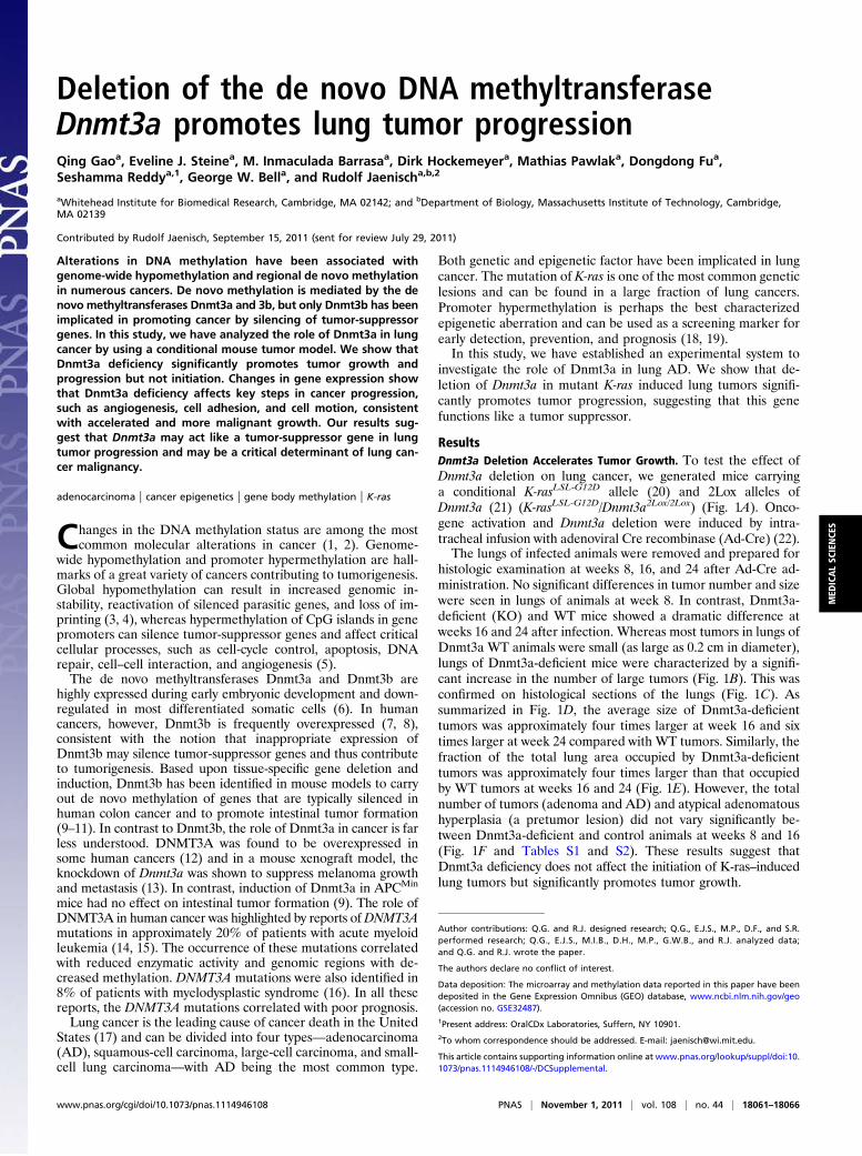
histologic examination at weeks 8, 16, and 24 after Ad-Cre ad-ministration. No significant differences in tumor number and sizewere seen in lungs of animals at week 8. In contrast, Dnmt3a-deficient (KO) and WT mice showed a dramatic difference atweeks 16 and 24 after infection. Whereas most tumors in lungs ofDnmt3a WT animals were small (as large as 0.2 cm in diameter),lungs of Dnmt3a-deficient mice were characterized by a signifi-cant increase in the number of large tumors (Fig. 1B). This wasconfirmed on histological sections of the lungs (Fig. 1C). Assummarized in Fig. 1D, the average size of Dnmt3a-deficienttumors was approximately four times larger at week 16 and sixtimes larger at week 24 compared with WT tumors. Similarly, thefraction of the total lung area occupied by Dnmt3a-deficienttumors was approximately four times larger than that occupiedby WT tumors at weeks 16 and 24 (Fig. 1E). However, the totalnumber of tumors (adenoma and AD) and atypical adenomatoushyperplasia (a pretumor lesion) did not vary significantly be-tween Dnmt3a-deficient and control animals at weeks 8 and 16(Fig. 1F and Tables S1 and S2). These results suggest thatDnmt3a deficiency does not affect the initiation of K-ras–inducedlung tumors but significantly promotes tumor growth.
Author contributions: Q.G. and R.J. designed research; Q.G., E.J.S., M.P., D.F., and S.R.performed research; Q.G., E.J.S., M.I.B., D.H., M.P., G.W.B., and R.J. analyzed data;and Q.G. and R.J. wrote the paper.
The authors declare no conflict of interest.
Data deposition: The microarray and methylation data reported in this paper have beendeposited in the Gene Expression Omnibus (GEO) database, www.ncbi.nlm.nih.gov/geo(accession no. GSE32487).1Present address: OralCDx Laboratories, Suffern, NY 10901.2To whom correspondence should be addressed. E-mail: [email protected].
This article contains supporting information online at www.pnas.org/lookup/suppl/doi:10.1073/pnas.1114946108/-/DCSupplemental.
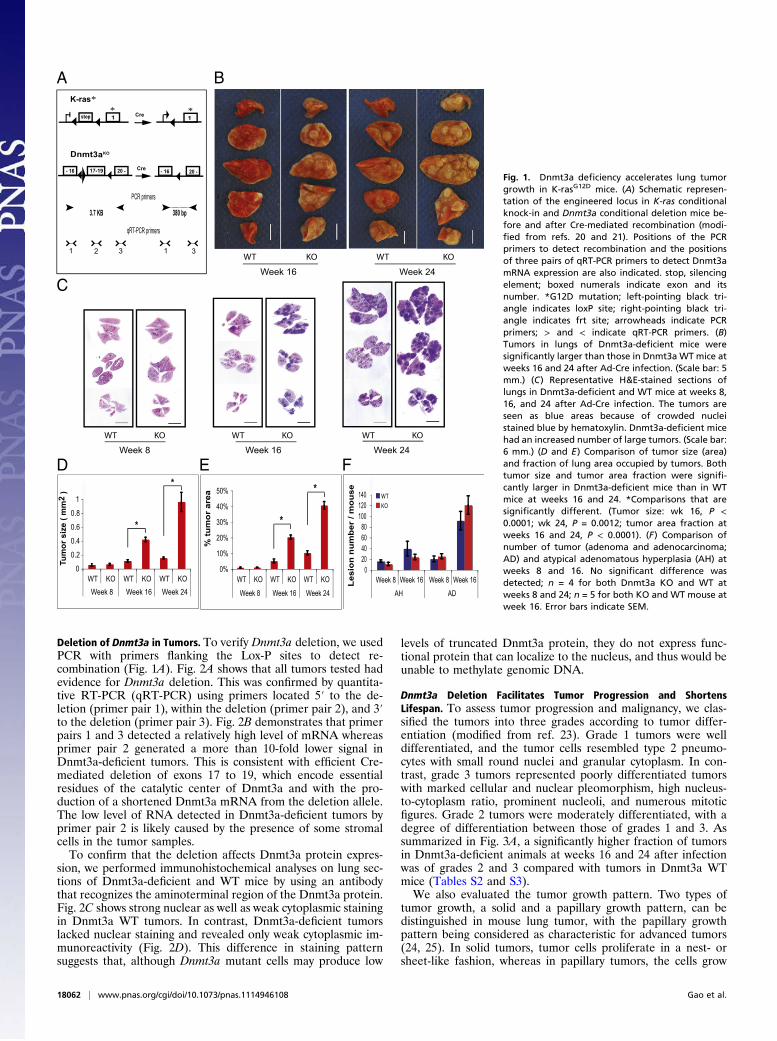
Deletion of Dnmt3a in Tumors. To verify Dnmt3a deletion, we usedPCR with primers flanking the Lox-P sites to detect re-combination (Fig. 1A). Fig. 2A shows that all tumors tested hadevidence for Dnmt3a deletion. This was confirmed by quantita-tive RT-PCR (qRT-PCR) using primers located 5′ to the de-letion (primer pair 1), within the deletion (primer pair 2), and 3′to the deletion (primer pair 3). Fig. 2B demonstrates that primerpairs 1 and 3 detected a relatively high level of mRNA whereasprimer pair 2 generated a more than 10-fold lower signal inDnmt3a-deficient tumors. This is consistent with efficient Cre-mediated deletion of exons 17 to 19, which encode essentialresidues of the catalytic center of Dnmt3a and with the pro-duction of a shortened Dnmt3a mRNA from the deletion allele.The low level of RNA detected in Dnmt3a-deficient tumors byprimer pair 2 is likely caused by the presence of some stromalcells in the tumor samples.To confirm that the deletion affects Dnmt3a protein expres-
sion, we performed immunohistochemical analyses on lung sec-tions of Dnmt3a-deficient and WT mice by using an antibodythat recognizes the aminoterminal region of the Dnmt3a protein.Fig. 2C shows strong nuclear as well as weak cytoplasmic stainingin Dnmt3a WT tumors. In contrast, Dnmt3a-deficient tumorslacked nuclear staining and revealed only weak cytoplasmic im-munoreactivity (Fig. 2D). This difference in staining patternsuggests that, although Dnmt3a mutant cells may produce low
levels of truncated Dnmt3a protein, they do not express func-tional protein that can localize to the nucleus, and thus would beunable to methylate genomic DNA.
Dnmt3a Deletion Facilitates Tumor Progression and ShortensLifespan. To assess tumor progression and malignancy, we clas-sified the tumors into three grades according to tumor differ-entiation (modified from ref. 23). Grade 1 tumors were welldifferentiated, and the tumor cells resembled type 2 pneumo-cytes with small round nuclei and granular cytoplasm. In con-trast, grade 3 tumors represented poorly differentiated tumorswith marked cellular and nuclear pleomorphism, high nucleus-to-cytoplasm ratio, prominent nucleoli, and numerous mitoticfigures. Grade 2 tumors were moderately differentiated, with adegree of differentiation between those of grades 1 and 3. Assummarized in Fig. 3A, a significantly higher fraction of tumorsin Dnmt3a-deficient animals at weeks 16 and 24 after infectionwas of grades 2 and 3 compared with tumors in Dnmt3a WTmice (Tables S2 and S3).We also evaluated the tumor growth pattern. Two types of
tumor growth, a solid and a papillary growth pattern, can bedistinguished in mouse lung tumor, with the papillary growthpattern being considered as characteristic for advanced tumors(24, 25). In solid tumors, tumor cells proliferate in a nest- orsheet-like fashion, whereas in papillary tumors, the cells grow
WT KO
Week 16
3
E F
KO
Week 24
WTWT KO
Week 16
A B
C
D
Tum
or s
ize
( mm
2 )
*
*
% tu
mor
are
a
0%
10%
20%
30%
40%
50%
WT KO WT KO WT KO
Week 8 Week 16 Week 24
*
*
WT KO
Week 8
WT KO
Week 24
-16
1*
- 16 20 -
Dnmt3aKO
PCR primers
qRT-PCR primers
1*
Cre
17-19 20 - - 16
K-ras*
stop
Cre
380 bp3.7 KB
020406080
100120140
Week 8 Week 16 Week 8 Week 16
AH AD
WTKO
Lesi
on n
umbe
r / m
ouse
0
0.2
0.4
0.6
0.8
1
WT KO WT KO WT KOWeek 8 Week 16 Week 24
1 12 33
Fig. 1. Dnmt3a deficiency accelerates lung tumorgrowth in K-rasG12D mice. (A) Schematic represen-tation of the engineered locus in K-ras conditionalknock-in and Dnmt3a conditional deletion mice be-fore and after Cre-mediated recombination (modi-fied from refs. 20 and 21). Positions of the PCRprimers to detect recombination and the positionsof three pairs of qRT-PCR primers to detect Dnmt3amRNA expression are also indicated. stop, silencingelement; boxed numerals indicate exon and itsnumber. *G12D mutation; left-pointing black tri-angle indicates loxP site; right-pointing black tri-angle indicates frt site; arrowheads indicate PCRprimers; > and < indicate qRT-PCR primers. (B)Tumors in lungs of Dnmt3a-deficient mice weresignificantly larger than those in Dnmt3a WT mice atweeks 16 and 24 after Ad-Cre infection. (Scale bar: 5mm.) (C) Representative H&E-stained sections oflungs in Dnmt3a-deficient and WT mice at weeks 8,16, and 24 after Ad-Cre infection. The tumors areseen as blue areas because of crowded nucleistained blue by hematoxylin. Dnmt3a-deficient micehad an increased number of large tumors. (Scale bar:6 mm.) (D and E) Comparison of tumor size (area)and fraction of lung area occupied by tumors. Bothtumor size and tumor area fraction were signifi-cantly larger in Dnmt3a-deficient mice than in WTmice at weeks 16 and 24. *Comparisons that aresignificantly different. (Tumor size: wk 16, P <0.0001; wk 24, P = 0.0012; tumor area fraction atweeks 16 and 24, P < 0.0001). (F) Comparison ofnumber of tumor (adenoma and adenocarcinoma;AD) and atypical adenomatous hyperplasia (AH) atweeks 8 and 16. No significant difference wasdetected; n = 4 for both Dnmt3a KO and WT atweeks 8 and 24; n = 5 for both KO and WT mouse atweek 16. Error bars indicate SEM.
18062 | www.pnas.org/cgi/doi/10.1073/pnas.1114946108 Gao et al.
around a fibrovascular core (Fig. 3B). Compared with the solidgrowth pattern, the papillary structure enables tumor cells toaccess blood circulation more efficiently, which is critical fortumor growth and progression. Fig. 3B shows that Dnmt3a-deficient mice had a significantly higher percentage of tumorswith papillary structure (i.e., papillary tumors) at weeks 16 and24 after infection than control mice (Tables S2 and S3). Finally,we observed tumor invasion in four Dnmt3a-deficient tumors(Fig. 3C), but no invasion was detected in WT tumors.Consistent with the high tumor load and the more malignant
tumor phenotype, the lifespan of Dnmt3a-deficient mice wassignificantly shorter than that of Dnmt3a heterozygous or WTmice (Fig. 3D). No significant difference was observed betweenDnmt3a heterozygous and WT mice. Autopsies of approximately70 animals revealed lung tumors as the most likely cause of death.
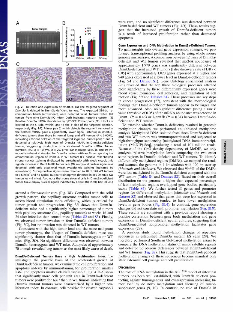
Dnmt3a-Deficient Tumors Have a High Proliferation Index. Toinvestigate the possible basis of the accelerated growth ofDnmt3a-deficient tumors, we examined the cell proliferation andapoptosis indexes by immunostaining for proliferation markerKi67 and apoptosis marker cleaved caspase-3. Fig. 4 A–C showthat significantly more cells per unit area in Dnmt3a-deficienttumors were positive for Ki67 than in WT tumors, indicating thatDnmt3a mutant tumors were characterized by a higher pro-liferation index. In contrast, cells positive for cleaved caspase-3
were rare, and no significant difference was detected betweenDnmt3a-deficient and WT tumors (Fig. 4D). These results sug-gest that the increased growth of Dnmt3a-deficient tumorsis a result of increased proliferation rather than decreasedapoptosis.
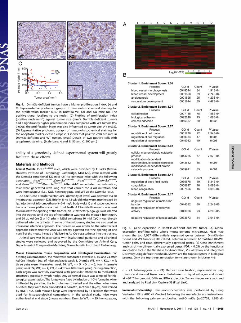
Gene Expression and DNA Methylation in Dnmt3a-Deficient Tumors.To gain insights into overall gene expression changes, we per-formed transcriptional profiling analyses by using whole mousegenome microarrays. A comparison between 12 pairs of Dnmt3a-deficient and WT tumors revealed that mRNA abundance ofapproximately 1,970 genes was significantly different betweenDnmt3a-deficient and WT tumors [false discovery rate (FDR) <0.05] with approximately 1,020 genes expressed at a higher and940 genes expressed at a lower level in Dnmt3a-deficient tumors(Fig. 5A and Dataset S1). Gene Ontology enrichment analysis(26) revealed that the top three biological processes affectedmost significantly by these differentially expressed genes wereblood vessel formation, cell adhesion, and regulation of cellmotion (Fig. 5B and Dataset S1). These processes are key stepsin cancer progression (27), consistent with the morphologicalfindings that Dnmt3a-deficient tumors appear to be larger andmore advanced. Also, no significant difference (using a raw Pvalue threshold of 0.05) of the mRNA abundance was detected inDnmt1 (P = 0.46) or Dnmt3b (P = 0.34) between Dnmt3a-de-ficient and WT tumors.To examine whether Dnmt3a deficiency resulted in genomic
methylation changes, we performed an unbiased methylomeanalysis. Methylated DNA isolated from three Dnmt3a-deficientand two WT tumors was immunoprecipitated and subjected tohigh-throughput sequencing [methylated DNA immunoprecipi-tation (MeDIP)-Seq], producing a total of 101 million reads.Because of the CpG density dependency of MeDIP, we onlyanalyzed relative methylation levels comparing the two exactsame regions in Dnmt3a-deficient and WT tumors. To identifydifferentially methylated regions (DMRs), we mapped the readsand scanned the genome in 1-kb windows with 100 bp overlap(Materials and Methods). We observed that 99.6% of the DMRswere less methylated in the Dnmt3a-deficient compared with theWT tumors (Table S4 and Dataset S2). Based on their overalldistribution on the genome, a higher than expected percentageof less methylated regions overlapped gene bodies, particularlyexons (Table S4). We further tested all genes and promoterregions for differential methylation (Materials and Methods andDataset S2) and observed that genes that were less expressed inDnmt3a-deficient tumors tended to have lower methylationlevels in gene bodies (Fig. S1A). In contrast, gene expressionchanges did not correlate with promoter methylation (Fig. S1B).These results are consistent with a previous report showing apositive correlation between gene body methylation and geneexpression in Dnmt3a-deficient neural cultures, suggesting thatDnmt3a-mediated nonpromoter methylation facilitates geneexpression (28).A previous study found methylation changes of repetitive
sequences in established Dnmt3a mutant ES cells (29). Wetherefore performed Southern blot-based methylation assays tocompare the DNA methylation status of minor satellite repeatsand detected no obvious differences between Dnmt3a-deficientand WT tumors (Fig. S2). This suggests that Dnmt3a-dependentmethylation changes of these sequences become manifest onlyafter extensive cell passage and cell proliferation.
DiscussionThe role of DNA methylation in the APCMin model of intestinaltumors has been well established, with Dnmt3b deletion pro-tecting against tumorigenesis and overexpression increasing tu-mor load by de novo methylation and silencing of tumor-suppressor genes (9, 10). In contrast, no role of Dnmt3a in
_Tumor samples
0.0
0.2
0.4
0.6
0.8
1.0
KO-tumor WT-tumor WT-lung
PP1PP2PP3
Relative m
RN
A ab
un
dan
ce
WT KO
A B
C D
Fig. 2. Deletion and expression of Dnmt3a. (A) The targeted segment ofDnmt3a is deleted in Dnmt3a-deficient tumors. The expected 380-bp re-combination bands (arrowhead) were detected in all tumors tested (40tumors from nine Dnmt3a-KO mice). Dash indicates negative control. (B)Relative Dnmt3a mRNA abundance by qRT-PCR. Primer pairs (PP) 1 to 3 arelocated to the 5′ side, within, and to the 3′ side of the targeted deletion,respectively (Fig. 1A). Primer pair 2, which detects the segment removed inthe deleted mRNA, gave a significantly lower signal (asterisk) in Dnmt3a-deficient tumors than those in normal lungs and WT tumors (P < 0.0001),indicating efficient deletion of the targeted segment. Primer pairs 1 and 3detected a relatively high level of Dnmt3a mRNA in Dnmt3a-deficienttumors, suggesting production of a shortened Dnmt3a mRNA. Tumornumbers: KO, n = 19; WT, n = 20. Error bar indicates SEM. (C and D) Im-munohistochemical staining for Dnmt3a protein with an Ab recognizing theaminoterminal region of Dnmt3a. In WT tumors (C), positive cells showedstrong nuclear staining (indicated by arrowheads) with weak cytoplasmicsignals, whereas in Dnmt3a-KO tumor cells (D), no typical nuclear signal wasdetected, with only occasional weak cytoplasmic staining (indicated byarrowheads). Strong nuclear signals were observed in 78 of 170 WT tumors(n = 6 mice) and no typical nuclear staining was detected in 160 Dnmt3a-KOtumors (n = 6 mice). Also note that some stromal cells in Dnmt3a-deficienttumor tissue display nuclear signals indicated by arrows (D). (Scale bar: 50 μm.)
Gao et al. PNAS | November 1, 2011 | vol. 108 | no. 44 | 18063
APCMin tumor formation has been established. Here we find thatdeletion of Dnmt3a promotes tumor growth and progression butnot tumor initiation, suggesting that this gene, counter to ex-pectation, acts like a tumor suppressor rather than like a cancer-promoting gene, as has been shown for Dnmt3b. Expression ofgenes involved in processes such as angiogenesis, cell adhesion,and cell motion was significantly altered in Dnmt3a-deficienttumors, consistent with an effect on tumor growth and progression.The methylation status of DNA may affect cancer by several
mechanisms: (i) global hypomethylation may increase genomicinstability (3, 4), and (ii) hypermethylation of promoters canmediate tumor suppressor gene silencing (5, 9). However, themechanism of how Dnmt3a affects gene expression and tumorformation is unclear. The genome of embryonic stem cells, incontrast to that of somatic cells, has methylated cytosine residuesat non-CpG contexts, which has been suggested to result fromthe activity of Dnmt3a (30). More recently, whole-genome pro-files of DNA methylation at single-base pair resolution of EScells detected non-CpG methylation in gene bodies, which waspositively correlated with gene expression (31). Another studydemonstrated that Dnmt3a-dependent gene body methylationcorrelated with expression of genes involved in neural differen-tiation (28). Consistent with this observation, we found that genebodies were less methylated in Dnmt3a-deficient tumors than inWT tumors, which also correlated with lower gene expression.Given that tumors in the mutant K-ras lung cancer model mayarise from stem cells giving rise to more differentiated cells in thetumor (32), it is possible that Dnmt3a-dependent gene body
methylation may be important for expression of genes thatpromote differentiation in a fraction of the tumor cells. Althoughmechanistic insights are lacking, our observation that Dnmt3a-deficient mice harbored more poorly differentiated and more ad-vanced tumors is consistent with the notion that Dnmt3a deficiencyinterferes with the differentiation process in tumor cells, pro-moting the formation of less mature and more malignant tumors.The majority of DNMT3a mutations found in myeloid neo-
plasm patients are missense mutations, and some of the mutationshave been shown or predicted to result in reduced translation(14–16). However, because nearly all patients are heterozygousfor the mutant allele it is not clear whether the DNMT3a muta-tions have dominant-negative effects or cause hemizygous in-sufficiency. In this context, our data using aDnmt3a-null allele areof interest, as they argue against the possibility that hemizygousinsufficiency affects lung tumors.Based on the well established role of de novo DNAmethylation-
mediated gene silencing in cancer, inhibitors of methyltransferasesare being actively investigated (33) and two drugs, azacitidineand decitabine, have been approved by the Food and DrugAdministration for treatment of myelodysplastic syndrome (34,35). Our data raise the possibility that such treatments, in ad-dition to activating silenced tumor-suppressor genes, may havethe unintended consequence of inhibiting DNMT3A, therebyaffecting its proposed tumor-suppressor function. Therefore, itwill be of great importance to elucidate the molecular mecha-nisms of how this gene affects cancer progression. The avail-
0 10 20 30 40
0
20
40
60
80
100
WT
Hetero
Homo
Age (weeks)
Pe
rc
en
ts
urv
iv
al
Solid Papillary
Grade 1 Grade 2 Grade 3
0%
20%
40%
60%
80%
100%
WT KO WT KO WT KO
Week 8 Week 16 Week 24
PapillarySolid
% tu
mor
**
0%
20%
40%
60%
80%
100%
WT KO WT KO WT KO
Week 8 Week 16 Week 24
Grade 3Grade 2Grade 1
% tu
mo
r
* *
A
B
C D
Fig. 3. Dnmt3a deficiency leads to moreadvanced tumors. (A) Dnmt3a-deficientmice had more grade 2 and grade 3 tumorsthan WT mice. Photomicrographs show themorphology of tumors of the three histo-logical grades (arrowhead, mitotic figure).Bar graph demonstrates that Dnmt3a-de-ficient mice had a significantly higher per-centage of grade 2 and 3 tumors at weeks16 and 24 (week 16, P = 0.0019; week 24,P < 0.0001) and grade 3 tumors at week24 (P = 0.0034). (B) Dnmt3a-deficient micehad more papillary tumors than WT mice.Photomicrographs show the morphology ofsolid and papillary growth pattern. Bargraph demonstrates that Dnmt3a-deficientmice had a significantly higher percentageof papillary tumors at weeks 16 and 24(week 16, P = 0.026; week 24, P = 0.0062);n = 4 for Dnmt3a KO and WT at week 8 and24; n = 5 for KO andWT mice at week 16. (C)Representative section showing invasion in-to a bronchiole (arrow) in a Dnmt3a-de-ficient tumor. (D) Dnmt3a-deficient (i.e.,homozygous KO) mice have a significantlyshorter lifespan than Dnmt3a heterozygousand WT mice (P < 0.0001). No significantdifference was detected between Dnmt3aheterozygous and WT mice (P = 0.63). WT,Dnmt3a WT, n = 29; Homo, Dnmt3a homo-zygous KO, n = 23; Hetero, Dnmt3a het-erozygous, n = 24. *Comparisons that aresignificantly different (at aforementioned Pvalues). Photomicrographs show H&E stain-ing. (Scale bars: A and B, 50 μm; C, 200 μm.)
18064 | www.pnas.org/cgi/doi/10.1073/pnas.1114946108 Gao et al.
ability of a genetically defined experimental system will greatlyfacilitate these efforts.
Materials and MethodsAnimal Models. K-rasLSL-G12D mice, which were provided by T. Jacks (Massa-chusetts Institute of Technology, Cambridge, MA) (20), were crossed withthe Dnmt3a conditional KO mice (21) to generate mice with the followinggenotypes: K-rasLSL-G12D/WT/Dnmt3a2lox/2lox, K-rasLSL-G12D/WT/Dnmt3aWT/WT,and K-rasLSL-G12D/WT/Dnmt3a2lox/WT. After Ad-Cre–mediated recombination,mice were generated with lung cells that carried the K-ras mutation andwere homozygous (i.e., KO), heterozygous, and WT at the Dnmt3a locus.
Ad-Cre (Gene Transfer Vector Core, University of Iowa) was delivered via anintratracheal approach (22). Briefly, 8- to 12-wk-old mice were anesthetized byi.p. injection of tribromoethanol (∼0.4 mg/g body weight) and suspended on abar of a mouse platform via their front teeth. A Fiber-lite Illuminator was usedto illuminate the openingof the trachea, an i.v. catheter (22gauge)was insertedinto the trachea until the top of the catheter was near the mouse’s front teeth,and 60 uL Ad-Cre (5 × 10⁷ pfu in MEM containing 10 mM CaCl2) was directlydelivered into the catheter. In some of the microarray studies, we also used anintranasal infection approach. The procedure was similar to the intratrachealapproach except that the virus was directly pipetted over the opening of onenostril of themouse instead of delivering Ad-Cre via a catheter into the trachea.
Animal care was in accordance with institutional guidance and all animalstudies were reviewed and approved by the Committee on Animal Care,Department of ComparativeMedicine,Massachusetts Institute of Technology.
Mouse Examination, Tissue Preparation, and Histological Examination. Forhistological comparison, themicewere euthanized atweeks 8, 16, and 24 afterAd-Cre infection (no. of mice analyzed: week 8, Dnmt3a WT, n = 4; KO, n = 4;three pairs were littermates; week 16, WT, n = 5; KO, n = 5; four littermatepairs; week 24, WT, n = 4; KO, n = 4; three littermate pairs). During dissection,each organ was carefully examined with particular attention to mediastinalstructures, especially lymph nodes. Any abnormal tissue was sampled for his-tological examination. The lungswerefixed by infusion of 10% formalin. Afterinfiltrated by paraffin, the left lobe was trisected and the other lobes werebisected; they were then embedded in paraffin, sectioned (4 μm), and stainedby H&E. Thus, each mouse’s lungs were represented by 11 sections that wereused for histopathological comparisons. In the survival study, mice wereeuthanized at end stage (mouse numbers: Dnmt3a WT, n = 29; homozygous,
n = 23; heterozygous, n = 24). Before tissue fixation, representative lungtumors and normal tissue were flash-frozen in liquid nitrogen and storedat −80 °C for genomic DNA and RNA extraction. Tumor images were capturedand analyzed by Pixel Link Capture SE (Pixel Link).
Immunohistochemistry. Immunohistochemistry was performed by usingVectastain Elite ABC kit (Vector) following the manufacturer’s instructions,with the following primary antibodies: anti-Dnmt3a (sc-20703, 1:200 di-
0.0 0.6 1.2 1.80
200
400
600
800KOWT
Tumor area(mm2)
Pos
itive
nuc
lei /
mm
2
C DD
AA
WT
BB
KO
Fig. 4. Dnmt3a-deficient tumors have a higher proliferation index. (A andB) Representative photomicrographs of immunohistochemical staining forthe proliferation marker Ki-67 in Dnmt3a WT (A) and KO mice (B). Thepositive signal localizes to the nuclei. (C) Plotting of proliferation index(positive nuclei/mm2) against tumor size (mm2). Dnmt3a-deficient tumorshad a significantly higher proliferation index compared with WT tumors (P =0.0006; the proliferation index was also influenced by tumor size, P = 0.032).(D) Representative photomicrograph of immunohistochemical staining forthe apoptosis marker cleaved caspase-3 shows that positive cells are rare inDnmt3a-deficient and WT tumors. (Inset) Details of two positive cells withcytoplasmic staining. (Scale bars: A and B, 50 μm; C, 200 μm.)
Cluster 1. Enrichment Score: 3.50
Process GO id Count P Valueblood vessel morphogenesis 0048514 34 1.91E-04blood vessel development 0001568 39 2.74E-04angiogenesis 0001525 25 4.23E-04vasculature development 0001944 39 4.47E-04
Cluster 2. Enrichment Score: 3.01
Process GO id Count P Valuecell adhesion 0007155 75 1.58E-04biological adhesion 0022610 75 1.68E-04cell-cell adhesion 0016337 30 0.035
Cluster 3. Enrichment Score: 2.67
Process GO id Count P Valueregulation of cell motion 0051270 22 2.94E-04regulation of cell migration 0030334 17 0.005regulation of locomotion 0040012 19 0.006
Cluster 4. Enrichment Score: 2.62
Process GO id Count P Valuecellular macromolecule catabolic process 0044265 77 7.07E-04modification-dependent macromolecule catabolic process 0043632 65 0.001modification-dependent protein catabolic process 0019941 65 0.001
Cluster 5. Enrichment Score: 2.41
Process GO id Count P Valueregulation of body fluid levels 0050878 19 5.34E-04coagulation 0050817 16 8.09E-04blood coagulation 0007596 16 8.09E-04
Cluster 6. Enrichment Score: 2.17
Process GO id Count P Valuenegative regulation of molecular function 0044092 30 2.24E-06negative regulation of catalytic activity 0043086 23 4.20E-05
negative regulation of kinase activity 0033673 14 3.04E-04
A
Blog (KO/WT) -2 0 1-1 2
2
1 2 3 4 5 6 7 8 9 10 11 12
Fig. 5. Gene expression in Dnmt3a-deficient and WT tumor. (A) Globalexpression profiling using whole mouse-genome microarrays. Heat mapshows the top 1,967 differentially expressed genes between Dnmt3a-de-ficient and WT tumors (FDR < 0.05). Columns represent 12 matched KO/WTtumor pairs, and rows differentially expressed genes. (B) Gene enrichmentanalysis of the differentially expressed genes (FDR < 0.05) by the functionalannotation tool in the Database for Annotation, Visualization, and IntegratedDiscovery using default thresholds. Shown are the top six clusters in biologicalprocess. Only the top three annotation terms are shown in cluster 4–6.
Gao et al. PNAS | November 1, 2011 | vol. 108 | no. 44 | 18065
MED
ICALSC
IENCE
S
lution; Santa Cruz), anti–Ki-67 (clone TEC-3, 1:50 dilution; DakoCytomation),and anti–cleaved caspase-3 (no. 9661, 1:1,000 dilution; Cell Signaling).Dnmt3a staining was performed on 160 tumors in six Dnmt3a-deficient miceand 170 tumors in six WT mice. Ki-67 and cleaved caspase-3 staining wereperformed on 57 Dnmt3a-deficient tumors from two mice and 69 WT tumorsfrom two mice.
PCR and RT-PCR. We used regular PCR to detect Cre-mediated recombinationin tumors of Dnmt3a KO mice. The sequences of the PCR primes (Fig. 1A)were as follows: sense, 5′ggcttttcctcagacagtgg3′; antisense, 5′ tcaatcat-cacggggttaga3′. PCR program was 95 °C for 2 min, 95 °C for 30 sec, 60 °C for30 sec, 72 °C for 45 sec, 30 cycles; 72 °C for 6 min. Forty tumors with diam-eters from 0.1 cm to 0.4 cm were tested from nine Dnmt3a-KO mice. Ge-nomic DNA (for large tumors) or tumor lysis (for small tumors) was used astemplate. Genomic DNA was isolated with AllPrep DNA/RNA mini kit (Qia-gen). Lysis solution was proteinase K 0.5 mg/mL, 50 mM KCl, 10 mM Tris-HCl(pH 8.3), 2 mM MgCl2, 0.1 mg/mL gelation, 0.45% Nonidet P-40, and 0.45%Tween 20.
The Dnmt3a mRNA abundance was analyzed in 19 tumors in four KO miceand 20 tumors in sixWTmice by qRT-PCR. Three pairs of qRT-PCR primers weredesigned to detect Dnmt3a mRNA using primers located 5′ to the deletion(primer pair 1), within the deletion (primer pair 2), and 3′ to the deletion(primer pair 3). The primer sequences are as follows: primer pair 1, 5′ggagccaccagaagaagaga3′, 5′gctctttctgggtttcttgg3′; primer pair 2, 5′cctgcaatgacctctccatt3′, 5′cggccagtaccctcataaag3′; and primer pair 3, 5′gaac-gagaggacggagaaaa3′, 5′tcctcctctgtggtttgctt3′. Total RNA was isolated withAllPrep DNA/RNA mini kit (Qiagen). First-strand cDNA was synthesized bySuperScript III First-Strand Synthesis SuperMix (Invitrogen). Real-time PCRwas performed on 7900HT Fast Real-Time PCR System (Applied Biosystems)using SYBR Green Master (Roche), following manufacturer protocols. Theexpression data were analyzed by comparative CT method.
Microarray. Total RNAwas isolated from 12matched Dnmt3a KO ∼WT tumorpairs from eight pairs of Dnmt3a KO ∼ WT mice. Each pair of mice shared atleast one parent. These 12 pairs of tumors included six pairs of large tumors(>0.4 cm in diameter) and six pairs of small tumors (<0.25 cm in diameter).The hybridization was performed on Agilent Whole Mouse Genome 4 × 44Kmicroarrays (two-channel) by Whitehead Genome Technology Core.
The two-color microarray raw data were normalized across channels byloess (locally weighted scatter-plot smoothing, using spot quality weights)and between arrays by quantile normalization of average intensities(“Aquantile”) using Bioconductor. Following summarization of replicateprobes by median, differential expression was assayed by moderated t testand corrected for FDR, as implemented by the limma package in Bio-conductor.
For gene enrichment analysis, we used the Database for Annotation,Visualization, and Integrated Discovery (26) to analyze the differentiallyexpressed genes list between Dnmt3a-deficient and WT tumors (FDR < 0.05).
DNA Methylation Assays. MeDIP-seq was performed on three Dnmt3a-de-ficient and twoWT tumors by using theMagmedip kit (Diagenode) accordingto the manufacturer’s protocol. Libraries were sequenced on the genomeanalyzer II (Illumina). Data analysis was similar to that described by Bocket al. (36).
Details of MeDIP-Seq and Southern blot analysis are provided in SIMaterials and Methods.
Statistical Analysis. We used Prism 5 (GraphPad Software) to perform sta-tistical analysis, with two-tailed Student t test for the comparison of tumornumber, size, grade, growth pattern, invasion, and fraction of tumor area;Kaplan–Meier survival analysis for comparison of lifespan; and two-wayANOVA for comparison of proliferation and apoptosis indexes. Unless in-dicated otherwise, 0.05 was used as the P value threshold for statisticalsignificance.
ACKNOWLEDGMENTS. We thank J. Dausman, R. Flannery, and K. Ganz formaintenance of the mouse colony and F. Soldner, L. Medeiros, G. Welstead,S. Sarkar, D. Faddah, and Y. Buganim for helpful discussions and criticalcomments on the manuscript. We thank T. Jacks (Massachusetts Institute ofTechnology) for providing K-ras mutant mice and A. Cheung and A. Dooleyfor technical assistance on intratracheal virus delivery. We also thankB. Yuan, R. Bronson, and D. Crowley for advice in statistics or histopathology;D. Cook, A. Leshinski, and C. Whittaker for running the Solexa pipeline; andJ. Kwon, J. Love, and S. Gupta for performing microarray hybridization. Thiswork was supported by a grant from Philip Morris International andNational Institutes of Health Grant R01-CA087869.
1. Feinberg AP (2007) Phenotypic plasticity and the epigenetics of human disease. Na-ture 447:433–440.
2. Jones PA, Baylin SB (2007) The epigenomics of cancer. Cell 128:683–692.3. Gaudet F, et al. (2003) Induction of tumors in mice by genomic hypomethylation.
Science 300:489–492.4. Chen RZ, Pettersson U, Beard C, Jackson-Grusby L, Jaenisch R (1998) DNA hypo-
methylation leads to elevated mutation rates. Nature 395:89–93.5. Sharma S, Kelly TK, Jones PA (2010) Epigenetics in cancer. Carcinogenesis 31:27–36.6. Okano M, Xie S, Li E (1998) Cloning and characterization of a family of novel mam-
malian DNA (cytosine-5) methyltransferases. Nat Genet 19:219–220.7. Mizuno S, et al. (2001) Expression of DNA methyltransferases DNMT1, 3A, and 3B in
normal hematopoiesis and in acute and chronic myelogenous leukemia. Blood 97:1172–1179.
8. Nosho K, et al. (2009) DNMT3B expression might contribute to CpG island methylatorphenotype in colorectal cancer. Clin Cancer Res 15:3663–3671.
9. Linhart HG, et al. (2007) Dnmt3b promotes tumorigenesis in vivo by gene-specific denovo methylation and transcriptional silencing. Genes Dev 21:3110–3122.
10. Lin H, et al. (2006) Suppression of intestinal neoplasia by deletion of Dnmt3b.Mol CellBiol 26:2976–2983.
11. Steine EJ, et al. (2011) Genes methylated by DNA methyltransferase 3b are similar inmouse intestine and human colon cancer. J Clin Invest 121:1748–1752 10.1172/JCI43169.
12. Robertson KD, et al. (1999) The human DNAmethyltransferases (DNMTs) 1, 3a and 3b:Coordinate mRNA expression in normal tissues and overexpression in tumors. NucleicAcids Res 27:2291–2298.
13. Deng T, et al. (2009) An essential role for DNA methyltransferase 3a in melanomatumorigenesis. Biochem Biophys Res Commun 387:611–616.
14. Ley TJ, et al. (2010) DNMT3A mutations in acute myeloid leukemia. N Engl J Med 363:2424–2433.
15. Yan XJ, et al. (2011) Exome sequencing identifies somatic mutations of DNA meth-yltransferase gene DNMT3A in acute monocytic leukemia. Nat Genet 43:309–315.
16. Walter MJ, et al. (2011) Recurrent DNMT3A mutations in patients with myelodys-plastic syndromes. Leukemia 25:1153–1158.
17. Jemal A, Siegel R, Xu J, Ward E (2010) Cancer statistics, 2010. CA Cancer J Clin 60:277–300.
18. Brock MV, et al. (2008) DNA methylation markers and early recurrence in stage I lungcancer. N Engl J Med 358:1118–1128.
19. Belinsky SA (2004) Gene-promoter hypermethylation as a biomarker in lung cancer.Nat Rev Cancer 4:707–717.
20. Jackson EL, et al. (2001) Analysis of lung tumor initiation and progression using
conditional expression of oncogenic K-ras. Genes Dev 15:3243–3248.21. Nguyen S, Meletis K, Fu D, Jhaveri S, Jaenisch R (2007) Ablation of de novo DNA
methyltransferase Dnmt3a in the nervous system leads to neuromuscular defects and
shortened lifespan. Dev Dyn 236:1663–1676.22. DuPage M, Dooley AL, Jacks T (2009) Conditional mouse lung cancer models using
adenoviral or lentiviral delivery of Cre recombinase. Nat Protoc 4:1064–1072.23. Jackson EL, et al. (2005) The differential effects of mutant p53 alleles on advanced
murine lung cancer. Cancer Res 65:10280–10288.24. Nikitin AY, et al. (2004) Classification of proliferative pulmonary lesions of the mouse:
Recommendations of the mouse models of human cancers consortium. Cancer Res 64:
2307–2316.25. Renne R, et al. (2009) Proliferative and nonproliferative lesions of the rat and mouse
respiratory tract. Toxicol Pathol 37(suppl):5S–73S.26. Huang W, Sherman BT, Lempicki RA (2009) Systematic and integrative analysis of
large gene lists using DAVID bioinformatics resources. Nat Protoc 4:44–57.27. Talmadge JE, Fidler IJ (2010) AACR centennial series: The biology of cancer metastasis:
Historical perspective. Cancer Res 70:5649–5669.28. Wu H, et al. (2010) Dnmt3a-dependent nonpromoter DNA methylation facilitates
transcription of neurogenic genes. Science 329:444–448.29. Liang G, et al. (2002) Cooperativity between DNA methyltransferases in the mainte-
nance methylation of repetitive elements. Mol Cell Biol 22:480–491.30. Ramsahoye BH, et al. (2000) Non-CpGmethylation is prevalent in embryonic stem cells
and may be mediated by DNA methyltransferase 3a. Proc Natl Acad Sci USA 97:
5237–5242.31. Lister R, et al. (2009) Human DNA methylomes at base resolution show widespread
epigenomic differences. Nature 462:315–322.32. Kim CF, et al. (2005) Identification of bronchioalveolar stem cells in normal lung and
lung cancer. Cell 121:823–835.33. Belinsky SA, et al. (2011) Combination therapy with vidaza and entinostat suppresses
tumor growth and reprograms the epigenome in an orthotopic lung cancer model.
Cancer Res 71:454–462.34. Taby R, Issa JP (2010) Cancer epigenetics. CA Cancer J Clin 60:376–392.35. Rodríguez-Paredes M, Esteller M (2011) Cancer epigenetics reaches mainstream on-
cology. Nat Med 17:330–339.36. Bock C, et al. (2010) Quantitative comparison of genome-wide DNA methylation


![Cytosine methylation is a conserved epigenetic feature ...€¦ · and repetitive element silencing [6]. Metazoan DNA methyltransferases (DNMT1, DNMT2, DNMT3a/3b [7]) catalyse this](https://static.documents.pub/doc/80x56/5eab33730f2ba76ce938ef9e/cytosine-methylation-is-a-conserved-epigenetic-feature-and-repetitive-element.jpg)


