Delirium in the Acute Care Setting: Characteristics, Diagnosis and Treatment Jose´ R. Maldonado, MD, FAPM, FACFE Departments of Psychiatry and Medicine, Stanford University School of Medicine, 401 Quarry Road, Suite 2317, Stanford, CA 94305, USA Delirium is an acute or subacute organic mental syndrome characterized by disturbance of consciousness, global cognitive impairment, disorienta- tion, the development of perceptual disturbance, attention deficits, decreased or increased psychomotor activity (depending on the type of de- lirium), disordered sleep-wake cycle, and fluctuation in presentation (eg, waxing and waning). The term ‘‘delirium,’’ from the Latin roots de (meaning ‘‘away from’’) and lira (meaning ‘‘furrow in a field’’) and ium (Latin for singular), literally means ‘‘a going off the ploughed track, a mad- ness.’’ The term ‘‘delirium’’ is reported to have been coined by the lay Roman writer Celsus (1AD) and described in his compendium De Medicina [1,2]. Clear descriptions of the syndrome are contained in Hippocrates’s writings, who called the syndrome by the term phrenitis [3]. In 1813, the British physician Thomas Sutton introduced the term delirium tremens to designate delirium caused by the withdrawal from central nervous system (CNS) depressant agents, but which is almost exclusively applied in modern times to delirium resulting from alcohol withdrawal [4]. In the acute care setting, many names are used to describe the acute men- tal status changes associated with delirium. Commonly used terms include ‘‘intensive care unit (ICU) psychosis’’ or ‘‘sundowning.’’ The first describes the fact that mental status changes are often seen in the ICU, the second is a descriptor of a pattern by which subjects tend to experience confusion more frequently during periods of decreased or inappropriate stimulation, such as at night or ‘‘sun down.’’ The psychiatric literature uses other terms that usually describe common characteristics or features of the syndrome, such as ‘‘acute confusional state’’ (ie, acute, confusion) and ‘‘acute brain failure’’ to describe the gravity of the situation. Yet, neurologists and inter- nists prefer the term ‘‘encephalopathy,’’ which literally means ‘‘disease of E-mail address: [email protected]0749-0704/08/$ - see front matter Ó 2008 Elsevier Inc. All rights reserved. doi:10.1016/j.ccc.2008.05.008 criticalcare.theclinics.com Crit Care Clin 24 (2008) 657–722
Transcript
Delirium in the Acute Care Setting:Characteristics, Diagnosis and Treatment
Jose R. Maldonado, MD, FAPM, FACFEDepartments of Psychiatry and Medicine, Stanford University School of Medicine,
401 Quarry Road, Suite 2317, Stanford, CA 94305, USA
Delirium is an acute or subacute organic mental syndrome characterizedby disturbance of consciousness, global cognitive impairment, disorienta-tion, the development of perceptual disturbance, attention deficits,decreased or increased psychomotor activity (depending on the type of de-lirium), disordered sleep-wake cycle, and fluctuation in presentation(eg, waxing and waning). The term ‘‘delirium,’’ from the Latin roots de(meaning ‘‘away from’’) and lira (meaning ‘‘furrow in a field’’) and ium(Latin for singular), literally means ‘‘a going off the ploughed track, a mad-ness.’’ The term ‘‘delirium’’ is reported to have been coined by the layRoman writer Celsus (1AD) and described in his compendium De Medicina[1,2]. Clear descriptions of the syndrome are contained in Hippocrates’swritings, who called the syndrome by the term phrenitis [3]. In 1813, theBritish physician Thomas Sutton introduced the term delirium tremens todesignate delirium caused by the withdrawal from central nervous system(CNS) depressant agents, but which is almost exclusively applied in moderntimes to delirium resulting from alcohol withdrawal [4].
In the acute care setting, many names are used to describe the acute men-tal status changes associated with delirium. Commonly used terms include‘‘intensive care unit (ICU) psychosis’’ or ‘‘sundowning.’’ The first describesthe fact that mental status changes are often seen in the ICU, the second isa descriptor of a pattern by which subjects tend to experience confusionmore frequently during periods of decreased or inappropriate stimulation,such as at night or ‘‘sun down.’’ The psychiatric literature uses other termsthat usually describe common characteristics or features of the syndrome,such as ‘‘acute confusional state’’ (ie, acute, confusion) and ‘‘acute brainfailure’’ to describe the gravity of the situation. Yet, neurologists and inter-nists prefer the term ‘‘encephalopathy,’’ which literally means ‘‘disease of
the brain.’’ The term encephalopathy is meant to convey a brain mal-function in the face of systemic metabolic derangements (eg, metabolicencephalopathy), cardiopulmonary or vascular problems (eg, hypoxic or hy-pertensive encephalopathy), renal disease (eg, uremic encephalopathy), liverdisease (eg, hepatic encephalopathy), or endocrine disease (eg, Hashimoto’sencephalopathy); or to be a consequence of toxic factors (eg, toxic ence-phalopathy or Wernicke’s encephalopathy) or problems with oxygenation(eg, hypoxic encephalopathy). Unfortunately, the use of these various terms,even if accurate, may add to the confusion and difficulties of identifying andtreating the syndrome of delirium.
Epidemiology of delirium
Delirium is the most common psychiatric syndrome found in the generalhospital setting. Its prevalence surpasses most commonly known and iden-tified psychiatric syndromes and varies depending on the medical setting.Table 1 compares the incidence of delirium in different medical settingsand various psychiatric disorders [5]. The incidence of delirium among med-ically ill patients ranges from 10% in the general medicine ward to 85% inadvanced cancer [6–11]. This wide range is associated with the organ systemand disease process under consideration. For example, in the adult generalmedicine population the incidence of delirium ranges from 10% to 24%: asreported by Speed and colleagues 10.9% [12], Maldonado and colleagues14% [13], Ritchie and colleagues 14.6% [14], and Gonzalez and colleagues24% [15]. As expected, the incidence goes up with increased severity of ill-ness, rising to 13% to 48% in after-stroke victims [16], 20% to 40% amongHIV/AIDS patients [17,18], 60% in frail-elderly patients [19], 60% to 80%among patients in the medical ICU [20], and as high as 80% to 90% in ter-minally ill cancer patients [21]. One study found that 89% of survivors ofstupor or coma progressed to delirium [22].
A European multinational study (n ¼ 3,608) by Valdes and colleagues[23] found a delirium prevalence rate of 9.1% in the general hospital popu-lation. A Spanish study by Gonzalez and colleagues [15] confirmed findingsin the United States and similarly suggested that the average hospital stay isprolonged from 12 days to 17.5 days when delirium is present. Similarly,a study conducted in Western Australia found a 10.9% prevalence rate ofdelirium among patients admitted to two general medicine wards(n ¼ 1,209) [12].
Similarly, in the general surgical population the incidence of delirium isabout 37% to 46% [24], and postoperative delirium has been described tooccur in 10% to 60% of patients [25]. Again, the range in incidence of post-operative delirium depends on the type of surgery and the population stud-ied: 25% to 32% among patients undergoing coronary artery bypassgrafting (CABG); 50% to 67% among patients undergoing cardiotomy(eg, cardiac valve replacement) [26–29]; about 20% of elderly patients after
Table 1
A comparison of the incidence of psychiatric disorder in the general population and delirium
among medically ill patients
Incidence of
psychiatric
disorders
% of general adult
us population [5]
Incidence of delirium
in selected
medical populations %
Major depression 6.7 General medicine wards 10–18
Dysthymic disorder 1.5 HIV/AIDS 30–40
Bipolar disorder 2.6 Medical-ICU 60–80
All mood disorders 9.5 General surgical
wards (range)
37–46 (10–60)
After stroke 13–48
Panic disorder 2.7 After CABG 25–32
OCD 1 After cardiotomy 50–67
PTSD 3.5 Elderly
GAD 3.1 Out-patient minor
(cataract) surgery
4.4
Social phobia 6.8 At time of hospitalization 10–15
Agoraphobia 8.7 In nursing homes 15–60
All anxiety disorders 18.1 After hip replacement 21–63
In cancer patients
Schizophrenia 1.1 General prevalence 25–40
Anorexia nervosa 0.5–3.7 Hospitalized cancer patients 25–50
surgery for gynecologic malignancies [30]; 33% of patients undergoingabdominal aneurysm repair [31]; 12.5% in patients undergoing spine surgery[32]; 41% after bilateral knee replacement [33]; and 25% of elderly patientsundergoing elective hip or knee replacement, compared with 65% after fem-oral neck fracture repair [34–36]. Acute mental status changes, neuropsychi-atric dysfunction, and neurocognitive deficits are common after cardiacsurgery [37]. Delirium and other forms of acute organic mental syndromeoccurred in 32% to 80% of patients undergoing cardiac surgery [29,38,39].
The incidence of delirium is well documented in the acutely medically illpatient. A study by Ely and colleagues [20] involving patients admitted tothe medical intensive care unit (MICU), 50% of which were receivingmechanical ventilation, found that 81.3% of MICU patients developeddelirium during the course of their ICU stay. The mean onset of deliriumwas 2.6 days (standard deviation or SD � 1.7), and the mean durationwas 3.4 days (SD � 1.9). The duration of delirium was associated withlength of stay in the ICU (r ¼ 0.65, P ¼ .0001) and total length of hospital
stay (LOS) (r ¼ 0.68, P ¼ .0001). Multivariate analysis demonstrated thatdelirium was the strongest predictor of LOS in the hospital (P ¼ .006),even after adjusting for severity of illness, age, gender, race, and days of ben-zodiazepine and narcotic drug administration.
Maldonado and colleagues [13] found an 18% incidence of delirium in anacute ICU (eg, combined medical and surgical patients) based on Diagnosticand Statistical Manual of Mental Disorders, Fourth Edition (DSM-IV)criteria. As in previous studies, the average delirious patient age was over65 years old and mostly male (60%). The presence of delirium significantlyextended the overall length of stay (ie, 15 days in delirious patients, com-pared with 11 days in nondelirious counterparts).
Finally, delirium has been found to be the most common clinical neuro-psychiatric condition in specialized palliative care units. It has been reportedto occur in 26% to 44% of cancer patients admitted to hospital or hospice.As the disease progresses, over 80% of all advanced cancer patients eventu-ally experience delirium in their final days [21,40].
Etiology of delirium
The syndrome of delirium is better thought of as having a multifactorialetiology, as is often the case in most medically ill patients. Patients in theICU are usually critically ill, which makes them more susceptible to devel-oping delirium. There are many risk factors known to contribute to thedevelopment of delirium.
Delirium clinical risk factors
Age (greater than 75 years old)Baseline cognitive functioning:
25% delirious are demented40% demented in hospital develop delirium
Male genderSensory impairmentUse of intravenous lines, bladder catheters, and physical restraintsSevere illness
Infections (particularly urinary tract infections and pneumonias, inolder persons)
Hip fractureHyperthermiaHypothermiaHypotension and hypoperfusionHypoxia or anoxiaMalnutrition and nutritional deficiencies (eg, thiamine deficiency lead-
ing to Wernicke’s encephalopathy)
661DELIRIUM IN THE ACUTE CARE SETTING
Metabolic disorders
Acute metabolic encephalopathies (eg, cardiac, hepatic and renal
embolism)Endocrinopathies (eg, hyper- and hypothyroidism)Water and electrolyte abnormalitiesHypo- or hyperglycemiaHypo- or hypernatremiaHypo- or hyperkalemiaDehydration
Elevation in serum cortisol levelsCNS pathology (ie, stroke, intracranial hemorrhages, normal pressurehydrocephalus)
Trauma (eg, severe physical trauma or surgery)Exogenous substances
Medication side effects:
Polypharmacy (more than three medications)Psychoactive medicationsSerotonergic agentsAnticholinergic agentsOver-the-counter substances
Substance abuse and withdrawal
AlcoholismCNS-depressant substances (both prescribed and illegal)CNS-depressant withdrawal (eg, delirium tremens)CNS-stimulant substances (both prescribed and illegal)HallucinogensOver-the-counter substances
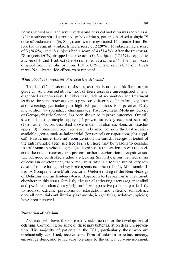
Two of the known risk factors for delirium include the patient’s age andthe presence of a baseline cognitive disorder (eg, dementia, stroke). Studieshave suggested that increasing age was an independent predictor of transi-tioning to delirium. A study of mehanically ventilated adults (n ¼ 275) sug-gests that there is an incremental risk for transitioning into delirium forpatients older than 65 years (odds ratio or OR of transitioning to deliriumfor age was 1.02 [1.00–1.03; P ¼ .04]). In fact, the results suggest that foreach additional year after age 65, the probability of transitioning to deliriumincreased by 2% (multivariable P values ! 0.05) (Fig. 1) [41]. Similarly,a study of elderly patients undergoing hip surgery, found that mini-mental
Fig. 1. Age and the probability of transitioning to delirium.Themost notable finding related to age
was that probability of transitioning to delirium increased dramatically for each year of life after 65
years. Adjusted OR 1.01 (1.00, 1.02) (P¼ .03). Y-axis¼ Probability; X-axis¼ Age in years. (From
Pandharipande P, Shintani A, Peterson J, et al. Lorazepam is an independent risk factor for transi-
tioning to delirium in intensive care unit patients. Anesthesiology 2006;104(1):23; with permission.)
662 MALDONADO
state examination (MMSE) scores were identified as an independent predic-tor of postoperative delirium [42].
Milstein and colleagues [43] reported on the development of deliriumamong the elderly patient undergoing relatively simple outpatient surgery.They studied elderly patients (n ¼ 296) undergoing cataract surgery andfound a 4.4% incidence of postoperative delirium. As others have suggested,those developing delirium were older (82.1 versus 73.06 years; P ! .001) andreceived higher benzodiazepine doses as pre-medication for surgery (69%versus 39.9%; P ! .002).
In a prospective study evaluating neuropsychologic performance in olderpatients (ie, O70 years), subjects (n ¼ 100) who were free of dementia andadmitted for elective orthopedic surgery underwent a series of neuropsychi-atric testing pre- and postoperatively [44]. Findings suggest that subtlepreoperative attention deficits were closely associated with postoperativedelirium. Patients who developed postsurgical delirium had significantlyslower mean reaction times (P % .011) and greater variability of reactiontime (P ¼ .017) preoperatively. A four- to fivefold increased risk of deliriumwas observed for people one standard deviation above the sample means onthese variables.
A study by Wahlund and Bjorlin [45] found that approximately 70% ofelderly patients admitted to a specialized delirium ward had a pre-existingcognitive disorder, either dementia or mild cognitive impairment. Bergmannand Eastham [46] studied elderly patients (n ¼ 100) admitted to an acutemedical unit in a general hospital for the presence of psychiatric morbidity.They found that 7% suffer from dementia, while 16% suffered from acute
663DELIRIUM IN THE ACUTE CARE SETTING
delirious states. Demented patients or patients suffering from other condi-tions associated with deficient brain function (ie, traumatic brain injury,drug and alcohol abuse and withdrawal) have a lower threshold for devel-oping delirium and do so with greater frequency. Similarly, a study of elderlysubjects undergoing hip or knee replacement (n ¼ 572) demonstrated thatthe presence of dementia increased the occurrence of delirium [36]. Twentyfour percent of subjects had preoperative dementia. Postoperatively, all(100%) of demented subjects developed delirium, compared with 31.8%in the nondemented population.
Poor oxygenation (ie, hypoperfusion and hypoxemia) has long been asso-ciated with the development of delirium, both because of medical problemsas well as postoperatively. Severe illness processes, combined with both de-creased oxygen supply and increased oxygen demand may lead to the samecommon end problem, namely decreased oxygen availability to brain tissue[47–50]. Inadequate oxidative metabolism may be one of the underlyingcauses of the basic metabolic problems initiating the cascade that leads tothe development of delirium, namely: inability to maintain ionic gradientscausing cortical spreading depression (ie, spreading of a self-propagatingwave of cellular depolarization in the cerebral cortex) [51–56]; abnormalneurotransmitter synthesis, metabolism and release [57–65]; and a failureto effectively eliminate neurotoxic by-products [58,59,63].
A study of postthoracotomy patients demonstrated that 21% of thepatients developed clinically significant postoperative delirium [66]. In thissample, delirium occurred in all patients who had inadequate oxygenation.The treatment of choice was supplementary oxygen, with a near perfecttreatment success. Others have similarly linked delirium to the presence ofpoor oxygenation associated with untreated obstructive sleep apnea [67]and to the presence of occult hypoxia after total hip arthroplasty [68].
Of note, animal studies have suggested that subjects with baseline organiccerebral disorders, such as cerebrovascular disease, may be particularly sen-sitive to hypoxic injury. Miyamoto and colleagues [69] submitted laboratoryanimals to hypocapnia during surgical anesthesia, causing tissue damage inthe caudoputamen. This model may suggest that a similar mechanism maybe responsible for long-lasting postoperative delirium in patients with strokeor dementia.
Sleep is another factor that seems to play a significant role in developingdelirium in the ICU. Sleep deprivation has long been linked to the develop-ment of delirium [70] and psychosis [71]. Studies have found that the averageamount of sleep in ICU patients is limited to 1 hour and 51 minutes per24-hour period [72]. Many factors may affect sleep in the ICU, including fre-quent therapeutic interventions, the nature of diagnostic procedures, pain,fear, and the noisy environment. Similarly, oversedation has been foundto be an independent predictor of prolonged mechanical ventilation. Ina prospective, controlled study (n ¼ 128) of adults undergoing mechanicalventilation, subjects were randomized to either continuous sedation or daily
664 MALDONADO
awakenings [73]. They found that the median duration of mechanical venti-lation was 4.9 days in the intervention group (ie, daily awakening), ascompared with 7.3 days in the control group (P ¼ .004), and the medianLOS in the intensive care unit was 6.4 days as compared with 9.9 days,respectively (P ¼ .02).
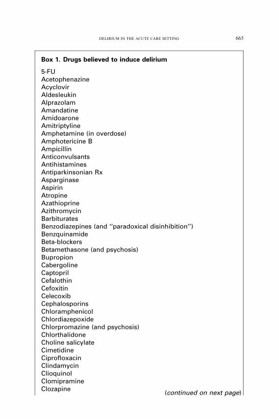
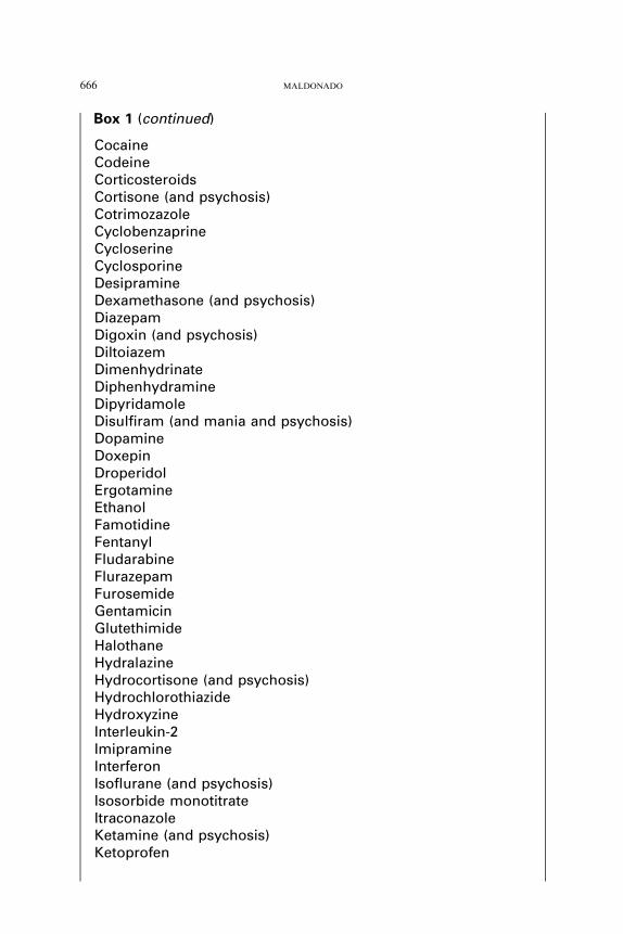
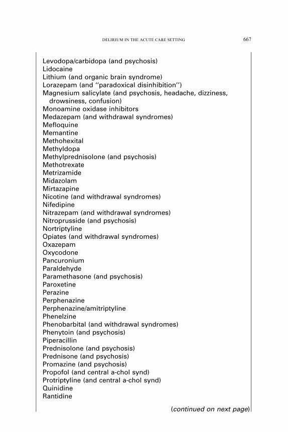
A great number of medications have been associated with an increasedrisk of delirium (Box 1). The highest incidence medication-induced deliriumhas been observed in patients taking more than three medications [74],medications with high psychoactive activity [75], and when drugs havehigh anticholinergic potential [76].
Medications with significant psychoactive effects have long been identifiedas a frequent cause of delirium. Several studies have linked the use of psycho-active agents to the etiology of 15% to 75% of delirium cases [19,21,77–81].More specifically, opioids, corticosteroids, and benzodiazepines have beenidentified as major contributors to delirium in several studies (Fig. 2) [75].Other medications, such as nonsteroidal anti-inflammatory agents, andchemotherapeutic agents, were also identified as causes of delirium.
There is significant evidence to suggest that there is a direct associationbetween a medication’s anticholinergic potential and their incidence of caus-ing delirium [74,76,82–86]. Some drugs (eg, diphenhydramine, atropine) areeasier to identify as having a high anticholinergic load. On the other hand,others are not so obvious. Several studies have demonstrated a direct rela-tionship between a drug’s anticholinergic potential (as measured by serumanticholinergic activity) and the development of delirium [76,85,87–90].Tune has conducted several studies looking at the cumulative effect of drugswith subtle anticholinergic potential and their serum anticholinergic activity(Box 2, Table 2) [76,83,84,86,90,91].
Blazer and colleagues [92] conducted a study of the potential for anticho-linergic toxicity among long-term care residents. Their study included resi-dents aged 65 years and older (n ¼ 5,902) who continuously resided ina nursing home for 1 year and determined drug administration and drugquantity. The survey revealed that 60% of residents received drugs withsignificant anticholinergic properties and nearly 10% of the residentsreceived three or more medications with high anticholinergic load. Finally,Han and colleagues [93] followed medical inpatients (n ¼ 278) and measuredtheir exposure to anticholinergic medications. They found that exposure toanticholinergic agents was an independent risk factor for the development ofdelirium, and specifically associated with a subsequent increase in deliriumsymptom severity.
As suggested by many others, many gamma amino-butyric acid (GABA)-ergic medications have been implicated in the development of delirium[20,94–97]. It is now beginning to be understood that agents commonlyused for achieving postoperative sedation may in fact contribute to deliriumby (a) interfering with physiologic sleep patterns and (b) causing a centrallymediated acetylcholine deficient state (ie, interruption of central cholinergic
irritability, palpitations, nightmares, or seizures)VancomycinVincristineWarfarinZolpidemZotepine (and anxiety, agitation)
Data from Electronic Physicians Desk Reference, 2007.
Box 1 (continued)
668 MALDONADO
muscarinic transmission at the level of the basal forebrain and hippocam-pus) [95–97]. A study of blood and urine melatonin levels revealed anabolition of the circadian rhythm of melatonin release in deeply sedatedICU patients [98]. This suggests that sedative agents may contribute tothe development of delirium by more than one mechanism (ie, disruptionof sleep patterns; central acetylcholine inhibition; disruption of melatonincircadian rhythm). Therefore, it appears that commonly used sedative (eg,propofol, midazolam) may promote the development of delirium.
The irony is that these are the same medications physicians often use tomanage agitated or delirious patients. This practice, even if immediatelyeffective in tranquilizing a patient may, in the long run, aggravate and per-petuate the syndrome of delirium. One of the first studies to demonstrate the
Fig. 2. Delirium cases potentially caused by opioids, corticosteroids and benzodiazepines in six
case-series. aBreitbart and colleagues [78], bMorita and colleagues [80], cTuma and DeAngelis [79],dLawlor and colleagues [21], eOlofsson and colleagues [81], fFrancis and colleagues [19]. (FromGau-
dreau JD,Gagnon P, RoyMA, et al. Association between psychotogenic medications and delirium
in hospitalized patients: a critical review. Psychosomatics 2005;46(4):304; with permission.)
669DELIRIUM IN THE ACUTE CARE SETTING
relationship between benzodiazepine use and delirium was conducted byMarcantonio and colleagues [94]. They found that development of deliriumwas significantly associated with postoperative exposure to benzodiazepines(OR, 3.0; 95% confidence interval or CI, 1.3–6.8). These findings have beenconfirmed by Pandharipande and colleagues [41], who studied adult venti-lated patients (n ¼ 275) in the ICU for the development of delirium. Theyfound that lorazepam was an independent risk factor for daily transitionto delirium (OR, 1.2; 95% CI, 1.1–1.4; P ¼ .003) (Fig. 3). These findingsconfirm many others who have previously suggested benzodiazepines tobe culprits in the development of delirium and other cognitive impairmentin medically ill patients [20,94,99,100]. Being aware of what types of medi-cations a patient is taking and eliminating unnecessary medications canhelp reduce the potential for anticholinergic side effects.
As in the case with sleep, both pain and medications used for the treat-ment of pain have been associated with the development of delirium. Vaurioand colleagues [25] demonstrated that presence of postoperative pain is anindependent predictor of delirium after surgery. Furthermore, they founda direct relationship between levels of preoperative pain and the risk forthe development of postoperative delirium. On the other hand, the use ofopioid agents has been implicated in the development of delirium [101–103]. Opioids are blamed for nearly 60% of the cases of delirium in patientswith advanced cancer [40]. A study of cancer patients (n¼ 114) showed a sig-nificant associations between opioids and delirium, after controlling forother medications used [104]. Several studies have reported that patients
Box 2. Commonly used medicines that have anticholinergiceffects
Central nervous systemAlprazolamAmitriptylineChlordiazepoxideCodeineDesipramineDiazepamDoxepinFlurazepamImipramineOxazepamOxycodonePhenelzinePhenobarbital
Data from Tune LE. Anticholinergic effects of medication in elderly patients.J Clin Psychiatry 2001;62 Suppl 21:13.
671DELIRIUM IN THE ACUTE CARE SETTING
who used oral opioid analgesics as their sole means of postoperative paincontrol were at decreased risk of developing delirium in comparison withthose who used opioid analgesics via intravenous (IV) patient-controlledanalgesia technique (OR, 0.4; 95% CI, 0.2–0.7) [25,103].
There is some data that suggests that some opioid agents may havegreater deliriogenic potential than others. For example, several reports sug-gest that meperidine has a greater deliriogenic potential than other opioids[94,101,105]. Other studies have suggested that an opioid rotation frommorphine to fentanyl has been associated with improved pain managementand lower delirium rating scores [106]. Similarly, at least one case reportsuggests that the use of acetylcholinesterase inhibitors successfully reversedopioid-induced hypoactive delirium [107]. This may implicate an anticholin-ergic mechanism of opioid induced delirium.
Besides their potential anticholinergic effect or their disruption of sleeppatterns, medications may cause delirium by disrupting thalamic gating
Table 2
Anticholinergic drug levels in 25 medications ranked by the frequency of their prescription for
elderly patients
MedicationaAnticholinergic drug level (ng/mL
of atropine equivalents)b
1. Furosemide 0.22
2. Digoxin 0.25
3. Dyazide 0.08
4. Lanoxin 0.25
5. Hydrochlorothiazide 0.00
6. Propranolol 0.00
7. Salicylic acid 0.00
8. Dipyridamole 0.11
9. Theophylline anhydrous 0.44
10. Nitroglycerin 0.00
11. Insulin 0.00
12. Warfarin 0.12
13. Prednisolone 0.55
14. Alpha-methyldopa 0.00
15. Nifedipine 0.22
16. Isosorbide dinitrate 0.15
17. Ibuprofen 0.00
18. Codeine 0.11
19. Cimetidine 0.86
20. Diltiazem hydrochloride 0.00
21. Captopril 0.02
22. Atenolol 0.00
23. Metoprolol 0.00
24. Timolol 0.00
25. Ranitidine 0.22
a At a 10–8 M concentration.b ¼ Threshold for delirium ¼ 0.80ng/mL.
Data from TuneL, Carr S,HoagE, et al. Anticholinergic effects of drugs commonly prescribed
for the elderly: potentialmeans for assessing riskof delirium.AmJPsychiatry 1992;149(10):1393–4.
672 MALDONADO
function (ie, the thalamus ability to act as a filter, allowing only relevant in-formation to travel to the cortex). The cholinergic and the dopaminergic sys-tems interact not only with each other but with glutamatergic and GABApathways. Besides the cerebral cortex, critical anatomic substrates of psy-chotic pathophysiology would comprise the striatum, the substantia nigra/ventral tegmental area, and the thalamus. The thalamus can be understoodas acting as a filter, usually allowing only relevant information to travel tothe cortex. On the other hand, drugs of abuse (eg, phencyclidine, Ecstasy),as well as psychoactive medications frequently prescribed to hospitalizedpatients (eg, benzodiazepines, opioids, sympathomimetics, steroids) couldcompromise the thalamic gating function, leading to sensory overload andhyperarousal. Gaudreau and Gagnon [108] have propose that drug-induceddelirium would result from such transient thalamic dysfunction caused byexposure to medications that interfere with central glutamatergic, GABAer-gic, dopaminergic, and cholinergic pathways at critical sites of action.
Fig. 3. Lorazepam and the probability of transitioning to delirium. The probability of transi-
tioning to delirium increased with the dose of lorazepam administered in the previous 24 hours.
This incremental risk was large at low doses and plateaued at around 20 mg per day. Y-axis ¼Delirium risk; X-axis¼ Lorazepam dose (in mg). (From Pandharipande P, Shintani A, Peterson J,
et al. Lorazepam is an independent risk factor for transitioning to delirium in intensive care unit
patients. Anesthesiology 2006;104(1):21–6; with permission.)
673DELIRIUM IN THE ACUTE CARE SETTING
There are several surgical procedures known to increase the risk of devel-oping delirium, presumably because of the complexity of the surgical proce-dure, the extensive use and type of intraoperative anesthetic agents, andpotential postoperative complications [109]. For example, in cases of cardiacsurgery the following factors have been associated with the increased risk fordelirium: the use of cardio-pulmonary by-pass (CPB) (eg, hypoperfusion,embolic load), management strategies (eg, pH stat versus alpha stat, on-pump versus off-pump) or to the type of procedure (eg, intracardiacversus extracardiac) [39,110–112]. In the case of orthopedic procedures,fat embolism, blood loss, older age, and the type of anesthetic agent usedhave all been associated with a greater risk of delirium [105,113,114].
Certain psychiatric diagnoses, including a history of alcohol and other sub-stance abuse (6.9%), as well as schizophrenia and bipolar disorder (up to14.6%) have also been associated with a higher incidence of delirium [14,115].
Finally, the severity of the patient’s underlying medical problems hasa significant role in the development and progression of delirium. Pandhar-ipande and colleagues [41] found that increased severity of illness, as mea-sured by the modified Acute Physiology and Chronic Health Evaluation(APACHE) II (ie, removing the Glasgow Coma Scale) is associated witha greater probability of transitioning to delirium. Furthermore, it indicatedthat the incremental risk becomes larger until reaching a plateau APACHEscore of 18 (Fig. 4). The adjusted odds ratio of transitioning to delirium forAPACHE II score was 1.06 (1.02–1.11; P ¼ .004). This odd ratio suggeststhat for each additional APACHE II score, the probability of transitioningto delirium increased by 6%. Similarly, in a study of elderly patients
Fig. 4. Severity of illness and the probability of transitioning to delirium. The probability of
transitioning to delirium increased dramatically for each additional point in APACHE II sever-
ity of illness score until reaching a plateau APACHE score of 18. (From Pandharipande P, Shin-
tani A, Peterson J, et al. Lorazepam is an independent risk factor for transitioning to delirium in
intensive care unit patients. Anesthesiology 2006;104(1):21–6; with permission.)
674 MALDONADO
undergoing hip surgery, APACHE II scores were identified as an indepen-dent predictor of delirium [42].
Mortality and morbidity of delirium
According to the latest statistics (2006) from the Society of Critical CareMedicine, there are 5,980 ICUs in the United States, caring for approxi-mately 55,000 patients per day [116]. The incidence of delirium in theICU has been reported to be as high as 81.3% [20]. Several studies havefound that patients who developed delirium fare much worse than their non-delirious counterparts when controlling for all other factors. One study [19]found that the mortality rate was higher among delirious patients, as high as8% (compared with 1% in nondelirious patients). In another study, ICU-patients who developed delirium had higher 6-month mortality rates (34%versus 15%, P ¼ .03) (Fig. 5) [117]. Similarly, another study found thatthe 90-day mortality was as high as 11% among delirious patients,compared with only 3% among nondelirious elderly patients [118].
Not only is delirium associated with an increased mortality, but the rate ofmorbidity is also increased.Multiple studies have demonstrated that deliriouspatients have prolonged hospital stays (ie, average 5–10 days longer), com-pared with patients suffering from the same medical problem who do notdevelop delirium as a complication [13,19,20,117]. Similarly, a study ofpsychiatric inpatients demonstrated that the hospital stays of patients withdelirium were 62.1% longer than those of patients without delirium [14].
Fig. 5. Analysis of delirium in the ICU and 6-month survival. (From Ely EW, Shintani A,
Truman B, et al. Delirium as a predictor of mortality in mechanically ventilated patients in
the intensive care unit. JAMA 2004;291(14):1758; with permission.)
675DELIRIUM IN THE ACUTE CARE SETTING
There are concerns regarding the long-term effects of delirium. It hasbeen estimated that about 40% of delirium cases develop some form ofchronic brain syndrome [118]. Some have suggested that the functionaldecline observed during the acute delirious state may persist 6 months orlonger after discharge from the hospital [119]. In fact, Maldonado and col-leagues [13] found that only about 14% of those patients who developeddelirium returned to their baseline level of cognitive functioning by thetime of discharge from the hospital. Levkoff and colleagues [120] foundan even lower rate of recovery. In their sample, only 4% of delirious patientsexperienced full resolution of all symptoms of delirium before dischargefrom the hospital. After following this sample longitudinally, they foundthat an additional 20.8% achieved resolution of symptoms by the thirdmonth after hospital discharge; and an additional 17.7% by the sixth monthafter discharge from the hospital. Furthermore, a study by Newman andcolleagues [121] reported that cognitive deficits at discharge were signifi-cantly associated with poor long-term cognitive functioning for up to 5 yearsafter cardiac surgery. This may explain why patients who develop deliriumwhile in the hospital have a greater need for placement in nursing homes orrehabilitation facilities instead of returning home (16% versus 3%) [19,122].Others have also suggested that elderly patients who develop delirium ‘‘arenever the same’’ even after they recover from the acute event [118,120,123].
Fann and colleagues [124] looked at the impact of delirium on cognitionin myeloablative hematopoietic stem-cell transplantation (HSCT) patients(n ¼ 90). All patients completed a comprehensive battery of neuropsychiat-ric testing before receiving their HSCT and were subsequently followed for30 and 80 days after transplantation. After adjusting for confounding fac-tors, patients who experienced delirium after HSCT had significantly worseexecutive functioning (beta ¼ �1.1; P ! .02), and worse attention and
676 MALDONADO
processing speed postoperatively (beta ¼ �4.7 and �5.4, respectively)compared with those who did not experience delirium.
In addition to a patient’s increased morbidity and mortality, increasedrisk of delivery of care to medical and nursing staff, and causing distressto the patient, the family, and medical caregivers, the development of post-operative delirium has been associated with greater care costs, poor func-tional and cognitive recovery, and prolonged hospital stays [117,125,126].An increasingly recognized phenomena is the development of posttraumaticstress disorder (PTSD) secondary to the dramatic and bizarre delusional andhallucinatory experiences that occur during a delirious state. The theorybehind this phenomenon is that the strong emotional tone of the frighteningdelusions may have contributed to the development of PTSD, particularly inindividuals with no factual recall of their ICU stay [78,127–130].
The economic impact of delirium is substantial, rivaling the health carecosts of falls and diabetes mellitus. Maldonado and colleagues [13]conducted a retrospective chart review of all patients who experienced delir-ium on a step-down critical care unit. The sample of medical and surgicalpatients (n ¼ 254) included all subjects admitted to the unit over a predeter-mined, 60-day period. Delirious patients were initially identified from a nurs-ing log of patients who manifested symptoms of delirium. Medical recordswere extensively reviewed to validate whether delirium occurred, and regis-tered the duration of symptoms and the treatment regimen applied in eachcase. Supporting data included two or more of the following: administrationof antipsychotic agents or a benzodiazepine for the management of agitationor psychosis, use of a sitter or physical restraints for the management ofconfusion or agitation, and results of cognitive function assessment methods(eg, MMSE, Delirium Rating Scale or DRS). Overall, 14% of patientsdeveloped delirium during their ICU stay. Collectively, all patients hada total of 1,471 inpatient days. Delirious patients were reported to be symp-tomatic for a total of 318 days. Thus, even though they were only 14% ofthe entire critical care unit population, they used 22% of the total inpatientdays. Men were over-represented among all admissions to the unit (61%);however, the proportion of men manifesting delirium was statistically iden-tical to that of the nondelirious patient group (chi square ¼ 0.757, P ¼ .38).The average number of days from symptomatic onset to resolution was10.8 days for untreated patients and 6.3 days for treated patients. Asa group, delirious patients were older (71.3 versus 63.6 years), remained hos-pitalized longer (16.4 versus 6.6 days), and represented greater total costsper case ($63,900 versus $30,800).
Similarly, Leslie and colleagues [131] studied hospitalized elderly patientsand looked at the difference in health care costs for those developingdelirium. Regression models were used to determine costs associated withdelirium after adjusting for patient sociodemographic and clinical character-istics. In their sample (n ¼ 841), 13% of patients developed delirium duringthe index hospitalization. Patients with delirium had significantly higher
677DELIRIUM IN THE ACUTE CARE SETTING
unadjusted health care costs and survived fewer days. After adjusting forpertinent demographic and clinical characteristics, average costs per daysurvived among patients with delirium were more than 2.5 times the costsamong patients without delirium. Total cost estimates attributable to delir-ium ranged from $16,303 to $64,421 per patient. Another study demon-strated that in patients who developed delirium in the ICU, the healthcare costs were 31% higher than for patients with similar medical problemsbut without delirium ($41,836 versus $27,106) [126]. The national burden ofdelirium on the health care system has been estimated to range from $38 bil-lion to $152 billion each year [131].
Diagnosing delirium
Despite its high prevalence, delirium remains unrecognized by most ICUclinicians in as many as 66% to 84% of patients experiencing this complica-tion [19,132]. Several studies have demonstrated that hospital staff in generaland physicians in particular are not good at identifying delirium. Often,mental status changes associated with delirium are misattributed to demen-tia, depression, or just an expected occurrence in the critically ill patient.A study by Farrell and Ganzini [133] found that about 41.8% of subjectsreferred to the psychiatry consultation service for depression were in factdelirious, highlighting how easy it is to misdiagnose this condition. Simi-larly, Kishi and colleagues [134] looked at the rate of missed diagnosis ofdelirium by general medicine and surgical services. Again, they found theseservices missed the diagnosis of delirium in 46% of requested psychiatricconsultations (ie, they called psychiatric consultations for reasons otherthan delirium, but delirium was the cause for the behavior for which theconsult was requested). The factors associated with their failure to identifydelirium accurately were first, the presence of a past psychiatric diagnosis,which the primary team used to explain delirium symptoms; and second,the presence of pain.
Eissa and colleagues [111] followed patients (n ¼ 48) after cardiac surgeryfor signs of postoperative confusion. Subjects were assessed by a nonstruc-tured physician interview, and by the short portable mental status question-naire (SPMSQ). The ‘‘ward interviews’’ involved informal dialogue betweenthe patients and medical staff during routine ward visits. There was no struc-tured format to the questions asked by the physician, although standardclinical management includes assessment of the subject’s orientation totime, place, person, and dialogue. Ultimately, the presence or absence ofconfusion was based solely on the medical staff’s subjective decisions. Thenonstructured physician interview detected confusion in only 2% of the sub-jects, whereas the SPMSQ diagnosed confusion in 31% of them. Thenonstructured ward interviews failed to detect confusion in 14 of the 15 sub-jects (93%) detected by the SPMSQ and also provided no standardizedmeans by which to classify the degree of confusion. This study highlights
678 MALDONADO
the need to actively assess for the presence of delirium in medically ill pa-tients. These findings are similar to those of Rolfson and colleagues [135],who followed 71 patients after cardiac surgery to detect the incidence of de-lirium using the Confusion Assessment Method (CAM) [136], the MMSE[137], the clock drawing technique [138,139], and DSM-III-R (revised) crite-ria [140]. They found that delirium was present in 32.4% of subjects.
The lack of recognition may be worsened by medical personnel’sunawareness of the patient’s pre-existing cognitive deficits. In a study ofelderly patients (ie, older than 65) (n ¼ 165) admitted to the ICU,researchers assessed patients and interviewed their families for evidence ofpre-existing cognitive deficits. They found that the prevalence of pre-existingcognitive impairment was 38%. Yet ICU attending physicians were unawareof the existence of these in 53% of the cases. The number was similar (59%)for resident physicians [141]. As previously discussed, the presence of cogni-tive deficits predicts a greater occurrence of delirium; thus, it is importantfor physicians to know the substrate they are working with and institutetechniques that would minimize delirium in populations at risk.
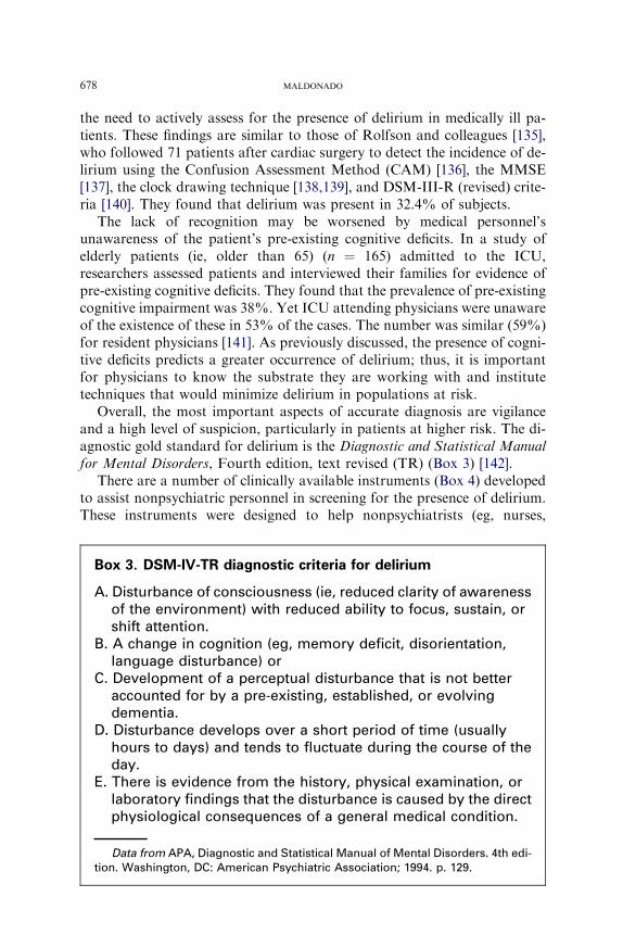
Overall, the most important aspects of accurate diagnosis are vigilanceand a high level of suspicion, particularly in patients at higher risk. The di-agnostic gold standard for delirium is the Diagnostic and Statistical Manualfor Mental Disorders, Fourth edition, text revised (TR) (Box 3) [142].
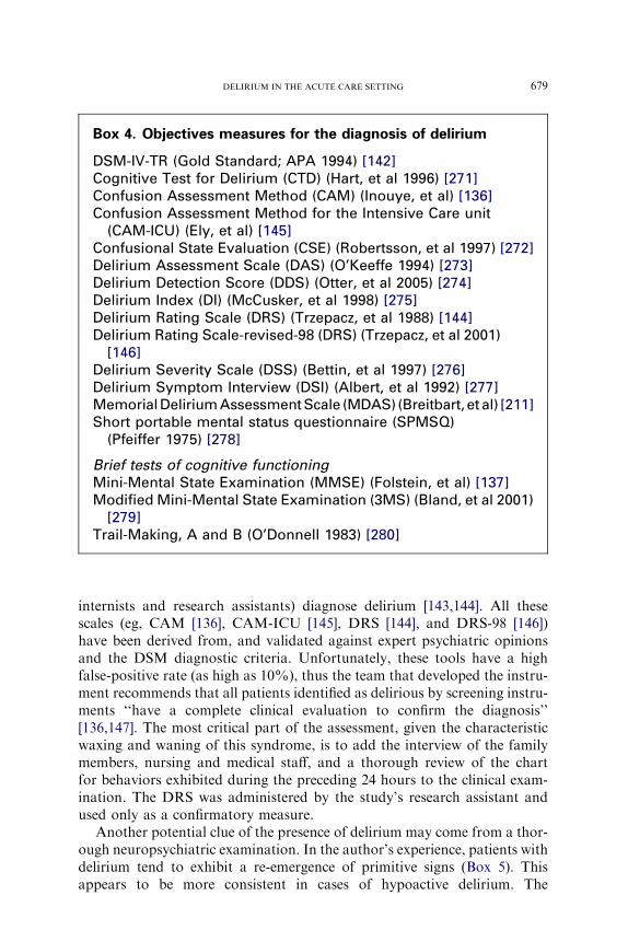
There are a number of clinically available instruments (Box 4) developedto assist nonpsychiatric personnel in screening for the presence of delirium.These instruments were designed to help nonpsychiatrists (eg, nurses,
Box 3. DSM-IV-TR diagnostic criteria for delirium
A. Disturbance of consciousness (ie, reduced clarity of awarenessof the environment) with reduced ability to focus, sustain, orshift attention.
B. A change in cognition (eg, memory deficit, disorientation,language disturbance) or
C. Development of a perceptual disturbance that is not betteraccounted for by a pre-existing, established, or evolvingdementia.
D. Disturbance develops over a short period of time (usuallyhours to days) and tends to fluctuate during the course of theday.
E. There is evidence from the history, physical examination, orlaboratory findings that the disturbance is caused by the directphysiological consequences of a general medical condition.
Data from APA, Diagnostic and Statistical Manual of Mental Disorders. 4th edi-tion. Washington, DC: American Psychiatric Association; 1994. p. 129.
Box 4. Objectives measures for the diagnosis of delirium
DSM-IV-TR (Gold Standard; APA 1994) [142]Cognitive Test for Delirium (CTD) (Hart, et al 1996) [271]Confusion Assessment Method (CAM) (Inouye, et al) [136]Confusion Assessment Method for the Intensive Care unit
(CAM-ICU) (Ely, et al) [145]Confusional State Evaluation (CSE) (Robertsson, et al 1997) [272]Delirium Assessment Scale (DAS) (O’Keeffe 1994) [273]Delirium Detection Score (DDS) (Otter, et al 2005) [274]Delirium Index (DI) (McCusker, et al 1998) [275]Delirium Rating Scale (DRS) (Trzepacz, et al 1988) [144]Delirium Rating Scale-revised-98 (DRS) (Trzepacz, et al 2001)
[146]Delirium Severity Scale (DSS) (Bettin, et al 1997) [276]Delirium Symptom Interview (DSI) (Albert, et al 1992) [277]Memorial Delirium Assessment Scale (MDAS) (Breitbart, et al) [211]Short portable mental status questionnaire (SPMSQ)
(Pfeiffer 1975) [278]
Brief tests of cognitive functioningMini-Mental State Examination (MMSE) (Folstein, et al) [137]Modified Mini-Mental State Examination (3MS) (Bland, et al 2001)
[279]Trail-Making, A and B (O’Donnell 1983) [280]
679DELIRIUM IN THE ACUTE CARE SETTING
internists and research assistants) diagnose delirium [143,144]. All thesescales (eg, CAM [136], CAM-ICU [145], DRS [144], and DRS-98 [146])have been derived from, and validated against expert psychiatric opinionsand the DSM diagnostic criteria. Unfortunately, these tools have a highfalse-positive rate (as high as 10%), thus the team that developed the instru-ment recommends that all patients identified as delirious by screening instru-ments ‘‘have a complete clinical evaluation to confirm the diagnosis’’[136,147]. The most critical part of the assessment, given the characteristicwaxing and waning of this syndrome, is to add the interview of the familymembers, nursing and medical staff, and a thorough review of the chartfor behaviors exhibited during the preceding 24 hours to the clinical exam-ination. The DRS was administered by the study’s research assistant andused only as a confirmatory measure.
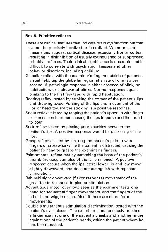
Another potential clue of the presence of delirium may come from a thor-ough neuropsychiatric examination. In the author’s experience, patients withdelirium tend to exhibit a re-emergence of primitive signs (Box 5). Thisappears to be more consistent in cases of hypoactive delirium. The
Box 5. Primitive reflexes
These are clinical features that indicate brain dysfunction but thatcannot be precisely localized or lateralized. When present,these signs suggest cortical disease, especially frontal cortex,resulting in disinhibition of usually extinguished or suppressedprimitive reflexes. Their clinical significance is uncertain and isdifficult to correlate with psychiatric illnesses and otherbehavior disorders, including delirium.
Glabellar reflex: with the examiner’s fingers outside of patient’svisual field, tap the glabellar region at a rate of one tap persecond. A pathologic response is either absence of blink, nohabituation, or a shower of blinks. Normal response equalsblinking to the first few taps with rapid habituation.
Rooting reflex: tested by stroking the corner of the patient’s lipsand drawing away. Pursing of the lips and movement of thelips or head toward the stroking is a positive response.
Snout reflex: elicited by tapping the patient’s upper lip with fingeror percussion hammer causing the lips to purse and the mouthto pout.
Suck reflex: tested by placing your knuckles between thepatient’s lips. A positive response would be puckering of thelips.
Grasp reflex: elicited by stroking the patient’s palm towardfingers or crosswise while the patient is distracted, causing thepatient’s hand to grasps the examiner’s fingers.
Palmomental reflex: test by scratching the base of the patient’sthumb (noxious stimulus of thenar eminence). A positiveresponse occurs when the ipsilateral lower lip and jaw moveslightly downward, and does not extinguish with repeatedstimulation.
Babinski sign: downward (flexor response) movement of thegreat toe in response to plantar stimulation.
Adventitious motor overflow: seen as the examiner tests onehand for sequential finger movements, and the fingers of theother hand wiggle or tap. Also, if there are choreiformmovements.
Double simultaneous stimulation discrimination: tested with thepatient’s eyes closed. The examiner simultaneously brushesa finger against one of the patient’s cheeks and another fingeragainst one of the patient’s hands, asking the patient where hehas been touched.
680 MALDONADO
681DELIRIUM IN THE ACUTE CARE SETTING
relationship between poor cognitive status and primitive reflexes has been de-scribed in patients suffering fromHIV-related cognitive disorders [148] and incases of dementia [149]. There is at least one study describing the presence ofprimitive reflexes in postcardiotomy patients suffering from postoperativeneuropsychiatric complications [150]. Further studies are needed to deter-mine whether an assessment for the presence of primitive reflexes may addto the diagnostic accuracy for delirium, or at least assist in the characteriza-tion of delirium type, or whether it has any prognostic value.
Some have advocated the use of the electroencephalogram (EEG) asa way to identify and diagnose delirium. Engel and colleagues [151] werethe first to describe the relationship between delirium and the diffuse slowingand progressive disorganization of rhythm seen in the EEG. The most com-mon EEG findings in delirium include slowing of peak and average frequen-cies, and decreased alpha activity but increased theta and delta waves.Studies suggest that EEG changes correlate with the degree of cognitive def-icit, but there does not appear to be a relationship between EEG patternsand delirium motoric type [152–160]. The clinical usefulness of EEG inthe diagnosis of delirium may be limited by its limited specificity (given thereare a number of conditions and medications that may affect the EEG) andthe practicality of conducting the test (particularly in the case of agitatedand combative patients). Still, the EEG may provide useful in differentiatingdelirium from other psychiatric and neurologic conditions, such as catatonicstates, seizure activity, somatoform disorders, and malingering.
The most critical part of the assessment, given the characteristic waxingand waning of this syndrome, is to obtain as much information and from asmany sources as possible (eg, interview of family members, nursing andother medical staff), coupled with a thorough review of the chart for behav-iors exhibited during the preceding 24 hours to the clinical examination.
Delirium subtypes
Liptzin and Levkoff [147] were the first to characterize the different typesof delirium based on behavioral characteristics (Table 3). Others have con-firmed the presence of these motoric subtypes. According to these studies,there are at least three types of delirium based on their clinical manifesta-tions: hyperactive, hypoactive, and mixed (Fig. 6) [161,162]. The most com-mon type is the mixed form (46%), followed by the hyperactive (30%) andthe hypoactive (24%). To most physicians, the most clear and recognizableform is the hyperactive type. Most clinicians agree that a confused, disori-ented patient who does not have a pre-existing psychiatric diagnosis, whosuddenly becomes agitated, combative, or assaultive, is probably sufferingfrom the hyperactive or ‘‘agitated type’’ of delirium. The term ‘‘mixedtype’’ is used to describe the classic ‘‘waxing and waning’’ pattern, com-monly seen in medically ill patients who appear agitated and combative attimes, with alternating episodes of somnolence and hypoactivity.
Table 3
Delirium subtypes
Hyperactive (three or more) Hypoactive (four or more)
Hypervigilance Unawareness
Restlessness Lethargy
Fast/loud speech Decreased alertness
Anger/irritability Staring
Combativeness Sparse/slow speech
Impatience Apathy
Uncooperative Decreased motor activity
Laughing
Swearing/singing
Euphoria
Wandering
Easy startling
Distractibility
Nightmares
Persistent thoughts
Data from Liptzin B, Levkoff SE. An empirical study of delirium subtypes. Br J Psychiatry
1992;161:843–5.
682 MALDONADO
The most difficult type of delirium to identify is the hypoactive type. Clas-sically, these patients present with symptoms that are commonly associatedwith depression [147]. These include unawareness of the environment,lethargy, apathy, decreased level of alertness, psychomotor retardation,decreased speech production, and episodes of unresponsiveness or staring.Patients with hypoactive delirium often endorse depressive symptoms,such as low mood (60%), worthlessness (68%), and frequent thoughts ofdeath (52%) [133]. Studies have demonstrated that a large percentage ofthese patients are inappropriately diagnosed and treated as depressed[133]. The author’s own experience at Stanford University Hospital parallelsthat of others [133,134]. Maldonado and colleagues [13] found that 42% of
24%
30%
46%
Hypoactive Hyperactive Mixed
Fig. 6. Motoric subtype of delirium. (Data fromMeagher DJ, O’Hanlon D, O’Mahony E, et al.
Relationship between symptoms and motoric subtype of delirium. J Neuropsychiatry Clin
Neurosci 2000;12(1):51–6.)
683DELIRIUM IN THE ACUTE CARE SETTING
the time when the psychiatry consultation service was called to treat a patientfor ‘‘depression,’’ the patient’s correct diagnosis was hypoactive delirium.The same study found that nearly 80% of these patients had been inappro-priately prescribed antidepressant medications.
Management of delirium
Clinicians have three potential approaches when it comes to the manage-ment of delirium: (1) managing delirium (ie, symptomatically managingbehavioral dyscontrol, such as agitation and psychosis); (2) treatment ofdelirium (ie, directly addressing either the underlying causes and the neuro-chemical cascade triggered by the underlying cause itself); or (3) preventionof delirium (ie, use of techniques or methods, either pharmacologic orbehavioral, with the purpose of avoiding the development of delirium).This section covers the first; the following section addresses the third. Thesecond section is covered in the article by Dr. Maldonado, elsewhere inthis issue.
The adequate treatment of delirium includes the following steps: (1) accu-rate diagnosis of the condition (eg, hypoactive delirium versus depression),(2) management of the behavioral and psychiatric manifestations and symp-toms to prevent the patient from self-harm or harming of others, (3) identi-fication of the etiologic causes of delirium, and (4) treatment of underlyingmedical problems. Adequate medical management begins with timely diag-nosis and early intervention, as shown in the following algorythm for theprevention and management of delirium.
Algorithm for the prevention and management of delirium
I. Be vigilant for the possibility of delirium.A. Obtain baseline level of cognitive functioning information from ac-
cessory sources.B. Screen for the development of delirium in high risk groups, either by
the use of psychiatric consultants or objective scales (eg, DRS-98;CAM).
C. Use psychiatric consultants to help with assessment and design of thetreatment plan, if available.
II. Identify and treat underlying medical causes.III. Non-Pharmacological Treatment Strategies:
A. Correct malnutrition, dehydration and electrolyte abnormalitiesshould be corrected as quickly and safely as possible.
B. Remove immobilizing lines and devices (ie, IV lines, chest tubes,bladder catheters and physical restraints) as early as possible.
C. Correct sensory deficits (ie, eyeglasses, hearing aids).D. Promote as normal a circadian light rhythm as possible. Better if this
can be achieved by environmental manipulations, such as light
684 MALDONADO
control (ie, lights on & curtains drawn during the day; off at night)and noise control (ie, provide ear plugs, turn off TVs, minimize nightstaff chatter), rather than by the use of medications.
E. Provide adequate intellectual and environmental stimulation as earlyas possible.
F. Minimize environmental isolation.IV. Pharmacological Treatment Strategies:
A. Conduct an inventory of all pharmacological agents been adminis-tered to the patient. Any medication or agent known to cause delir-ium (see Table C) or to have high anticholinergic potential (see TableG) should be discontinued, if possible, or a suitable alternativeinstituted.
B. Avoid using GABAergic agents to control agitation, if possible. Ex-ceptions: cases of CNS-depressant withdrawal (ie, alcohol, benzodi-azepines, barbiturates) or when more appropriate agents havefailed and sedations is needed to prevent patient’s harm.
C. Adequately assess and treat pain.D. Avoid the use of opioids for behavioral control of agitation.E. For the pharmacological management of delirium (all types) consider
using:i. Acetylcholinesterase inhibitor (eg, rivastigmine, donepezil, physo-stigmine, rivastigmine) for correction of central anticholinergicsyndrome.
ii. Serotonin antagonist (eg, ondansetron) to control toxic elevationsof 5-HT usually associated with hypoactive delirium, althoughsome studies have suggested its use may be indicated in all typesof delirium.
iii. Rotate opioids from morphine and meperidine to fentanyl orhydromorphone.
iv. Melatonin or melatonin agonists (eg, ramelteon) to promotea more natural sleep.
v. Dopamine agonists to manage the theorized abnormally elevatedlevels of dopamine, and provide restoration of putative hippo-campal functions (eg, short-term memory) and reversal of otherregional brain disturbances (eg, agitation, psychosis, primitive re-flexes), as well as to protect neurons against hypoxic stress andinjury. The dose of dopamine antagonist use may depend onthe type of delirium been treated.
vi. Alpha-2 agonists (eg, dexmedetomidine, clonidine), for protec-tion against the acute NE released secondary to hypoxia or ische-mia, leads to further neuronal injury and the development ofworsening of delirium.
vii. NMDA-receptor blocking agents, to minimize glutamine inducedneuronal injury (eg, amantadine, memantine).
685DELIRIUM IN THE ACUTE CARE SETTING
F. In case of hyperactive delirium:i. Use low to moderate dose haloperidol (eg, ! 20mg/24hr), if thepatient’s cardiac condition allows it and there are no significantelectrolyte abnormalities.
a. Before using haloperidol: obtain 12-lead ECG; measureQTc & electrolytes. Correct Kþ & Mgþ, if needed.
b. If possible avoid other medications known to increase QTcand/or inhibitors of CPY3A4.
c. Discontinue its use if QTc increases to O25% of baseline orO500msec.
ii. When the use of haloperidol is contraindicated or not desirable,atypical antipsychotics should be considered:
a. Better evidence for: risperidone, quetiapine.b. Limited data for: olanzepine, aripripazole, perospirone.c. Avoid: clozapine, ziprasidone.
G. In case of hypoactive delirium:i. Evidence suggests that DA antagonists may still have a placegiven the excess DA theory.
a. If haloperidol is use, recommended doses are in the very lowrange (ie, 0.25 to 1mg / 24hr).
b. If an atypical is preferred, consider an agent with low seda-tion (ie, risperidone); unless a sedative agent is needed to re-store sleep-wake cycle not responding to E-iv (see above).
ii. In cases of extreme psychomotor retardation or catatonic fea-tures, in the absence of agitation or psychosis, consider the useof psychostimulant agents (eg, methylphenidate, dextroamphet-amine, modafinil) or conventional dopamine agonists (eg, bromo-criptine, amantadine, memantine).
Nonpharmacologic treatment strategies
Given the findings reported by Inouye and colleagues [74,163], a multi-component approach is recommended; targeting identified, treatable con-tributing factors must be undertaken early. As mentioned above, giventhe high rate of under and missed diagnosed cases, vigilance and a high levelof suspicion is essential, particularly in high-risk patients. The routine use ofassessment scales or diagnostic interviews by properly trained personnel iskey both in prevention and timely treatment. Early involvement of thePsychosomatic Medicine team (Psychiatry) or a Geropsychiatric servicehas been shown to be extremely valuable, both in prevention and earlyintervention. An active search for possible etiologies of delirium must firstattempt to rule out the common causes of the syndrome (see list titled‘‘Delirium clinical risk factors’’ above). This must include a review of allmedications and identification and possible discontinuation of agents with
686 MALDONADO
high deliriogenic potential (see Box 1). Appropriate diagnostic tests andassays should be ordered and reviewed in a timely fashion, and all abnormalfindings addressed accordingly.
Immobilizing lines and devices (eg, chest tubes, IV lines, bladder catethers)should be removed as early as possible. Similarly, physical restraints should beavoided and eliminated as soon as it is safe to do so. Early correction of sen-sory deficits should be undertaken. That is, eyeglasses and hearing aids shouldbe replaced or fitted (if not using them before the hospitalization) as soon aspossible. This will allow patients to familiarize themselves with the environ-ment and reorient themselves early on. It will also minimize the occurrenceof misperceptions or misinterpretation of environmental cues and stimuli.Environmental isolation should be minimized if possible. Family membersand loved ones should be encouraged to visit and provide a familiar andfriendly environment, as well as provide appropriate orientation and stimula-tion to patients, especially those with baseline cognitive deficits.
Dehydration and electrolyte abnormalities should be corrected as quicklyand safely as possible. Malnutrition should be corrected, unless there aregood reasons not to (eg, terminal dementia).
Early correction of sleep disturbance, preferably by nonpharmacologicmeans, should occur, although the use of nonbenzodiazepine agents, such asmelatonin or melatonin agonists (ie, ramelteon) or sedating antidepressantagents (eg, trazodone or mirtazapine) should be considered. On the otherhand, cliniciansmust consider factors, such as drug–drug interaction andmed-ication half-lives when prescribing. For example, mirtazapine and trazodonemay indeed promote night sleep, but their effects may last well into the nextday, interfering with cognition, attention, and concentration. Sedative agentswith high anticholinergic load, such as antihistaminic agents (eg, diphenhydra-mine, hydroxizine) or tricyclic antidepressants (eg, amitriptyline) should beavoided, as theywill aggravatedeliriumeven if immediately effective inpromot-ing sleep. Similarly, benzodiazepines should also be avoided if at all possible.
Finally, conduct an inventory of all pharmacologic agents being admin-istered to the patient. Any medication or agent known to cause delirium(see Box 1) or to have high anticholinergic potential (see Box 2) should bediscontinued, if possible, or a suitable alternative instituted.
Pharmacologic treatment strategies
It cannot be overstated that the definitive treatment of delirium is theaccurate identification and treatment of its underlying causes. Nevertheless,pharmacologic intervention with various psychoactive agents is oftenneeded to help manage agitated patients. Following the Hippocratic princi-ple of ‘‘first, do no harm,’’ clinicians should first avoid the use of GABAer-gic agents, if at all possible. As described above, all such agents(ie, benzodiazepines, propofol) may cause or aggravate delirium and itsbehavioral manifestations [20,41,94]. The use of benzodiazepines in the
687DELIRIUM IN THE ACUTE CARE SETTING
management of delirium should be limited to: (a) patients experiencingdelirium related to the withdrawal from a CNS-depressant agent (ie, alco-hol, barbiturates, benzodiazepines); or (b) when other more appropriateagents (see below) have failed and the level of agitation and need for behav-ioral control outweighs the potential detrimental effects of benzodiazepines.Similarly, clinicians should do everything possible to avoid the use of opioidagents to tranquilize agitated patients, as opioids have been implicated inthe development of delirium in many patient populations [25,40,101–106].On the other hand, opioids should be administered when there is evidencethat pain may be a contributor to the patient’s agitation.
The literature has long recognized that intravenous neuroleptic agents arethe recommended emergency treatment for agitated and mixed type delirium[164–169]. The intravenous administration of haloperidol has always beenthought of as superior to oral administration because the IV route hasmore reliable absorption, even in cases of systemic organ failure. Intrave-nous haloperidol use has the added advantage of requiring no patient’scooperation, thus facilitating its use even in uncooperative and agitatedpatients. Studies suggest that the IV use of high-potency neuroleptic agentsis associated with minimal effects on blood pressure, respiration, and heartrate [167,170–175].
Further research suggests a decreased incidence of extrapyramidal symp-toms (EPS) when the intravenous route versus the oral route is used [176].This study consisted of a retrospective chart review of all patients admittedto a large university hospital receiving haloperidol in any form over a 90-dayperiod. A total of 238 subjects receiving haloperidol were identified duringthe index period, using data obtained through the digital pharmacy distribu-tion system (Pyxis). Only patients with a known pre-existing movementdisorder (eg, Parkinson disease) were excluded. In this sample, 51% of thesubjects were women and the mean age was 62 years for women and 55 yearsfor men. The most common reasons for which haloperidol was prescribedincluded delirium (69%), psychosis (11%), nausea or vomiting (9%), affec-tive disorder (6%), and dementia (5%). Haloperidol doses ranged from0.5 mg to 90 mg per day for subjects receiving intravenous administration,and from 0.5 mg to 20 mg per day for those receiving oral administration.Results show that patients receiving IV-haloperidol experienced much lowerEPS than patients receiving the oral form (7.2% versus 22.6%; P ! .01).In this sample, the most common forms of EPS observed included medica-tion-induced Parkinsonism (50%), akathisia (32%), and acute dystonicreactions (14%). The investigators found no cases of significant respiratorydepression or Torsade de Pointes (TdP) deemed to have been caused by hal-operidol use. These findings are similar to those previously reported, alsosuggesting a lower incidence of EPS when haloperidol is administered intra-venously [168].
Maldonado and Dhami [177] conducted a prospective study, involving allpatients (n ¼ 225) admitted to the critical care unit during a 6-month period.
688 MALDONADO
Subjects were monitored throughout their hospital stay to assess the effec-tiveness of a protocol-based management of delirium among critical carepatients. Subjects were followed daily by the study research assistant, usingobjective methods to assess delirium (ie, the MMSE [137] and the DRS[144]). There were slightly more surgical cases (n ¼ 129), than medical cases(n ¼ 96). A total of 18% of the subjects were identified as being delirious byDRS-criteria during the index period. Consultations to the PsychosomaticMedicine Service (PMS) were called in only 42% of the cases. On average,the surgical team consulted psychiatry 2.8 days after the onset of manifesta-tion of delirium, whereas medicine services called after 4.2 days. Pharmaco-logic management varied significantly between the two groups (ie, standardof care versus study protocol). Medical and surgical services managed theirdelirious patients with varying combinations of medications, including opi-oids, benzodiazepines (ie, primarily midazolam or lorazepam), propofol,and various neuroleptic agents, usually on an as-needed basis. On the otherhand, the treatment used by the PMS consisted of the routine use of IV hal-operidol given throughout the day, on a regular schedule every 0400-, 1000-,1600-, and 2200-hours. Lorazepam was used in cases of agitated deliriumnot responding to haloperidol alone, in cases of primary CNS-depressantagent withdrawal (ie, alcohol, benzodiazepines), or at night only to helppromote sleep. The treatment regime doses were adjusted every 24 hoursand titrated to effect. The dosing difference maintained a haloperidol-to-lorazepam ratio of at least two-to-one (the H2A protocol) to avoid thepossibility of disinhibition by the benzodiazepines. That is, when used, thelorazepam dose was always less than half the haloperidol dose in milligrams.Nevertheless, because of the possibility that benzodiazepines themselvesmay contribute to delirium, the lowest effective dose was always used.Whenever possible, no benzodiazepines were used.
The results demonstrated that the PMS-management approach (ie, sched-uled IV haloperidol use) was superior to the ‘‘standard approach’’ (ie, as-needed use of sedatives and antipsychotics) at treating delirium [177]. Thelength of stay (15 versus 11 days) (Fig. 7A), total duration of delirium(13 versus 6 days) (Fig. 7B), and percentage time being delirious (86% ver-sus 58%) (Fig. 7C) were all shorter on patients treated by the PMS protocol.In addition, a significant improvement in cognitive functioning wasobserved in patients treated with the PMS-protocol. Finally, complete reso-lution of delirium (as measured by a MMSE greater than 26 and a DRS lessthan 10) at the time of discharge home was greater for patients treated withthe PMS-protocol than (90% in the psychiatry group versus 14% in themedical/surgical group) (Fig. 7D). As many previous studies have indicated,these results suggest that a rational and controlled approach to the earlyidentification and treatment of delirium in critical care patients results ina more accurate and prompt diagnosis, shorter hospital stays, a reductionin the use of restraints, faster recovery, and a substantially greater resolutionof symptoms of delirium at the time of hospital discharge. Even though the
15
11
02468
10121416
Med/Surg Psych
A- Total Length of Stay B- Average Total Delirious Days
D- Delirium Resolution
13
6
02468
101214
Med/Surg Psych
Med/Surg Psych
87
54
14
90
0102030405060708090
0102030405060708090
Med/Surg Psych
C- Percentage of Delirious Days
Fig. 7. Effects of the early identification and treatment of delirium according to protocol. (A)
total length of stay; (B) average total delirious days; (C) percentage of delirious days. Final bar
graph represents complete resolution; (D) percentage of complete resolution of delirium on
discharge home by treatment groups. Red ¼ treatment as usual by medical/surgical teams;
Blue ¼ psychological protocol treatment. (From Maldonado JR, Dhami N. ‘‘Recognition
and Management of Delirium in the Medical and Surgical Intensive Care Wards.’’ Poster pre-
sentation. 17th World Congress on Psychosomatic Medicine, Waikoloa, Hawaii. August 27,
2003; with permission; Data from Maldonado JR, Dhami N. Recognition and management
of delirium in the medical and surgical intensive care wards. Journal of Psychosomatic
Research 2003;55(2):150.)
689DELIRIUM IN THE ACUTE CARE SETTING
number of patients treated by ‘‘standard or conventional’’ approach achiev-ing a complete resolution of their symptoms of delirium appears dismal,these findings may represent more of the rule than the exception.
Levkoff and colleagues [120] followed all older patients (n ¼ 325) admit-ted to the medical and surgical services of a teaching hospital. During theindex period, 10.5% of all patients fulfilled DSM-III [178] criteria for delir-ium on admission and an additional 31.1% developed delirium during theindex hospitalization. Similar to previous studies, development of deliriumwas associated with prolonged hospital stay and an increased risk of institu-tional placement among community-dwelling older persons. In their sample,only 4% of delirious patients in their study experienced full resolution ofdelirium symptoms before discharge from the hospital. On longitudinal fol-low-up, an additional 20.8% had resolution of all symptoms by 3 months,and an additional 17.7% had resolution of all symptoms by 6 months afterdischarge from the hospital.
Despite the widespread use of IV haloperidol and multiple reports inthe literature describing its safety [166,167,169–172,174,175,179], evenwhen used at fairly high doses, some reports suggesting a range of
690 MALDONADO
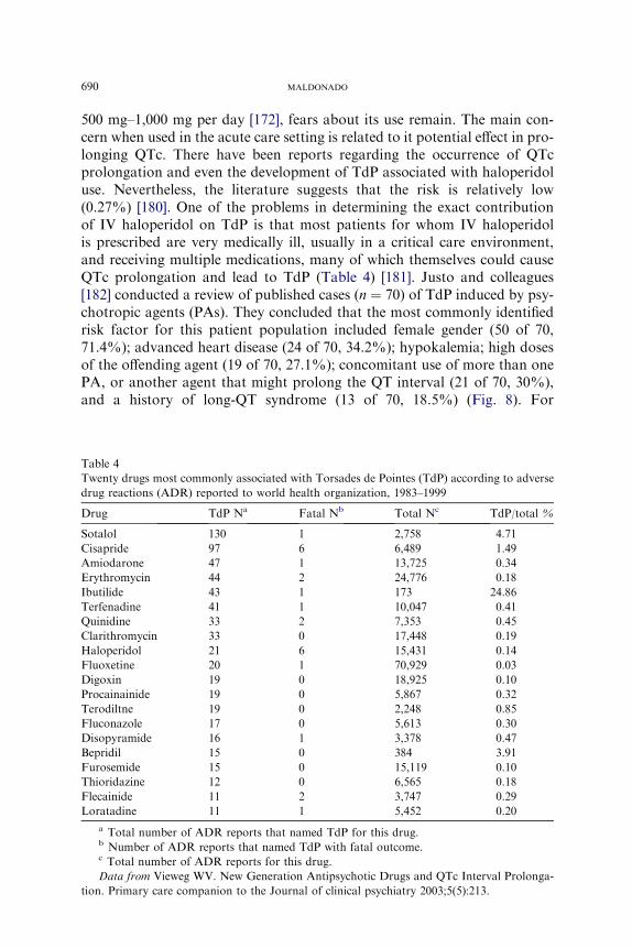
500 mg–1,000 mg per day [172], fears about its use remain. The main con-cern when used in the acute care setting is related to it potential effect in pro-longing QTc. There have been reports regarding the occurrence of QTcprolongation and even the development of TdP associated with haloperidoluse. Nevertheless, the literature suggests that the risk is relatively low(0.27%) [180]. One of the problems in determining the exact contributionof IV haloperidol on TdP is that most patients for whom IV haloperidolis prescribed are very medically ill, usually in a critical care environment,and receiving multiple medications, many of which themselves could causeQTc prolongation and lead to TdP (Table 4) [181]. Justo and colleagues[182] conducted a review of published cases (n ¼ 70) of TdP induced by psy-chotropic agents (PAs). They concluded that the most commonly identifiedrisk factor for this patient population included female gender (50 of 70,71.4%); advanced heart disease (24 of 70, 34.2%); hypokalemia; high dosesof the offending agent (19 of 70, 27.1%); concomitant use of more than onePA, or another agent that might prolong the QT interval (21 of 70, 30%),and a history of long-QT syndrome (13 of 70, 18.5%) (Fig. 8). For
Table 4
Twenty drugs most commonly associated with Torsades de Pointes (TdP) according to adverse
drug reactions (ADR) reported to world health organization, 1983–1999
Drug TdP Na Fatal Nb Total Nc TdP/total %
Sotalol 130 1 2,758 4.71
Cisapride 97 6 6,489 1.49
Amiodarone 47 1 13,725 0.34
Erythromycin 44 2 24,776 0.18
Ibutilide 43 1 173 24.86
Terfenadine 41 1 10,047 0.41
Quinidine 33 2 7,353 0.45
Clarithromycin 33 0 17,448 0.19
Haloperidol 21 6 15,431 0.14
Fluoxetine 20 1 70,929 0.03
Digoxin 19 0 18,925 0.10
Procainainide 19 0 5,867 0.32
Terodiltne 19 0 2,248 0.85
Fluconazole 17 0 5,613 0.30
Disopyramide 16 1 3,378 0.47
Bepridil 15 0 384 3.91
Furosemide 15 0 15,119 0.10
Thioridazine 12 0 6,565 0.18
Flecainide 11 2 3,747 0.29
Loratadine 11 1 5,452 0.20
a Total number of ADR reports that named TdP for this drug.b Number of ADR reports that named TdP with fatal outcome.c Total number of ADR reports for this drug.
Data from Vieweg WV. New Generation Antipsychotic Drugs and QTc Interval Prolonga-
tion. Primary care companion to the Journal of clinical psychiatry 2003;5(5):213.
Fig. 8. Prevalence of risk factors for Torsade de Pointes among patients with TdP induced
by psychotropic drugs. (From Justo D, Prohorov V, Heller K, et al. Torsade de Pointes in-
duced by psychotropic drugs and the prevalence of its risk factors. Acta Psychiatr Scand
2005;111(3):171–6; with permission.)
691DELIRIUM IN THE ACUTE CARE SETTING
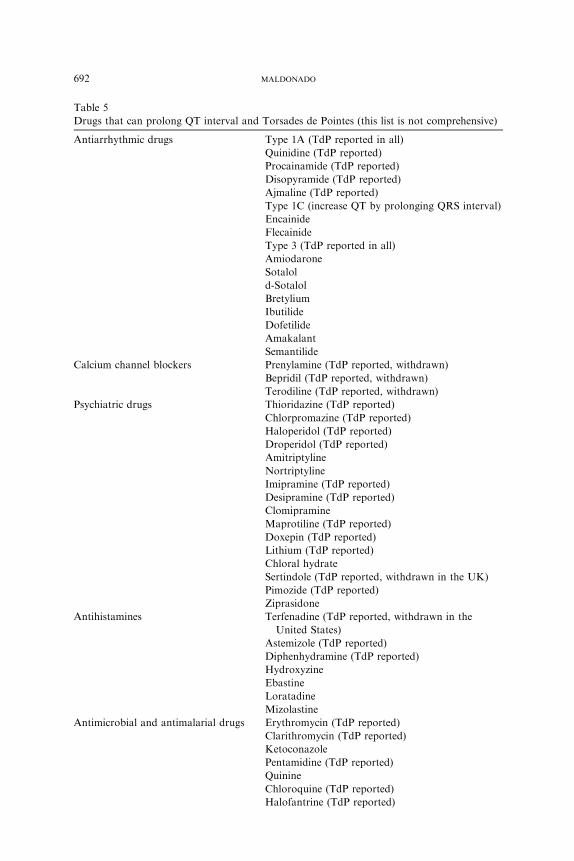
a comprehensive review of medications that cause QTc prolongation andTdP, see Yap and Camm (Table 5) [183].
A MEDLINE and manual search of the literature published between1966 and 1996 was conducted looking for cases of conduction disturbancesassociated with the use of butyrophenone antipsychotics [184]. They foundonly 18 patients described and concluded that, ‘‘it seems reasonable to sug-gest that the incidence of adverse cardiovascular effects due to droperidoland haloperidol is small.’’ The investigators made several recommendationsregarding the use of haloperidol in the critically ill patient. Before initiatingtherapy with haloperidol, a baseline QTc interval and serum magnesium andpotassium concentrations should be measured. Electrolytes should be cor-rected, if necessary, before initiation of treatment. If the baseline QTc inter-val is greater than or equal to 440 msec, and patients are receiving otherdrugs that may prolong the QTc interval or in the presence of significantelectrolyte disturbances, a butyrophenone antipsychotic should be usedwith caution. Once treatment has been initiated, all critically ill patientsreceiving haloperidol should undergo regular electrocardiograph monitor-ing and QTc interval measurement. Special attention should be given tothose receiving doses greater than 50 mg every 24 hours. Based on the cur-rently available literature, any critically ill patient receiving droperidol or
Table 5
Drugs that can prolong QT interval and Torsades de Pointes (this list is not comprehensive)
Antiarrhythmic drugs Type 1A (TdP reported in all)
Antihistamines Terfenadine (TdP reported, withdrawn in the
United States)
Astemizole (TdP reported)
Diphenhydramine (TdP reported)
Hydroxyzine
Ebastine
Loratadine
Mizolastine
Antimicrobial and antimalarial drugs Erythromycin (TdP reported)
Clarithromycin (TdP reported)
Ketoconazole
Pentamidine (TdP reported)
Quinine
Chloroquine (TdP reported)
Halofantrine (TdP reported)
692 MALDONADO
Sparfloxacin
Grepafloxacin (TdP reported, withdrawn in the UK
and United States)
Pentavalent antimonial meglumine
Ketanserin (TdP reported)
Serotonin agents Amantadine (TdP reported)
Promotility agents Cisapride (TdP reported, withdrawn in the UK
and United States)
Immunosuppressant Tacrolimus (TdP reported)
Antidiuretic hormone Vasopressin (TdP reported)
Other agents Adenosine
Organophosphates
Probucol (TdP reported)
Papaverine (TdP reported)
Cocaine
Data from Yap YG, Camm AJ. Drug induced QT prolongation and Torsades de Pointes.
Heart 2003;89(11):1367.
693DELIRIUM IN THE ACUTE CARE SETTING
haloperidol therapy, whose QTc interval lengthens by greater than or equalto 25% over baseline, should undergo dose reduction or should be switchedto a different agent.
Despite these concerns, in 1995 a task force of more than 40 experts indisciplines related to the use of analgesic and sedative agents in the ICUwas convened from the membership of the American College of CriticalCare Medicine and the Society of Critical Care Medicine (SCCM) [185].This consensus of experts provided six recommendations with supportingdata for intravenous analgesia and sedation in the ICU setting:
Morphine sulfate is the preferred analgesic agent for critically ill patients.Fentanyl is the preferred analgesic agent for critically ill patients with
hemodynamic instability, for patients manifesting symptoms of hista-mine release with morphine, or morphine allergy.
Hydromorphone can serve as an acceptable alternative to morphine.Midazolam or propofol are the preferred agents only for the short-term
(! 24 hours) treatment of anxiety in the critically ill adult.Lorazepam is the preferred agent for the prolonged treatment of anxiety
in the critically ill adult.Haloperidol is the preferred agent for the treatment of delirium in the
critically ill adult.
Similarly, the use of IV haloperidol as the agent of choice for critically illpatients was reinforced by the SCCM’s most recent guidelines, published in2002 [186]. Since then, a ‘‘best evidence topic in cardiac surgery’’ was writtenaccording to a structured protocol, addressing the issue of haloperidol safetyfor critically ill patients. Their search included 294 articles and concludedthat haloperidol should be considered the first-line drug for agitated patientsafter cardiac surgery [187].
694 MALDONADO
Of note, in September 2007, the Food and Drug Administration (FDA)issued a ‘‘black-box’’ warning for the ‘‘off-label’’ clinical practice of using IVhaloperidol [188]. It is important to remember that haloperidol has neverbeen approved by the FDA for IV use.
Alternatives to haloperidol
Because of the stigma and potential side effects associated with typicalantipsychotics, atypical agents (also known as second-generation antipsy-chotics, or SGA) have been used at increasing rates over the last few yearsfor management of psychiatric symptoms (eg, agitation, psychosis, delirium)in medically ill patients. Large studies, particularly head-to-head compari-son between SGA and more conventional agents (ie, haloperidol) are lack-ing. At least one study suggested that SGA may have a greater incidence ofadverse effects than typical agents, excluding EPS [189]. Leucht and col-leagues [190] conducted a meta-analysis of all randomized, controlled trialsin which new generation antipsychotics (ie, SGA) had been compared withconventional drugs. The study included studies that met quality criteria A orB in the Cochrane Collaboration Handbook, and assessed quality with theJadad scale. The investigators included in their analysis 31 studies witha total of 2,320 participants. The findings concluded that of the new gener-ation drugs, only clozapine was associated with significantly fewer EPS (riskdifference or RD ¼ �0.15, 95% CI, �0.26 to �0.4, P ¼ .008) and higherefficacy than low-potency conventional drugs. The reduced frequency ofEPS seen with olanzapine was of borderline significance (RD ¼ �0.15,CI, �0.31 to �0.01, P ¼ .07). Similarly, they identified only one inconclusivetrial of quetiapine and risperidone, and no investigations of ziprasidone andsertindole. They concluded that as a group, new generation drugs were mod-erately more efficacious than low-potency antipsychotics, largely irrespectiveof the comparator doses used; and that optimum doses of low-potency con-ventional antipsychotics might not induce more EPS than new generationdrugs.
Other problems to consider when choosing an alternative agent include thefact that SGA may be associated with weight gain, dyslipidemia, high bloodpressure, and ultimately with cardiovascular disease, diabetes, and metabolicsyndrome [191]. As when considering the use of typical agents (ie, haloperi-dol), clinicians must consider these factors and weigh potential risks and ben-efits before prescribing these agents to a critically ill patient. Finally, there isalso evidence that some atypical agents may aggravate or cause delirium(eg, clozapine, olanzepine), probably because of their anticholinergic poten-tial [189]. Data on most atypical agents are limited to small case reports.
Horikawa and colleagues [192] conducted a prospective open trial on ris-peridone for the treatment of delirium among medically ill patients (n ¼ 10).They reported an overall effectiveness of 80%, using doses of risperidonebetween 0.5 mg to 2 mg per day. Side effects included sedation in 30%
695DELIRIUM IN THE ACUTE CARE SETTING
and EPS in 10% of subjects. Mittal and colleagues [193] reported similarresults (n ¼ 10) with risperidone, using a mean daily dose of 0.75 mg. Thelargest, open-label risperidone study was reported by Parellada and col-leagues [194], who followed subjects hospitalized for a medical condition(n ¼ 64). Once the diagnosis of delirium was established (based on theDRS) treatment with risperidone was initiated. The investigators reportedimprovements on all studied measures (ie, Clinical Global Impressions orCGI scale, DRS, and MMSE) after 7 days of treatment. There was a verylow incidence of overall adverse effects (3.1%) and no EPS reported. Onthe other hand, there are at least four publications reporting risperidone-induced delirium [195–198].
There are six publications reporting on the use of quetiapine for the treat-ment of delirium. Torres and colleagues [199] reported improvements onMMSEandDRS-R-98 in two subjects. Similarly, Al-Samarrai and colleagues[200] reported on another two delirious subjects responding to quetiapine.Sasaki and colleagues [201] reported on a prospective, open-label study(n ¼ 12) of delirious subjects treated with a mean daily dose of quetiapine of45mg plus or minus 31mg per day. They found amean duration of symptomsof 4.8 plus or minus 3.5 days and improvements on MMSE, and reported nosignificant side effects. Similarly, Kim and colleagues [202] reported onanother 12 subjects treated with a mean daily dose of quetiapine of 94 mgplus or minus 23 mg per day. The mean duration of symptoms was 5.9 plusor minus 2.2 days, as well as improvements on the clock drawing test andMMSE. Pae and colleagues [203] treated 22 subjects with a mean daily doseof quetiapine of 127.1 mg plus or minus 72.2 mg per day. In this group, themeandurationof symptomswas 8.5 plus orminus 4.5 days, aswell as improve-ments onDRS-R-98 andCGI.Again, no significant side effects were reported.Maneeton and colleagues [204] studied the effectiveness of quetiapine in themanagement of delirium (n ¼ 22) in an open-label study. The means (SDs)dose and duration (SD) of quetiapine treatment were 45.7 (28.7) mg per dayand 6.5 (2.0) days, respectively. Results show that the DRS and CGI-S scoresof days two to sevenwere significantly lower than those of day 0 (P! .001) forall comparisons. The incidence of side effects was minimal. Finally, Balit andcolleagues [205] and Sim and colleagues [206] reported on a case each in whichquetiapine was the suspected cause of delirium.
There are several publications reporting the use of olanzapine for thetreatment of delirium. Kim and colleagues [207] reported on an open trial(n ¼ 20) on the use of olanzapine for the treatment of delirium caused bymultiple medico-surgical conditions. The average olanzapine dose was5.9 mg plus or minus 1.5 mg per day and the average duration of treatmentwas 6.6 plus or minus 1.7 days. Their data showed improvement in the DRSat relatively low doses (5.9 mg � 1.5 mg per day) and no evidence of signif-icant side effects. Passik and Cooper [208] and Halil and colleagues [209]reported a single case report each in which olanzepine was successfullyused in the treatment of delirium associated to a medical problem. Breitbart
696 MALDONADO
and colleagues [210] conducted an open, prospective trial of olanzapine forthe treatment of delirium in hospitalized cancer patients (n ¼ 79). In thissample, olanzepine was effective in treating 76% of delirium patients as evi-denced by the Memorial Delirium Assessment Scale (MDAS) [211], butrecorded problems with excessive sedation in 30% of patients. They alsodescribed several factors significantly associated with poorer response toolanzapine treatment for delirium, including age greater than 70 years, his-tory of dementia, central nervous system spread of cancer and hypoxia asdelirium etiologies, hypoactive delirium, and delirium of ‘‘severe’’ intensity(as measured by an MDAS greater than 23). Robinson and colleagues[212], Steil [213], Morita and colleagues [214], Samuels and Fang [215],Prommer [216], Arora and Praharaj [217], and Lim and colleagues [218]all reported on cases associated with olanzapine-induced delirium at thera-peutic doses. Delirium has been reported as a side effect in 54% of the caseswhen large doses of olanzepine have been ingested in an overdose. Patientswith olanzapine-induced delirium had an increased length of hospital stayand ICU admission rate (50%), and 70% of them required physical orchemical restraint [219].
Aripiprazole has been described as an effective treatment of delirium in twocase reports [220]. Straker and colleagues [221] reported on an open-labelseries of subjects (n ¼ 14) treated with aripiprazole for management of delir-ium used in a flexible dosing range, from 5 mg per day to 15 mg per day,titrated over a 7-day period, with dose increases on day 3 and day 7, as clin-ically indicated. DRS-R-98 scores declined from 25.1 (�5.2) on initial evalu-ation to 9.4 (4.9) at treatment end-point. Fifty percent of the subjects (7 out of14) had improved significantly (ie, R 50% reduction in DRS-R-98 scores) byday 5, while 12 of the 14 subjects had a reduction in their DRS-R-98 scoresgreater than or equal to 50% by treatment end-point.
To date, there are two single case reports on the use of ziprasidone for themanagement of delirium [222,223]. There is at least one open-label study(n¼ 38) on the use of perospirone, a recently developed atypical antipsychoticwith potent serotonin 5-HT2 and dopamine D2 antagonist activity. Perospir-one was effective in 86.8% of patients (based onDRS-98 assessments) and theeffect appearedwithin several days (5.1� 4.9 days). The initial dosewas 6.5mgplus or minus 3.7 mg per day and maximum dose of perospirone was 10.0 mgplus or minus 5.3 mg per day. Reported side effects included fatigue (15.2%),sleepiness (6.1%), akathisia (3.0%), and hypotension (3.0%) [224].
There is little published data regarding controlled studies of atypicalantipsychotics for the treatment of delirium. Sipahimalani and Masand[225] conducted a single-blind study using olanzepine versus haloperidol.Eleven subjects with delirium were treated, using a mean daily dose ofolanzepine of 8.2 mg plus or minus 3.4 mg versus haloperidol 5.1 mg plusor minus 3.5 mg per day. Peak response (ie, the number of days the patientreceived the neuroleptic before achieving maximum improvement) wassimilar in both groups (mean � SD: 6.8 � 3.5 days for olanzapine and
697DELIRIUM IN THE ACUTE CARE SETTING
7.2 � 4.9 days for haloperidol, P ¼ .8279). Mean plus or minus SD pretreat-ment DRS scores were comparable in the olanzepine (17.9 � 4.4) and thehaloperidol (20.1 � 5.2) groups (P ¼ .2968). Mean plus or minus SD af-ter-treatment DRS scores were 10.3 plus or minus 4.8 for the olanzapinegroup and 11.1 plus or minus 7.1 for the haloperidol group (P ¼ .7601).The mean improvement was 7.6 for the olanzapine group and 10 for the hal-operidol group. Five of the olanzapine subjects and six of the haloperidolsubjects showed a greater than 50% reduction in their DRS scores.
Schwartz and Masand [226] performed a single-blind study of quetiapineversus haloperidol in delirious subjects (n¼ 11). The quetiapine average dailydosewas 200mg per day. The investigators reported an effectiveness of greaterthan or equal to 50% in reducing DRS scores. When compared with haloper-idol, there was no difference in onset of symptom resolution, duration oftreatment, and overall clinical improvement. Skrobik and colleagues [227]conducted an open-label, prospective randomized trial, comparing the use ofenteral olanzepine (dosed at 5 mg per day) or haloperidol (dosed at 2.5 mg–5mgevery 8hours) in the treatment of delirium in a critical care setting.DeliriumIndex decreased over time in both groups, as did the administered dose of ben-zodiazepines. Clinical improvement was similar in both treatment arms. Thedose of rescue haloperidol, opiates, sedatives other than benzodiazepines,Ramsay scores, vital signs, and liver function tests were no different betweengroups. Thus, no significant clinically effective difference was appreciatedbetween groups. Liu and colleagues [228] conducted a single-blind risperidoneversus haloperidol study. They treated 41 subjects with a mean daily dose ofrisperidone of 1.2 mg plus or minus 0.75 mg per day. The investigators foundno significant difference in the efficacy or frequency of response rate betweenhaloperidol and risperidone on any of the measures (ie, DRS, MDAS).
The only published double-blind, randomized study looked at 28 subjectswith delirium who were randomly assigned to receive a flexible-dose regimenof haloperidol or risperidone over a 7-day treatment period [229]. The sever-ity of delirium was assessed by using the MDAS and the DRS. The studyinvestigators found no significant difference in the efficacy, frequency, orrate of response between haloperidol and risperidone on any of the mea-sures. Similarly, there were no clinically significant side effect differencesamong study groups.
A Cochrane Database review study looking at the use antipsychotics forthe treatment of delirium was conducted and included haloperidol and allatypical antipsychotics for which data has been published [230]. Only threestudies met the design criteria. These compared haloperidol with risperi-done, olanzapine, and placebo in the management of delirium and the inci-dence of adverse drug reactions. The authors concluded that the decreases indelirium scores were not significantly different comparing the effect of lowdose haloperidol (! 3.0 mg per day) with the atypical antipsychotics olan-zapine and risperidone (OR 0.63; 95% CI, 10.29–1.38; P ¼ .25), and thatlow-dose haloperidol did not have a higher incidence of adverse effects
698 MALDONADO
than the atypical antipsychotics. Finally, low-dose haloperidol may be effec-tive in decreasing the degree and duration of delirium in postoperativepatients, compared with placebo.
Ozbolt and colleagues [231] conducted a search of the published literatureon atypical antipsychotic agents for the treatment of delirium using MED-LINE and PubMed for articles (including review articles, randomized con-trolled trials, clinical trials, or meta-analyses) written in English. They foundthat risperidone was the most thoroughly studied atypical antipsychotic forthe management of delirium. In most studies, risperidone was found to beapproximately 80% to 85% effective in treating the behavioral disturbancesof delirium at doses of 0.5 mg to 4 mg per day. The search indicates thatolanzapine was approximately 70% to 76% effective in treating the behav-ioral manifestations of delirium at doses of 2.5 mg to 11.6 mg per day. Therewere very few studies conducted using quetiapine, although available datasuggests that it also appears to be a safe and effective alternative to high-potency antipsychotics. In the limited number of trials comparing atypicalantipsychotics to haloperidol, haloperidol consistently produced a higherrate (an additional 10% to 13%) of extrapyramidal side effects.
Antipsychotics are widely used to manage behavioral disorders, includingdelirium, in patients with dementia. Recently, serious concerns have beenraised about the stroke and mortality risk of atypical antipsychotics whenadministered to patients with dementia. Schneider and colleagues [232]reviewed 15 clinical trials, including 16 contrasts of atypical antipsychoticdrugs with placebo (aripiprazole [n ¼ 3], olanzapine [n ¼ 5], quetiapine[n ¼ 3], risperidone [n ¼ 5]) and a total of 3,353 subjects randomized tostudy drug versus 1,757 randomized to placebo. The investigators foundthat death occurred more often among patients randomized to drugs (118or 3.5% versus 40 or 2.3%; the OR by meta-analysis was 1.54; 95% CI,1.06–2.23; P ¼ .02; RD 0.01; 95% CI, 0.004–0.02; P ¼ .01). The results sug-gested that atypical antipsychotic drugs may be associated with a smallincreased risk for death compared with placebo.
Yet, an even more recent study by Raivio and colleagues [233] examinedthe use of antipsychotic agents to manage behavioral disorders in patients(n ¼ 254) with dementia. In this sample, nearly half (48.4%) of the patientswere administered antipsychotic medication. A total of 37.4% received con-ventional neuroleptics (n ¼ 95), while only 11.0% received atypical antipsy-chotics (n ¼ 28). The mean number of hospital admissions was higheramong the non-users than among the users of conventional or atypicalantipsychotics. Among the users of atypical antipsychotics (eg, risperidone,olanzapine), 32.1% died within 2 years, compared with 45.3% in the con-ventional neuroleptics group, and 49.6% in the non-neuroleptic user group.In the Cox proportional hazard model, a high number of medications andthe use of physical restraint predicted higher mortality at 2 years. On theother hand, the use of atypical antipsychotics showed lower risk of mortal-ity, if any. The investigators concluded that neither the use of atypical
699DELIRIUM IN THE ACUTE CARE SETTING
antipsychotics, nor the use of conventional neuroleptics increased mortalityor hospital admissions.
One study pooled QTc interval data from acutely agitated patients acrossfour double-blind trials and showed that when all of the intramuscular olan-zapine data were considered, QTc interval changes were small, variable, andgenerally symmetric at around 0, suggesting that these values were reflectiveof normal and random intra-individual variability [234]. A series of casereports by Balit and colleagues [205] implicated that quetiapine poisoningwas associated with an increase in the mean QTc interval. The FDA haspublished data on the effect of atypical antipsychotics on QTc interval(Table 6). Yet, no new generation antipsychotic drug has been associatedwith Torsade de Pointes. All of them have been associated with QTc intervalprolongation. In order of degree, QTc interval prolongation is greatest withziprasidone and least with olanzepine [235].
Finally, one must consider the fact that newer antipsychotic agents(SGA) have a wider range of pharmacologic affinity (ie, affects a greaternumber of neurotransmitters and receptors) than older agents. AlthoughSGA may have lower EPS side effects, they have other undesirable side ef-fects, such as high sedation and anticholinergic activity (Fig. 9). The sedativeeffect may be considered desirable in the case of agitated agents, althoughgiven the agents relatively long half-lives this may later affect attentionand cognition and be detrimental in cases of hypoactive delirium. On theother hand, anticholinergic side effects are never desirable when it comesto delirium and this may be a consideration when making treatment choices.
Nonantipsychotic agents
Addressing the theory that proposes delirium is caused by a central cho-linergic deficiency state, some researchers and clinicians have experimentedwith the use acetylcholinesterase inhibitor agents. Most of the publisheddata consists of small series of case reports associated with the use of riva-stigmine in the treatment of delirium in older persons [236,237]. There havebeen at least 19 articles, mostly case reports, suggesting that
Table 6
Effects of orally-administered antipsychotics on the QT interval
Drug Mean increase in QTc (ms) % of subjects with O 60 ms increase in QTc
Thioridazine 35.8 29
Ziprasidone 20.6 21
Quetiapine 14.5 11
Risperidone 10.0 4
Olanzapine 6.4 4
Haloperidol 4.7 4
Data from Huffman JC, Stern TA. QTc Prolongation and the use of antipsychotics: A case
discussion. Primary Care Companion to the Journal of Clinical Psychiatry 2003;5(6):278–81.
Fig. 9. Binding of atypical antipsychotics at dopamine 2 (D2) and muscarinic receptors (MR).a
700 MALDONADO
acetylcholinesterase inhibitor agents (eg, donepezil, galantamine, physostig-mine, rivastigmine) may be effective in the treatment of delirium (Box 6).
Some have theorized that an impaired serotonin metabolism may playa role in the development of delirium. At least one report suggests thatthe antiemetic agent ondansetron (ie, a selective serotonin 5-HT3-type recep-tor antagonist) may be effective in the treatment of delirium. Bayindir andcolleagues [238] conducted a prospective study of patients (n ¼ 35) whodeveloped delirium in the intensive care unit after coronary artery bypassgraft surgery. The investigators developed a behavioral scoring scale, with
Box 6. Case reports suggesting a positive effectof acetylcholinesterase inhibitors in the treatment of delirium
Burt 2000 [281]Bruera, et al 2003 [282]Dautzenberg, et al 2004 [236]Fisher, et al 2001 [283]Gleason 2003 [284]Hasse and Rundshagen 2007 [285]Hori, et al 2003 [286]Kaufer, et al 1998 [287]Kobayashi, et al 2004 [82]Logan and Stewart 2007 [288]Moretti, et al 2007 [266]Palmer 2004 [289]Rabinowitz 2002 [290]Weizberg, et al 2006 [291]Wengel, et al 1998 [292]Wengel, et al 1999 [293]
701DELIRIUM IN THE ACUTE CARE SETTING
normal scored as 0, and severe verbal and physical agitation was scored as 4.After a subject was determined to be delirious, patients received a single IVdose of ondansetron (ie, 8 mg), and were re-evaluated 10 minutes later. Be-fore the treatment, 7 subjects had a score of 2 (20%); 10 subjects had a scoreof 3 (28.6%); and 18 subjects had a score of 4 (51.4%). After the treatment,28 subjects (80%) dropped their score to 0; 6 subjects (17.1%) dropped toa score of 1, and 1 subject (2.9%) remained at a score of 4. The mean scoredropped from 3.20 plus or minus 1.01 to 0.29 plus or minus 0.75 after treat-ment. No adverse side effects were reported.
What about the treatment of hypoactive delirium?
This is a difficult aspect to discuss, as there is no available literature toguide us. As discussed above, most of these cases are unrecognized or mis-diagnosed as depression. In either case, lack of recognition and treatmentleads to the same poor outcomes previously described. Therefore, vigilanceand screening, particularly in high-risk populations is imperative. Earlyintervention by specialized clinicians (eg, Psychosomatic Medicine Serviceor Geropsychiatric Service) has been shown to improve outcomes. Overall,several clinical principles apply: (1) prevention is key (see next section);(2) all other factors described above under nonpharmacologic approachesapply; (3) if pharmacologic agents are to be used, consider the least sedatingavailable agents, such as haloperidol (for typical) or risperidone (for atypi-cal). Furthermore, take into consideration the anticholinergic potential ofthe antipsychotic agent use (see Fig. 9). There may be reasons to consideruse of nonantipsychotic agents (as described in the section above) to accel-erate the rate of recovery and prevent further deterioration of cognitive sta-tus, but good controlled studies are lacking. Similarly, given the mechanismof delirium development, there may be a rationale for the use of very lowdoses of nonsedating antipsychotic agents (see the article by Maldonado ti-tled, A Comprehensive Multifoactorial Understanding of the Neurobiologyof Delirium and an Evidence-based Approach to Prevention & Treatment,elsewhere in this issue). Similarly, the use of activating agents (eg, modafiniland psychostimulants) may help mobilize hypoactive patients, particularlyto address extreme psychomotor retardation and extreme somnolenceonce all potential contributing pharmacologic agents (eg, sedatives, opioids)have been removed.
Prevention of delirium
As described above, there are many risks factors for the development ofdelirium. Controlling for some of these may better assist on delirium preven-tion. The majority of patients in the ICU, particularly those who aremechanically ventilated, receive some form of sedation to reduce anxiety,encourage sleep, and to increase tolerance to the critical care environment,
702 MALDONADO
including multiple lines, pain management, endotracheal tubes, and ventila-tors. Sedative and analgesic drugs are among the most commonly prescribedmedications in the ICU [239]. As discussed above (see etiology section), sed-ative agents (mostly GABAergic) and opioids may contribute to the devel-opment of delirium by one of five mechanisms: (1) interfering withphysiologic sleep patterns; (2) interfering with central cholinergic function;(3) increasing compensatory up-regulation of N-methyl D-aspartate andkainite receptors and Ca2þ channels; (4) disrupting the circadian rhythmof melatonin release; and (5) disrupting thalamic gating function. To tryto prevent delirium altogether, Maldonado and colleagues [240] were thefirst to report on the use of novel agents as alternative sedation in order min-imize delirium by avoid the use of benzodiazepines and related agents(eg, midazolam, propofol) during the postoperative state. Postcardiotomypatients were selected, given the high incidence of delirium in postcardiot-omy patients (around 57%) nationwide [26].
In the final analysis, Maldonado and colleagues [241] studied patients(n ¼ 118) undergoing cardiac surgery (ie, repair or replacement) with CPB.Intraoperative anesthesia for the surgical procedures was standardized forall subjects.All procedureswere performedviamedian sternotomy in conjunc-tionwithCPBand inductionofmoderate hypothermia.After successfulwean-ing from CPB, subjects were started on one of three randomly assigned,postoperative sedation regimens: dexmedetomidine, propofol, or midazolam.Upon arrival at the ICU, a standardized protocol for postoperative care wasimplemented for all subjects. Study results show there were no significantpreoperative or intraoperative differences between treatment groups (eg,age, sex, American Society of Anesthesiologists classes, bypass time, clamptime, or lowest temperature achieved). The only real difference inmanagementbetween groups was the type of postoperative sedation. Final results demon-strated an incidence of delirium of 3% (1 out of 30) for subjects on dexmede-tomidine, 50% (15 out of 30) for propofol, and 50% (15 out of 30) formidazolam (P ! .01) (Fig. 10). The absolute risk reduction in the incidenceof delirium associated with using dexmedetomidine was 47% (95% CI,28%–66%) corresponding to anNTT (number needed to treat) of 2.1 subjects(95% CI, 1.5–3.6). As in other studies, subjects who developed postoperativedelirium experienced significantly longer intensive care stays (4.1 versus1.9 days, P ! .001) and longer total hospitalization (10.0 versus 7.1 days,P ! .001) compared with subjects without delirium. The average age of sub-jects who developed delirium was significantly older than those who did not(64.9 � 15.9 versus 52.9 � 16.1 years, P ! .001) (Table 7).
Even though previous reports have suggested that the cognitive declineobserved after cardiac surgery could be attributed to the use of the CPBpump [110,242,243], Van Dijk and colleagues [244] found no difference incognitive outcomes in cardiac patients operated with the aid of CPBand without (off-pump), suggesting that factors other than CPB may beresponsible for cognitive decline after cardiac surgery. Maldonado and
50 50
30
10
20
30
40
50
60
70
80
90
100
midazolam propofol dexmedetomidine
Fig. 10. Dexmedetomidine prophylaxis in postsurgical valve disease patients versus dexmedeto-
midine. P !.01, adjusted for comparing multiple group means. (From Maldonado J, Wysong
A, van der Starre PJA, et al. Dexmedetomidine and the reduction of postoperative delirium
after cardiac surgery. Accepted for publication. Psychosomatics. 2008; with permission.
Copyright � 2008, American Psychiatric Association.)
703DELIRIUM IN THE ACUTE CARE SETTING
colleagues [241] study results support this theory, and suggest that postop-erative sedation, not the CPB, is an independent negative factor for mentalstatus changes (ie, delirium) in cardiac surgery patients.
The investigators proposed at least two sets of theories used to explainthe fact that patients in the dexmedetomidine group experienced a lowerincidence of postoperative delirium [241]. The first theory suggests that dex-medetomidine has intrinsic ‘‘delirium-sparing effects.’’ Several specific char-acteristics of the drug may account for this effect. First, studies havesuggested that the likelihood of delirium is increased with the number ofneurotransmitter pathays disrupted [245–247]. Dexmedetomidine assertsits sedative effects by blocking a single neurotransmitter, norepinephrine,via aB2B-adrenoceptor binding. The second characteristic is its effect in pre-synaptic noradrenergic transmission. Changes in the noradrenergic systemhave been described as potential causative factors in delirium, with increasedlevels of plasma free-MHPG (3-methoxy-4-hydrophenylglycol) concentra-tion observed in some delirious states [246,248]. Third, dexmedetomidineproduces sedation without respiratory depression [249]. Studies have dem-onstrated that hypoxia and anoxia in the central nervous system are criticalevents leading to the biomolecular derangements in delirium [245,250], whileothers [66] have reported lower postoperative oxygen saturation in postthor-acotomy patients who developed delirium, compared with patients who didnot develop delirium with the resolution of mental status changes afteroxygen supplementation. Fourth, dexmedetomidine lacks clinically signifi-cant anticholinergic effects and, in fact, has some mild cholinergic activation[251]. A strong association has been documented between medications withanticholinergic potential and the development of delirium [86,88,90]. Fifth,several studies have suggested that postoperative sedation with dexmedeto-midine has been associated with lower opioid requirements, an average of
Table 7
Selected postoperative outcome variables for cardiac patients with cardiopulmonary bypass by intervention group
a Of patients who developed delirium.b Sum of average morphine equivalents (fentanyl, oxycodone, and hydrocodone) received in postoperative days 1 to 3.c Number of patients who received dolasetron mesylate or promethazine HCl in postoperative day 1.d Average amount over 3 days. None of these medications were given until a diagnosis of delirium was established.
Data from Maldonado J, Wysong A, van der Starre PJA, et al. Dexmedetomidine and the reduction of postoperative delirium after cardiac surgery.
Accepted for publication, Psychosomatics 2008, in press; with permission.
704
MALDONADO
705DELIRIUM IN THE ACUTE CARE SETTING
40% lower [252,253]. This is significant, as studies have demonstrateda direct relationship between opiate use and development of delirium[254,255]. Sixth, dexmedetomidine is believed to promote a more physio-logic sleep-wake cycle in the ICU setting [249,256]. This is important, assleep deprivation and disruption have been implicated in the onset and per-petuation of delirium [166]. Finally, dexmedetomidine has been shown tohave neuroprotective effects [257] in animal models of ischemia [258] andin human beings undergoing cardiac surgery [259].
The second theory suggests that the reason subjects had significantly lessdelirium in the dexmedetomidine group was not because of its use per se, butbecause those subjects were not exposed to other sedative agents with muchgreater delirium potential. As suggested by many others, GABAergic agents(ie, propofol, midazolam) have been implicated in the development of delir-ium. In fact, GABAergic medications and narcotics are among the factorsassociated with the onset and worsening of delirium [20,41,95,96], by inter-fering with physiologic sleep patterns and causing a centrally mediatedacetylcholine deficient state, via interruption of central cholinergic musca-rinic transmission at the level of the basal forebrain and hippocampus[95,97,245]. These may be mechanisms by which midazolam or propofolmay contribute to higher rates of delirium [41,96]. Midazolam and propofolwere specifically chosen as comparators, given these agents are customarilyused in routine medical practice throughout critical and intensive caresettings, and are both commonly used for postoperative sedation after car-diac surgery.
Antipsychotic agents for prevention of delirium
Antipsychotic agents have longbeenused for the treatment of thebehavioralsymptomsofdelirium.Somesuspect that they couldbeused topreventdeliriumaswell. At least one randomized, controlled trial addressed the issue of prophy-lactic haloperidol. In at-risk patients aged greater than 70 years, oral haloper-idol 0.5 mg twice a day was administered from up to 72 hours preoperativelyuntil the third postoperative day. The study found that prophylactic haloperi-dol use did not alter the incidence of postoperative delirium (15.1%) comparedwith placebo (16.5%; relative risk or RR 0.91; 95% CI, 0.59–1.44) [260].
On the other hand, in another study, elderly patients (n ¼ 430) undergo-ing hip surgery were given 1.5-mg haloperidol per day or placebo, startedpreoperatively and continued for up to 3 days postoperatively [261]. Neuro-psychiatric evaluations demonstrated that the overall incidence of postoper-ative delirium was 15.8%, but that subjects in the haloperidol group hada slightly lower incidence compared with placebo (15.1% versus 16.5%)(RR 0.91, 95% CI, 0.6–1.3); the mean highest DRS-R-98 score plus or mi-nus SD was 14.4 plus or minus 3.4 and 18.4 plus or minus 4.3, respectively(mean difference 4.0, 95% CI, 2.0–5.8; P ! .001); delirium duration was5.4 versus 11.8 days, respectively (mean difference 6.4 days, 95% CI,
706 MALDONADO
4.0–8.0; P ! .001); and the mean number of days in the hospital was17.1 plus or minus 11.1 and 22.6 plus or minus 16.7, respectively (mean dif-ference 5.5 days, 95% CI, 1.4–2.3; P ! .001). No haloperidol-related sideeffects were noted. Thus, the study suggests that although prophylactictreatment with low-dose haloperidol had no efficacy in reducing the inci-dence of postoperative delirium, it did have a positive effect on the severityand duration of delirium and shortened the length of hospital stay.
Prakanrattana and Prapaitrakool [262] conducted a randomized, double-blinded, placebo controlled trial (n ¼ 126) of patients undergoing cardiacsurgery with CPB. Subjects were randomly assigned to receive either 1-mgrisperidone or placebo sublingually when they regained consciousness(ie, immediately after surgery). They found that the incidence of postopera-tive delirium in the risperidone group was lower than the placebo group(11.1% versus 31.7% respectively, P ¼ .009, RR 0.35, 95% CI, 0.16–0.77). A recently presented abstract reported a significant decreased in theincidence of postoperative delirium following orthopedic joint replacementsurgery (n ¼ 400). The study compared olanzepine (5-mg Zydis formulation,administered just preoperatively, and 5 mg administered immediately aftersurgery upon awakening) to placebo. Researchers found the incidence ofdelirium in the intervention group was 15%, compared with 41% in theplacebo-controlled group (P ! .0001) [263].
Acetylcholinesterase inhibitors in delirium prevention
Despite the logical premise behind the prophylactic use of acetylcholines-terase inhibitor agents, two studies have failed to demonstrate efficacy in theprevention of postoperative delirium. The first study was a randomized,double-blind, placebo-controlled trial involving elderly patients undergoingelective total joint replacement surgery (n ¼ 80) [264]. Each participant wasevaluated before surgery and then received donepezil or placebo for 14 daysbefore surgery and 14 days afterward. Delirium, diagnosed by DSM-IV cri-teria, was found in 18.8% of subjects, but there were no significant differ-ences between the donepezil and placebo groups. Subsyndromal deliriumwas found in 68.8% of subjects, but again, there was no difference betweengroups.
A second study also failed to demonstrate efficacy of donepezil in pre-venting postoperative delirium after elective total hip replacement surgeryin older people without pre-existing dementia (n ¼ 33) [265]. The investiga-tors randomized (double-blind, placebo controlled) subjects to receive eitherplacebo or donepezil (5 mg) immediately postoperatively and every 24 hoursthereafter for the first 3 postoperative days, with no serious adverse eventsreported. The overall incidence of postoperative delirium was 21.2% in allsubjects, but there was no significant difference between the groups. Theunadjusted risk ratio (donepezil versus placebo) for delirium was 0.29(95% CI, 0.06, 1.30). The mean length of hospital stay was 9.9 days for
707DELIRIUM IN THE ACUTE CARE SETTING
the donepezil group versus 12.1 days in the placebo group; difference inmeans equals �2.2 days (95% CI, �0.39, 4.78).
There have been some positive trials involving other agents. A study ofdementia patients (n ¼ 366) demonstrated that the chronic rivastigmine(a slowly reversible inhibitor of acetylcholinesterase and butyrylcholinester-ase) group had a much lower incidence of delirium (45.5%), compared withthe control group (88.9%) (P ! .05) [236]. Another study has also demon-strated a decrease in the occurrence and duration of delirium in elderlypatients (n ¼ 246) suffering from vascular dementia [266]. Subjects weredivided into two homogenous groups (matched for age and educationlevels): Group A received 3-mg to 6-mg rivastigmine per day, while Group Breceived 100-mg cardioaspirin per day. Both groups presented episodes ofdelirium, which occurred during a concomitant medical illness. During thefollow-up period, the incidence of delirium was 40% in Group A versus62% in group B (P ! .001). Moreover, the mean duration of the deliriumwas shorter in Group A (mean duration 4 � 1.71 days) compared withGroup B (7.86 � 2.73 days; P ! .01).
Other pharmacologic agents as prevention strategies
A randomized, double-blind study involving children (n ¼ 85) undergo-ing dental repair studied the effectiveness of ketamine (versus placebo) forthe prevention of delirium in sevoflurane-induced anesthesia using the Pedi-atric Anesthesia Emergence Delirium scale. The study demonstrated a sub-stantially lower incidence of emergence agitation in the ketamine group(16.6%) compared with the placebo group (34.2%). There was no differencein time to meet recovery room discharge criteria between the two groups[267].
Nonpharmacologic prevention strategies
Still, not all proposed prophylactic methods are pharmacologic. Inouyeand colleagues [166] conducted a landmark study of hospitalized patients(n ¼ 852) and assessed for manifestations of delirium in response to the cor-rection of environmental factors commonly associated with increased riskfor delirium. The intervention consisted of simple techniques applied bythe hospital staff, including reorientation, appropriate cognitive stimulationthree times a day, the implementation of a nonpharmacologic sleep protocolto help normalize a patient’s sleep-wake cycle, early mobilization aftersurgery or extubation, timely removal of catheters and restraints, correctionof sensory deficiencies (ie, eyeglasses and hearing aids), and early cor-rection of dehydration and electrolyte abnormalities. As a result to these en-vironmental manipulations, they observed an astonishing 40% reduction inodds for delirium (Fig. 11).
Another study looked at the effectiveness of proactive geriatric consulta-tion compared with usual care (ie, control group) in reducing delirium in
Fig. 11. A multicomponent intervention to prevent delirium in hospitalized older patients.
(From Inouye SK, Bogardus ST, Charpentier PA, et al. A multicomponent intervention to
prevent delirium in hospitalized older patients. N Engl J Med. 1999;340(9):674; with permission.
Copyright � 1999, Massachusetts Medical Society.)
708 MALDONADO
a group of patients 65 and older (n ¼ 126) admitted emergently for surgicalrepair of hip fracture [114]. There were no statistical differences betweenintervention and control groups regarding baseline measures and character-istics. The results suggest a reduction in the occurrence of delirium in theintervention group (32%) compared with usual care (50%) (P ¼ .04), repre-senting a relative risk of 0.64 (95% CI, 0.37–0.98) for the consultationgroup. One case of delirium was prevented for every 5.6 subjects in thegeriatrics consultation group. There was an even greater reduction in casesof ‘‘severe delirium,’’ occurring in 12% of intervention subjects and 29% ofusual-care subjects, with a relative risk of 0.40 (95% CI, 0.18–0.89). Despitethis reduction in delirium, length of stay did not significantly differ betweenintervention and usual-care groups (median � interquartile range ¼ 5 �2 days in both groups), likely because protocols and pathways predeter-mined length of stay.
Lundstrom and colleagues [268] randomly assigned elderly patients(n ¼ 190) after femoral neck fracture repair to postoperative care in a spe-cialized geriatric ward (ie, intervention group) or a conventional orthopedicward. The intervention consisted of staff education focusing on the assess-ment, prevention, and treatment of delirium and associated complications.As a result of the intervention, the number of days of postoperative deliriumwas fewer (5.0 � 7.1 days versus 10.2 � 13.3 days, P ¼ .009) compared withcontrols. A lower proportion of intervention subjects were delirious postop-eratively than controls (54.9% versus 75.3%, P ¼ .003). Similarly, subjectsin the intervention group suffered from fewer complications (eg, decubitusulcers, urinary tract infections, nutritional complications, sleeping problems,and falls). Overall, the total postoperative hospitalization was shorter in theintervention ward (28.0 � 17.9 days versus 38.0 � 40.6 days, P ¼ .028),suggesting that prevention methods can have a significant impact on
709DELIRIUM IN THE ACUTE CARE SETTING
postoperative delirium, resulting in fewer days of delirium, lower incidenceof medical complications, and shorter length of hospitalization.
Others have studied the use of light therapy as a method of maintainingor restoring the natural circadian rhythm [269]. The investigators followedpatients (n ¼ 11) after esophageal cancer and after removal of the endotra-cheal tube. Subjects were either exposed to therapeutic lighting (ie, 5,000 lxat a distance from the light source of 100 cm; study group), or placed ina natural lighting environment (control group) after extubation. The studyfound that the incidence of delirium was 16% in the study group comparedwith 40% in the control, group suggesting that alterations in circadianrhythm may serve as a possible contributor to the development of delirium.It also suggests that light therapy may serve as potential prophylaxis ortreatment option.
Finally, a Cochrane database review study was conducted (searching theSpecialized Register of the Cochrane Dementia and Cognitive ImprovementGroup and searches in MEDLINE, EMBASE, CINAHL and PsycINFOfor delirium prevention trials; searched on October 28, 2005) to assess theeffectiveness of interventions for preventing delirium in hospitalized patients[270]. The final analysis included only six randomized, controlled trials. Theresearchers found there was heterogeneity in methods, participants, and out-comes examined. The investigators concluded that at the time of their searchthere was little evidence from delirium prevention studies to guide clinicalpractice. In summary, there was no trial evidence available on the effectivenessof pharmacologic strategies. Based on a single study, the investigators suggestthat prophylactic low-dose haloperidol may reduce severity and duration ofdelirium episodes and shorten length of hospital admission in hip surgery,but that further studies of delirium prevention are needed. A study on theproactive use of geropsychiatric consultations showed favorable results inreducing the severity and duration of postoperative hip surgery.
Summary
Delirium is a neurobehavioral syndrome caused by the transient disrup-tion of normal neuronal activity secondary to systemic disturbances. It isalso the most common psychiatric syndrome found in the general hospitalsetting, its prevalence surpassing most commonly known psychiatric syn-dromes. In addition to causing distress to patients, families, and medicalcaregivers, the development of delirium in general, and postoperative delir-ium in particular, has been associated with increased morbidity and mortal-ity, increased cost of care, increased hospital-acquired complications, poorfunctional and cognitive recovery, decreased quality of life, prolonged hos-pital stays, and increased placement in specialized intermediate and longterm care facilities. Given increasing evidence that delirium is not alwaysreversible and the many sequelae associated with its development, physi-cians must do everything possible to prevents its occurrence or shorten its
710 MALDONADO
duration by recognizing its symptoms early, correcting underlying contrib-uting causes, and using treatment strategies proven to help recover func-tional status.
References
[1] Adamis D, et al. APOE and cytokines as biological markers for recovery of prevalent
delirium in elderly medical inpatients. Int J Geriatr Psychiatry 2007;22(7):688–94.
[2] Lindesay J. The concept of delirium. Dement Geriatr Cogn Disord 1999;10(5):310–4.
[3] Lipowski ZJ. Delirium: how its concept has developed. Int Psychogeriatr 1991;3(2):115–20.
[4] Sydenham Society Lexicon of Medicine. Online etymology dictionary 2001 November
2001 [cited]. Available at: http://www.etymonline.com/index.php?search¼delirium&
searchmode¼none. Accessed March 24 2008.
[5] NIMH. The numbers count: mental disorders in America. Stat 2008.. Available at: http://