Dementia with criminal or socially inappropriate behavior - prevalence and consequence Liljegren, Madeleine 2018 Document Version: Publisher's PDF, also known as Version of record Link to publication Citation for published version (APA): Liljegren, M. (2018). Dementia with criminal or socially inappropriate behavior - prevalence and consequence. Lund: Lund University: Faculty of Medicine. General rights Copyright and moral rights for the publications made accessible in the public portal are retained by the authors and/or other copyright owners and it is a condition of accessing publications that users recognise and abide by the legal requirements associated with these rights. • Users may download and print one copy of any publication from the public portal for the purpose of private study or research. • You may not further distribute the material or use it for any profit-making activity or commercial gain • You may freely distribute the URL identifying the publication in the public portal Take down policy If you believe that this document breaches copyright please contact us providing details, and we will remove access to the work immediately and investigate your claim.
Transcript
LUND UNIVERSITY
PO Box 117221 00 Lund+46 46-222 00 00
Dementia with criminal or socially inappropriate behavior - prevalence andconsequence
Liljegren, Madeleine
2018
Document Version:Publisher's PDF, also known as Version of record
Link to publication
Citation for published version (APA):Liljegren, M. (2018). Dementia with criminal or socially inappropriate behavior - prevalence and consequence.Lund: Lund University: Faculty of Medicine.
General rightsCopyright and moral rights for the publications made accessible in the public portal are retained by the authorsand/or other copyright owners and it is a condition of accessing publications that users recognise and abide by thelegal requirements associated with these rights.
• Users may download and print one copy of any publication from the public portal for the purpose of private studyor research. • You may not further distribute the material or use it for any profit-making activity or commercial gain • You may freely distribute the URL identifying the publication in the public portalTake down policyIf you believe that this document breaches copyright please contact us providing details, and we will removeaccess to the work immediately and investigate your claim.
Lund University, Faculty of Medicine Doctoral Dissertation Series 2018:120
ISBN 978-91-7619-688-5 ISSN 1652-8220
Dementia with criminal or socially inappropriate behavior- prevalence and consequenceMADELEINE LILJEGREN
FACULTY OF MEDICINE | LUND UNIVERSITY
Dementia with criminal or socially inappropriate behavior
Madeleine Liljegren graduated from the School of Medicine at Lund University in 2013. She had her two years of internship at Karolinska University Hospital in Stockholm and worked four years as physician in psychiatry. She is currently speciali-zing in psychiatry at Psykiatri Nordväst, Stockholm, with the intention of doing a double specialization in neurology and psychiatry.
In this thesis criminal and socially inappropriate behaviors in people with dementia are being in-
vestigated. The thesis examines prevalence as well as consequences of the behaviors and discusses future directions of the current research field.
9789176
196885
Prin
ted
by M
edia
-Try
ck, L
und
2018
N
ORD
IC S
WA
N E
CO
LABE
L 3
041
0903
1
Dementia with criminal or socially inappropriate behavior
- prevalence and consequence
2
3
Dementia with criminal or socially inappropriate behavior
- prevalence and consequence
Madeleine Liljegren
DOCTORAL DISSERTATION by due permission of the Faculty of Medicine, Lund University, Sweden. To be defended at Segerfalksalen. Date: October 19, 2018 time: 13.00.
Faculty opponent Magnus Sjögren, University of Copenhagen, Denmark
Title and subtitle Dementia with criminal or socially inappropriate behavior – prevalence and consequence
Abstract Dementia is an umbrella term for neurological disorders resulting in a cognitive decline, severe enough to interfere with independence and daily life and not primarily attributable to another mental disorder. It is a result of neuronal cell death with typical neuropathological hallmarks post-mortem. Dementia emerges when the six cognitive domains (complex attention, executive function, language, learning and memory, perceptual-motor function, and social cognition) are affected by the disease process. Symptoms less frequently acknowledged in dementia are behavioral problems and personality changes, symptoms that could be early signs of the disease. The general aim of this thesis is to study criminal and socially inappropriate behavior among patients diagnosed with dementia of different types. The current thesis is built on two different patient cohorts: one from San Francisco, USA (2397 individuals), and one from Lund, Sweden (303 individuals). Summarizing the results of this thesis, it can be concluded that criminal as well as socially inappropriate behavior is more prevalent in frontotemporal dementia (FTD) than in other neurodegenerative disorders. It can be an early manifestation of the disease, often before a diagnosis is made. Non-tau pathology is strongly associated with criminal behavior in FTD patients. Physical aggression is more frequent in Alzheimer’s disease (AD) than in vascular dementia+mixed dementia and FTD. It is, however, exhibited earlier in the disease course in FTD than in AD and vascular dementia+mixed dementia. Qualitative results include that physical aggression exerted by FTD patients is often more brutal when compared to physical aggression exerted by AD patients. The situations eliciting physically aggressive behavior seem to differ between the respective patient groups. On the one hand, it is more common among FTD patients than AD patients to interact with the police, and also more often to do so due to criminal behavior. Alzheimer’s disease patients, on the other hand, interact with the police earlier in the disease course. Hopefully the results of this thesis can contribute to an increased awareness in the healthcare as well as the judicial system and society in general about less frequently acknowledged symptoms of dementia such as criminal and socially inappropriate behaviors. This could potentially lead to a shorter time from symptom onset to diagnosis and, hence, proper care in time. Key words Frontotemporal dementia, Alzheimer’s disease, vascular dementia, mixed dementia, criminal behavior, socially inappropriate behavior, neuropathology
Classification system and/or index terms (if any)
Supplementary bibliographical information Language English
ISSN and key title1652-8220 Faculty of Medicine Doctoral Dissertation Series 2018:120
ISBN 978-91-7619-688-5
Recipient’s notes Number of pages 68 Price
Security classification
I, the undersigned, being the copyright owner of the abstract of the above-mentioned dissertation, hereby grant to all reference sources permission to publish and disseminate the abstract of the above-mentioned dissertation.
Signature Date 2018-09-28
5
Dementia with criminal or socially inappropriate behavior
- prevalence and consequence
Madeleine Liljegren
6
Front cover photo and illustration on page 19 by Magdalena Fischerström
Department of Clinical Sciences Lund ISBN 978-91-7619-688-5
ISSN 1652-8220
Printed in Sweden by Media-Tryck, Lund University Lund 2018
Media-Tryck is an environmentallycertified and ISO 14001 certifiedprovider of printed material.Read more about our environmentalwork at www.mediatryck.lu.se
Dissecting the origin of criminal and socially inappropriate behavior using a neuroscientific approach ............................................................................................... 15
Underlying brain structures and processes thought to be involved in regulation of behavior .........................................................................................................16
The frontal lobes .......................................................................................16 The temporal lobes ...................................................................................18
Study I ................................................................................................................35 Comments .................................................................................................35
Study II ..............................................................................................................36 Comments .................................................................................................36
Study III .............................................................................................................37 Comments .................................................................................................37
Study IV .............................................................................................................38 Comments .................................................................................................39
AD Alzheimer’s disease bvFTD behavioral variant of frontotemporal dementia CB criminal behavior CBD corticobasal degeneration CBS corticobasal syndrome FDG-PET fluorodeoxyglucose-positron emission tomography FTD frontotemporal dementia FTLD frontotemporal lobar degeneration FUS fused in sarcoma HD Huntington’s disease LU Lund University MD mixed dementia MRI magnetic resonance imaging OFC orbitofrontal cortex PA physical aggression PET positron emission tomography PFC prefrontal cortex PI police interaction PNFA progressive nonfluent aphasia PSP progressive supranuclear palsy SD semantic dementia SIB socially inappropriate behavior svPPA semantic variant of primary progressive aphasia TDP-43 transactive response DNA-binding protein 43 ToM theory of mind UCSF University of California San Francisco VaD vascular dementia
11
Introduction
Figure 1 Phineas Gage (1823-1861)
Ever since the curious case of Phineas Gage1 in 1848, doctors and scientists have been intrigued by the complex brain and its connection to people’s personalities and behaviors. Mr. Gage, at the time a 25-year-old railroad foreman, suffered a severe accident where an iron rod was suddenly propelled from below, damaging his prefrontal brain regions.
12
Despite his major physical injuries Mr. Gage astonishingly recovered. A few months after the accident, a personality change was noted in the former very efficient and capable workman. He became impulsive and disorganized and started using profane language. It is said that his friends told the doctor that they found him “no longer Gage”. Twenty years after the accident, Dr. John M. Harlow, the physician initially summoned to the place of the accident, reported to the Massachusetts Medical Society that:
Mentally the recovery certainly was only partial, his intellectual faculties being decidedly impaired, but not totally lost; nothing like dementia, but they were enfeebled in their manifestations, his mental operations being perfect in kind, but not in degree or quantity.
Dr. Harlow encountered resistance from fellow colleagues who questioned the veracity of the case report and the consequent conclusions regarding Mr. Gage’s personality being affected by the injury.
Almost 30 years later, the Czech-born neuropsychiatrist Arnold Pick described several cases with dementia, progressive aphasia and behavioral disturbances. At autopsy, atrophy of the frontal and temporal lobes was found.2,3
In 2018, 150 years after Dr. Harlow’s report on the accident of Phineas Gage, the role of the brain’s frontal lobes in regulating people’s emotions, impulse control, empathy and personality is hardly disputed.
Dementia, also called major neurocognitive disorder,4 is an umbrella term for neurological disorders resulting in a cognitive decline, severe enough to interfere with independence and daily life and not primarily attributable to another mental disorder. Well aware of this new term, I have chosen to use the long-established terminology and will hereinafter refer to “dementia” instead of “major neurocognitive disorder”. Dementia is a result of neuronal cell death with typical neuropathological hallmarks post-mortem. The most prevalent forms are Alzheimer’s disease and vascular dementia, closely followed by Lewy body dementia and frontotemporal dementia. Mixed forms do exist and are highly prevalent. Approximately 47 million people are affected by dementia worldwide and the numbers are expected to increase to 132 million by the year 2050.5
Dementia emerges when the six cognitive domains (complex attention, executive function, language, learning and memory, perceptual-motor function, and social cognition) are affected by the disease process.4 The domains further have subdomains such as processing speed, decision-making, word finding, long-term memory, visual perception, and recognition of emotions that all in turn influence which symptoms are being exhibited in the affected individual.6 Symptoms less frequently acknowledged in dementia are behavioral problems and personality changes, symptoms that could be early signs of the disease.7,8 Reports and studies
13
have been published on individuals with dementia exhibiting socially inappropriate and even criminal behaviors, although the latter have not been extensively mapped.
Why do some people with dementia commit crimes? What types of crimes do they commit? At what stage of the disease do they commit crimes? Is there a difference when comparing different types of dementia and what are the consequences of affected individuals’ criminal and socially inappropriate behaviors? These were questions considered important at the start of this project.
14
15
Dissecting the origin of criminal and socially inappropriate behavior using a neuroscientific approach
Profound knowledge about the brain and its relationship to people’s behavior has emerged since the fascinating case of Phineas Gage and the subsequent groundbreaking findings of Dr. Pick, which his colleague Dr. Alois Alzheimer later pointed out.2,3,9 Today it can be stated with confidence that the brain’s frontal and temporal lobes do play a crucial role when it comes to relating to others, controlling impulses, and making appropriate social contributions. Besides Mr. Gage, several other famous incidents and original case studies have contributed to the desire to further study and elucidate the complex brain and its connection to socially inappropriate, immoral, and criminal behaviors.
In 1966, CH, then a 25-year-old top student, murdered 16 people, including his own mother and his wife, and wounded 31 others. Before the assassinations, CH had started to develop headaches, violent thoughts and a compulsion for writing. He was killed by the police and at the autopsy a prominent and malignant tumor was found in his temporal lobe.10 The finding raised questions as to whether the tumor had contributed to his personality change and the horrific crime he had committed.
Almost 30 years later, HB, an advertising executive with no prior history of violence, brutally strangled his wife during an argument and then tossed her body out of the window of his apartment in an attempt to make it look like she had committed suicide. A magnetic resonance imaging (MRI) brain scan revealed a large cyst in his left frontotemporal region and fluorodeoxyglucose-positron emission tomography (FDG-PET) indicated decreased glucose metabolism in both the affected frontal and temporal lobes.11 This was further used as “evidence” in court that Mr. HB lacked criminal responsibility; his defense attorney claimed that the cyst had been the underlying cause of his impaired ability to reason.
In 2015 the famous American football player, AH, was convicted of murder after killing a man in 2013. He was sentenced to life in prison. On April 19, 2017, Mr. AH was found dead in his cell, after committing suicide. His autopsy, which became public, revealed severe brain damage to the hippocampus and the frontal lobe, and
16
signs of the medical condition known as “chronic traumatic encephalopathy”.12,13 Mr. AH’s wife and daughter filed a lawsuit against the National Football League, since they believed that his homicidal as well as suicidal actions were a direct consequence of his long-term football career and the resultant brain damage he had suffered. The organization subsequently implemented several changes in an attempt to improve players’ safety and to prevent injuries. The more recent changes have been aimed at protecting the head and neck.14
Underlying brain structures and processes thought to be involved in regulation of behavior
Despite the brain’s enormous complexity, there is evidence that some parts of the brain play a more prominent role than others in regulating people’s emotions and steering their behavior.15-18 This is of special importance when studying criminal, immoral, and socially inappropriate behaviors. Two of the more important brain lobes in this respect are the frontal and temporal ones. Following a short description of these two, some of the more significant anatomical structures or regions located within, or in close proximity to, the frontal and temporal lobes will be described, as well as their relation to behaviors.
The frontal lobes
The frontal lobes are the most anterior anatomical structures in the brain. They are delineated posteriorly by the central sulcus and are further divided into the motor, premotor and prefrontal sections. The frontal lobes are responsible for voluntary movements, speech and language production, and cognitive functions such as attention, planning, social and moral reasoning, and emotional contributions to others.19 With recent advances in neuroscience, different networks within the brain, rather than specific brain regions per se, have been discovered to be essential for different behaviors, owing to connectivity studies. Two of these, the salience and executive control networks are involved in stressor-associated anxiety and behavior as well as directing attention and controlling oneself.20 Dysfunction of the salience network has been indicated in disorders such as autism, behavioral variant of frontotemporal dementia (bvFTD) and schizophrenia.21,22
17
The prefrontal cortex
The ability to draw conclusions about other people’s affective and cognitive mental states and realize that they might differ from one’s own is called theory of mind (ToM).23,24 There is evidence that the prefrontal cortex (PFC) is impaired in people with abnormal reactions to harming others or problems with ToM. It has been proposed that evaluation of emotions of the self and others depends on a brain network centered on the PFC and lesions within the medial PFC have been associated with ToM deficits.25,26 It is thought that people with damage to the PFC have the ability to understand social situations on a cognitive level but lack the affective component which makes it difficult for them to make appropriate social contributions, thus have an impaired ToM.27,28 Studies have indicated a relationship between atrophy of the PFC and high caregiver ratings of socially inappropriate behaviors, for example making sexual remarks to others or acting in an impulsive manner without reflecting on the consequences.29-31
Damage to the PFC has also been associated with deviant decisions in moral decision-making tasks, including breaking moral rules, harming others for self-benefit, and a tendency to respond to moral dilemmas in a calculated fashion and exhibiting increased utilitarianism when faced with “personal” harm scenarios.32-34 In addition to the putatively impaired ToM, damage to the PFC seems to contribute to a less emotionally aversive response to the potential harming of others. Young people with damage to the ventromedial PFC have been demonstrated to exhibit defective social and moral reasoning, resulting in a syndrome mirroring psychopathy.35
The orbitofrontal cortex
One of the most studied brain regions when it comes to behavior is the orbitofrontal cortex (OFC). The OFC is particularly involved when it comes to learning from reward and punishment. Previous research has demonstrated that when patients with damage to the OFC participate in gambling tasks, they exhibit maladaptive decision-making.36-38 Patients with damage to the OFC tend to make more risky decisions compared to healthy controls and do not learn from their mistakes to the same extent as the healthy controls, despite losing in the gambling task. Decreased metabolic activity in the OFC has also been coupled to disinhibition in frontotemporal dementia (FTD) patients.39 When caregivers of FTD patients rate the prevalence of socially inappropriate and disinhibited behaviors in the affected person there is often evidence of damage to the OFC.40,41
The anterior cingulate cortex and the anterior insula
The anterior cingulate cortex (ACC) and the anterior insula (AI) are involved in people’s ability to demonstrate empathy with others.42 When studying youths with psychopathic traits, reduced activity in areas associated with affective empathy,
18
including the ACC, was found.43 The characteristics of the ACC are closely coupled to those of the AI. Several studies have indicated a decrease in empathy and the ability to both take notice of and respond to the emotional states of others when either one of these two brain regions are damaged. It is argued that the AI serves as the input station receiving sensations and the ACC as the output station exerting voluntary control.44 Both the ACC and the AI contain a certain type of neuron, called von Economo neurons. These neurons are thought to play an important role in social-emotional functioning in animals such as humans, great apes, and elephants and are concentrated in the ACC and AI.45-47 A loss of von Economo neurons is seen in FTD patients. The von Economo neurons are more selectively degenerated than other neurons in deep and superficial layers in bvFTD.48,49 Neuropathological observations have revealed an association with high neurofibrillary tangle density in the ACC and agitation in Alzheimer’s disease cases.50
The inferior frontal gyrus
Several studies have indicated the role of the inferior frontal gyrus when it comes to inhibiting automatic responses, using a number of established laboratory tests that measure and evaluate both verbal and motor responses.51-54 There are reports of social-emotional disinhibition when using a caregiver-rated scale in bvFTD patients with atrophy of the inferior frontal gyrus.55
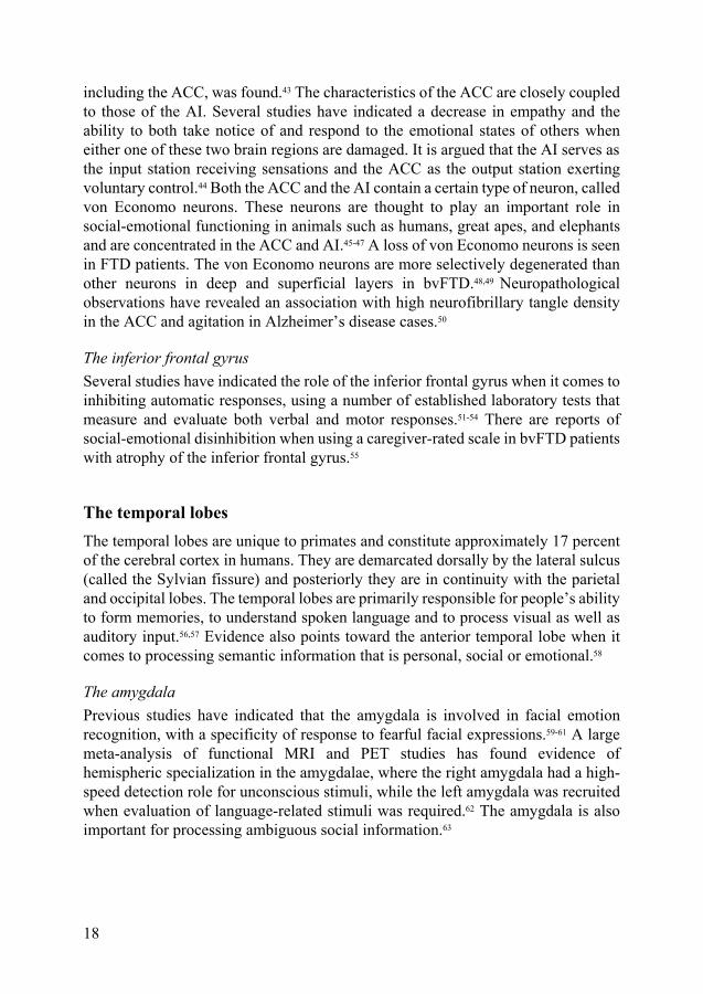
The temporal lobes
The temporal lobes are unique to primates and constitute approximately 17 percent of the cerebral cortex in humans. They are demarcated dorsally by the lateral sulcus (called the Sylvian fissure) and posteriorly they are in continuity with the parietal and occipital lobes. The temporal lobes are primarily responsible for people’s ability to form memories, to understand spoken language and to process visual as well as auditory input.56,57 Evidence also points toward the anterior temporal lobe when it comes to processing semantic information that is personal, social or emotional.58
The amygdala
Previous studies have indicated that the amygdala is involved in facial emotion recognition, with a specificity of response to fearful facial expressions.59-61 A large meta-analysis of functional MRI and PET studies has found evidence of hemispheric specialization in the amygdalae, where the right amygdala had a high-speed detection role for unconscious stimuli, while the left amygdala was recruited when evaluation of language-related stimuli was required.62 The amygdala is also important for processing ambiguous social information.63
19
The temporal parietal junction
As with the PFC, the temporal parietal junction is strongly connected to ToM.64 It is especially associated with the ability to adopt a third-person perspective and understand and reason about other people’s aspirations, as well as evaluate the mental states of others in social contexts.65,66 The temporal parietal junction is thought to be involved in representing the world from different visual perspectives and making behavioral predictions, including predicting behavioral consequences.67,68
The posterior superior temporal sulcus
The posterior superior temporal sulcus is partly responsible for the perception of human faces, voices, and motion, as well as the integration of these different emotional signals.69 It is also involved in understanding social contexts and damage to this region has been demonstrated to contribute to the genesis of anosognosia or “loss of insight” and the dysfunction of knowledge of self versus others.70
Alzheimer’s disease (AD) is the most common form of dementia worldwide, accounting for 60% to 80% of all dementias, and has an estimated prevalence of 10% to 30% among people over 65 years of age.71 The incidence is around 1% to 3%.72,73 There are genetic variants, although the great majority of AD patients (>95%) have the sporadic form, which usually presents later in life (80-90 years of age).73 Alzheimer’s disease is characterized by memory loss, spatial difficulties, and word-finding problems. Sometimes symptoms manifest years before a diagnosis is made and common manifestations include depression, progressive aphasia, and memory loss, although some variants manifest with focal neurological symptoms such as gait disturbances and myoclonus.74-76
Neuropathological picture
Macroscopically, an often symmetrical cortical atrophy and widening of the ventricles are seen.77 Neuronal cell degeneration is seen globally in the brain, accentuated in the post-central and temporal-limbic areas.78,79 Regions affected are primarily the hippocampi, the amygdalae, and the temporal and parietal lobes. Aggregates consisting of two different pathological proteins, abnormal tau and amyloid-β, are central in the disease process, commonly called neurofibrillary tangles and plaques.80-83 Degeneration of the locus coeruleus and decreased cortical levels of noradrenaline are also regular findings in AD.84,85
22
Figure 3 Microphotograph, neocortex in a case of Alzheimer’s disease, hematoxylin-eosin staining. Gliosis is seen in the molecular layer, as is neuronal reduction in layer II and amyloid plaque cores.
Figure 4 Same case as Figure 3, Alzheimer’s Disease and AT8-immunostaing showing tau-positive tangles and neurites.
23
Vascular dementia
Epidemiology and clinical picture
Vascular dementia (VaD) is the second most common form of dementia and is caused by cerebrovascular damage, such as focal infarcts or diffuse ischemic neuronal loss and demyelination.86 Risk factors for VaD include aging, hypertension, diabetes, obesity, smoking and atherosclerosis.87,88 It is hard to accurately estimate the prevalence and incidence of VaD due to different methods being used in different studies. The clinical symptoms depend on where the damage is located in the brain but can include motor and cognitive dysexecutive slowing, forgetfulness, dysarthria, mood changes, urinary symptoms, and a short-stepped gait.89,90 The cognitive deficits can be attributed to disruption of cortical-subcortical circuits, and information processing and complex attention speed, including frontal-executive function, are likely to be affected.87,91
Neuropathological picture
The neuropathological signs of VaD include ischemic and hypoxic-hypoperfusive lesions, grey and white matter infarcts of variable size, smaller lacunar infarcts in the basal ganglia and white matter, different forms of cerebral angiopathies, ischemic white matter attenuation and ischemic hippocampal sclerosis.77,92-95 Infarcts located in certain anatomical regions such as the thalamus and hippocampi often lead to cognitive impairment disproportionate to the size of the infarct.77 Macroscopically, there may or may not be a marked atrophy on the exterior, however often a central atrophy resulting in ventricular widening. Vascular dementia may often occur as a frontally accentuated disorder, the frontal white matter being particularly attenuated, with or without focal infarcts.96
24
25
Mixed dementia
Epidemiology and clinical picture
As the name suggests, mixed dementia (MD) is the coexistence of AD and VaD. It is considered a difficult diagnostic challenge due to its neurodegenerative and vascular damages and the diagnosis itself remains controversial.97 Neuropathological studies indicate that MD accounts for approximately 22% of all dementia.98 The symptoms vary depending on the localization of the AD component and the vascular component, respectively. There is evidence that frontal white matter changes and vascular disease is associated with noncognitive behavioral changes in AD.99,100
Neuropathological picture
The neuropathological signs of MD are the same as in AD and VaD. There is still some debate as to whether MD should be considered a separate form of dementia or not.97 Neurofibrillary tangles, plaques as well as lacunar infarcts and white matter lesions are commonly present and are all considered contribute to the disease. Some argue that the risk factors for VaD would also contribute to the development of AD, but new research points to different pathogenic origins.101
26
27
Frontotemporal dementia
Epidemiology and clinical picture
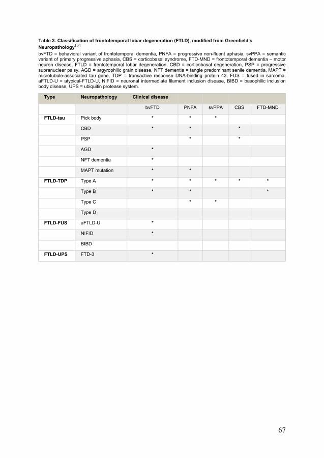
Frontotemporal dementia is a heterogeneous disorder and a leading cause of dementia in people <65 years of age.102 The average age of onset is between 45 and 65 years, although the disease occurs in people both younger and older than that.103 It has several clinical variants: bvFTD, progressive nonfluent aphasia (PNFA) and semantic dementia (SD)/semantic variant of primary progressive aphasia (svPPA). Increasingly, corticobasal degeneration (CBD)/corticobasal syndrome (CBS) and progressive supranuclear palsy (PSP) are judged to be part of the FTD spectrum.104
The symptoms vary from behavioral disturbances, personality changes, and apathy to apraxia of speech and motor symptoms.105,106
Neuropathological picture
The clinical term “FTD” corresponds to the neuropathological term “frontotemporal lobar degeneration” (FTLD).107,108 It is characterized by neuronal degeneration accentuated in the frontotemporal regions and to a variable extent also involves the parietal regions and/or the basal ganglia and the thalamus, as well as the brain stem. With few exceptions, protein depositions of tau, transactive response DNA-binding protein 43 (TDP-43) or fused in sarcoma (FUS) are found at autopsy. These proteins accumulate in the neuronal soma, glial cells or degenerated neuronal neurites – as inclusions and threads, respectively. As the disease progresses, other parts are affected as well.
28
Figure 5 The neocortex in a case of frontotemporal lobar degeneration, hematoxylin-eosin staining. Gliosis is seen in the molecular layer, as is neuronal loss in layer II and microvacuolisation.
Figure 6 Same case as Figure 5, frontotemporal lobar degeneration. TDP-43-immunostaining shows neuronal inclusions and TDP-neurites.
29
Aims of the thesis
General aim
The general aim of this thesis is to study criminal and socially inappropriate behavior among patients diagnosed with dementia of different types.
Specific aims
Study I
Ascertain the frequency and type of criminal behavior exhibited by patients diagnosed with dementia.
Study II
Specifically study the frequency of physical aggression among dementia patients and try to map potential differences in clinical traits.
Study III
Scrutinize the frequency and reasons behind police interaction with patients diagnosed with dementia.
Study IV
Map criminal and socially inappropriate behavior exhibited by dementia patients and look for potential differences in protein pathology in frontotemporal dementia patients exhibiting criminal behavior.
30
31
Patients and methods
The University of California San Francisco cohort
The University of California, San Francisco (UCSF) cohort consisted of 2,397 patients who were either referred, or sought admission themselves, to the Memory and Aging Center between 1999 and 2012 due to a suspicion of dementia diagnosis. They were evaluated either at the department’s outpatient clinic or through one of its parent research projects. This cohort was the foundation for study I. A large database containing 13,477 patient notes from the above-mentioned patients were searched for specific keywords denoting criminal behavior (CB). The keywords were chosen with the aim to capture as many different types of CB as possible. The term “CB” was defined as acts that violate the law, as well as those that deviate from traditional social decorum and could potentially lead to legal ramifications. Examples of keywords used in the study were “jail”, “theft”, and “violence”. We further categorized the different CBs into categories such as traffic violations, public urination and sexual advances. The majority of individuals had been clinically diagnosed, whereas a small subgroup of deceased individuals had been neuropathologically ascertained. In this subgroup (n=31), we studied the clinicopathological concordance.
The Lund University cohort
The Lund University (LU) cohort included a total of 303 patients with a neuropathological diagnosis of dementia, the different diagnoses being AD (n=101), VaD (n=43), MD (n=40), and FTLD, (n=119). The autopsies had been performed between 1967 and 2017 by neuropathologists working at the Department of Clinical Genetics and Pathology, Region Skåne/Lund University.
All patients had been referred to and followed at the Memory Clinic (previously Psychogeriatric Department) in Lund by specialists in geriatric psychiatry/cognitive medicine during their entire disease course. The majority had been included in one of two prospective longitudinal studies: Lund Longitudinal Dementia Study or Lund Prospective Frontotemporal Dementia Study. All available notes from the patients
32
(including referral letters, radiology findings and laboratory data) were scrutinized for signs of physical aggression (PA), police interaction (PI), CB or socially inappropriate behavior (SIB).
The term “FTD” was used for the neuropathological term “FTLD”, which in turn correlates to the spectrum of clinical diagnoses bvFTD, SD/svPPA, PNFA, CBD/CBS, and PSP.
For studies II and III we included 97 cases with FTD/FTLD (autopsies performed between 1969 and 2013), whereas in study IV we had the possibility to add 22 cases (autopsies performed between 2013 and 2017). For studies III and IV the dominating (and generally sole) protein pathology (tau, TDP-43, FUS or other) from the FTLD patients’ neuropathology reports was noted.
Physical aggression was defined as a behavior, observed and interpreted by medical staff or people caring for the dementia patient, exerted to harm another living person or creature. We analyzed the prevalence, frequencies, and first time of symptom onset of PA and also noted the victims of the aggression being exerted.
The retrospective review procedure in the third study was the same as in the second study. The first aim was to investigate and compare the prevalence and recurrence of PI in the dementia cohort. The second aim was to study the reasons behind PI (judged to be either due to criminal behavior or not due to criminal behavior), the time of occurrence of the PI (during the course of the disease), and potential consequences of the PI. We chose to define PI as an encounter (direct or indirect) with the police department/officials. The definition of CB was the same as in study I, however defined in terms of Swedish law. The prevalence of PI was calculated among the different diagnostic groups. The prevalence of PI due to CB, as opposed to PI of other cause, was then further studied.
In study IV we focused specifically on neuropathologically verified cases with a diagnosis of AD (n=101) or a diagnosis within the FTLD spectrum (n=119), where autopsies had been performed between 1967 and 2017. The first aim of study IV was to investigate and compare the prevalence, recurrence, and type of CB (except for PA, since we already examined this trait in the second study), and prevalence and type of SIB among dementia patients with AD and FTD. The second aim was to study the time of occurrence of the CB (during the course of the disease; whether it occurred during the first or second half of the disease duration). The third aim was to investigate whether, within the FTLD group, there was a certain type of protein pathology that was associated with CB more frequently than with others. The definition of CB was the same as in study III, except that we omitted PA.
33
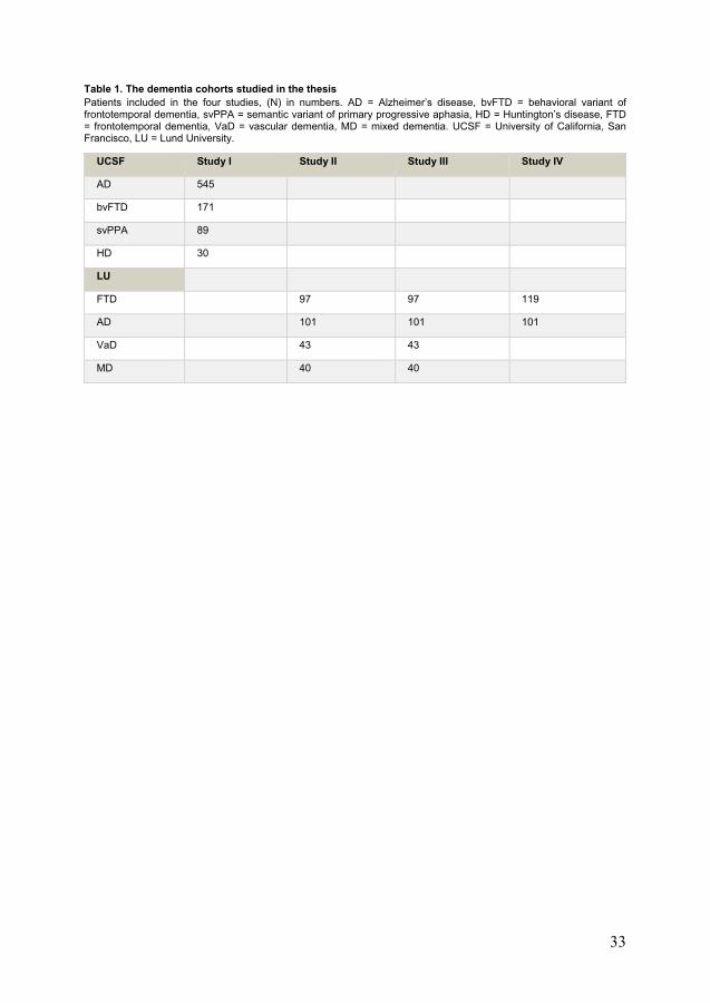
Table 1. The dementia cohorts studied in the thesis Patients included in the four studies, (N) in numbers. AD = Alzheimer’s disease, bvFTD = behavioral variant of frontotemporal dementia, svPPA = semantic variant of primary progressive aphasia, HD = Huntington’s disease, FTD = frontotemporal dementia, VaD = vascular dementia, MD = mixed dementia. UCSF = University of California, San Francisco, LU = Lund University.
UCSF Study I Study II Study III Study IV
AD 545
bvFTD 171
svPPA 89
HD 30
LU
FTD 97 97 119
AD 101 101 101
VaD 43 43
MD 40 40
34
35
Results
Study I
A total of 204 (8,5%) patients had exhibited behavior that could be considered criminal. The major diagnostic groups were bvFTD (n=64), svPPA (n=24), AD (n=42), and Huntington’s disease HD (n=6). We chose to exclude the other diagnoses due to their small numbers. Among these we found VaD, CBS, PSP, mild cognitive impairment (MCI), and others. The diagnostic group with the highest criminal frequency was bvFTD (37,4%), followed by svPPA (27,0%), HD (20,0%), and AD (7,7%). There was a statistically significant difference when comparing the criminal frequencies among bvFTD, svPPA and AD patients (p<0.001). Of the bvFTD patients who had exhibited CB, 14,0% had exhibited this as the first symptom of the disease. The respective numbers for the svPPA and AD patients were 7,8% and 2,0% (p<0.001). The clinicopathological concordance among the 31 autopsied patients was 93%. Sexual advances, traffic violations, theft, and violence were common criminal manifestations in bvFTD patients. These were also common in the svPPA group, except for sexual advances. Among the AD patients who had exhibited CB, traffic violations and trespassing were more common due to cognitive impairment and confusion rather than disinhibition or impulsivity.
Comments
Our study revealed that new CBs emerged in association with specific neurocognitive disorders but not with others. The US law states109 that if a person knows what he or she is doing when committing a crime, he or she could be held culpable for his or her actions. Frontotemporal dementia patients often have the ability to verbalize that their actions are wrong and hence they are not protected by the so-called insanity defense (see Appendix) that states that a person cannot be held culpable if he or she does not understand that his or her actions are wrong.110 Since FTD patients become ill earlier in life than AD patients and, according to our study, exhibit CB earlier in the disease course than AD patients, they are particularly vulnerable with respect to the legal system. Previous research has indicated that people with FTD have to wait a longer time from first symptom to diagnosis in comparison to other neurodegenerative disorders.111,112 We suggest a screening for
36
neurocognitive disorders in all first-time offenders over 55 years of age. It is important to find these individuals as early as possible in their disease course to be able to help them in the best possible way.
Study II
Of the 281 patients, 97 (35%) had a history of PA during the disease course. Physical aggression was distributed in the different diagnostic groups in the following way: 42/101 AD patients (42%), 29/97 FTD patients (30%), 12/43 VaD patients (28%), and 14/40 MD patients (35%). The AD patients, when adjusting for age and disease onset, had more frequent reports of PA than FTD patients (p=0.04), and overall males were more aggressive than females (p=0.015). We decided not to include the VaD and MD patients in the statistical analysis, due to a different pathogenesis (significant vascular component compared to pure neurodegenerative disorders) and the fact that these groups were considerably smaller than the AD and FTD groups, that were of comparable size. The FTD patients had a higher PA score, a finding not statistically significant (p=0.059), and were exhibiting PA earlier in the disease course compared to AD patients (p=0.017). The victims of the PA were predominantly health staff, followed by other patients, but the PA also affected family members, and people and animals unknown to the dementia patient. The PA ranged from being exhibited in nursing care situations to unprovoked assault and the consequences varied from mild to life threatening.
Comments
Physical aggression was more frequent among AD than FTD patients, an unexpected finding. The methods in the first and second studies differ and could be one of the explanations for this disparity. The first study is based on notes written by physicians at the Memory and Aging Center, while the second study is based on information from several sources, such as doctors’ offices at LU, nursing homes and caregivers. We did, however, confirm in the second study that the FTD patients did exhibit PA earlier in the disease course than the AD patients did. In this study we investigated PA during the entire disease period, in contrast to the previous study (the UCSF cohort), where period prevalence until the doctor’s visit was recorded. There seemed to be some difference between FTD and AD patients with regard to situations eliciting PA. The FTD group more often exhibited unprovoked and unexpected PA, while the AD patients mostly reacted with PA in nursing care situations, where their personal space was being invaded. This could be due to difficulties in understanding and interpreting intentions, thereby eliciting a sense of fear. Previous research has indicated that caregivers who become victims of PA may
37
distance themselves from the patient and this may in turn impair the quality of care.113 There is also a risk that caregivers act aggressively towards the dementia patient and it is a well-known fact that abuse of the elderly does occur.114,115 It is important to continuously educate people working with dementia patients on how to act when violent situations occur.
Study III
A history of PI was noted for 50 individuals (18%) during the course of their disease. The respective numbers distributed as follows: 9/101 AD patients (9%) and 25/97 FTD patients (26%). The difference was statistically significant (p=0.002). Police interaction initiated by criminal behavior occurred in 2 of the 9 patients with AD (22%) and in 16 of the 25 patients with FTD (64%) (p=0.052), thus demonstrating criminal behavior in 2% of all AD and 16% of all FTD individuals. The FTD group exhibited a higher recurrence of PI than the other groups; the difference was, however, not statistically significant (p=0.242). The FTD group was also the sole group including patients with >3 reports of PI. Among the 25 FTD patients with PI, 18 were of the TDP-43 type, 6 of them had tau pathology, and one was indeterminate (p=0.137). With regard to the time point of PI during the course of disease, the 9 AD patients were subject to PI predominantly early in the disease, while in the FTD and VaD+MD groups, the PI occurred later (and was also more prevalent). A number of patients were involved in lawsuits; however, most of the lawsuits were withdrawn, primarily because a physician had written a statement declaring that the patient suffered from a neurodegenerative disease. Other consequences included that a driver’s license was revoked and one of the patients incurred some mild injuries due to difficulties cooperating with the police officers.
Comments
This is the first study to examine the prevalence, recurrence, and reasons behind PI among patients with clinically and neuropathologically confirmed and subtyped dementia. The statistically significant differences when comparing PI among AD and FTD patients did not come as a surprise to us. If we had studied larger groups, we might also have found statistically significant differences when studying the reasons behind PI (due to criminal behavior or not due to criminal behavior). This is in line with previous research indicating that FTD patients, to a greater extent than AD patients, exhibit CB and hence they may also interact with the police more frequently.116-119
38
This study revealed that the AD patients interacted with the police earlier in the disease course than the FTD patients. The earlier prevalence of PI in the AD group may be explained by the fact that many AD patients move to nursing homes as the disease progresses, whereas FTD patients tend to live at home for a longer time than AD patients.120-122 A study focusing on police contact with older persons indicated a drop in PI prevalence when the older person was placed in a nursing home.123 The primary reason for PI among the AD patients was wandering. It seems logical that AD patients wander off earlier in their disease course, before being admitted to nursing homes, and hence interact with the police early in the disease course. The initial deviant behavior exhibited by the FTD patients may arise before a diagnosis is made, since in FTD patients there is a long time delay from the first symptom to when a diagnosis is made.124 Relatives might not want to call the police the first times CB is exhibited. Today none of the various universities and schools for higher education providing police education in Sweden offers specific training in meeting or handling people with dementia. We believe that a basic training program for police officers, focusing on age-related health and dementia, would be useful in Sweden to increase the police force’s knowledge and skills and to avoid harmful situations. It is important that the judiciary system and especially the police force are vigilant when it comes to socially deviant, criminal, and antisocial behavior in middle-aged and older adults.
Study IV
Of the 220 patients studied, 65 (30%) exhibited behavior that could be considered criminal during the disease course. The distribution of CB was 15 (15%) in the AD patient group and 50 (42%) in the FTD patient group, yielding a statistically significant difference between the two (p<0.001). Recurrence of CB also differed between the diagnostic groups: of the 15 AD patients with CB, 7 (47%) committed a crime once and 8 (53%) did so more than once. Corresponding numbers for FTD patients were 9 (18%) and 41 (82%), once again yielding a statistically significant difference (p=0.039). The criminal behavior included verbal (including homicidal) threats, vandalism, traffic violations, sexual advances, pyromania, theft, and stalking of others.
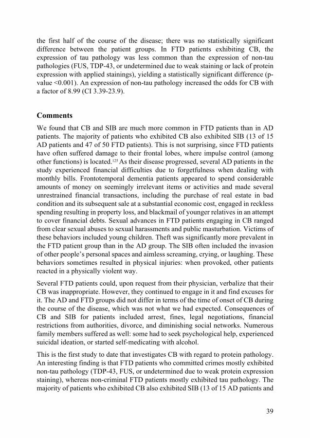
A total of 57 (56%) AD patients and 89 (75%) FTD patients exhibited SIB during the course of the disease, once again yielding a statistically significant difference between the patient groups (p=0.004). The FTD patients exhibited a greater prevalence of SIB in every category being studied except for public urination/defecation, where AD patients dominated. The time of onset of CB was evenly spread between the groups. Almost half the patients exhibiting CB (47% of the 15 AD patients and 46% of the 50 FTD patients) did so for the first time during
39
the first half of the course of the disease; there was no statistically significant difference between the patient groups. In FTD patients exhibiting CB, the expression of tau pathology was less common than the expression of non-tau pathologies (FUS, TDP-43, or undetermined due to weak staining or lack of protein expression with applied stainings), yielding a statistically significant difference (p-value <0.001). An expression of non-tau pathology increased the odds for CB with a factor of 8.99 (CI 3.39-23.9).
Comments
We found that CB and SIB are much more common in FTD patients than in AD patients. The majority of patients who exhibited CB also exhibited SIB (13 of 15 AD patients and 47 of 50 FTD patients). This is not surprising, since FTD patients have often suffered damage to their frontal lobes, where impulse control (among other functions) is located.125 As their disease progressed, several AD patients in the study experienced financial difficulties due to forgetfulness when dealing with monthly bills. Frontotemporal dementia patients appeared to spend considerable amounts of money on seemingly irrelevant items or activities and made several unrestrained financial transactions, including the purchase of real estate in bad condition and its subsequent sale at a substantial economic cost, engaged in reckless spending resulting in property loss, and blackmail of younger relatives in an attempt to cover financial debts. Sexual advances in FTD patients engaging in CB ranged from clear sexual abuses to sexual harassments and public masturbation. Victims of these behaviors included young children. Theft was significantly more prevalent in the FTD patient group than in the AD group. The SIB often included the invasion of other people’s personal spaces and aimless screaming, crying, or laughing. These behaviors sometimes resulted in physical injuries: when provoked, other patients reacted in a physically violent way.
Several FTD patients could, upon request from their physician, verbalize that their CB was inappropriate. However, they continued to engage in it and find excuses for it. The AD and FTD groups did not differ in terms of the time of onset of CB during the course of the disease, which was not what we had expected. Consequences of CB and SIB for patients included arrest, fines, legal negotiations, financial restrictions from authorities, divorce, and diminishing social networks. Numerous family members suffered as well: some had to seek psychological help, experienced suicidal ideation, or started self-medicating with alcohol.
This is the first study to date that investigates CB with regard to protein pathology. An interesting finding is that FTD patients who committed crimes mostly exhibited non-tau pathology (TDP-43, FUS, or undetermined due to weak protein expression staining), whereas non-criminal FTD patients mostly exhibited tau pathology. The majority of patients who exhibited CB also exhibited SIB (13 of 15 AD patients and
40
47 of 50 FTD patients). Interestingly, there was a clear majority of cases that exhibited CB in the TDP-43 and FUS groups. Approximately 60% in these groups had committed crimes during the course of the disease. This is in stark contrast to only 14% of tau cases. It appears that non-tau pathology is associated with more severe behavioral problems potentially leading to CB. The association between non-tau pathology and higher prevalence of CB probably relates more to the regional spread of brain damage than to the type of protein pathology in FTLD spectrum disorders: CBD/CBS and PSP (tau diseases) affect more central parts of the brain, while svPPA/SD (mostly non-tau), PNFA and bvFTD (tau and non-tau diseases, i.e. TDP-43 and FUS) affect the cortex, which is the locus of behavioral control, empathy, and appropriate emotional responses.18
41
Conclusions: studies I-IV
Criminal as well as socially inappropriate behavior is more prevalent in FTD than in other neurodegenerative disorders. It can be an early manifestation of the disease, often before a diagnosis is made. Non-tau pathology is strongly associated with criminal behavior in FTD patients.
Physical aggression is more frequent in AD than in VaD+MD, and FTD. It is, however, exhibited earlier in the disease course in FTD than in AD and VaD+MD. Qualitative results include that physical aggression exerted by FTD patients is often more brutal when compared to physical aggression exerted by AD patients. The situations eliciting physically aggressive behavior seem to differ between the respective patient groups.
On the one hand, it is more common among FTD patients than AD patients to interact with the police, and also more often to do so due to criminal behavior. Alzheimer’s disease patients, on the other hand, interact with the police earlier in the disease course.
42
43
Discussion and future perspectives
Summarizing the results of this thesis, it can be concluded that the initial questions posed when starting this thesis project have, to some extent, been answered.
Why do some people with dementia commit crimes?
It seems likely that some parts of the brain are more affected by the disease than others when analyzing people with dementia who commit crimes. “Why” they commit crimes is almost a philosophical question. However, there is no doubt that certain parts of the brain do play a role in decision-making, impulse control, theory of mind, relating to others, and learning from reward and punishment. These parts of the brain are often damaged in several of the dementing disorders studied, especially in FTD. Recent research has called more attention to a single connected brain network (including the OFC, the ventromedial PFC and the anterior temporal lobes), rather than specific brain regions, that seems to be associated with criminal behavior having a known temporal association with a brain lesion. This network is unique to lesions affiliated with criminal behavior compared with lesions causing other syndromes.126
What types of crimes do people with dementia commit?
The current thesis has demonstrated that patients affected by dementia commit a wide range of different crimes, the most prevalent forms being theft, traffic violations, sexual advances, trespassing, and violence, but also mismanagement of personal finances leading to difficulties with the authorities.
At what stage of the disease do people with dementia commit crimes?
According to our studies, criminal behavior can be an early manifestation of a dementing disorder, especially in FTD. It is more prevalent among FTD patients to exhibit violence in the first half of their disease course when compared to the violence exhibited by AD patients. We could also see that FTD patients, to a greater extent than AD patients, committed crimes repeatedly during the disease course.
44
Is there a difference when comparing different types of dementia?
It seems that there is a difference when comparing different types of dementia. The disorder that stood out the most in this thesis was FTD (in study I bvFTD, followed by svPPA). Patients with AD do not seem to commit crimes to the same extent as FTD patients, especially not in the early stages of the disease, according to the results of studies I and II. We noted that there was a disparity between the two patient groups in terms of how they committed crimes. Frontotemporal dementia patients were more prone to actively commit crimes, probably due to their lack of inhibitory control and ability to understand the nature and consequences of their behaviors, while AD patients more often exhibited CB related to cognitive impairment, when, for example, they forgot to pay for groceries or missed a red light when driving. This was also evident when studying SIB, where FTD patients dominated as well. In the UCSF cohort, we found a high prevalence of CB in HD patients. This patient group was not included in the studies from the LU cohort. Patients with HD do not usually seek help at memory units in Sweden; rather, they are being taken care of at neurology departments or, in more severe cases with behavioral disturbances, in close cooperation with specialists in psychiatry. We can therefore not draw any conclusions regarding Swedish patients with HD and the potential exhibition of CB or SIB.
What are the consequences of the criminal and socially inappropriate behaviors of patients with dementia?
When studying the individual cases in this thesis, it became apparent that the consequences of the patients’ criminal and socially inappropriate behaviors were extensive. In the UCSF cohort there were patients who had to spend some time in jail. A number of patients in the LU cohort had received orders from prosecutors instructing them to appear in court. Some of the lawsuits were withdrawn, most often because a physician had written a statement declaring that the patient suffered from a neurodegenerative disease. Patients with FTD are in general younger at disease onset than AD patients. It is not unusual for the patients to be employed, have a family and a social network surrounding them. When the disease strikes and behavioral symptoms emerge, it is therefore common among the (often undiagnosed) affected individuals to lose their jobs, get divorced and experience diminished social networks. Numerous family members suffered as well: some had to seek psychological help, experienced suicidal ideation, or started self-medicating with alcohol. To map all the consequences in these individual cases would constitute the subject for a thesis in itself. While researching this thesis, and having met affected patients and their families, it became apparent to me that more support to caregivers and families is needed.
45
Costs for society
In addition to the social consequences of the patients’ behaviors, dementia is associated with a financial burden for society. Every time police officers respond to a call or are required to help citizens in an emergency situation, it results in a cost for the taxpayers. In a report on the cost of dementia in Wales127 the total financial burden to society due to dementia was estimated as £1.4 billion per year, of which £6 million was spent on the police, among other costs, owing to missing person enquiries. An American research group quantified the socioeconomic burden of FTD compared to previous research on AD, using an Internet survey administered to caregivers of patients with FTD. The economic burden of FTD was substantial and 6% of the respondents reported the need for police intervention during the previous year.128 Legal costs were further reported at 9.6%, with one of the leading reasons for court appearances being the bringing of criminal charges (3.2%). Criminal behavior in general is associated with enormous costs to society, where there are estimates of $1 trillion per year.129 We believe that the CB evidenced in our studies also contributed to an economic burden on society, although we did not specifically study this aspect.
The question of liability when people with dementia commit crimes
In Sweden there was a case before the Court of Appeal involving a patient with dementia who was suspected of murder.130 The suspect could describe details about the crime, such as where he had hidden the weapon and where he went after the crime. He initially seemed cognitively intact. As it became apparent later in the investigation that he suffered from an early form of dementia, the penalty became forensic psychiatric care. The case was never heard in the Supreme Court. United States law states109 that if a person knows what he or she is doing when committing a crime, he or she could be held culpable for his or her actions. Frontotemporal dementia patients often have the ability to verbalize that their actions are wrong and hence they are not protected by the so-called insanity defense. The insanity defense states that the person cannot be held culpable if he or she does not understand that his or her actions are wrong.110 The ability to understand the criminal nature of an action and proceed regardless is problematic, especially when an FTD patient is facing charges or is interacting with the surrounding community or family members.
The need for future prospective studies
It appears that CB and SIB are more prevalent in FTD than in other neurodegenerative disorders. Criminal behavior is also more common to be one of the earlier symptoms of the disease. The CB and SIB do affect people around the patient in negative ways. These findings may help in the differential diagnostic process ante-mortem. We did not have access to criminal records and could therefore not search for more extensive details surrounding patients’ CB. There is a
46
possibility that the numbers of criminal incidents are indeed higher, since many patients or their families might not have wanted to, or had forgotten to, inform the physician about CB. Our numbers should thus be seen as minimum figures when it comes to CB. We believe this argument is also applicable when studying SIB. Our interpretation of patients’ behavior and whether it should be considered criminal or socially inappropriate was partly a result of subjective opinion, which in turn was based on our experience of life in Sweden and the US, and our knowledge of what is legal or deemed socially appropriate there. To aid in our evaluation of these matters, we consulted with a lawyer who specialized in mental health. Sometimes the question as to whether behavior could be considered criminal became strictly hypothetical, since the majority of the CBs included in this study were never tried in court, hence neither we, nor the lawyer who helped us could state the potential judicial outcome. It would be interesting to study the phenomenon of CB from “the other side”, in other words, screening criminal records from the police departments in search of perpetrators with a neurocognitive disorder diagnosis. We suggest that middle-aged and older individuals exhibiting CB or SIB for the first time should be screened for neurodegenerative disorders. Prospective studies on this matter, including neuropathological follow-up post-mortem, are needed.
The end to free will?
Will the advances in neuroscience be the end of the expression of free will, often called “the basis for moral responsibility”? As mentioned at the beginning of the thesis, there are court cases where imaging of perpetrators’ brains has been used as evidence that the prosecuted person could not be held culpable for his or her actions.
The doctrine that all events, including human action, are ultimately determined by causes regarded as external to the will is called determinism. Some philosophers have taken determinism to imply that individual human beings have no free will and cannot be held morally responsible for their actions. This view is highly controversial and challenges traditional ideas and views on morality and personal responsibility. Some argue that free will is an illusion generated by our cognitive architecture and that the more advances we make in neuroscience, the more we will have to redefine how we look at judicial outcomes.131 Others, however, are strongly against this view and claim that neuroscientific evidence can instead reveal how free will works and what gives rise to our sense of responsibility for our actions by elucidating the underlying neural machinery.132 Another important aspect is the methodological problem of applying scientific research on group findings to individual cases. In Germany, for instance, preventive detention can be ordered instead of a prison sentence, thereby risking the wrongful use of biological data for the purpose of restraining individuals.133
This discussion is, I believe, highly interesting when studying people with dementia who commit crimes. The patients included in this thesis were, initially, law-abiding
47
citizens, but as their disease progressed, they developed deviant behaviors and committed crimes. Would we say that they chose to act this way? Or were the decisions they made only a result of their disease? Some of them, as mentioned, could verbalize that their actions were wrong but decided to proceed anyway. I believe that the answer to these questions lies somewhere in between.
Another interesting question that has arised during this project is: When does a disease start? Is it when the individual starts to exhibit symptoms? Is it years before, when changes in the brain could have been found if the patient had been examined with an MRI scan or a brain biopsy revealing pathological transformations?
Is it when the individual is born, due to his or her genetic predisposition and vulnerability to developing dementia? Exciting research challenges our view on dementia as a disease of the elderly. There is evidence that tau-positive material is present in a high proportion of children, and in the absence of amyloid-β accumulation. These findings may indicate that the AD-related pathological process leading to neurofibrillary tangle formation may begin in subcortical nuclei, and that it may start early, before puberty or in early young adulthood.134,135 Would this change our view on who could be held responsible for his or her actions?
I will, of course, not be able to answer these questions, but I am convinced that regardless of what view one has on moral responsibility and brain disease, the advances of neuroscience will influence the future in terms of how we look at criminal and socially inappropriate behaviors. It is with great interest that I will continue to follow scientific breakthroughs on this matter.
48
49
Populärvetenskaplig sammanfattning på svenska (General summary in Swedish)
Under lång tid har vetenskapen fascinerats och förundrats över den komplexa hjärnan och dess koppling till vår personlighet. Ett av de mer kända medicinhistoriska fallen är historien om Phineas Gage. En järnvägsarbetare som råkade ut för en allvarlig arbetsplatsolycka, där ett långt järnrör for rakt igenom hans skallben och pannlob. Han överlevde mirakulöst men kort efter olyckan noterades en tydlig förändring i personligheten. Otaliga exempel efter Phineas Gage vittnar om människor med hjärnskador orsakade av trauma, tumörer eller slaganfall som i sin tur har lett till förändringar i personligheten. Modern forskning visar att hjärnans pann- och tinninglober har stor påverkan när det gäller personlighet, empati, social förmåga och moraliska resonemang.
Demens, eller neurokognitiv störning, är ett samlingsbegrepp för en rad olika sjukdomar som drabbar hjärnan. Gemensamt för demenssjukdomarna är att hjärnans nervceller förtvinar. Beroende på var i hjärnan som förlusten av nervceller sker uppvisar den drabbade individen olika symtom. Den vanligaste demenssjukdomen är Alzheimers sjukdom. Sjukdomen karakteriseras av minnessvårigheter, försämrat språk och en oförmåga att orientera sig i tid och rum. Vaskulär demens är den näst vanligaste demenssjukdomen. Orsaken är försämrat blodflöde i hjärnan, vilket kan bero på t.ex. blödning eller infarkt. Detta leder i sin tur till nervcellsdöd och en uttunning av myelinet, som omger nervcellernas utskott. Vanliga symtom är nedsatt initiativförmåga, en generell psykomotorisk förlångsamning och svårigheter med planering. Symtomen varierar beroende på var i hjärnan skadan sitter. Blanddemens är en kombination av Alzheimers sjukdom och vaskulär demens. En av de mer ovanliga demenssjukdomarna är frontotemporal demens. Frontotemporal demens drabbar hjärnans pann- och tinninglober och är en vanlig orsak till demens hos yngre personer (<65 år). Framträdande symtom hos drabbade individer är beteendestörningar, personlighetsförändring men också påverkan på språk och motorik. Det tar ofta lång tid från första symtom till korrekt diagnos och omhändertagande.
50
Denna avhandlings syfte är att undersöka prevalens och konsekvenser av kriminellt och socialt avvikande beteende hos personer med demenssjukdom. Avhandlingens första studie baseras på en grupp bestående av 2 397 individer som sökt vård vid en stor minnesklinik vid University of California, San Francisco, USA mellan åren 1999 och 2012. Avhandlingens andra, tredje och fjärde studie baseras på ett material om totalt 303 avlidna individer där samtliga fått en demensdiagnos efter döden, baserat på obduktion gjord i Lund mellan åren 1967-2017.
I den första studien fann vi att 37,4 procent av personer som fått diagnosen beteendevariant av frontotemporal demens hade begått en kriminell handling under sin sjukdomsperiod. I gruppen Alzheimers sjukdom var det enbart 7,7 procent som hade gjort samma sak. Denna skillnad var statistiskt signifikant. Kriminellt beteende visade sig i 14 procent av fallen med beteendevarianten av frontotemporal demens vara det första tecknet på demenssjukdom. Vanliga brott var stöld, våld, trafiköverträdelser och sexuella närmanden.
Vi valde att studera fysisk aggressivitet i avhandlingens andra studie. Resultaten visade att 35 procent av individerna hade varit fysiskt aggressiva under sin sjukdomsperiod. Vanligaste var det bland patienter med Alzheimers sjukdom (42 procent), följt av blanddemens (35 procent), frontotemporal demens (30 procent) och vaskulär demens (28 procent). Patienterna med Alzheimers sjukdom var fysiskt aggressiva oftare än de andra diagnosgrupperna, men patienter med frontotemporal demens uppvisade fysisk aggressivitet tidigare i sjukdomsförloppet och våldet de utövade var grövre och mer oprovocerat i jämförelse med patienter med Alzheimers sjukdom. Offren var till största del vårdpersonal och medpatienter, men även närstående, djur och främlingar drabbades. Konsekvenserna varierade från milda till livshotande.
I avhandlingens tredje studie tittade vi närmare på polisinteraktion hos personer med demenssjukdom. Det framkom att 9 procent av personer med Alzheimers sjukdom hade varit i kontakt med polis under sin sjukdomsperiod, i jämförelse med 26 procent av personer med frontotemporal demens. Denna skillnad var statistiskt signifikant. Det var vanligare bland personer med frontotemporal demens att komma i kontakt med polis på grund av kriminellt beteende, i jämförelse med personer med Alzheimers sjukdom. Antalet gånger personerna med frontotemporal demens kom i kontakt med polis var också högre än hos personer med Alzheimers sjukdom även om dessa skillnader inte var statistiskt signifikanta. Några patienter fick böter, en del blev kallade till domstolsförhandlingar och några blev av med sina körkort.
I den fjärde och sista studien fokuserade vi enbart på Alzheimer sjukdom och frontotemporal demens när vi granskade kriminellt och socialt avvikande beteende. Liksom i den första studien, baserat på ett stort amerikanskt material, fann vi i detta arbete en statistiskt signifikant skillnad gällande kriminellt beteende hos personer
51
med Alzheimers sjukdom (15 procent) respektive frontotemporal demens (42 procent). Det var vanligare hos personer med frontotemporal demens att begå upprepade kriminella handlingar, även detta en statistiskt signifikant skillnad i jämförelse med Alzheimers sjukdom. De kriminella handlingarna yttrade sig i form av verbala hot (inklusive dödshot), vandalisering, trafiköverträdelser, sexuella närmanden, pyromani, stalkning och stöld. Kriminellt beteende uppvisades både tidigt och sent i sjukdomsförloppet. Här fann vi ingen statistiskt signifikant skillnad mellan grupperna. Socialt avvikande beteende kunde konstateras i 56 respektive 75 procent av fallen hos Alzheimers sjukdom respektive frontotemporal demens (en statistiskt signifikant skillnad). Vi fann att vissa typer av proteininlagringar i hjärnan hos individer med frontotemporal demens var mer associerade med kriminellt beteende än andra, även detta en statistisk signifikant skillnad.
Sammanfattningsvis har denna avhandling visat att kriminellt och socialt avvikande beteende kan vara ett tidigt tecken på demenssjukdom, oftast frontotemporal demens. Det är viktigt att yngre och medelålders individer som börjar bete sig avvikande eller till och med börjar begå kriminella handlingar uppmärksammas och får hjälp till läkarkontakt. Förhoppningsvis kan resultaten presenterade i denna avhandling hjälpa sjukvården, rättsväsendet och allmänheten i att uppmärksamma individer som riskerar att lida av en demenssjukdom och hjälpa dem till medicinsk utredning och vård i tid.
52
53
Acknowledgments
I would like to start by expressing my sincerest gratitude towards the patients and their families for making this research possible. Thank you also to the Hennerlöfska Foundation (Swedish Society of Medicine), the Trolle-Wachtmeister Foundation for Medical Research, and Region Skåne for financial support.
This thesis would not have been written without the encouragement and support of many people who are very important to me.
First of all, thank you, Elisabet Englund, my main supervisor for introducing me to the exciting field of medical research, and the borderline between neuropathology, psychiatry, and neurology, and for the inspiration. I am grateful for your never-ending support and enthusiasm, even when times were tough and I doubted myself. Thank you for all the fun memories and laughter, ranging from San Francisco bars and hipster stores to late nights discussing our work over a glass of wine at your house with your adorable dogs running around our legs. Also, thank you for bringing your machete to cut away all the unnecessary words and reasoning in my drafts.
Maria Landqvist Waldö, my co-supervisor: thank you for being the more careful and reasonable person when I have drifted away in terms of ideas, text, or reasoning. Your feedback has been invaluable to me, and your genuine commitment to patients and research, including your hard work, is inspiring. Thank you also for being a fun shopping and socializing partner at conferences in different cities around the world!
Thank you, Robert Rydbeck, my co-supervisor, for making me see things from a different perspective. I am grateful for your artful and tricky questions that made me rethink how I look at behaviors, personalities, the brain, and life in general. I will never forget when you spontaneously said: “When you become a professor in psychiatry in the future...” That meant a great deal to me, to hear you believe in me and my capacity, like it was obvious, not potential, that I one day would become one.
I have also been encouraged and inspired by Bruce Miller, my main supervisor during my stay at the UCSF Memory and Aging Center in San Francisco. Thank you for showing me how much one can accomplish if surrounded with ambitious, creative and devoted people. Your leadership and mentoring has been crucial in my inner process of deciding that I want to pursue a career in psychiatry and neurology.
54
Your profound knowledge of the brain, and FTD in particular, is impressive and I am very grateful for the opportunity to have been one of the MAC scholars. I hope I can put some of what I learned into practice in my future work.
Thank you, my co-author Alexander Frizell Santillo, for generously sharing your knowledge about FTD and giving me wise and thoughtful advice on future directions in my research. Thank you for sharing coffee breaks and laughter when talking about life as a PhD student.
I would also like to express my gratitude to the inspiring, kind, and humble Lars Gustafson, one of the “fathers” of FTD, Christer Nilsson, Ulla Passant, Karin Nilsson, and Susanna Vestberg for your hard, thorough, and systematic work, which is the foundation of this thesis.
Thank you, my fellow PhD student colleagues Erik Blennow Nordström, Martin Salö and Emil Ygland for liberating discussions and sharing of experiences, frustrations and thoughts on combining clinical work with PhD studies.
I would not have been able to complete this thesis without the invaluable help from both the Neurology and the Pathology departments in Lund for giving me temporary access to the buildings and letting me use your workspace while I was in Lund for weeks at a time. Thank you also to the staff at the Neurology department for nice lunch and coffee breaks, and thank you, Anette Persson, Camilla Lidman, and Eva Olsson at the Neuropathology department for always being so kind and welcoming to me and helping me out with practical issues. Thank you, Susann Ullén, for patiently and generously giving me advice and help when doing statistics.
The talented Magdalena Fischerström made the beautiful cover photo and the other illustration in this thesis. Thank you for doing this for me. The thesis has been printed with help and support from Media-Tryck, and especially Jonas Palm, thank you. Anette Saltin, research coordinator at the Medical Faculty at Lund University, provided me with inestimable administrative support during these years – thank you. Scribbr.com has proofread and given me essential feedback on the language in this thesis and in some of my papers.
Thank you, Ola Kronkvist and Maria Nyström Agback, for necessary guidance in the world of the Swedish police force and the Swedish judiciary system.
My time at the Memory and Aging Center and my stay in San Francisco would not have resulted in such fond memories if it were not for all the inspiring colleagues, co-authors and new friends that I made during my time there. Especially I would like to thank Kate Rankin, David Perry, Lea Grinberg, Bill Seeley, Allen Lee, Eduardo Caverzasi, Zach Miller, Edgar Busovaca, Richard Binney, Laura Jastrzab, Leslie Goss, Eleanor O’Brien, Yuan-Yu Kristy Liao, Smita Yadav, Sarinporn Manitsirikul Tipudom, Jumbo Pongsakorn Tanayapong, Magdalena Rowe, Marta Huglen Revheim, Britt Moore, and Frida Thomsen.
55
My half-time reviewers Mats Lindström and Sara Hall sharpened my scientific arguments and indirectly gave me advice on how to proceed with my thesis. Thank you, Mats, for entertaining lectures in psychiatry in medical school and thank you, Sara, for meaningful and fun discussions on research and life in general.
Thank you, my former and current co-workers at Norra Stockholms psykiatri, Karolinska University Hospital, and Psykiatri Nordväst, especially Amy Goldman, Anna Malmqvist, Tove Mogren, Anne Wandel, Isidora Bubak, Alessandra Hedlund, Nataliya Alegrund, Peter Bergmark, Anton Sendel, Idun Holmdahl, Tim Hansson, Dafne Jansson, Martin Gärde, Shahram Bahmanyar, Clara Frumiere, Maximilian Zoltek, Anastasia Poyiadji, Basil Ali, Susanna Brauner, Staffan Nyström, and Petter Norman. Thank you also to Lotta Renström Koskela, Sten Friberg, and Tove Gunnarsson for believing in me and providing me with the opportunity to combine clinical work and research. Thank you, Jurgita Kundrotiené, my clinical supervisor, for giving me essential clinical advice and making me laugh when I needed it the most.
To Thomas Lindén and Mia Fahlén, my informal mentors and dear friends, thank you for being such great role models to me, and for interesting and fun discussions on so many different aspects of life.
During my weeks or even months spent in Lund with my suitcase as my only companion I would not have survived without the warm hospitality from Maria Valeur, Karin Dalene Skarping and Pär Bengtsson, Lars Lundstedt and Anna Callmer, thank you.
Thank you, my dear friend and intermittent roommate, Karolina Fischerström, for generously opening up your different homes in Lund and Malmö during these years, for bubbles on the balcony and for watching and laughing at bad TV series while eating ice cream.
My faithful and supportive friends who always encourage me make me feel like a truly privileged person, having so many warm-hearted and caring people around me. Thank you for making me laugh and focus on things that have nothing to do with the thesis, things that really matter in life: Anna Rognli, Catharina Klingspor, Cecilia Anvret, Ebba Lindqvist, Emma Milerad, Frida Henkow, Rebecka Casselbrant, and Sofie Veraeus.
My wonderful sisters, Marie, Louise, and Frédérique Liljegren, I would be no one without you. You mean the world to me and I am so proud of being your older sister. Thank you for always being there for me, no matter what.
My fantastic, loyal, creative, adventurous and caring true love, Eyvind Johansson, the support and love from you these past years have been essential to me. Thank you for standing by my side, confident and strong. Thank you for making me feel
56
loved and appreciated and for always keeping your sense of humor when I have lost mine. I love you endlessly.
This thesis is dedicated to the people that have made this possible from the start, my dear and very much loved parents, Lars and Suzanne Liljegren. Thank you for always believing in me, encouraging me to pursue my dreams, for being the biggest supporters of what I do, no matter what, and for showing me how it feels to be unconditionally loved. Words cannot describe my gratitude and love for you.
57
References
1. Harlow JM. Passage of an iron rod through the head. 1848. J Neuropsychiatry Clin Neurosci. 1999;11(2):281-283.
2. Pick A. Uber die Beziehungen der senilen Hirnatrophie zur Aphasie. Prag Med Wchnschr. 1892;17:165-167.
3. Pick A. Über einen weiteren Symptomenkomplex im Rahmen der Dementia senilis, bedingt durch umschriebene stärkere Hirnatrophie (gemischte Apraxie). European Neurology. 1906;19(2):97-108.
4. American Psychiatric Association. Diagnostic and statistical manual of mental disorders (5th ed.). Washington, DC: Author; 2013.
5. World Health Organization. The epidemiology and impact of dementia: current state and future trends. 2015.
6. Sachdev PS, Blacker D, Blazer DG, et al. Classifying neurocognitive disorders: the DSM-5 approach. Nat Rev Neurol. 2014;10(11):634-642.
7. Lindau M, Almkvist O, Kushi J, et al. First symptoms--frontotemporal dementia versus Alzheimer's disease. Dement Geriatr Cogn Disord. 2000;11(5):286-293.
8. Shinagawa S, Ikeda M, Fukuhara R, et al. Initial symptoms in frontotemporal dementia and semantic dementia compared with Alzheimer's disease. Dement Geriatr Cogn Disord. 2006;21(2):74-80.
9. Alzheimer A. Über eigenartige Krankheitsfälle des späteren Alters. Zeitschrift für die gesamte Neurologie und Psychiatrie. 1911(4):356-385.
10. Batts S. Brain lesions and their implications in criminal responsibility. Behav Sci Law. 2009;27(2):261-272.
11. Rojas-Burke J. PET scans advance as tool in insanity defense. J Nucl Med. 1993;34(1):13N-16N, 25N-26N.
12. Carey B. Yes, Aaron Hernandez Suffered Brain Injury. But That May Not Explain His Violence. The New York Times. September 22, 2017, 2017.
13. New images show Aaron Hernandez suffered from extreme case of CTE. The Guardian. November 9, 2017, 2017.
14. Brantley K. Could the NFL ever be safe? As football is blamed for crippling players' brains, we explain the medical scandals - and how the game could change to protect its stars. Daily Mail. December 29, 2017.
15. Bzdok D, Schilbach L, Vogeley K, et al. Parsing the neural correlates of moral cognition: ALE meta-analysis on morality, theory of mind, and empathy. Brain Struct Funct. 2012;217(4):783-796.
58
16. Young L, Dungan J. Where in the brain is morality? Everywhere and maybe nowhere. Soc Neurosci. 2012;7(1):1-10.
17. Eres R, Louis WR, Molenberghs P. Common and distinct neural networks involved in fMRI studies investigating morality: an ALE meta-analysis. Soc Neurosci. 2018;13(4):384-398.
18. Roberts S, Henry JD, Molenberghs P. Immoral behaviour following brain damage: A review. J Neuropsychol. 2018.
20. Krudop WA, Pijnenburg YA. Historical evolution of the frontal lobe syndrome. Psychopathology. 2015;48(4):222-229.
21. Menon V. Large-scale brain networks and psychopathology: a unifying triple network model. Trends Cogn Sci. 2011;15(10):483-506.
22. Zhou J, Seeley WW. Network dysfunction in Alzheimer's disease and frontotemporal dementia: implications for psychiatry. Biol Psychiatry. 2014;75(7):565-573.
23. Saxe R. Uniquely human social cognition. Curr Opin Neurobiol. 2006;16(2):235-239.
24. Saxe R, Baron-Cohen S. The neuroscience of theory of mind. Soc Neurosci. 2006;1(3-4):i-ix.
25. Ochsner KN, Knierim K, Ludlow DH, et al. Reflecting upon feelings: an fMRI study of neural systems supporting the attribution of emotion to self and other. J Cogn Neurosci. 2004;16(10):1746-1772.
26. Shamay-Tsoory SG, Tomer R, Berger BD, et al. Impaired "affective theory of mind" is associated with right ventromedial prefrontal damage. Cogn Behav Neurol. 2005;18(1):55-67.
27. Gregory C, Lough S, Stone V, et al. Theory of mind in patients with frontal variant frontotemporal dementia and Alzheimer's disease: theoretical and practical implications. Brain. 2002;125(Pt 4):752-764.
28. Shamay-Tsoory SG, Tibi-Elhanany Y, Aharon-Peretz J. The ventromedial prefrontal cortex is involved in understanding affective but not cognitive theory of mind stories. Soc Neurosci. 2006;1(3-4):149-166.
29. Liu W, Miller BL, Kramer JH, et al. Behavioral disorders in the frontal and temporal variants of frontotemporal dementia. Neurology. 2004;62(5):742-748.
30. Rosen HJ, Allison SC, Schauer GF, et al. Neuroanatomical correlates of behavioural disorders in dementia. Brain. 2005;128(Pt 11):2612-2625.
31. Yi DS, Bertoux M, Mioshi E, et al. Fronto-striatal atrophy correlates of neuropsychiatric dysfunction in frontotemporal dementia (FTD) and Alzheimer's disease (AD). Dement Neuropsychol. 2013;7(1):75-82.
33. Thomas BC, Croft KE, Tranel D. Harming kin to save strangers: further evidence for abnormally utilitarian moral judgments after ventromedial prefrontal damage. J Cogn Neurosci. 2011;23(9):2186-2196.
59
34. Taber-Thomas BC, Asp EW, Koenigs M, et al. Arrested development: early prefrontal lesions impair the maturation of moral judgement. Brain. 2014;137(Pt 4):1254-1261.
35. Anderson SW, Bechara A, Damasio H, et al. Impairment of social and moral behavior related to early damage in human prefrontal cortex. Nat Neurosci. 1999;2(11):1032-1037.
36. Fellows LK, Farah MJ. Different underlying impairments in decision-making following ventromedial and dorsolateral frontal lobe damage in humans. Cereb Cortex. 2005;15(1):58-63.
37. Weller JA, Levin IP, Shiv B, et al. Neural correlates of adaptive decision making for risky gains and losses. Psychol Sci. 2007;18(11):958-964.
38. Studer B, Manes F, Humphreys G, et al. Risk-sensitive decision-making in patients with posterior parietal and ventromedial prefrontal cortex injury. Cereb Cortex. 2015;25(1):1-9.
39. Peters F, Perani D, Herholz K, et al. Orbitofrontal dysfunction related to both apathy and disinhibition in frontotemporal dementia. Dement Geriatr Cogn Disord. 2006;21(5-6):373-379.
40. Massimo L, Powers C, Moore P, et al. Neuroanatomy of apathy and disinhibition in frontotemporal lobar degeneration. Dement Geriatr Cogn Disord. 2009;27(1):96-104.
41. Hornberger M, Geng J, Hodges JR. Convergent grey and white matter evidence of orbitofrontal cortex changes related to disinhibition in behavioural variant frontotemporal dementia. Brain. 2011;134(Pt 9):2502-2512.
42. Singer T, Seymour B, O'Doherty J, et al. Empathy for pain involves the affective but not sensory components of pain. Science. 2004;303(5661):1157-1162.
43. Marsh AA, Finger EC, Fowler KA, et al. Empathic responsiveness in amygdala and anterior cingulate cortex in youths with psychopathic traits. J Child Psychol Psychiatry. 2013;54(8):900-910.
44. Medford N, Critchley HD. Conjoint activity of anterior insular and anterior cingulate cortex: awareness and response. Brain Struct Funct. 2010;214(5-6):535-549.
45. Nimchinsky EA, Vogt BA, Morrison JH, et al. Spindle neurons of the human anterior cingulate cortex. J Comp Neurol. 1995;355(1):27-37.
46. Allman JM, Watson KK, Tetreault NA, et al. Intuition and autism: a possible role for Von Economo neurons. Trends Cogn Sci. 2005;9(8):367-373.
47. Bernhardt BC, Singer T. The neural basis of empathy. Annu Rev Neurosci. 2012;35:1-23.
48. Seeley WW, Carlin DA, Allman JM, et al. Early frontotemporal dementia targets neurons unique to apes and humans. Ann Neurol. 2006;60(6):660-667.
49. Santillo AF, Nilsson C, Englund E. von Economo neurones are selectively targeted in frontotemporal dementia. Neuropathol Appl Neurobiol. 2013;39(5):572-579.
50. Tekin S, Mega MS, Masterman DM, et al. Orbitofrontal and anterior cingulate cortex neurofibrillary tangle burden is associated with agitation in Alzheimer disease. Ann Neurol. 2001;49(3):355-361.
60
51. Thompson-Schill SL, Jonides J, Marshuetz C, et al. Effects of frontal lobe damage on interference effects in working memory. Cogn Affect Behav Neurosci. 2002;2(2):109-120.
52. Aron AR, Fletcher PC, Bullmore ET, et al. Stop-signal inhibition disrupted by damage to right inferior frontal gyrus in humans. Nat Neurosci. 2003;6(2):115-116.
53. Molenberghs P, Gillebert CR, Schoofs H, et al. Lesion neuroanatomy of the Sustained Attention to Response task. Neuropsychologia. 2009;47(13):2866-2875.
54. Aron AR, Robbins TW, Poldrack RA. Inhibition and the right inferior frontal cortex: one decade on. Trends Cogn Sci. 2014;18(4):177-185.
55. Krueger CE, Laluz V, Rosen HJ, et al. Double dissociation in the anatomy of socioemotional disinhibition and executive functioning in dementia. Neuropsychology. 2011;25(2):249-259.
56. Squire LR, Stark CE, Clark RE. The medial temporal lobe. Annu Rev Neurosci. 2004;27:279-306.
57. Kiernan JA. Anatomy of the temporal lobe. Epilepsy Res Treat. 2012;2012:176157.
58. Wong C, Gallate J. The function of the anterior temporal lobe: a review of the empirical evidence. Brain Res. 2012;1449:94-116.
59. Adolphs R, Tranel D, Damasio H, et al. Impaired recognition of emotion in facial expressions following bilateral damage to the human amygdala. Nature. 1994;372(6507):669-672.
60. Morris JS, Frith CD, Perrett DI, et al. A differential neural response in the human amygdala to fearful and happy facial expressions. Nature. 1996;383(6603):812-815.
61. Sergerie K, Chochol C, Armony JL. The role of the amygdala in emotional processing: a quantitative meta-analysis of functional neuroimaging studies. Neurosci Biobehav Rev. 2008;32(4):811-830.
62. Costafreda SG, Brammer MJ, David AS, et al. Predictors of amygdala activation during the processing of emotional stimuli: a meta-analysis of 385 PET and fMRI studies. Brain Res Rev. 2008;58(1):57-70.
63. de Gelder B, Terburg D, Morgan B, et al. The role of human basolateral amygdala in ambiguous social threat perception. Cortex. 2014;52:28-34.
64. Molenberghs P, Johnson H, Henry JD, et al. Understanding the minds of others: A neuroimaging meta-analysis. Neurosci Biobehav Rev. 2016;65:276-291.
65. Saxe R, Kanwisher N. People thinking about thinking people. The role of the temporo-parietal junction in "theory of mind". Neuroimage. 2003;19(4):1835-1842.
66. Van Overwalle F. Social cognition and the brain: a meta-analysis. Hum Brain Mapp. 2009;30(3):829-858.
67. Frith CD, Frith U. The neural basis of mentalizing. Neuron. 2006;50(4):531-534.
68. Aichhorn M, Perner J, Kronbichler M, et al. Do visual perspective tasks need theory of mind? Neuroimage. 2006;30(3):1059-1068.
69. Kreifelts B, Ethofer T, Shiozawa T, et al. Cerebral representation of non-verbal emotional perception: fMRI reveals audiovisual integration area between voice- and face-sensitive regions in the superior temporal sulcus. Neuropsychologia. 2009;47(14):3059-3066.
61
70. Zamboni G, Grafman J, Krueger F, et al. Anosognosia for behavioral disturbances in frontotemporal dementia and corticobasal syndrome: A voxel-based morphometry study. Dement Geriatr Cogn Disord. 2010;29(1):88-96.
71. Garre-Olmo J. [Epidemiology of Alzheimer's disease and other dementias]. Rev Neurol. 2018;66(11):377-386.
72. Ballard C, Gauthier S, Corbett A, et al. Alzheimer's disease. Lancet. 2011;377(9770):1019-1031.
74. Galton CJ, Patterson K, Xuereb JH, et al. Atypical and typical presentations of Alzheimer's disease: a clinical, neuropsychological, neuroimaging and pathological study of 13 cases. Brain. 2000;123 Pt 3:484-498.
75. Lyketsos CG, Carrillo MC, Ryan JM, et al. Neuropsychiatric symptoms in Alzheimer's disease. Alzheimers Dement. 2011;7(5):532-539.
76. Bature F, Guinn BA, Pang D, et al. Signs and symptoms preceding the diagnosis of Alzheimer's disease: a systematic scoping review of literature from 1937 to 2016. BMJ Open. 2017;7(8):e015746.
77. Brunnström H. Neuropathological findings and staging in dementia. Lund, Sweden: Department of Pathology, Lund University; 2011.
78. Brun A, Gustafson L. Distribution of cerebral degeneration in Alzheimer's disease. A clinico-pathological study. Arch Psychiatr Nervenkr (1970). 1976;223(1):15-33.
79. Brun A, Englund E. Regional pattern of degeneration in Alzheimer's disease: neuronal loss and histopathological grading. Histopathology. 1981;5(5):549-564.
80. Braak H, Braak E. Neuropathological stageing of Alzheimer-related changes. Acta Neuropathol. 1991;82(4):239-259.
81. Mirra SS, Heyman A, McKeel D, et al. The Consortium to Establish a Registry for Alzheimer's Disease (CERAD). Part II. Standardization of the neuropathologic assessment of Alzheimer's disease. Neurology. 1991;41(4):479-486.
82. Consensus recommendations for the postmortem diagnosis of Alzheimer's disease. The National Institute on Aging, and Reagan Institute Working Group on Diagnostic Criteria for the Neuropathological Assessment of Alzheimer's Disease. Neurobiol Aging. 1997;18(4 Suppl):S1-2.
83. Braak H, Del Tredici K. The preclinical phase of the pathological process underlying sporadic Alzheimer's disease. Brain. 2015;138(Pt 10):2814-2833.
84. Palmer AM, Wilcock GK, Esiri MM, et al. Monoaminergic innervation of the frontal and temporal lobes in Alzheimer's disease. Brain Res. 1987;401(2):231-238.
85. Haglund M, Sjobeck M, Englund E. Locus ceruleus degeneration is ubiquitous in Alzheimer's disease: possible implications for diagnosis and treatment. Neuropathology. 2006;26(6):528-532.
86. Rizzi L, Rosset I, Roriz-Cruz M. Global epidemiology of dementia: Alzheimer's and vascular types. Biomed Res Int. 2014;2014:908915.
87. O'Brien JT, Thomas A. Vascular dementia. Lancet. 2015;386(10004):1698-1706.
62
88. Akinyemi RO, Mukaetova-Ladinska EB, Attems J, et al. Vascular risk factors and neurodegeneration in ageing related dementias: Alzheimer's disease and vascular dementia. Curr Alzheimer Res. 2013;10(6):642-653.
89. Roman GC, Erkinjuntti T Fau - Wallin A, Wallin A Fau - Pantoni L, et al. Subcortical ischaemic vascular dementia. Lancet Neurol. 2002(1474-4422 (Print)).
90. Grinberg LT. Vascular dementia: Current concepts and nomenclature harmonization. Dement Neuropsychol. 2012;6(3):122-126.
91. Sachdev P, Kalaria R, O'Brien J, et al. Diagnostic criteria for vascular cognitive disorders: a VASCOG statement. Alzheimer Dis Assoc Disord. 2014;28(3):206-218.
92. Roman GC, Tatemichi TK, Erkinjuntti T, et al. Vascular dementia: diagnostic criteria for research studies. Report of the NINDS-AIREN International Workshop. Neurology. 1993;43(2):250-260.
93. Kalaria RN, Kenny RA, Ballard CG, et al. Towards defining the neuropathological substrates of vascular dementia. J Neurol Sci. 2004;226(1-2):75-80.
94. Alafuzoff I, Gelpi E, Al-Sarraj S, et al. The need to unify neuropathological assessments of vascular alterations in the ageing brain: multicentre survey by the BrainNet Europe consortium. Exp Gerontol. 2012;47(11):825-833.
96. Englund E. The matter of white matter. Clinical and pathophysiological aspects of white matter disease related to cognitive decline and vascular dementia. Vol 10. Utrecht, The Netherlands: Academic Pharmaceutical Productions; 2000.
97. Zekry D, Hauw JJ, Gold G. Mixed dementia: epidemiology, diagnosis, and treatment. J Am Geriatr Soc. 2002;50(8):1431-1438.
98. Custodio N, Montesinos R, Lira D, et al. Mixed dementia: A review of the evidence. Dement Neuropsychol. 2017;11(4):364-370.
99. Englund E, Brun A, Gustafson L. A white�matter disease in dementia of Alzheimer's type–clinical and neuropathological correlates. International Journal of Geriatric Psychiatry. 1989;4(2):87-102.
100. Victoroff J, Lin FV, Coburn KL, et al. Noncognitive Behavioral Changes Associated With Alzheimer's Disease: Implications of Neuroimaging Findings. J Neuropsychiatry Clin Neurosci. 2018;30(1):14-21.
101. Javanshiri K, Waldo ML, Friberg N, et al. Atherosclerosis, Hypertension, and Diabetes in Alzheimer's Disease, Vascular Dementia, and Mixed Dementia: Prevalence and Presentation. J Alzheimers Dis. 2018.
102. Onyike CU, Diehl-Schmid J. The epidemiology of frontotemporal dementia. Int Rev Psychiatry. 2013;25(2):130-137.
104. Greenfield's Neuropathology Ninth Edition. Boca Raton, Florida: Taylor & Francis Group; 2015.
63
105. Rascovsky K, Hodges JR, Knopman D, et al. Sensitivity of revised diagnostic criteria for the behavioural variant of frontotemporal dementia. Brain. 2011;134(Pt 9):2456-2477.
106. Poole ML, Brodtmann A, Darby D, et al. Motor Speech Phenotypes of Frontotemporal Dementia, Primary Progressive Aphasia, and Progressive Apraxia of Speech. J Speech Lang Hear Res. 2017;60(4):897-911.
107. Cairns NJ, Bigio EH, Mackenzie IR, et al. Neuropathologic diagnostic and nosologic criteria for frontotemporal lobar degeneration: consensus of the Consortium for Frontotemporal Lobar Degeneration. Acta Neuropathol. 2007;114(1):5-22.
108. Mackenzie IR, Neumann M, Bigio EH, et al. Nomenclature and nosology for neuropathologic subtypes of frontotemporal lobar degeneration: an update. Acta Neuropathol. 2010;119(1):1-4.
109. Insanity defense, US Department of Justice, Vol 18 U.S.C. § 17. U.S.A.
110. Mendez MF. The unique predisposition to criminal violations in frontotemporal dementia. J Am Acad Psychiatry Law. 2010;38(3):318-323.
111. Luscombe G, Brodaty H, Freeth S. Younger people with dementia: diagnostic issues, effects on carers and use of services. Int J Geriatr Psychiatry. 1998;13(5):323-330.
112. Rosness TA, Haugen PK, Passant U, et al. Frontotemporal dementia: a clinically complex diagnosis. Int J Geriatr Psychiatry. 2008;23(8):837-842.
113. Isaksson U, Graneheim UH, Astrom S. Female caregivers' experiences of exposure to violence in nursing homes. J Psychiatr Ment Health Nurs. 2009;16(1):46-53.
114. Ryden MB. Aggressive behavior in persons with dementia who live in the community. Alzheimer Dis Assoc Disord. 1988;2(4):342-355.
115. Yon Y, Mikton CR, Gassoumis ZD, et al. Elder abuse prevalence in community settings: a systematic review and meta-analysis. Lancet Glob Health. 2017;5(2):e147-e156.
116. Mendez MF, Chen AK, Shapira JS, et al. Acquired sociopathy and frontotemporal dementia. Dement Geriatr Cogn Disord. 2005;20(2-3):99-104.
117. Diehl-Schmid J, Perneczky R, Koch J, et al. Guilty by suspicion? Criminal behavior in frontotemporal lobar degeneration. Cogn Behav Neurol. 2013;26(2):73-77.
118. Liljegren M, Naasan G, Temlett J, et al. Criminal behavior in frontotemporal dementia and Alzheimer disease. JAMA Neurol. 2015;72(3):295-300.
119. Shinagawa S, Shigenobu K, Tagai K, et al. Violation of Laws in Frontotemporal Dementia: A Multicenter Study in Japan. J Alzheimers Dis. 2017;57(4):1221-1227.
120. Heyman A, Peterson B, Fillenbaum G, et al. Predictors of time to institutionalization of patients with Alzheimer's disease: the CERAD experience, part XVII. Neurology. 1997;48(5):1304-1309.
121. Knopman DS, Berg JD, Thomas R, et al. Nursing home placement is related to dementia progression: experience from a clinical trial. Alzheimer's Disease Cooperative Study. Neurology. 1999;52(4):714-718.
122. Knapp M, Chua KC, Broadbent M, et al. Predictors of care home and hospital admissions and their costs for older people with Alzheimer's disease: findings from a large London case register. BMJ Open. 2016;6(11):e013591.
64
123. Pasquier F, Richard F, Lebert F. Natural history of frontotemporal dementia: comparison with Alzheimer's disease. Dement Geriatr Cogn Disord. 2004;17(4):253-257.
124. Grasbeck A, Horstmann V, Englund E, et al. Evaluation of predictors of mortality in frontotemporal dementia-methodological aspects. Int J Geriatr Psychiatry. 2003;18(7):586-593.
125. Desmarais P, Lanctot KL, Masellis M, et al. Social inappropriateness in neurodegenerative disorders. Int Psychogeriatr. 2018;30(2):197-207.
126. Darby RR, Horn A, Cushman F, et al. Lesion network localization of criminal behavior. Proc Natl Acad Sci U S A. 2018;115(3):601-606.
127. Hu B, Wittenberg R, Knapp M. The hidden cost of dementia in Wales. London: London School of Economics; 2015.
128. Galvin JE, Howard DH, Denny SS, et al. The social and economic burden of frontotemporal degeneration. Neurology. 2017;89(20):2049-2056.
129. Anderson DA. The Aggregate Burden of Crime. The Journal of Law & Economics. 1999;42(2):611-642.
130. Mål nummer B 6660-07, November 21, 2007 (Svea Hovrätt 2007).
131. Greene J, Cohen J. For the law, neuroscience changes nothing and everything. Philos Trans R Soc Lond B Biol Sci. 2004;359(1451):1775-1785.
132. Nestor PG. In defense of free will: Neuroscience and criminal responsibility. Int J Law Psychiatry. 2018.
133. Schleim S. Brains in context in the neurolaw debate: the examples of free will and "dangerous" brains. Int J Law Psychiatry. 2012;35(2):104-111.
134. Braak H, Del Tredici K. The pathological process underlying Alzheimer's disease in individuals under thirty. Acta Neuropathol. 2011;121(2):171-181.
135. Duyckaerts C. Tau pathology in children and young adults: can you still be unconditionally baptist? Acta Neuropathol. 2011;121(2):145-147.
65
Appendix
Insanity defense
18 U.S.C. § 17 TITLE 18 - CRIMES AND CRIMINAL PROCEDURE PART I – CRIMES CHAPTER 1 - GENERAL PROVISIONS § 17. Insanity defense
(a) Affirmative Defense. — It is an affirmative defense to a prosecution under any Federal statute that, at the time of the commission of the acts constituting the offense, the defendant, as a result of a severe mental disease or defect, was unable to appreciate the nature and quality or the wrongfulness of his acts. Mental disease or defect does not otherwise constitute a defense.
(b) Burden of Proof. — The defendant has the burden of proving the defense of insanity by clear and convincing evidence.
Frontotemporal dementia diagnostic criteria
Table 2. International consensus criteria for behavioural variant FTD, from Rascovsky et al. 2011105
bvFTD = behavioral variant of frontotemporal dementia, FTD = frontotemporal dementia, MRI = magnetic resonance imaging, CT = computed tomography, PET = positron emission tomography, SPECT = single photon emission computed tomography, FTLD = frontotemporal lobar degeneration
I. Neurodegenerative disease
The following symptom must be present to meet criteria for bvFTD
A. Shows progressive deterioration of behaviour and/or cognition by observation or history (as provided by a knowledgeable informant).
II. Possible bvFTD
Three of the following behavioural/cognitive symptoms (A–F) must be present to meet criteria. Ascertainment requires that symptoms be persistent or recurrent, rather than single or rare events.
A. Early behavioural disinhibition [one of the following symptoms (A.1–A.3) must be present]:
66
A.1. Socially inappropriate behaviour
A.2. Loss of manners or decorum
A.3. Impulsive, rash or careless actions
B. Early apathy or inertia [one of the following symptoms (B.1–B.2) must be present]:
B.1. Apathy
B.2. Inertia
C. Early loss of sympathy or empathy [one of the following symptoms (C.1–C.2) must be present]:
C.1. Diminished response to other people’s needs and feelings
C.2. Diminished social interest, interrelatedness or personal warmth
D. Early perseverative, stereotyped or compulsive/ritualistic behaviour [one of the following symptoms (D.1–D.3) must be present]:
D.1. Simple repetitive movements
D.2. Complex, compulsive or ritualistic behaviours
D.3. Stereotypy of speech
E. Hyperorality and dietary changes [one of the following symptoms (E.1–E.3) must be present]:
E.1. Altered food preferences
E.2. Binge eating, increased consumption of alcohol or cigarettes
E.3. Oral exploration or consumption of inedible objects
F. Neuropsychological profile: executive/generation deficits with relative sparing of memory and visuospatial functions [all of the following symptoms (F.1–F.3) must be present]:
F.1. Deficits in executive tasks
F.2. Relative sparing of episodic memory
F.3. Relative sparing of visuospatial skills
III. Probable bvFTD
All of the following symptoms (A–C) must be present to meet criteria.
A. Meets criteria for possible bvFTD
B. Exhibits significant functional decline (by caregiver report or as evidenced by Clinical Dementia Rating Scale or Functional Activities Questionnaire scores)
C. Imaging results consistent with bvFTD [one of the following (C.1–C.2) must be present]:
C.1. Frontal and/or anterior temporal atrophy on MRI or CT
C.2. Frontal and/or anterior temporal hypoperfusion or hypometabolism on PET or SPECT
IV. Behavioural variant FTD with definite FTLD pathology
Criterion A and either criterion B or C must be present to meet criteria.
A. Meets criteria for possible or probable bvFTD
B. Histopathological evidence of FTLD on biopsy or at post-mortem
C. Presence of a known pathogenic mutation
V. Exclusionary criteria for bvFTD
Criteria A and B must be answered negatively for any bvFTD diagnosis. Criterion C can be positive for possible bvFTD but must be negative for probable bvFTD.
A. Pattern of deficits is better accounted for by other non-degenerative nervous system or medical disorders
B. Behavioural disturbance is better accounted for by a psychiatric diagnosis
C. Biomarkers strongly indicative of Alzheimer’s disease or other neurodegenerative process
Lund University, Faculty of Medicine Doctoral Dissertation Series 2018:120
ISBN 978-91-7619-688-5 ISSN 1652-8220
Dementia with criminal or socially inappropriate behavior- prevalence and consequenceMADELEINE LILJEGREN
FACULTY OF MEDICINE | LUND UNIVERSITY
Dementia with criminal or socially inappropriate behavior
Madeleine Liljegren graduated from the School of Medicine at Lund University in 2013. She had her two years of internship at Karolinska University Hospital in Stockholm and worked four years as physician in psychiatry. She is currently speciali-zing in psychiatry at Psykiatri Nordväst, Stockholm, with the intention of doing a double specialization in neurology and psychiatry.
In this thesis criminal and socially inappropriate behaviors in people with dementia are being in-
vestigated. The thesis examines prevalence as well as consequences of the behaviors and discusses future directions of the current research field.