27
Dental Follicle - The E- Journal Of Dentistry ISSN 2230 – 9489 (e) Vol VI / Issue 11 / November 2012 123 ISSN 2230-9489 (e) | Dr. Syed Nabeel
Dental Follicle - The E- Journal Of Dentistry ISSN 2230 – 9489 (e)
Vol VI / Issue 11 / November 2012 123
ISSN 2230-9489 (e) | Dr. Syed Nabeel
Dental Follicle - The E- Journal Of Dentistry ISSN 2230 – 9489 (e)
Vol VI / Issue 11 / November 2012 124
Contents
Scientific Editorial - Prosthetic Rehabilitation with Removable and Fixed Appliance: Pictorial. 125
Abstract: ...................................................................................................................................... 125
Case report: - ............................................................................................................................... 125
Conclusion – ................................................................................................................................ 127
Management of a gouged access cavity in a lateral incisor with a Talon Cusp: A case report ... 128
Abstract: ...................................................................................................................................... 128
Introduction ................................................................................................................................. 128
Case Report: ................................................................................................................................ 129
Discussion: ................................................................................................................................... 132
Conclusion: .................................................................................................................................. 132
References: .................................................................................................................................. 132
Improving Endodontic Success through Coronal Leakage Prevention ........................................ 134
Abstract: ...................................................................................................................................... 134
Introduction ................................................................................................................................. 134
Obturation ................................................................................................................................... 142
Conclusion ................................................................................................................................... 146
Author Information ..................................................................................................................... 146
References ................................................................................................................................... 146
Dental Follicle - The E- Journal Of Dentistry ISSN 2230 – 9489 (e)
Vol VI / Issue 11 / November 2012 125
Scientific Editorial - Prosthetic Rehabilitation with Removable and
Fixed Appliance: Pictorial. Dr.Syed Nabeel | Editor in Chief Dental Follicle - The E Journal of Dentistry | Founder and CEO DentistryUnited.com | Director: Smile Maker Clinics Pvt. Ltd INDIA
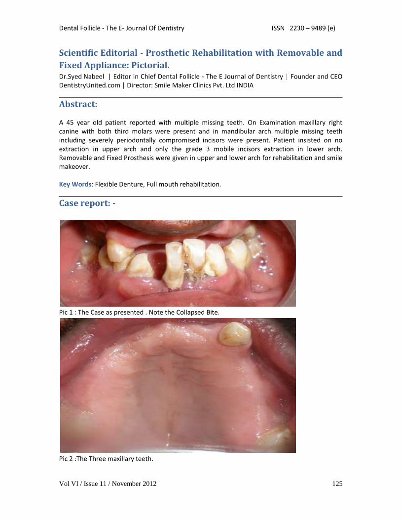
Abstract: A 45 year old patient reported with multiple missing teeth. On Examination maxillary right canine with both third molars were present and in mandibular arch multiple missing teeth including severely periodontally compromised incisors were present. Patient insisted on no extraction in upper arch and only the grade 3 mobile incisors extraction in lower arch. Removable and Fixed Prosthesis were given in upper and lower arch for rehabilitation and smile makeover. Key Words: Flexible Denture, Full mouth rehabilitation.
Case report: -
Pic 1 : The Case as presented . Note the Collapsed Bite.
Pic 2 :The Three maxillary teeth.
Dental Follicle - The E- Journal Of Dentistry ISSN 2230 – 9489 (e)
Vol VI / Issue 11 / November 2012 126
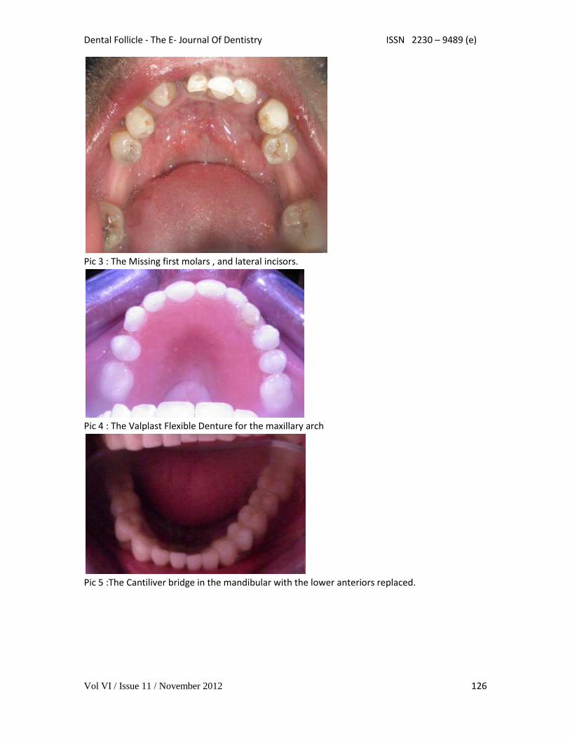
Pic 3 : The Missing first molars , and lateral incisors.
Pic 4 : The Valplast Flexible Denture for the maxillary arch
Pic 5 :The Cantiliver bridge in the mandibular with the lower anteriors replaced.
Dental Follicle - The E- Journal Of Dentistry ISSN 2230 – 9489 (e)
Vol VI / Issue 11 / November 2012 127
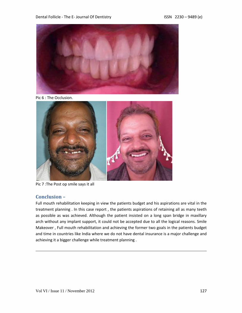
Pic 6 : The Occlusion.
Pic 7 :The Post op smile says it all
Conclusion – Full mouth rehabilitation keeping in view the patients budget and his aspirations are vital in the
treatment planning . In this case report , the patients aspirations of retaining all as many teeth
as possible as was achieved. Although the patient insisted on a long span bridge in maxillary
arch without any implant support, it could not be accepted due to all the logical reasons. Smile
Makeover , Full mouth rehabilitation and achieving the former two goals in the patients budget
and time in countries like India where we do not have dental insurance is a major challenge and
achieving it a bigger challenge while treatment planning .
Dental Follicle - The E- Journal Of Dentistry ISSN 2230 – 9489 (e)
Vol VI / Issue 11 / November 2012 128
Management of a gouged access cavity in a lateral incisor with a
Talon Cusp: A case report
Dr Imran Cassim|BDS |PG Dip Dent Endo|Private practice Pinetown Medicross |Durban
|South Africa
Abstract:
Mishaps such as perforation or gouging can occur during access cavity preparation when
endodontic therapy is initiated. The presence of anatomic anomalies of the crown of the tooth
can lead to complications during access preparation. This case report describes the correction of
a misaligned access cavity in a maxillary lateral incisor with a talon cusp.
Key-words: Talon Cusp, Access Cavity, Mishap.
Introduction Access cavity preparation is the most
important phase of nonsurgical endodontic
therapy. A well-designed access preparation
is important for an optimum endodontic
result. Without adequate access, adequate
instrumentation and obturation become
difficult in the complex and variable root
canal system. Inadequate access cavities
not only result in compromised preparation
and obturation but may also cause
procedural accidents such as chamber
perforation, canal ledging, gouging and root
perforation1. The ideal access cavity creates
a smooth, straight-line path to the canal
system and ultimately to the apex.
Sufficient tooth structure must be removed
to allow instruments to be placed easily into
each canal orifice without interference from
canal walls. Optimal access preparation
results in straight entry into the canal
orifice, with the line angles forming a
tapered funnel that drops smoothly into
one or more canals2.
An astute assessment of the inclination of
the tooth, root canal morphology, the
presence of caries, large restorations and
anomalies that may be present helps in
preventing mishaps during endodontic
therapy3. The maxillary lateral incisor may
present with developmental anomalies such
as Dens Invaginatus, peg shape, radicular
grooves and talon cusp4. The presence of
these anomalies could complicate access
cavity preparation. The talon cusp was first
reported by Mitchell in 19363 and
corresponds to abnormal development of
the cingulum of the maxillary incisor giving
the appearance of an extra cusp on the
palatal surface. The talon cusp has also
been described as Dens Evaginatus as it
appears to be the opposite of Dens
Invaginatus5. Nabeel et al. (2011) suggested
a precise and comprehensive classification
for talon cusp based on the extension of the
talon cusp and on the surface and anomaly
of the involved tooth6.

Dental Follicle - The E- Journal Of Dentistry ISSN 2230 – 9489 (e)
Vol VI / Issue 11 / November 2012 129
Case Report: A 26 year old Caucasian female patient was
referred for endodontic treatment of the
upper left lateral incisor. The referring
dentist stated that he had initiated access
cavity preparation but could not locate the
pulp chamber and was afraid of perforating
the tooth. Clinical examination revealed an
access cavity with a cotton pellet and the
outline of an evaginated cingulum on the
disto-palatal aspect of the 22 (Fig1.)Thermal
tests were negative, there was mild
tenderness to percussion and periodontal
probing depths were within normal limits.
Radiographic examination revealed the
presence of a periradicular radiolucency on
tooth 22, the radio-opaque outline of the
talon cusp and the outline of the misaligned
access cavity mesiodistally (Fig.2). From the
history the patient mentioned that she has
fallen off a slide and sustained trauma to
the front of her face and teeth when she
was twelve years old. A diagnosis of chronic
apical absess was made and the treatment
plan was to complete the endodontic
treatment over 2 visits with an inter-
appointment dressing of Calcium
Hydroxide.
Fig.1. A lingual view showing the access cavity with a cotton pellet in it and the black arrows
point to the outline of the remainder of the talon cusp.
Figure.2 The preoperative radiograph showing the periapical radiolucency, the outline of the
mis-aligned, gouged access cavity and the coronal radio-opacity depicting the remainder of the
talon cusp.

Dental Follicle - The E- Journal Of Dentistry ISSN 2230 – 9489 (e)
Vol VI / Issue 11 / November 2012 130
Looking at the inclination of the tooth with
respect to the neighbouring teeth and
alveolar contours and the radiograph, a line
was marked in pencil on the buccal aspect
of the tooth corresponding to the long axis
of the tooth. Next the distance from the
incisal edge to the the pulp chamber was
measured on the radiograph and this length
was transferred to an 016 tapered diamond
crown preparation bur (Komet, Germany)
using a permanent marker. Following
anaesthesia and rubber dam isolation the
hand piece with bur was aligned to the
marking on the buccal surface (fig. 4) and
then using a gentle pressure the access
cavity preparation was performed. At the
depth indicated on the bur a slight decrease
in apical pressure was felt, denoting the
penetration of the pulp chamber. This was
verified visually and then the access cavity
was flooded with 3% sodium hypochlorite
and a pre curved size 15 K-file(Dentsply
Bellaiguise, Switzerland) was used to scout
the canal and taken to resistance and
approximate working length. A radiograph
was taken to verify the placement of the file
(fig. 5). An apex locator (i-Pex, Nakanishi,
Japan) attached to a size 10 k-File was used
to verify and confirm the working length.
The canal was shaped using the Wave One
Large (Dentsply, Maillefer, Bellaiguise,
Switzerland). During shaping the Endovac
(Sybron, Endo, California) was used for
irrigation with the macro cannula during
preparation and the micro-cannula was
shaping was completed. A final rinse was
done with 17% EDTA and the canal dried
and Calcium Hydroxide placed (Ultracal,
Ultradent, USA).Duotemp (Coltene,
Whaledent)was used to seal the access
cavity and the patient was reappointed
after 6 weeks.
Figure 3. A line was made in pencil on the buccal surface of 22 corresponding to the
approximate midline and long axis of the tooth.
Dental Follicle - The E- Journal Of Dentistry ISSN 2230 – 9489 (e)
Vol VI / Issue 11 / November 2012 131
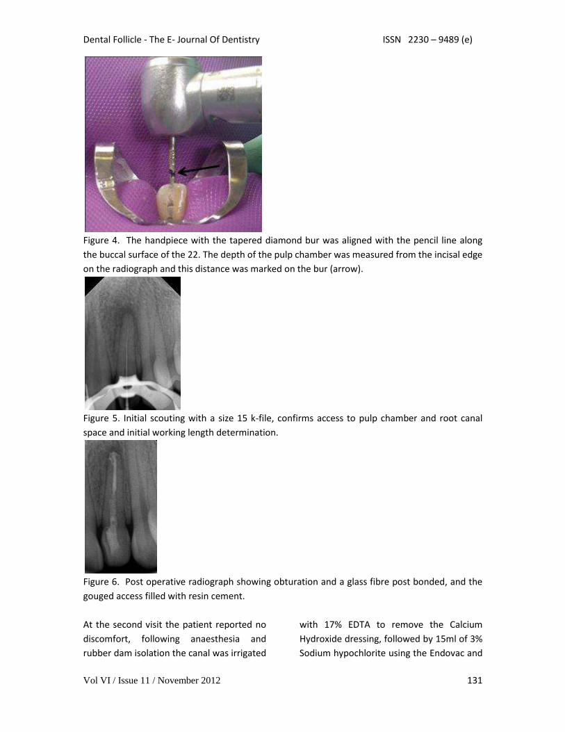
Figure 4. The handpiece with the tapered diamond bur was aligned with the pencil line along
the buccal surface of the 22. The depth of the pulp chamber was measured from the incisal edge
on the radiograph and this distance was marked on the bur (arrow).
Figure 5. Initial scouting with a size 15 k-file, confirms access to pulp chamber and root canal
space and initial working length determination.
Figure 6. Post operative radiograph showing obturation and a glass fibre post bonded, and the
gouged access filled with resin cement.
At the second visit the patient reported no
discomfort, following anaesthesia and
rubber dam isolation the canal was irrigated
with 17% EDTA to remove the Calcium
Hydroxide dressing, followed by 15ml of 3%
Sodium hypochlorite using the Endovac and

Dental Follicle - The E- Journal Of Dentistry ISSN 2230 – 9489 (e)
Vol VI / Issue 11 / November 2012 132
then a final rinse with 17% EDTA. The canal
was dried and obturated by multiple wave
warm vertical compaction using the
Calamus unit (Dentsply, Maillefer,
Bellaiguise, Switzerland). A post space was
created and a glass fibre post(Contec
Blanco, Hahnenkratt, Germany) was
bonded using RelyX Unicem
(3M,Germany).A post operative radiograph
was taken(fig. 6)
Discussion: According to Torabinejad and Lemon (2001)
in order to prevent mishaps during access
cavity preparation, an in depth knowledge
of tooth morphology, including both surface
and internal anatomy and their
relationships, is mandatory to prevent pulp
chamber perforations. Clinically, the
location and angulations of the tooth must
be related to adjacent teeth and alveolar
bone to avoid a misaligned access
preparation. Additionally, radiographs of
the teeth from different angles provide
better spatial information about the
orientation, size and extent of the pulp
chamber. The use of magnification and
illumination during endodontic therapy is a
useful if not essential adjunct to help avoid
mishaps because it greatly enhances
visibility of the working area 2, 7, 8. In
maxillary incisors the access cavity is
initiated by placing the bur occlusal to the
cingulum, almost perpendicular to the
palatal surface with a slight tilt towards the
long axis of the tooth. The cingulum is
chosen as a starting point, because, in
contrast to the gingival margin which can
retract and the incisal margin which can
undergo occlusal wear or erosion, this ridge
remains constant throughout the patient’s
life9. In lateral incisors with a talon cusp, the
evaginated cingulum can be cut back to
resemble shape of the cingulum of a normal
lateral incisor before beginning access
cavity preparation4. The common error of
perforating or gouging the gingivo-labial
aspect is usually due to two factors: not
allowing adequate access toward the incisal
aspect of the cavity preparation or not
properly aligning the bur vertically with the
long axis of the tooth. Another common
failure is not providing adequate access or
removal of the lingual shoulder10.
Conclusion: Careful assessment and meticulous
planning is important before access cavity
preparation. In Maxillary lateral incisors
with talon cusps, the cusp can be trimmed
down to resemble the cingulum of a normal
lateral incisor before endodontic treatment,
thereby establishing a familiar anatomy for
the clinician. Highlighting landmarks on the
tooth when anatomic anomalies are
present can help the clinician to orientate
themselves better and avoid mishaps during
access cavity preparation.
References: 1.Torabinejad M and Lemon RR. Procedural accidents.
In:Walton RE, TorabinejadM, eds. Principles and
practice of endodontics. 4thedn. Philadelphia: W. B.
Saunders, 2001.
2. Vertucci FJ, Haddix JE: Tooth morphology and
access cavity preparation In Cohen S, Hargreaves KM:
Pathways of the pulp .10th Ed. St Louis: The C.V.
Mosby Co. 2011.
Dental Follicle - The E- Journal Of Dentistry ISSN 2230 – 9489 (e)
Vol VI / Issue 11 / November 2012 133
3.Vertucci FJ and Walton RE: Internal Anatomy
In:Walton RE, TorabinejadM, eds. Principles and
practice of endodontics. 4thedn. Philadelphia: W. B.
Saunders, 2001.
4. Percora DJ, Sousa Neto MD, Saquy PC and Leite
APP. Endodontic treatment of a maxillary lateral
incisor with a Talon Cusp: Case report. Braz Dent J
1993; 4(2): 127-130
5. Percora JD, Vansan LP, Sousa Neto MD and Saquy
PC. Tratamento endodontico de um dens evaginatus.
Rev Ass Paul Cirurg Dent 1991; 45; 535-536.
6. Nabeel S, Hegde U, Mull P, Danish, G. Talon Cusp
Affecting Two Generations: Report of Two Cases and
Proposed Comprehensive Classification. International
Journal of Oral and Maxillofacial Pathology, North
America, 2, jun. 2011. Available at:
<http://journalgateway.com/index.php/ijomp/article
/view/2.3.10.36>. Date accessed: 17 Nov. 2012.
7. West J. Endodontic update. J Esthet Restor Dent
2006; 18: 280-300.
8. Castellucci A. Mgnification in endodontics: the use
of the operating microscope. Endod Practice 2003;
Sept:15-22
9. Castellucci A. Endodontics.1st ed, Vol 1. Florence,
Italy Odontoiatriche Il Tridente , 2005
10. Ingle JI. PDQ Endodontics. 1st ed. Hamilton,
London. BC Decker Inc, 2005
Dental Follicle - The E- Journal Of Dentistry ISSN 2230 – 9489 (e)
Vol VI / Issue 11 / November 2012 134
Improving Endodontic Success through Coronal Leakage
Prevention Dr. Gregori M. Kurtzman |DDS |MAGD |FACD |FPFA |FADI |DICOI |DADIA |
Abstract: Coronal leakage is a frequently overlooked cause for endodontic failure and relates to both the
restoration present in the coronal portion of the tooth and the materials used to obturate the
canal system. This article will address materials and techniques to prevent coronal leakage and
improve the long term prognosis of endodontic treatment.
Key words: coronal leakage, endodontics, obturation
Introduction Endodontic failure has been associated with
coronal leakage within the canal system
following obturation. The literature
suggests that coronal leakage is far more
likely a determinant of clinical success or
failure then apical leakage.1 Recent
advances in resin obturation materials have
been shown to provide superior sealing of
the canal system but without addressing
the coronal aspect of the tooth, failure
endodontically may occur. Studies confirm
that a sound coronal seal is of paramount
importance to the overall success of root
canal treatment.2, 3 Regardless of the
obturation method the best rule is: a
properly cleaned, shaped, and obturated
tooth should be permanently restored as
soon as possible.4
No matter what our intentions are following
obturation of the canal system, patients
may delay restoration of the tooth that has
been treated. Financial and time constraints
often influence when the final restoration is
completed. Additionally, between visits an
adhesive material will prevent leakage and
contamination of the canal.
Coronal leakage
Coronal leakage has been indicated in the
literature as the major determinant of
endodontic success or failure. No matter
what we place in the canal, if the coronal
portion of the tooth is not sealed with
materials that bond to tooth structure and
are resistant to dissolution by oral fluids,
then, over time endodontic failure may be
inevitable.
It is not unusual to have a patient present
with decay at the margin of a crown of a
tooth that had prior endodontic therapy.
Because the tooth was treated
endodontically, sensitivity that may indicate
a problem under the crown will not alert
the patient to seek dental care. Coronal
leakage for even a minimal amount of time
may quickly lead to apical migration of
bacteria. When the patient does present
coronal leakage may have been ongoing for
an extended period of time complicating
treatment or rendering the tooth non-
restorable necessitating extraction.
The literature indicates significant coronal
dye and bacterial leakage following
exposure of sealed root canals to artificial

Dental Follicle - The E- Journal Of Dentistry ISSN 2230 – 9489 (e)
Vol VI / Issue 11 / November 2012 135
and natural saliva leading to complete
bacterial leakage may occur within 2 days.5
Supported in an invitro study, found that
dye leakage can occur in as little as three (3)
days.6 It has been suggested that gutta-
percha does not offer an effective barrier to
crown-down leakage when exposed to the
oral environment.7 Additional studies using
gutta percha and various sealers, indicate
that gutta percha will allow bacterial
leakage. But use of an adhesive sealer can
significantly slow or stop coronal-apical
bacterial migration.8
The predominant bacteria found in root-
filled teeth with coronal leakage and
persistent apical periodontitis is the Gram-
positive facultative anaerobe
Staphylococcus. This is followed by the
groups Streptococcus and Enterococcus; all
normal salivary flora.9 Coronal leakage
provides a constant source of
microorganisms and nutrients that initiate
and maintain periradicular inflammation
and may well be the largest cause of failure
in endodontic therapy.10
Endodontic obturation materials do not
prevent coronal microleakage for an
indefinite period of time.11 In a sample of
937 root filled teeth which had not received
restorative treatment during the previous
year, the data showed that the technical
standard of both coronal restoration and
root filling were essential to periapical
health.12 It is not uncommon for coronal
leakage to occur following root canal
treatment as a result of the presence of a
deficient composite resin fillings and
secondary caries under restorations.13
Yet the endodontic materials utilized over
the past fifty (50) years have shown that
they do not prevent coronal leakage when
challenged. In yet another investigation,
forty-five root canals were cleaned, shaped,
and then obturated with gutta-percha and
root canal sealer, using a lateral
condensation technique. The coronal
portions of the root filling materials were
placed in contact with Staphylococcus
epidermidis and Proteus vulgaris. The
number of days required for these bacteria
to penetrate the entire root canals was
determined. Over 50% of the root canals
were completely contaminated after 19-day
exposure to S. epidermidis. Fifty percent of
the root canals were also totally
contaminated when the coronal surfaces of
their fillings were exposed to P. vulgaris for
42 days.14 When comparing AH-26 and
other commonly used sealers after 45 days
exposure to the oral cavity, none of the
sealers was capable of preventing leakage
and coronal dye penetration.15 So we can
see that the quality of both the coronal
restoration and obturation material are
essential to periapical health as none of the
present-day root canal sealers may
hermetically seal "the root canal wall—
gutta percha filling interface". In this
respect the importance of perfectly sealing
coronal restorations (both temporary and
permanent) needs to be emphasized.16
Pre-Endodontic Therapy Buildups (Canal
Projection)
Coronal leakage is a major contributor to
Endodontic failure.17 A bonded core placed
prior to disinfection and obturation of the
canal system of the tooth can greatly
diminish the leakage potential both during
and after Endodontic therapy.
Dental Follicle - The E- Journal Of Dentistry ISSN 2230 – 9489 (e)
Vol VI / Issue 11 / November 2012 136
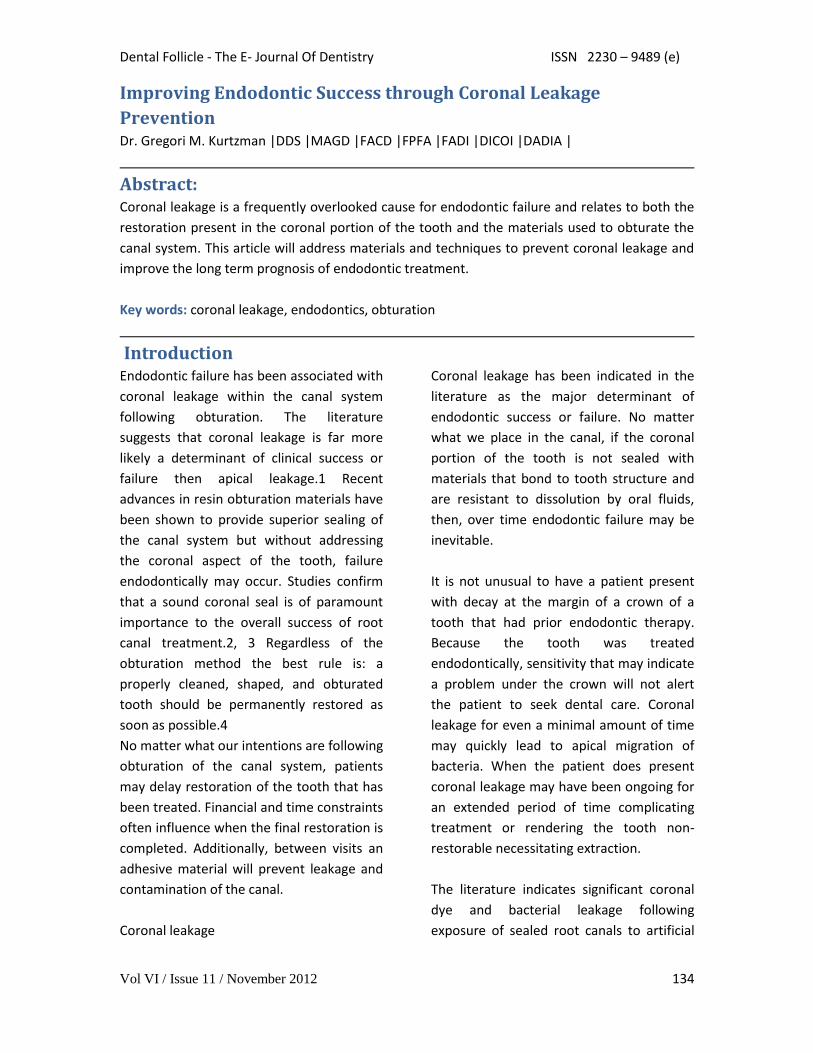
Isolation of the pulp chamber can be a
challenging task when minimal coronal
structure remains and Endodontic therapy
is required as part of the oral rehabilitation.
(Figure 1) Coronal reinforcement has
traditionally been addressed following the
Endodontic phase. But a coronal bonded
buildup can simplify the Endodontic phase
and strengthen the tooth, decreasing the
possibility of further damage to the tooth
due to the dam clamp or mastication before
a full coverage restoration can be placed.
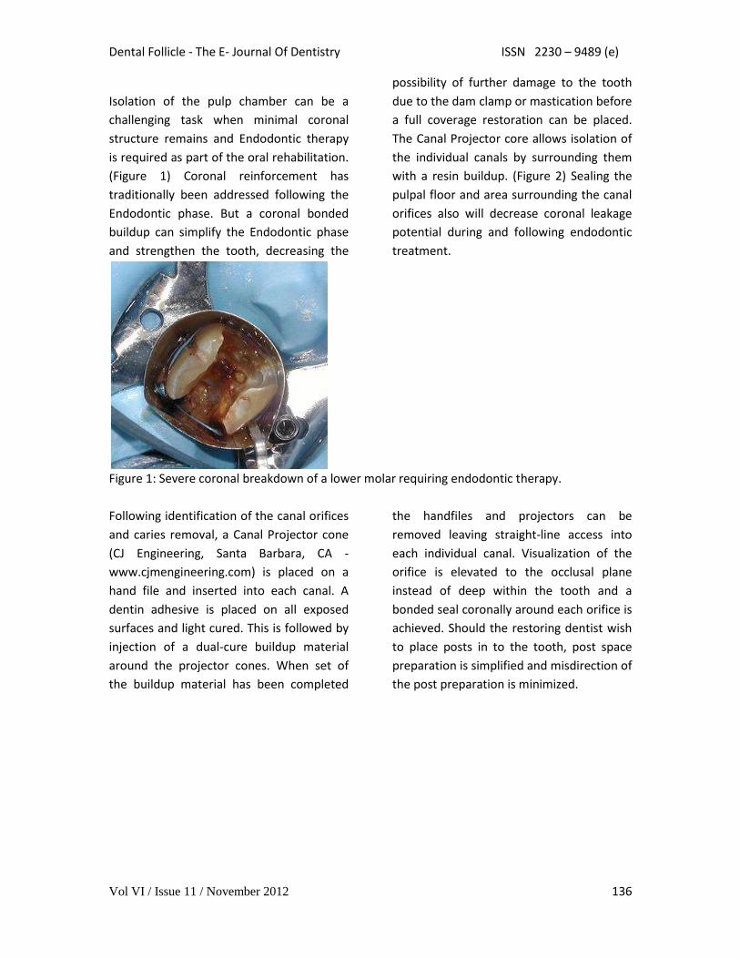
The Canal Projector core allows isolation of
the individual canals by surrounding them
with a resin buildup. (Figure 2) Sealing the
pulpal floor and area surrounding the canal
orifices also will decrease coronal leakage
potential during and following endodontic
treatment.
Figure 1: Severe coronal breakdown of a lower molar requiring endodontic therapy.
Following identification of the canal orifices
and caries removal, a Canal Projector cone
(CJ Engineering, Santa Barbara, CA -
www.cjmengineering.com) is placed on a
hand file and inserted into each canal. A
dentin adhesive is placed on all exposed
surfaces and light cured. This is followed by
injection of a dual-cure buildup material
around the projector cones. When set of
the buildup material has been completed
the handfiles and projectors can be
removed leaving straight-line access into
each individual canal. Visualization of the
orifice is elevated to the occlusal plane
instead of deep within the tooth and a
bonded seal coronally around each orifice is
achieved. Should the restoring dentist wish
to place posts in to the tooth, post space
preparation is simplified and misdirection of
the post preparation is minimized.
Dental Follicle - The E- Journal Of Dentistry ISSN 2230 – 9489 (e)
Vol VI / Issue 11 / November 2012 137
Figure 2: Coronal pre-endodontic buildup achieved with Canal Projectors providing individual
straight-line access into each canal.
Coronal Restoration (Access sealing)
Microorganisms can penetrate through
different temporary restorative materials
and supposedly well obturated root canals.
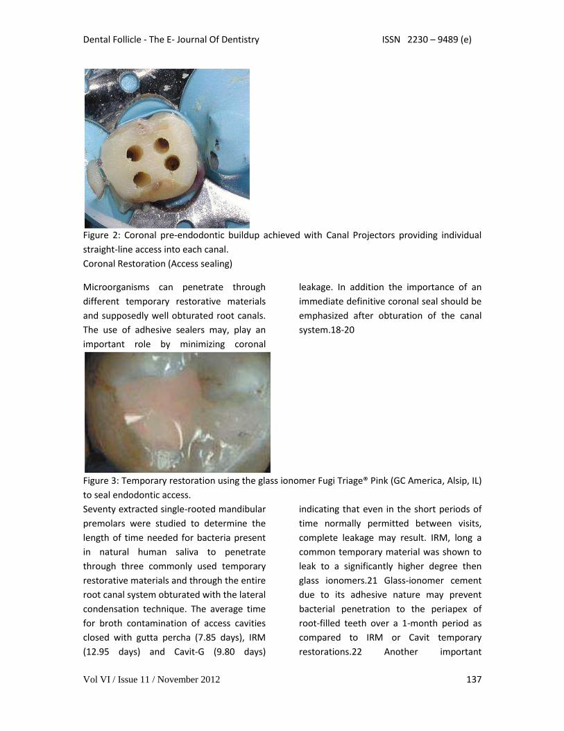
The use of adhesive sealers may, play an
important role by minimizing coronal
leakage. In addition the importance of an
immediate definitive coronal seal should be
emphasized after obturation of the canal
system.18-20
Figure 3: Temporary restoration using the glass ionomer Fugi Triage® Pink (GC America, Alsip, IL)
to seal endodontic access.
Seventy extracted single-rooted mandibular
premolars were studied to determine the
length of time needed for bacteria present
in natural human saliva to penetrate
through three commonly used temporary
restorative materials and through the entire
root canal system obturated with the lateral
condensation technique. The average time
for broth contamination of access cavities
closed with gutta percha (7.85 days), IRM
(12.95 days) and Cavit-G (9.80 days)
indicating that even in the short periods of
time normally permitted between visits,
complete leakage may result. IRM, long a
common temporary material was shown to
leak to a significantly higher degree then
glass ionomers.21 Glass-ionomer cement
due to its adhesive nature may prevent
bacterial penetration to the periapex of
root-filled teeth over a 1-month period as
compared to IRM or Cavit temporary
restorations.22 Another important
Dental Follicle - The E- Journal Of Dentistry ISSN 2230 – 9489 (e)
Vol VI / Issue 11 / November 2012 138
consideration with regard to the temporary
restoration’s ability to prevent coronal
leakage is how the material behaves under
mechanical load and thermocycling. Non-
adhesive temporaries show an increased
percentage of marginal breakdown and
increased microleakage after thermocycling
and loading. There was no significant
improvement with increased thickness of
the temporary material.23-25 When crowns
were sealed with IRM, recontamination was
detected within 13.5 days in the canals
medicated with chlorhexidine, after 17.2
days in the group medicated with CaOH2
and after 11.9 days in the group medicated
with both chlorhexidine and CaOH2. The
group with no medication, but sealed with
IRM, showed recontamination after 8.7
days. There were statistically significant
differences between the teeth with or
without coronal seal. The coronal seal
delayed but did not prevent leakage of
microorganisms.26 Other studies, confirm
that IRM started to leak after ten (10) days,
whereas Cavit and Dyract leaked after two
(2) weeks.27
The use of a resin based temporary
restorative material or glass ionomer over
partially removed resin composite
restorations could be beneficial in achieving
better resistance to marginal leakage.
(Figure 3) Maintaining partially removed
permanent restorations does not seem to
cause a problem with achieving marginal
seal.28 Glass ionomer provided a
statistically better coronal seal then bonded
composite or a bonded amalgam
preventing bacterial apical migration.29
This may be due to the glass ionomers
ability to adhere to the scerlotic dentin
found on the pulpal floor better then
adhesive resins. The key seems to be, lock
out the coronal bacteria and the apical area
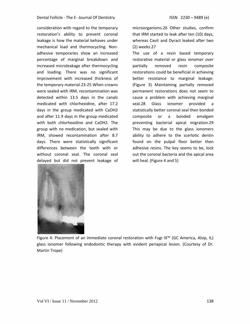
will heal. (Figure 4 and 5)
Figure 4: Placement of an immediate coronal restoration with Fugi IX™ (GC America, Alsip, IL)
glass ionomer following endodontic therapy with evident periapical lesion. (Courtesy of Dr.
Martin Trope)

Dental Follicle - The E- Journal Of Dentistry ISSN 2230 – 9489 (e)
Vol VI / Issue 11 / November 2012 139
Figure 5: Coronal seal has been maintained allowing apical healing of periapical lesion one year
following treatment. (Courtesy of Dr. Martin Trope)
Mineral Trioxide Aggregate (MTA) has since its introduction a few years ago been advocated as a
sealing material especially when perforation has occurred. But an investigation found mild
inflammation was observed in 17% and 39% of the roots with and without an orifice plug,
respectively without development of severe inflammation, the sealing efficacy of MTA orifice
plugs could not be determined.30
Should amalgam be the material of choice for the dentist, a bonded amalgam produced
significantly less leakage than did the non-bonded amalgams. To prevent the reinfection of the
endodontically treated molar, it may be preferable to restore the tooth immediately after
obturation by employing a bonded amalgam coronal-radicular technique.31 Whereas, core
buildup or access closure, with adhesive materials has shown good long term leakage resistance.
The "sandwich" technique (GI base with overlaying composite) and the composite resin
restorations allowed significantly less coronal leakage than glass ionomer cement restorations.
This may be because the composite resin prevents salivary dissolution of the glass ionomer long
term.32
Figure 6: The pulp chamber has been etched and an adhesive applied to all surfaces.
Dental Follicle - The E- Journal Of Dentistry ISSN 2230 – 9489 (e)
Vol VI / Issue 11 / November 2012 140
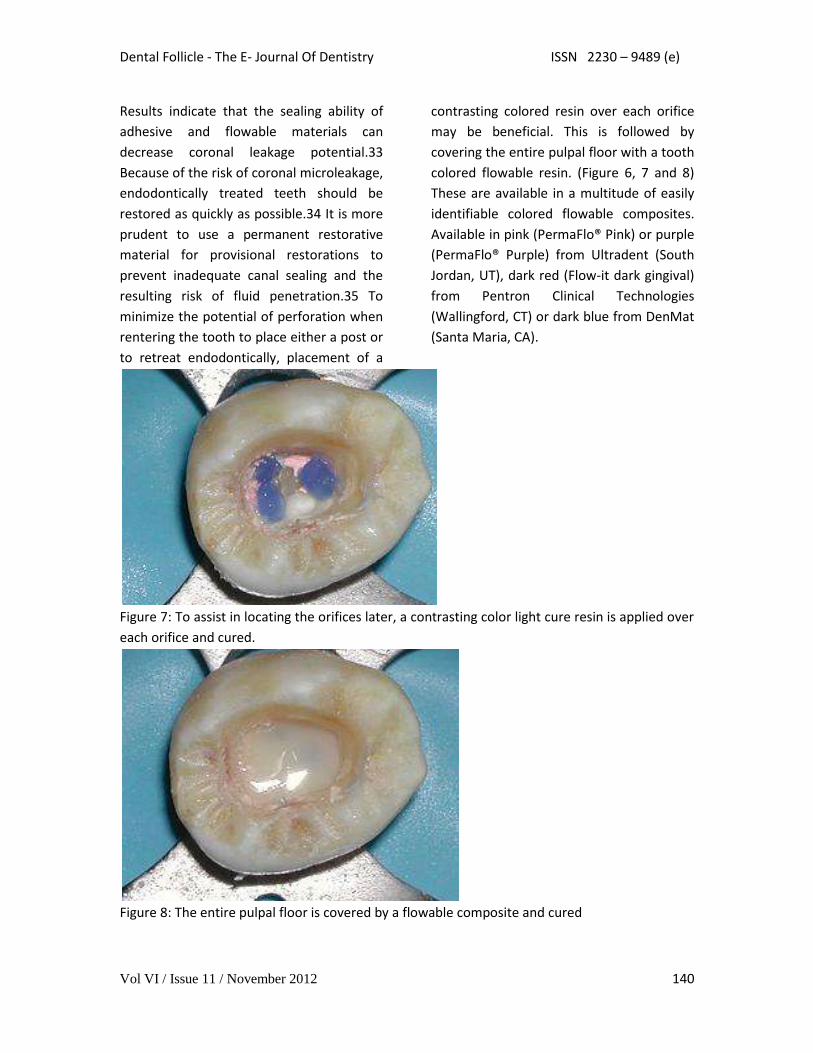
Results indicate that the sealing ability of
adhesive and flowable materials can
decrease coronal leakage potential.33
Because of the risk of coronal microleakage,
endodontically treated teeth should be
restored as quickly as possible.34 It is more
prudent to use a permanent restorative
material for provisional restorations to
prevent inadequate canal sealing and the
resulting risk of fluid penetration.35 To
minimize the potential of perforation when
rentering the tooth to place either a post or
to retreat endodontically, placement of a
contrasting colored resin over each orifice
may be beneficial. This is followed by
covering the entire pulpal floor with a tooth
colored flowable resin. (Figure 6, 7 and 8)
These are available in a multitude of easily
identifiable colored flowable composites.
Available in pink (PermaFlo® Pink) or purple
(PermaFlo® Purple) from Ultradent (South
Jordan, UT), dark red (Flow-it dark gingival)
from Pentron Clinical Technologies
(Wallingford, CT) or dark blue from DenMat
(Santa Maria, CA).
Figure 7: To assist in locating the orifices later, a contrasting color light cure resin is applied over
each orifice and cured.
Figure 8: The entire pulpal floor is covered by a flowable composite and cured

Dental Follicle - The E- Journal Of Dentistry ISSN 2230 – 9489 (e)
Vol VI / Issue 11 / November 2012 141
Coronal microleakage has received
considerable attention as a factor related to
failure of endodontic treatment and much
emphasis is placed on the quality of the
final restoration. Intracanal posts are
frequently used for the retention of coronal
restorations. Many authors have examined
coronal microleakage with respect to gutta-
percha root fillings and coronal
restorations, but few have investigated the
coronal seal afforded by various post
systems. The seal provided by a cemented
post depends on the seal of the cement
used. It appears that the dentine-bonding
cements (adhesive resins and glass
ionomers) have less microleakage than the
traditional, non-dentine-bonding cements
(i.e. zinc phosphates and
polycarboxolates).36 Resin-supported
polyethylene fiber and glass fiber dowels
showed the lowest coronal leakage when
compared with stainless steel and zirconia
dowels. This may be due to better adhesion
of the luting agent to these resin
impregnated posts then metal or ceramic
posts which do not allow adhesive
penetration into the surface of the post.
There were no significant differences
between resin-supported polyethylene fiber
and glass fiber dowels at any time period.
The initial leakage measurement in zirconia
dowel and stainless steel dowels were
similar but became significantly different at
3 and 6 months. Resin-supported
polyethylene fiber dowels and glass fiber
dowels tested exhibited less microleakage
compared to zirconia dowel systems.37
Cleansening the Canal (Smear Layers)
Coronal sealing ability is not the only factor
to influence the seal of the canal and
prevent apical leakage. How well the sealer
adheres to the canal walls is also important.
Smear layer can play a factor which may
prevent sealer penetration into the dentinal
tubules. The frequency of bacterial
penetration through teeth obturated with
intact smear layer (70%) was-significantly
greater than that of teeth from which the
smear layer had been removed (30%).
Removal of the smear layer enhanced
sealability as evidenced by increased
resistance to bacterial penetration. 38 The
incidence of apical leakage was reduced in
the absence of the smear and the
adaptation of gutta-percha was improved
no matter what obturation method was
used later.39-41 However, regardless of the
obturation technique (Thermoplastized,
lateral or vertical condensation or single
cone) when a non-adhesive sealer was used
leakage increased after 30 days.42

Dental Follicle - The E- Journal Of Dentistry ISSN 2230 – 9489 (e)
Vol VI / Issue 11 / November 2012 142
Figure 9: Periapical lesions present associated with lower premolar and molar obturated with
Resilon system at completion of endodontic treatment. (Courtesy of Dr. Joseph Maggio)
What is used to obturate the canals is
important, however the manner in which
the canal was prepared prior to obturation
also determines how well the canal is
sealed when therapy is completed. Rotary
instrumentation with NiTi files has shown
less microleakage then hand instrument
prepared canals irrespective of what was
used to obturate the canal.43 The
machining of the canal walls with NiTi
rotary instruments provides smoother canal
walls and shapes that are easier to obturate
then can be achieved with stainless steel
files. The better the adaptation of the
obturation material to the instrumented
dentinal walls, the less leakage is to be
expected along the entire root length. The
better the canal walls are prepared, the
more smear layer and organic debris is
removed which is beneficial to root canal
sealing.
Smear layer removal is best achieved by
irrigating the canals with NaOCL (sodium
hypochlorite) followed but 17% EDTA
solution.44 Whereas, the NaOCL dissolves
the organic component of the smear layer
exposing the dentinal tubules lining the
canal walls. EDTA, a chelating agent,
dissolves the inorganic portion of the dentin
opening the dentinal tubules. Alternating
between the two irrigants as the
instrumentation is being performed will
permit removal of more organic debris
further into the tubules, increasing
resistance to bacterial penetration once the
canal is obturated.45, 46
Obturation The purpose of the obturation phase of a
endodontic therapy is two-fold; to prevent
microorganisms from re-entering the root
canal system, and to isolate any
microorganisms that may remain within the
tooth from nutrients in tissue fluids. No
matter how well we seal the canal, if the
coronal portion of the tooth is not
thoroughly sealed then bacterial leakage
may be a matter of time. Accessory canals
maybe present in the pulp chamber leading
to the furcation area. This may be an
additional source of leakage that often goes
unaddressed either following obturation of
the canals or during the restorative phase.
Placement of a layer of resin-modified glass
ionomer cement or adhesive resin to seal
this area immediately following obturation
can prevent leakage prior to final
restoration of the tooth.47 But, it must
always be remembered that success will
only be achieved if the root canal system
has been as thoroughly debrided as possible
of infected material. Irrigation is key, to
removal of this smear layer lining the canal
walls.

Dental Follicle - The E- Journal Of Dentistry ISSN 2230 – 9489 (e)
Vol VI / Issue 11 / November 2012 143
Figure 10: Seven months post completion of endodontic treatment, showing lose of coronal
restorations, yet apical lesions seen previously have resolved significantly. (Courtesy of Dr.
Joseph Maggio)
The obturation material is a two pronged
sword. What sealer is used is as important
as which core material is placed within the
canal. Gutta percha has limitations in
resistance to coronal leakage which have
been overcome with the newer resin
alternatives. Although sealers can form
close adhesion to the root canal wall, none
is able to bond to the gutta percha core
material. Upon setting, shrinkage of the
sealer allows the sealer to pull away from
the gutta percha core, leaving a microgap
gap through which bacteria may pass.48
Several alternatives are available for core
material selection.
Resilon™, a resin gutta percha alternative
that is bondable with methacrylic sealers
such as Epiphany™ (Pentron Clinical
Technologies, Wallingford, CT) and
RealSeal™ (SybronEndo, Orange, CA) was
introduced three years ago after extensive
studies. The core material Resilon™, is
available in .02, .04 or .06 taper ISO sized
cones from Pentron Clinical Technologies
(Wallingford, CT) or SybronEndo (Orange
CA) and as sized apical plugs (Lightspeed
Technologies, San Antonio, TX).49, 50
Resilon™ showed significantly less leakage
than gutta percha. In studies performed at
University of North Carolina, the gutta
percha group demonstrated leakage in 80%
of specimens when and was not dependant
on obturation technique nor which sealer
was used.51 Because of these limitations
seen with gutta percha, the seal of a
coronal restoration may be as important as
the gutta percha fill in preventing
reinfection of the root canal. Studies have
shown that leakage of bacteria with
Resilon™ is significantly reduced compared
with gutta percha. The significance of this is
should the coronal break down the
adhesive obturation material may slow
down or prevent apical migration of
bacteria allowing healing to occur. (Figure 9
and 10) An additional benefit when filling
the canals with the new resin-based
obturation material an increase was
observed in the invitro resistance to
fracture of endodontically treated single-
canal extracted teeth when compared with
standard gutta percha techniques. Resilon™
demonstrated a twenty-five (25) percent
increase in root strength than gutta percha
samples.52
Dental Follicle - The E- Journal Of Dentistry ISSN 2230 – 9489 (e)
Vol VI / Issue 11 / November 2012 144
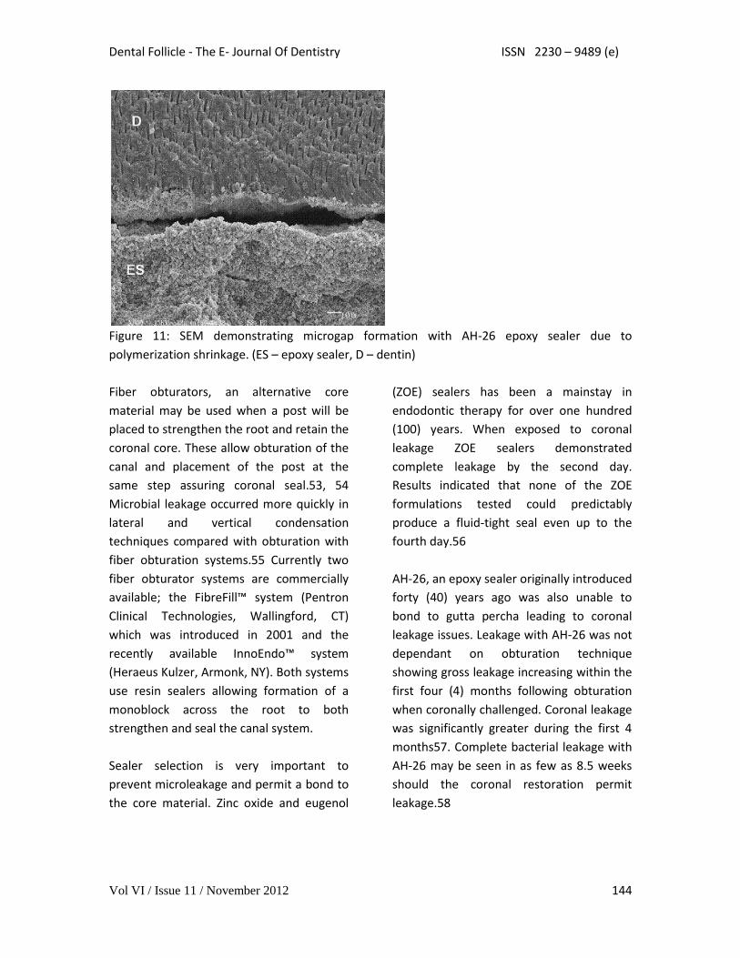
Figure 11: SEM demonstrating microgap formation with AH-26 epoxy sealer due to
polymerization shrinkage. (ES – epoxy sealer, D – dentin)
Fiber obturators, an alternative core
material may be used when a post will be
placed to strengthen the root and retain the
coronal core. These allow obturation of the
canal and placement of the post at the
same step assuring coronal seal.53, 54
Microbial leakage occurred more quickly in
lateral and vertical condensation
techniques compared with obturation with
fiber obturation systems.55 Currently two
fiber obturator systems are commercially
available; the FibreFill™ system (Pentron
Clinical Technologies, Wallingford, CT)
which was introduced in 2001 and the
recently available InnoEndo™ system
(Heraeus Kulzer, Armonk, NY). Both systems
use resin sealers allowing formation of a
monoblock across the root to both
strengthen and seal the canal system.
Sealer selection is very important to
prevent microleakage and permit a bond to
the core material. Zinc oxide and eugenol
(ZOE) sealers has been a mainstay in
endodontic therapy for over one hundred
(100) years. When exposed to coronal
leakage ZOE sealers demonstrated
complete leakage by the second day.
Results indicated that none of the ZOE
formulations tested could predictably
produce a fluid-tight seal even up to the
fourth day.56
AH-26, an epoxy sealer originally introduced
forty (40) years ago was also unable to
bond to gutta percha leading to coronal
leakage issues. Leakage with AH-26 was not
dependant on obturation technique
showing gross leakage increasing within the
first four (4) months following obturation
when coronally challenged. Coronal leakage
was significantly greater during the first 4
months57. Complete bacterial leakage with
AH-26 may be seen in as few as 8.5 weeks
should the coronal restoration permit
leakage.58
Dental Follicle - The E- Journal Of Dentistry ISSN 2230 – 9489 (e)
Vol VI / Issue 11 / November 2012 145
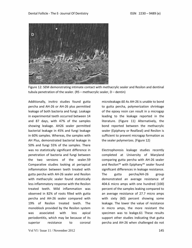
Figure 12: SEM demonstrating intimate contact with methacrylic sealer and Resilon and dentinal
tubula penetration of the sealer. (RS – methacrylic sealer, D – dentin)
Additionally, invitro studies found gutta
percha and AH-26 or AH-26 plus permitted
leakage of both bacteria and fungi. Leakage
in experimental teeth occurred between 14
and 87 days, with 47% of the samples
showing leakage. AH26 sealer permitted
bacterial leakage in 45% and fungi leakage
in 60% samples. Whereas, the samples with
AH Plus, demonstrated bacterial leakage in
50% and fungi 55% of the samples. There
was no statistically significant difference in
penetration of bacteria and fungi between
the two versions of the sealer.59
Comparative studies looking at periapical
inflammation between teeth treated with
gutta percha with AH-26 sealer and Resilon
with methacrylic sealer found statistically
less inflammatory response with the Resilon
treated teeth. Mild inflammation was
observed in 82% of roots filled with gutta
percha and AH-26 sealer compared with
19% of Resilon treated teeth. The
monoblock provided by the Resilon system
was associated with less apical
periodontitis, which may be because of its
superior resistance to coronal
microleakage.60 As AH-26 is unable to bond
to gutta percha, polymerization shrinkage
of the epoxy resin can result in a microgap
leading to the leakage reported in the
literature. (Figure 11) Alternatively, the
bond reported between the methacrylic
sealer (Epiphany or RealSeal) and Resilon is
sufficient to prevent microgap formation as
the sealer polymerizes. (Figure 12)
Electrophoresis leakage studies recently
completed at University of Maryland
comparing gutta percha with AH-26 sealer
and Resilon™ with Epiphany™ sealer found
significant differences in leakage resistance.
The gutta percha/AH-26 group
demonstrated an average resistance of
404.6 micro amps with one hundred (100)
percent of the samples leaking compared to
an average resistance of 27.7 micro amps
with sixty (60) percent showing some
leakage. The lower the value of resistance
in micro amps, the more resistant the
specimen was to leakge.61 These results
support other studies indicating that gutta
percha and AH-26 when challenged do not
Dental Follicle - The E- Journal Of Dentistry ISSN 2230 – 9489 (e)
Vol VI / Issue 11 / November 2012 146
offer resistance to coronal leakage. Should
the practitioner wish to continue using
these materials a permanent restoration
needs to be placed at the appointment
when endodontic therapy is completed?
Conclusion Of 41 articles published between 1969 and
1999 (the majority from the 1990s) the
literature suggests that the prognosis of
root canal-treated teeth can be improved
by sealing the canal and minimizing the
leakage of oral fluids and bacteria into the
periradicular areas as soon as possible after
the completion of root canal therapy62.
Endodontic success is a multifactoral issue.
Like a jigsaw puzzle, the full picture can only
be seen when all the pieces are fit together.
How the canals are instrumented is as
important as what is used to obturate the
canal system. This is also influenced by
what is placed coronally and when the
coronal aspect is sealed. NiTi rotary
instruments and an irrigation protocol that
includes NaOCL and EDTA will maximize the
sealing ability of glass ionomer or the newer
methacrylic resin sealers. The last piece of
the puzzle, sealing coronally should be
performed with adhesive permanent
restorative materials immediately at the
conclusion of the first endodontic
appointment to prevent apical migration of
bacteria and assure sealability of the canals.
Author Information Dr. Gregori Kurtzman is in private practice
in Silver Spring, Maryland and is an
Assistant Clinical Professor at the University
of Maryland Baltimore College of Dental
Surgery, Department of Endodontics,
Prosthetics and Operative Dentistry. He has
lectured both nationally and internationally
on the topics of Restorative dentistry,
Endodontics and dental implant surgery and
prosthetics and has had numerous journal
articles published in peer reviewed
publications. Dr. Kurtzman is on the
editorial board of numerous publications.
He is a consultant and clinical evaluator to
multiple dental manufacturers. He has
earned Fellowships in the Academy of
General Dentistry, the International
Congress of Oral Implantologists, the Pierre
Fauchard Academy, American College of
Dentists, Masterships in The Academy of
General Dentistry and the Implant
Prosthetic Section of the International
Congress of Oral Implantologists.
Additionally, a former Assistant Program
Director for a University based implant
maxi-course.
References 1. Sritharan A.: Discuss that the coronal seal is more
important than the apical seal for endodontic success.
Aust Endod J. 2002 Dec;28(3):112-5.
2. Begotka BA, Hartwell GR.: The importance of the
coronal seal following root canal treatment. Va Dent
J. 1996 Oct-Dec;73(4):8-10
3. Siqueira JF Jr, Rocas IN, Favieri A, Abad EC, Castro
AJ, Gahyva SM.: Bacterial leakage in coronally
unsealed root canals obturated with 3 different
techniques. Oral Surg Oral Med Oral Pathol Oral
Radiol Endod. 2000 Nov;90(5):647-50
Dental Follicle - The E- Journal Of Dentistry ISSN 2230 – 9489 (e)
Vol VI / Issue 11 / November 2012 147
4. Pommel L, Camps J.: In vitro apical leakage of
system B compared with other filling techniques. J
Endod. 2001 Jul;27(7):449-51
5. Khayat A, Lee SJ, Torabinejad M.: Human saliva
penetration of coronally unsealed obturated root
canals. J Endod 1993 Sep;19(9):458-61.
6. Swanson K, Madison S.: An evaluation of coronal
microleakage in endodontically treated teeth. Part I.
Time periods. J Endod. 1987 Feb;13(2):56-9.
7. Cohen S, Burns R.: Pathways to the Pulp. 8th
edition, CV Mosby, New York, 2001.
8. Britto LR, Grimaudo NJ, Vertucci FJ.: Coronal
microleakage assessed by polymicrobial markers. J
Contemp Dent Pract. 2003 Aug 15;4(3):1-10.
9. Adib V, Spratt D, Ng YL, Gulabivala K.: Cultivable
microbial flora associated with persistent periapical
disease and coronal leakage after root canal
treatment: a preliminary study. Int Endod J. 2004
Aug;37(8):542-51.
10. J.E. Leonard; J.L. Gutmann; I.Y. Guo.: Apical and
coronal seal of roots obturated with a dentine
bonding agent and resin. Inter Endod J 1996 29.76-83
11. Pisano D; DiFiore P; McClanahan S;
Lautenschlager E; Duncan J.: Intraorific Sealing of
Gutta-Percha Obturated Root Canal to Prevent
Coronal Microleakage. J Endod 1998 Oct;10.
12. De Moor R, Coppens C, Hommez G.: Coronal
leakage reconsidered. Rev Belge Med Dent.
2002;57(3):161-85.
13. Chong BS.: Coronal leakage and treatment failure.
J Endod. 1995 Mar;21(3):159-60.
14. Torabinejad M, Ung B, Kettering JD.: In vitro
bacterial penetration of coronally unsealed
endodontically treated teeth. J Endod. 1990
Dec;16(12):566-9.
15. Kopper PM, Figueiredo JA, Della Bona A, Vanni JR,
Bier CA, Bopp S.: Comparative in vivo analysis of the
sealing ability of three endodontic sealers in post-
prepared root canals. Int Endod J. 2003
Dec;36(12):857-63.
16. De Moor R, Coppens C, Hommez G.: Coronal
leakage reconsidered, Rev Belge Med Dent.
2002;57(3):161-85. De Moor R, Hommez G.: The
importance of apical and coronal leakage in the
success or failure of endodontic treatment, Rev Belge
Med Dent. 2000;55(4):334-44.
17. Kurtzman GM.: Restoring Teeth with Severe
Coronal Breakdown as a Prelude to Endodontic
Therapy. Endodontic Therapy, 2004.
18. Imura N, Otani SM, Campos MJA, Jardim EG, Zuolo
ML.: Bacterial penetration through temporary
restorative materials in root-canal-treated teeth in
vitro. Inter Endod J 1997 30,381-385
19. Uranga A, Blum JY, Esber S, Parahy E, Prado C.: A
comparative study of four coronal obturation
materials in endodontic treatment. J Endod. 1999
Mar;25(3):178-80.
20. Fox K, Gutteridge DL.: An in vitro study of coronal
microleakage in root-canal- treated teeth restored by
the post and core technique. Int Endod J 1997
Nov;30(6):361-8
21. Barthel CR, Zimmer S, Wussogk R, Roulet JF.:
Long-Term bacterial leakage along obturated roots
restored with temporary and adhesive fillings. J
Endod. 2001 Sep;27(9):559-62
22. Barthel CR, Strobach A, Briedigkeit H, Gobel UB,
Roulet JF.: Leakage in roots coronally sealed with
different temporary fillings. J Endod. 1999
Nov;25(11): 731-4
23. Mayer T, Eickholz P.: Microleakage of temporary
restorations after thermocycling and mechanical
loading. J Endod. 1997 May;23(5):320-2
24. Deveaux E, Hildelbert P, Neut C, Boniface B,
Romond C.: Bacterial microleakage of Cavit, IRM, and
TERM. Oral Surg Oral Med Oral Pathol. 1992
Nov;74(5):634-43
25. Deveaux E, Hildelbert P, Neut C, Romond C.:
Bacterial microleakage of Cavit, IRM, TERM, and
Fermit: a 21-day in vitro study. J Endod. 1999
Oct;25(10):653-9
26. Gomes BP, Sato E, Ferraz CC, Teixeira FB, Zaia AA,
Souza-Filho FJ.: Evaluation of time required for
recontamination of coronally sealed canals medicated
with calcium hydroxide and chlorhexidine. Int Endod
J. 2003 Sep;36(9):604-9.
Dental Follicle - The E- Journal Of Dentistry ISSN 2230 – 9489 (e)
Vol VI / Issue 11 / November 2012 148
27. Balto H.: An assessment of microbial coronal
leakage of temporary filling materials in
endodontically treated teeth. J Endod. 2002
Nov;28(11):762-4.
28. Tulunoglu O, Uctasli MB, Ozdemir S.: Coronal
microleakage of temporary restorations in previously
restored teeth with amalgam and composite. Oper
Dent. 2005 May-Jun;30(3):331-7.
29. Nup C, Boylan R, Bhagat R, Ippolito G, Ahn SH,
Erakin C, Rosenberg PA.: An evaluation of resin-
ionomers to prevent coronal microleakage in
endodontically treated teeth. J Clin Dent.
2000;11(1):16-9.
30. Mah T, Basrani B, Santos JM, Pascon EA,
Tjaderhane L, Yared G, Lawrence HP, Friedman S.:
Periapical inflammation affecting coronally-
inoculated dog teeth with root fillings augmented by
white MTA orifice plugs. J Endod. 2003 Jul;29(7):442-
6.
31. Howdle MD, Fox K, Youngson CC.: An in vitro
study of coronal microleakage around bonded
amalgam coronal-radicular cores in endodontically
treated molar teeth. Quintessence Int. 2002
Jan;33(1):22-9.
32. Kleitches AJ, Lemon RR, Jeansonne BG.: Coronal
microleakage in conservatively restored endodontic
access preparations. J Tenn Dent Assoc. 1995
Jan;75(1):31-4.
33. Shindo K, Kakuma Y, Ishikawa H, Kobayashi C,
Suda H.: The influence of orifice sealing with various
filling materials on coronal leakage. Dent Mater J.
2004 Sep;23(3):419-23.
34. de Souza FD, Pecora JD, Silva RG.: The effect on
coronal leakage of liquid adhesive application over
root fillings after smear layer removal with EDTA or
Er:YAG laser. Oral Surg Oral Med Oral Pathol Oral
Radiol Endod. 2005 Jan;99(1):125-8.
35. Uranga A, Blum JY, Esber S, Parahy E, Prado C.: A
comparative study of four coronal obturation
materials in endodontic treatment. J Endod. 1999
Mar;25(3):178-80.
36. Ravanshad S, Ghoreeshi N.: An in vitro study of
coronal microleakage in endodontically-treated teeth
restored with posts. Aust Endod J. 2003
Dec;29(3):128-33.
37. Usumez A, Cobankara FK, Ozturk N, Eskitascioglu
G, Belli S.: Microleakage of endodontically treated
teeth with different dowel systems. J Prosthet Dent.
2004 Aug;92(2):163-9.
38. Behrend GD, Cutler CW, Gutmann JL.: An in-vitro
study of smear layer removal and microbial leakage
along root-canal fillings. Int Endod J. 1996 Mar;
29(2):99-107
39. Karagoz-Kucukay I, Bayirli G.: An apical leakage
study in the presence and absence of the smear layer.
Int Endod J. 1994 Mar;27(2):87-93
40. Saunders WP, Saunders EM.: Influence of smear
layer on the coronal leakage of Thermafil and laterally
condensed gutta-percha root fillings with a glass
ionomer sealer. J Endod. 1994 Apr;20(4):155-8.
41. Gencoglu N, Samani S, Gunday M.: Dentinal wall
adaptation of thermoplasticized gutta-percha in the
absence or presence of smear layer: a scanning
electron microscopic study. J Endod. 1993
Nov;19(11): 558-62
42. Pommel L, Camps J.: In vitro apical leakage of
system B compared with other filling techniques. J
Endod. 2001 Jul;27(7):449-51
43. von Fraunhofer JA, Fagundes DK, McDonald NJ,
Dumsha TC.: The effect of root canal preparation on
microleakage within endodontically treated teeth: an
in vitro study. Int Endod J. 2000 Jul;33(4):355-60.
44. Behrend GD, Cutler CW, Gutmann JL.: An in-vitro
study of smear layer removal and microbial leakage
along root-canal fillings. Int Endod J 1996
Mar;29(2):99-107.
45. Clark-Holke D, Drake D, Walton R, Rivera E,
Guthmiller JM.: Bacterial penetration through canals
of endodontically treated teeth in the presence or
absence of the smear layer. J Dent. 2003
May;31(4):275-81.
46. Vivacqua-Gomes N, Ferraz CC, Gomes BP, Zaia AA,
Teixeira FB, Souza-Filho FJ.: Influence of irrigants on
the coronal microleakage of laterally condensed
gutta-percha root fillings. Int Endod J. 2002
Sep;35(9):791-5.
47. Carrotte P.: Endodontics: Part 8. Filling the root
canal system. Br Dent J. 2004 Dec 11;197(11):667-72.
Dental Follicle - The E- Journal Of Dentistry ISSN 2230 – 9489 (e)
Vol VI / Issue 11 / November 2012 149
48. Teixeira FB, Teixeira EC, Thompson J, Leinfelder
KF, Trope M.:Dentinal bonding reaches the root canal
system. J Esthet Restor Dent. 2004;16(6):348-54.
49. Maggio JD.: RealSeal--the real deal. Compend
Contin Educ Dent. 2004 Oct;25(10A):834, 836.
50. Chivian N.: Resilon--the missing link in sealing the
root canal. Compend Contin Educ Dent. 2004
Oct;25(10A):823-4, 826.
51. Shipper G, Orstavik D, Teixeira FB, Trope M.: An
evaluation of microbial leakage in roots filled with a
thermoplastic synthetic polymer-based root canal
filling material (Resilon). J Endod. 2004
May;30(5):342-7.
52. Teixeira FB, Teixeira EC, Thompson JY, Trope M.:
Fracture resistance of roots endodontically treated
with a new resin filling material. J Am Dent Assoc.
2004 May;135(5):646-52.
53. Kurtzman GM, Jones OJ, Lopez L,: Predictable
Endodontics: A fiber reinforced adhesively bonded
endodontic obturator and post system. Endodontic
Therapy, 2003.
54. Kurtzman GM, Jones OJ, Lopez L,: Fiberfill: A fiber
reinforced adhesively bonded Endodontic obturator
and post system. Oral Health Journal, 2003.
55. Shipper G, Trope M.: In vitro microbial leakage of
endodontically treated teeth using new and standard
obturation techniques. J Endod. 2004 Mar;30(3):154-
8.
56. Tewari S, Tewari S.: Assessment of coronal
microleakage in intermediately restored endodontic
access cavities. Oral Surg Oral Med Oral Pathol Oral
Radiol Endod. 2002 Jun;93(6):716-9.
57. De Moor RJ, Hommez GM.: The long-term sealing
ability of an epoxy resin root canal sealer used with
five gutta percha obturation techniques. Int Endod J.
2002 Mar;35(3):275-82.
58. Chailertvanitkul P, Saunders WP, MacKenzie D,
Weetman DA.: An in vitro study of the coronal
leakage of two root canal sealers using an obligate
anaerobe microbial marker. Int Endod J. 1996
Jul;29(4):249-55.
59. Miletic I, Prpic-Mehicic G, Marsan T, Tambic-
Andrasevic A, Plesko S, Karlovic Z, Anic I.: Bacterial
and fungal microleakage of AH26 and AH Plus root
canal sealers. Int Endod J. 2002 May;35(5):428-32.
60. Shipper G, Teixeira FB, Arnold RR, Trope M.:
Periapical inflammation after coronal microbial
inoculation of dog roots filled with gutta-percha or
resilon. J Endod. 2005 Feb;31(2):91-6.
61. von Fraunhofer JA, Kurtzman GM, Norby CE,:
Resin-based Sealing of Root Canals in Endodontic
Therapy. Submitted for publication.
62. Heling I, Gorfil C, Slutzky H, Kopolovic K, Zalkind
M, Slutzky-Goldberg I.: Endodontic failure caused by
inadequate restorative procedures: review and
treatment recommendations. J Prosthet Dent. 2002
Jun;87(6):674-8.


