102
Presented by Saloni Pathak (pg 1 st yr.) Department of Conservative Dentistry and Endodontics Jaipur dental college
| Date post: | 22-Jan-2018 |
| Category: |
Education |
| Upload: | saloni7pathak |
| View: | 602 times |
| Download: | 5 times |

Presented by
Saloni Pathak
(pg 1st yr.)Department of Conservative Dentistry and
Endodontics
Jaipur dental college

INTRODUCTION PHYSICAL AND CHEMICAL PROPERTIES DEVELOPMENT AND MINERALIZATION HISTOLOGY OF DENTIN STRUCTURAL LINES TYPES OF DENTIN AGE AND FUNCTIONAL CHANGES

Also called Substantia Eburnea.
Forms the main bulk and general form of the tooth.
Covered by enamel in crown and by cementum in root.
Determines the shape of the crown and the number and size of the roots.
It has tubules throught its thickness which are given off by odontoblasts.

Colour : light yellow-decidious dentition
pale yellow – permanent dentition
Hardness : less harder than enamel , more harder
than bone and cementum.
It is viscoelastic and subject to slight deformation.
More radiolucent than enamel.
Thickness : 3 to 3.5mm in coronal aspect.

Density 22.1gm/mm
KHN 68
Reflective index 1.54
Specific gravity 2.3

CHEMICAL PROPERTIESTencate has reported that :-
According to weight
According to volume
Inorganic 70%
Organic 20%
Water 10%
Inorganic 45%
Organic 33%
Water 22%
Organic components
Collagen fibrils embedded in the ground substance of mucopolysaccharides.
collagen fibers (82%)
ground substance (18%)
It consist of mainly type 1 collagen and type 3and type 5 collagen in small amount.
Lipids
Growth factors
Serum proteins

Constituents of Ground substance :-
Proteoglycans – Condroitin sulphates
Decorin
Biglycan
Glycoproteins – Dentin sialoprotein (DSP)
Osteonectin
Osteopontin
Phosphoproteins – Dentin phosphoprotein(DPP)
γ-carboxyglutamate

Inorganic components
Consist of hydroxyapatite crystals –plate
shaped.
Much smaller than crystals of enamel.
These crystals are calcium rich and carbon poor.
DIMENSIONS :-
length 60-70nm
width 20-30 nm
thickness 3-4nm

Dentin also contains small amount of sulfates, phosphates ,carbonates.

Development is divided into four stages:
1)Bud stage2)Cap Stage3)Early bell Stage4)Late bell stage


Dentin is formed by cells called odontoblasts, which
differentiate from ectomesenchymal cells of dental
papilla. Thus the dental papilla is the formative organ of
dentin and eventually becomes the pulp of the tooth.
Dentinogenesis is a 2 stage or phase sequence in
which the collagen matrix is formed first and then
calcified. Von Korff’s fibers described that the initial
deposition begins at the cusp tips after odontoblast
differentiation.

STAGES OF DENTINOGENESIS
Odontoblasts differentiation
Collagen matrix formation
Mineralization of matrix

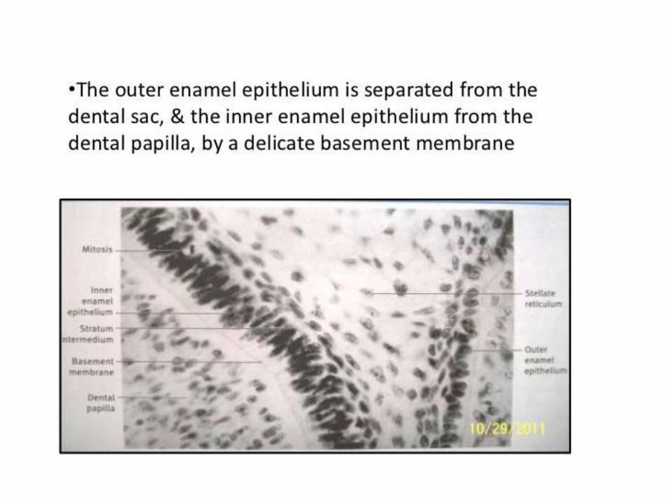
Odontoblast differentiation
UndifferentiatedMesenchymal cell
mitotic spindle perpendicular to basal lamina
Epithelial cell
Inner enamel epithelium
Under the influence of epithelium start differentiating
odontoblast
subodontoblast

Odontoblast differentiate , change from ovoid to columnar shape and nuclie get basally oriented.
One or several processes arrises from the apical end of cells in contact with basal lamina.
length increases to 40 microns and constant width 7 microns.
Proline appears in rough endoplasmic reticulum and golgi apparatus.
Cell recedes leaving behind a single extension.

As matrix formation continues, odontoblastic process
lengthens.
Initially 4 microns of dentin is formed daily till the crown
is formed and erupt into occlusion. after this it reduces to
1 microns.

Occurs by globular calcification.
Deposition of very fine plates of hydroxyapatite on the surface of collagen fibrils and ground substance.
Starts from common centre, so called spherulite form. These are first form of dentin.
Long axis of crystals are parallel to long axis of fibrils.

Important factors in mineralization are :-
Odontoblast secret dentin phosphoprotein (DPP) :-
it is highly ionic and binds to the minrelization front and
controls the growth of apatite crystals.
Osteopontin :- promotes mineralization.
Osteonectin :- inhibits the growth of apatite crystals
and promotes its binding to collagen.
Gamma carboxyglutamic acid (GLA) protein :- attract
the calcium.
Genes implicated in dentinogenesis :-
PHEX – for mineralization
MAP1B – for odontoblast differentiation.

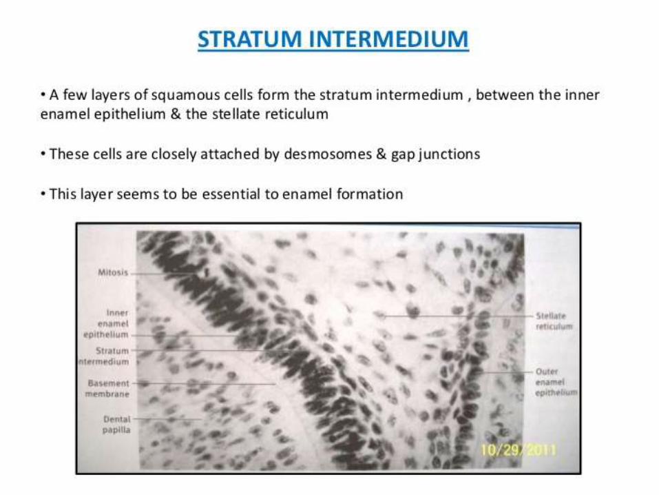
When viewed microscopically following structural features can be identified
Dentinal Tubules Peritubular Dentin Intertubular Dentin Predentin Odontoblastic processes Interglobular Dentin
Granular layer of Tomes Dentinoenamel junction

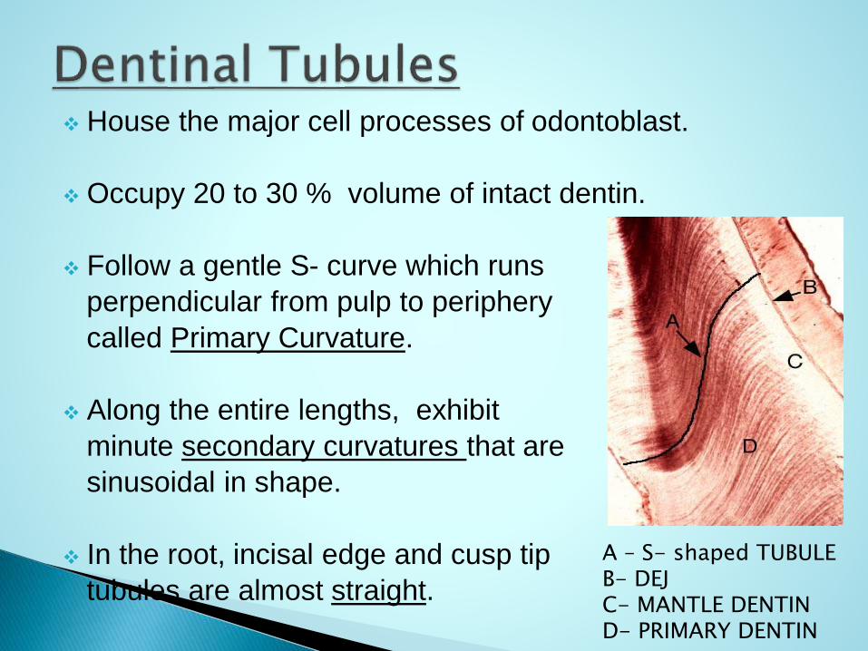
House the major cell processes of odontoblast.
Occupy 20 to 30 % volume of intact dentin.
Follow a gentle S- curve which runs
perpendicular from pulp to periphery
called Primary Curvature.
Along the entire lengths, exhibit
minute secondary curvatures that are
sinusoidal in shape.
In the root, incisal edge and cusp tip
tubules are almost straight.
A – S- shaped TUBULEB- DEJ C- MANTLE DENTIND- PRIMARY DENTIN

Terminal branches
Tubules are longer than the thickness of dentin.
Ratio of outer and inner surface dentin is 5:1.
No. of tubules/unit area on pulpal and outer dentin is 4:1.
Diameter is larger at the pulpal side (1.5-3 mm) than at the periphery(1mm).

Tubules/unit area in crown is more than in root.
Tubules are farther apart in peripheral layer and
more closely packed near the pulp.
No. of dentinal tubules decreases with age, thus
reducing sensitivity.

The dentinal tubules have lateral braches
throughout dentin, called
Canaliculi / microtubules,1 um in
diameter.
They originate at right angles to the
main tubule at every 1-2 um along
its length.
Some may enter adjacent tubules
while others end in intertubular dentin.
Thus form anastomosing canalicular system.
MICROTUBULE

Few dentinal tubules that extend through the DEJ in enamel
for several mm are called enamel spindle.
A – ENAMEL SPINDLEB – DENTINAL TUBULE

Dentinaltubules

Dentinal Tubules
Coronal dentin
Root dentin

Immediately surrounds the dentinal tubules, highly calcified
matrix and forms the walls of tubules .
Highly mineralized , harder, provide added structural
support for intertubular dentin , thus strengthening the
tooth.
It is twice as thick in outer dentin (750nm) than inner
dentin(44nm).
Contains few collagen fibrils and higher proportion of
sulphated proteoglycans.
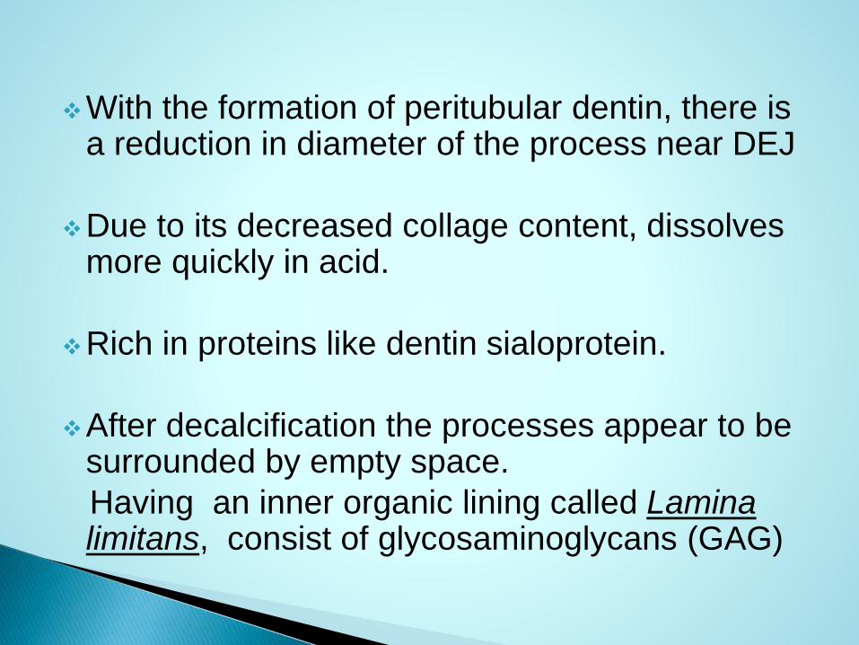
With the formation of peritubular dentin, there is a reduction in diameter of the process near DEJ
Due to its decreased collage content, dissolves more quickly in acid.
Rich in proteins like dentin sialoprotein.
After decalcification the processes appear to be surrounded by empty space.
Having an inner organic lining called Lamina limitans, consist of glycosaminoglycans (GAG)

A – INTERTUBULAR DENTIN
B – PERITUBULAR DENTIN
C – DENTINAL TUBULE

Located between the dentinal tubules or in other
terms between the zone of peritubular dentin.
Constitute main bulk of dentin.
Although highly mineralized, it is retained after
decalcification.
Its organic matrix consists mainly of interwoven network
of collagen fibrils having diameter of 50-200 um in which
apatite crystals are deposited. Fibrils are arranged
randomly at right angles to dentinal tubules.


Dentinal tubules
Peritubular dentin Intertubular dentin

First formed dentin.
Not mineralized.
Located adjacent to pulp tissue, 2-6 micron
wide.
Its thickness remains constant.
As the collagen fibers undergo mineralization at
the predentin- dentin junction, the predentin
becomes dentin & new layer of predentin forms
circumpulpally .

Thickest during dentinogenesis and diminishes
with age.
This predentin layer, secreted by odontoblasts, is
a protein carbohydrate complex consisting of
proteoglycans , phosphoproteins, glycoproteins
and collagen fibrils.
Its presence is important to maintain the integrity
of dentin.


Zone of hypomineralisation present between the mineralized globules which have failed to coalease into a homogenous mass.
Defect of mineralization & not of matrix formation.
It is seen in teeth with vitamin D deficiency or exposure of high level of floride during dentinogenesis , hypophosphatasia.
Shows higher concentration of sulfur.
It is seen in circumpulpal dentin just below mantle dentin. It follows incremental pattern.

Interglobuler Dentin
Inter-globulerdentin
DentinalTubules
DentinoEnamelJunction

Inter-globulerdentin

Cytoplasmic extensions of odontoblast cells.
The odontoblast cells lie in the peripheral pulp at the pulp- predentin border & their process extend into the dentinal tubules.
Diameter: 7 um
Length :- 40 um
Diameter largest near pulp (3-4 um) and tapers approx. 1 um in dentin


Odontoblasts and process
Odontoblast cellsOdontoblast process
Dentin Pulp
The processes narrow to about half the size of the cell as they enter the tubules
The processes is composed of microtubules of 20 micron in diameter & small filaments 5 – 7.5 micron in diameter
Divide near the DEJ & extend into the enamel as enamel spindles

Junction b/w enamel and dentin is called DEJ.
Irregular and scalloped.
Convexity of scallops are directed towards dentin and concavity towards enamel.
A membrane is seen b/w the enamel and dentin during tooth development called membrane performativa. After calcification this membrane disappers and the interface is called DEJ.

A ground section of root, when viewed under transmitted
light , a zone adjacent to the cementum appears
granular.This is called Tome’s Granular Layer.
This zone increases in amount from cementoenamel
junction to the root apex.
Appears dark in transmitted light and lighter in reflected
light.
Remain unmineralized.

Shows higher concentration of calcium and
phosphorous.
This granular layer represent the looped terminal
portions of the dentinal tubules in the root dentin.

Dentin
Cementum
Granular layer of Tomes


Formed prior to the root completion and eruption of teeth.
MANTLE DENIN –
First formed dentin in the crown underlying DEJ.
It is the outermost part of the primary dentin & is 20 -150 microm thick .
Bounded by DEJ & zone of interglobular dentin .
It consist of lager collagen fibers (type 3).
Less mineralized.


CIRCUMPULPAL DENTIN –
Forms remaining primary Dentin or bulk of tooth
Represents all Dentin formed before root completion.
Collagen fibrils are smaller in diameter 0.05 micron & are more closely packed than mantle dentin .
More mineral content than mantle dentin .


Narrow band of dentin bordering the pulp and
formed after the root completion and complete
eruption of teeth.
Continues to forms throughout the life of the tooth.
Contains fewer tubules than primary dentin.
Formed more slowly than primary dentin.
At the junction of primary and secondary
dentin a bend is seen in the dentinal tubules.


It is not formed uniformly and appears in greater amount on root and floor of the coronal pulp chambers , where it protects the pulp from exposure in older teeth.
Due to the regular arrangement of dentinal tubules, it is called regular sec.dentin.

With the growth of this dentin there is decrease in size of the pulp chamber with age. So called pulp recession.
Tubules of Secondary Dentin scleroses more rapidly than those of the Primary Dentin. Thus reduce overall permeability of dentin thereby protecting the pulp.

Primaryphysiologicaldentin
Secondaryphysiologicaldentin


Also referred as reparative dentin, response dentin,
irregular secondary dentin.
It is localized formation of Dentin on pulp dentin border ,
formed in reaction to trauma such as caries or
restorative procedures, attrition ,abrasion , erosion.
Formations depends on the intensity & duration of
stimulus.
Forms along entire pulp dentin border by cells directly
affected by the stimuli.
Tertiary dentin may have tubules continous with those of
sec. dentin , tubule spares in no. and irregurarly
arranged or no tubules at all.
This minimizes dentin permeability at the site of
deposition thus giving protection to the underlying dental
pulp.
It help seal off the area of injury causing resolution of
inflammation .
The cells forming tertiary dentin become included in the
dentin , and hence called Osteodentin.
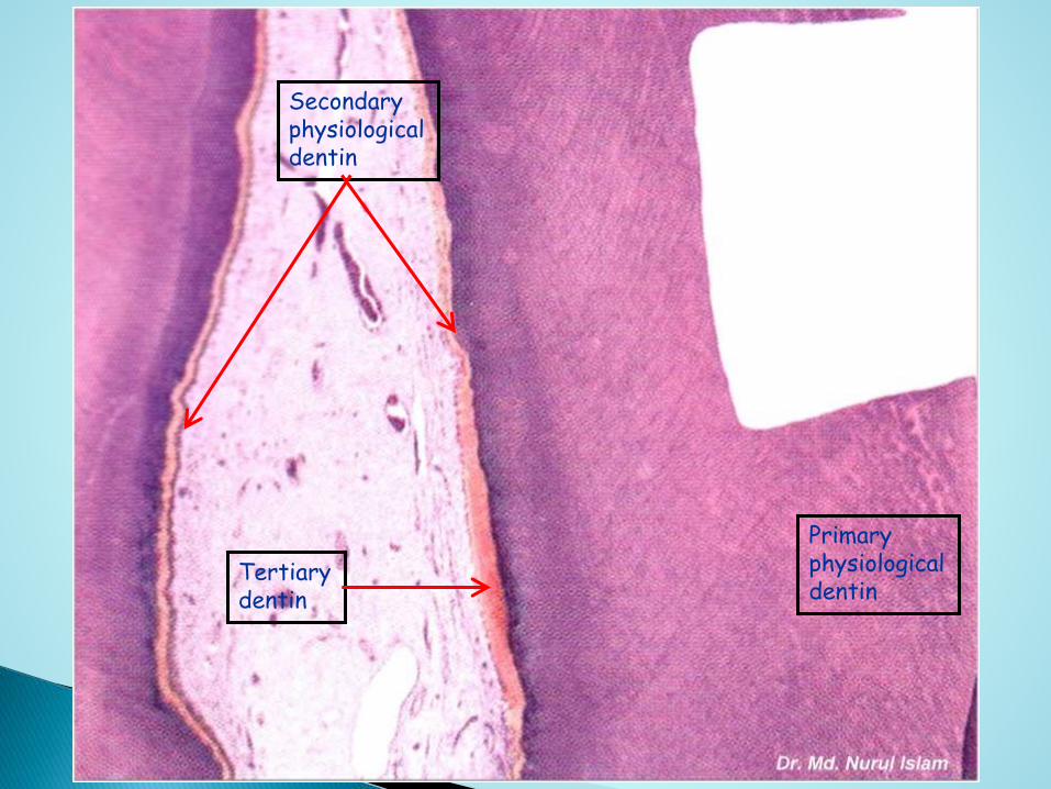
Primaryphysiologicaldentin
Secondaryphysiologicaldentin
Tertiarydentin

Primaryphysiologicaldentin
Secondaryphysiologicaldentin
Tertiarydentin
Dead tracts

Tertiary dentin is further classified into :-
• Reactionary dentin /Regenerated dentin
• Reparative dentin

By trauma stimuli odontoblast processes are exposed , cut/killed ,die or survive.
Those cells who survived – produce dentin called Reactionary/Regenerated dentin.
Those who are killed – are replaced by the migration of undifferentiated cells from the cell rich zone or undifferentiated perivascularcells present deeper inside the pulp.
The newely differentiated odontoblast than begin deposition of Reparative dentin

This action to seal off the zone of injury occurs as a healing process initiated by the pulp, resulting in resolution of the inflammatory process and removal of dead cells.
This zone of hard tissue thus formed is called Reparative Dentin.
Due to the irregular nature of dentinal tubules , these type of dentin is called irregular sec. dentin.
Tertiary dentin is differ from other form of dentin, as it contains dentin phosphophoryn.


Incremental lines of Von Ebner
Contour lines of Owen
Neonatal lines

They appear as fine lines or striations in dentin .
They run at right angles to the dentinal tubules.
These lines reflects daily rhythmic, recurrent deposition of dentin matrix in the daily formative process .
Distance between lines varies from 4 –8 micron in the crown to much less in the root .
Daily increment decreases as the tooth comes into functional occlusion.

Incremental line of von Ebner

Some of the incremental lines are accentuated because
of disturbances in the matrix & mineralization process
,these are called contour lines of Owen.
They lie at right angle
to the von ebner lines.
These lines represents
hypocalcified bands.
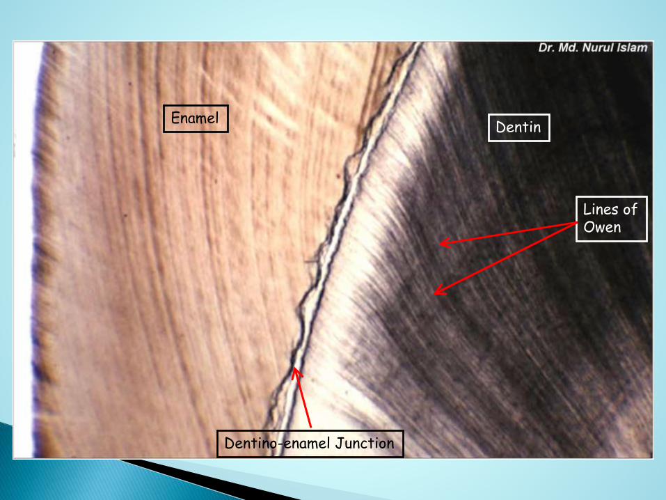
DentinEnamel
Lines ofOwen
Dentino-enamel Junction


In deciduous teeth & in first permanent molars, where dentin is formed partly before & partly after the birth, the prenatal & post natal dentin separated by accentuated contour line
Seen in enamel as well as dentin.
The line represents the disturbances in mineralization due to the abrupt changes in environment that occurs at birth.
It is a zone of hypocalcification.
The dentin matrix formed prior to birth is better quality than that formed after birth.

NEONATAL LINE IN ENAMEL
NEONATAL LINE IN DENTIN

Formation of secondary dentin
Formation of reperative dentin
Dead tracts
Sclerotic dentin

The presence of caries , attrision , abrasion , erosion, cavity preparation or any external stimuli may cause increase in dentin deposition as well as increase in mineralization of old existing dentin.
Which leads to excessive formation of collagen fibers and appatite crystals in dentinal tubules.
That leads to complete obliteration of dentinal tubules as a defensive reaction of dentin.

Apatite crystals are initially only sporadic in a dentinal tubules but gradually the tubule becomes fluid with a fine meshwork of crystals.
Gradually tubule lumen is obliterated with mineral, which appears very much like Peritubular dentin.
Due to the high mineral content, they appear
transparent.
Sclerotic dentin is observed in teeth of elderly people,
specially roots , also observed under slowly progressing
caries.

Sclerosis reduces the permeability of dentin & help to
prolong pulp vitality.
Appears dark in reflected light and transparent in
transmitted dentin.
Dentin formed due to aging is harder than that formed
below carious lesion.
It is harder than normal dentin and fracture toughness is
reduced, elastic properties are altered.
Crystals present in this dentin are smaller than those
present in normal dentin.

Dental cariesSclerotic dentin


In ground section of dentin, when viewed under transmitted light shows sometimes dark lines that follow the course of dentinal tubules.These are called Dead Tracts.
They are dead degenerated odontoblast processes in dentinal tubules.
These tubules are empty and are filled with air and that appears dark in transmitted light.
Occur in condition such as attrision , erosion, abrasion, cavity preperation.
It is often observed in areas of narrow pulp horn because of crowding of odontoblasts.

Appears greater in extent in older teeth due to aging
process. These areas demonstrate decreased
sensitivity.
They are probably the initial step in formation of sclerotic
dentin.
A – DEAD TRACTS
B – SECONDARY DENTIN
C – TERTIARY DENTIN


