Dermal-Fat Graft as a Primary Enucleation Technique WILLIAM R. NUNERY, MD, KATHY J. HETZLER Abstract: A retrospective study of 36 dermal-fat grafts was undertaken to determine the indications as a primary enucleation implant material. Twenty- two patients or 61 % required a reoperation in a three-year follow-up period. Sixty-seven percent required secondary prosthetic revision. We do not rec- ommend dermal-fat grafting for routine enucleation due to high reoperation rate and prosthetic revisions. [Key words: an ophthalmic socket, dermal-fat graft, en ucleation, ocular prosthesis, prosthetic fabrication.] Ophthalmology 92: 1256- 1261,1985 Following enucleation, the orbital surgeon has many implant materials from which to choose. These materials include sphere implants, irregularly surfaced motility im- plants, and autogenous dermis-fat composite grafting to the anophthalmic socket. Regardless of the implant ma- terial chosen, problems of an ophthalmic socket volume loss, implant extrusion, and reduced prosthetic motility continue to plague the ophthalmologist and ocularist. In order to minimize these problems, following enucleation and prosthetic fitting, evaluations and comparisons of various techniques should continually be undertaken to elucidate optimum surgical and prosthetic fabrication techniques. In a previous report, we discussed a modified technique of sphere implantation with attachment of extraocular muscles to the respective fornices, motility template im- plantation utilizing eyebank sclera, and routine deepening of the inferior fornix.' This technique, in our experience, has proven to be an effective technique for primary enu- cleation. It has been associated with a low incidence of implant extrusion, low incidence of unacceptable volume deficit, and good prosthetic motility. Despite the success with the modified sphere technique, other authors have reported success with autogenous der- mal-fat grafting as a primary enucleation technique. 2 - 5 We have therefore examined our previous three year ex- perience with autogenous dermal-fat grafting to the an- ophthalmic socket. We will consider relative indications From the Department of Ophthalmology, Indiana University, Indianapolis. Presented at the Eighty·ninth Annual Meeting of the American Academy of Ophthalmology, Atlanta, Georgia, November 11-15, 1984. Reprint requests to William R. Nunery, MD, Department of Ophthalmology, Indiana University, 702 Rotary Circle, Indianapolis, IN 46223. 1256 and relative contraindications of dermal-fat grafting as a primary enucleation technique. We will also discuss as- pects of prosthetic fitting indigenous to the use of autog- enous dermal-fat grafting. SURGICAL TECHNIQUE Autogenous composite grafts of dermis and subdermal fat are first obtained from the buttocks area. Epidermis from the donor site is elevated with a dermatome. An ellipse of dermis is elevated with a # 10 Bard-Parker blade (Fig I). An attached circular ball of fatty tissue is elevated while attached to the dermis. The diameter of the har- vested fatty ball measures approximately 35 mm in di- ameter initially. The autogenous graft is preserved in a moist sponge while the donor site is closed and the an- ophthalmic socket prepared for grafting. The donor site is closed with multiple interrupted 3-0 vicryl sutures placed through the fatty tissue and subcu- taneous tissue. A 6-0 Prolene® suture is used to close the dermis. The epidermal tissue previously elevated is then replaced over the closed dermal graft site, cut to fit the closed dermis site precisely, and closed with a running 6- o nylon vertical mattress suture around the perimeter of the epithelium. Attention is then turned to the anophthalmic socket. The four recti muscles are identified and tagged with dou- ble armed 5-0 vicryl suture. Tenon's capsule is released with multiple anterior to posterior relaxing incisions to allow grafted fatty tissue to come into maximal contact with orbital fatty tissue (Fig 2). The autogenous dermal- fat graft is trimmed to remove excess fat and to allow the tissue to be transferred into the an ophthalmic socket bed. An overcorrection of approximately 20% in orbital vol- ume is desirable.
Transcript
Dermal-Fat Graft as a Primary Enucleation Technique WILLIAM R. NUNERY, MD, KATHY J. HETZLER
Abstract: A retrospective study of 36 dermal-fat grafts was undertaken to determine the indications as a primary enucleation implant material. Twentytwo patients or 61 % required a reoperation in a three-year follow-up period. Sixty-seven percent required secondary prosthetic revision. We do not recommend dermal-fat grafting for routine enucleation due to high reoperation rate and prosthetic revisions. [Key words: an ophthalmic socket, dermal-fat graft, en ucleation, ocular prosthesis, prosthetic fabrication.] Ophthalmology 92: 1256-1261,1985
Following enucleation, the orbital surgeon has many implant materials from which to choose. These materials include sphere implants, irregularly surfaced motility implants, and autogenous dermis-fat composite grafting to the anophthalmic socket. Regardless of the implant material chosen, problems of an ophthalmic socket volume loss, implant extrusion, and reduced prosthetic motility continue to plague the ophthalmologist and ocularist. In order to minimize these problems, following enucleation and prosthetic fitting, evaluations and comparisons of various techniques should continually be undertaken to elucidate optimum surgical and prosthetic fabrication techniques.
In a previous report, we discussed a modified technique of sphere implantation with attachment of extraocular muscles to the respective fornices, motility template implantation utilizing eyebank sclera, and routine deepening of the inferior fornix.' This technique, in our experience, has proven to be an effective technique for primary enucleation. It has been associated with a low incidence of implant extrusion, low incidence of unacceptable volume deficit, and good prosthetic motility.
Despite the success with the modified sphere technique, other authors have reported success with autogenous dermal-fat grafting as a primary enucleation technique.2- 5
We have therefore examined our previous three year experience with autogenous dermal-fat grafting to the anophthalmic socket. We will consider relative indications
From the Department of Ophthalmology, Indiana University, Indianapolis.
Presented at the Eighty·ninth Annual Meeting of the American Academy of Ophthalmology, Atlanta, Georgia, November 11-15, 1984.
Reprint requests to William R. Nunery, MD, Department of Ophthalmology, Indiana University, 702 Rotary Circle, Indianapolis, IN 46223.
1256
and relative contraindications of dermal-fat grafting as a primary enucleation technique. We will also discuss aspects of prosthetic fitting indigenous to the use of autogenous dermal-fat grafting.
SURGICAL TECHNIQUE
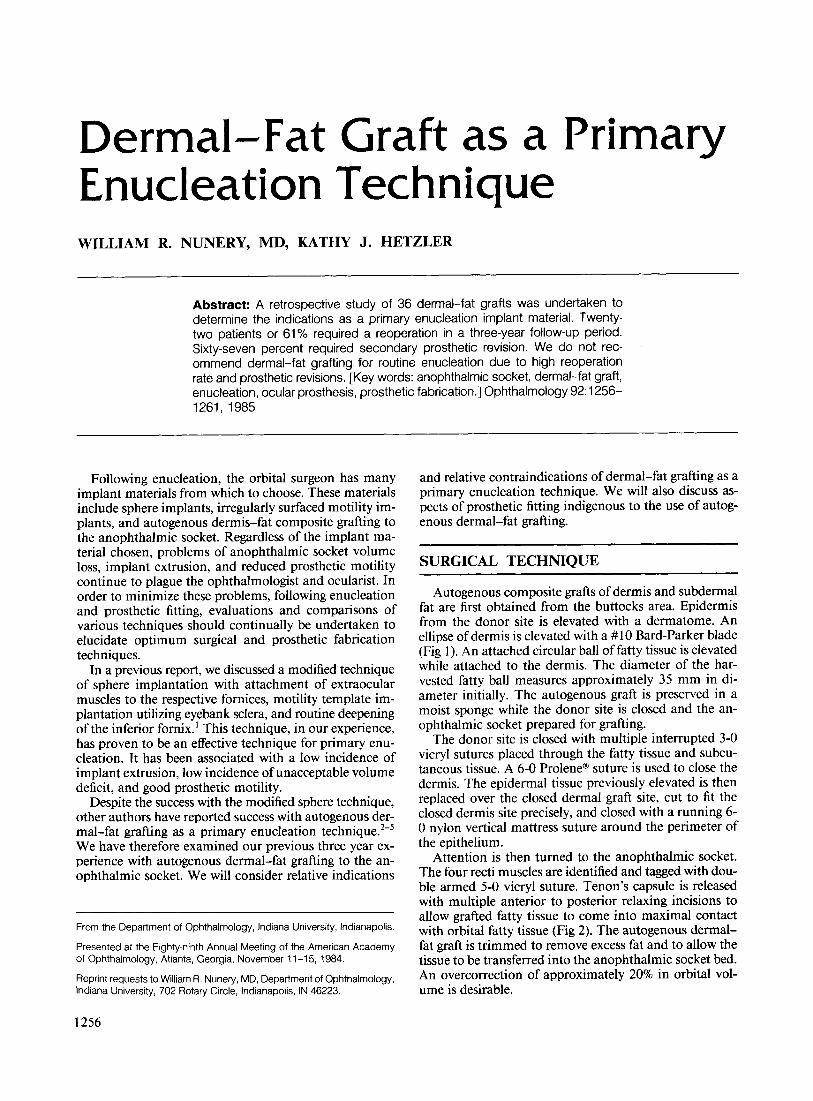
Autogenous composite grafts of dermis and subdermal fat are first obtained from the buttocks area. Epidermis from the donor site is elevated with a dermatome. An ellipse of dermis is elevated with a # 10 Bard-Parker blade (Fig I). An attached circular ball of fatty tissue is elevated while attached to the dermis. The diameter of the harvested fatty ball measures approximately 35 mm in diameter initially. The autogenous graft is preserved in a moist sponge while the donor site is closed and the anophthalmic socket prepared for grafting.
The donor site is closed with multiple interrupted 3-0 vicryl sutures placed through the fatty tissue and subcutaneous tissue. A 6-0 Prolene® suture is used to close the dermis. The epidermal tissue previously elevated is then replaced over the closed dermal graft site, cut to fit the closed dermis site precisely, and closed with a running 6-o nylon vertical mattress suture around the perimeter of the epithelium.
Attention is then turned to the anophthalmic socket. The four recti muscles are identified and tagged with double armed 5-0 vicryl suture. Tenon's capsule is released with multiple anterior to posterior relaxing incisions to allow grafted fatty tissue to come into maximal contact with orbital fatty tissue (Fig 2). The autogenous dermalfat graft is trimmed to remove excess fat and to allow the tissue to be transferred into the an ophthalmic socket bed. An overcorrection of approximately 20% in orbital volume is desirable.
NUNERY AND HETZLER • DERMAL-FAT GRAFT
Fig 1. Elevation of dermal-fat graft.
I, I
TENON'S FASCIA
Fig 2. Preparation of socket for grafting.
RECTI MUSCLE SUTURES ~~~~~~Z\'
Fig 3. Dermal-fat graft in socket.
The dermis is anchored into position by placing the four recti muscles to the corresponding margins of the dermis (Fig 3). The remainder of the dermis is re-ap-
CONJUNCTIVA
Fig 4. Final closure.
, .
PROSTHESIS
Fig S. Profile of graft in socket.
proximated to existing conjunctiva with multiple interrupted 5-0 vicryl sutures (Fig 4).
Two double armed 4-0 silk sutures are placed through a silicone conformer. The needles are cut free of the suture and the suture is passed through the inferior fornix and through the periosteum of the inferior orbital rim (Fig 5). The suture exits the skin of the cheek with the use of a large curved general closure cutting needle. The anophthalmic socket is covered with a moderate pressure eyepad dressing.
POSTOPERATIVE CARE AND PROSTHETIC FITTING
One week following grafting, the fornix sutures are removed and the silicone conformer is replaced with a clear acrylic conformer (Fig 6). Initially, the silicone conformer is valuable for the ease in which the conformer can be
1257
OPHTHALMOLOGY • SEPTEMBER 1985 • VOLUME 92 • NUMBER 9
Fig 6. Post-enucleation conformers.
Fig 7. Cicatricial bands at graft interface.
sutured to the inferior fornix. After removal of these sutures, the acrylic conformer is used to allow visibility of the healing dermis graft on the posterior surface of the socket (Table 1).
Eight to ten days following surgery, the socket and conformer are examined by the ocularist. A custom, clear acrylic conformer is fabricated. The purposes of the conformer are first to provide a slight vaulting over the dermis graft. Some vaulting is desirable to avoid abrasion of the regenerating mucosal edge covering the dermis. Excessive vaulting, however, is avoided so that infections will not be encouraged by dead space between the socket and the conformer.
Secondly, the conformer is fit securely into the medial and lateral canthi. Gentle pressure in these areas flattens mucosal tissue. Well-defined canthi may prevent later rotation of the prosthesis in the event of tissue reabsorption in the fornices.
At three to four weeks following surgery, the socket is reexamined. In most cases, reduction in postoperative edema and early reabsorption of graft tissue require refabrication of the conformer. If complete reepithelialization of the dermis surface has not occurred, the posterior surface is again slightly vaulted. Lid positions and fornices are evaluated by the surgeon and ocularist to determine if socket tissue is adequate to allow eventual prosthetic fitting with the desired outcome.
At six to eight weeks postoperatively, an impressionedfit, custom prosthesis is fabricated and delivered. The
1258
Fig 8. Lid retraction secondary to graft shrinkage.
posterior surface is fit flush with the socket tissue provided the epithelium has regenerated. Additional volume is added to the prosthesis to compensate for graft reabsorption.
At 12 to 16 weeks postoperatively, the prosthesis and socket are reexamined for excessive reabsorption, superior sulcus deformity, rotation of the prosthesis, altered lid position, or blunting of the inferior fornix.
Problem aspects of the socket at this stage may include cicatricial bands of tissue along the margins of the dermis graft (Fig 7), troughing or excessive rounding of the inferior fornix, eyelid retraction, and superior sulcus deformity. These abnormalities occur as the result of reduction of fatty volume. Following fat grafting, adhesions form between fat tissue and the superior and inferior recti muscles. As the fat reabsorbs, the shrinkage creates a posterior pull on the recti muscles and secondarily on the levator and inferior retractor muscles (Fig 8).
The trough-like effect on the inferior fornix is also due to fatty reabsorption with posterior pull and rounding of the inferior fornix tissue (Fig 9). The troughing of the fornix permits rotation of the prosthesis. Custom fitting around irregularities in the canthi may be utilized to prevent this undesirable rotation of the prosthesis.
If changes in the socket anatomy justify, the prosthesis is replaced with a new custom design. Similar reexamination, and refitting when necessary, is carried out at six months to one year following surgical implantation of the dermal-fat graft.
Table 1. Prosthetic Timetable after Dermal-Fat Graft
Time
10 days 4 weeks 6-8 weeks 16 weeks 6 months
Custom clear conformer Refashion conformer Impressioned prosthesis Modification of prosthesis Refabrication if necessary
NUNERY AND HETZLER • DERMAL-FAT GRAFT
Fig 9. Rounded fornix secondary to graft shrinkage.
Fig )0. Superior sulcus deformity due to graft reabsorption.
Table 2. Reoperations Following 36 Dermal-Fat Grafts
Volume replacement Volume reduction Lid recession Inferior fornix revision
Total
No.
14 1 4 3
22
(%)
(39) (3)
(11 ) (8)
61%
Fig 11. Eyelid retraction secondary to graft shrinkage.
Fig 12. Pyogenic granuloma at graft interface.
RESULTS
Dermal-fat grafts were placed in 36 post-enucleation sockets. Follow-up time ranged from three months to three years. The ages of the patients ranged from 2 years to 82 years with an average age of 45 years. During the same period, 22 reoperations for complications of dermal-fat grafting were performed in 20 of these same patients, for a reoperation rate of 61 %.
Fourteen patients required volume augmentation for excessive fatty reabsorption (Fig 10). One patient required volume reduction. Four patients underwent levator recession for post dermal-fat graft lid retraction (Fig 11). Three patients underwent inferior fornix deepening procedures (Table 2).
In addition to complications requiring reoperation, two cases of orbital infection following dermal-fat grafting were encountered. The minor complication of pyogenic granuloma formation of the dermis conjunctival interface occurred in 12 patients (Fig 12). None of these patients
1259
OPHTHALMOLOGY • SEPTEMBER 1985 • VOLUME 92 • NJMBER 9
Fig 14. Left. proptosis secondary to fatty growth in socket. Right. post -excision of excess fatty tissue.
required re-operation although excision of granulomas in the office was required prior to prosthetic fitting. Twentyfour patients or 67% required major revision or refabrication of the prosthesis between three to twelve months following surgery due to significant changes in socket anatomy (Table 3).
DISCUSSION
Autogenous dermal-fat grafting has been advocated by some surgeons following primary enucleation.5 Advan-
Table 3. Nonoperative Complications Following 36 Dermal-Fat Grafts
1260
Orbital cellulitis Granuloma Refabrication
No.
2 12 24
(%)
(6) (33) (67)
tages of dermal-fat grafted sockets include the use of autogenous tissue with no foreign implants, good motility when extraocular muscles are reattached to the dermalfat graft, maximal preservation of the inferior fornix, and their ability to expand with growth.
Our experience, however, suggests that the use of autogenous dermal-fat grafting as a primary enucleation technique has the disadvantage of a high reoperation rate and unpredictable volume reabsorption.
These disadvantages have prohibited the dermal-fat graft technique from becoming the procedure of choice as a routine primary enucleation technique, in our experience. This is especially true when compared to the predictable volume, low extrusion rate, and excellent motility associated with the modified sphere technique which we previously reported. 1
In addition to undesirable surgical aspects, undesirable prosthetic fabrication aspects encountered in dermal-fat grafted sockets include unpredictable or prolonged healing time prior to prosthetic fitting, excessive socket discharge prior to reepithelialization of the dermis, granuloma for-
NUNERY AND HETZLER • DERMAL-FAT GRAFT
mation affecting the prosthetic fit, rotation of the prosthesis, cicatricial bands at the dermis-conjunctival interface, and alteration of lid position due to retraction on extraocular muscles.
However, several cases have been isolated in our experience in which we have considered autogenous dermalfat grafting to be the implant material of choice following primary enucleation. These cases include patients who underwent limited exenteration following the removal of optic nerve sheath meningioma, patients who have sustained massive acute trauma with significant orbital tissue loss (Fig 13), and patients with cicatricial shortening of the fornices.
Theoretically, autogenous dermal-fat grafting may be useful in children less than six years of age. Since the autogenous fatty tissue may grow with increases in the patient's body weight, this may lead to progressive stimulus for orbital bony development. While this is theoretically attractive, there has been no laboratory evidence to verify the usefulness of dermal-fat grafting in bony growth stimulation.
Both of our patients who developed persistent postoperative wound infections following autogenous dermalfat grafting were diabetic. We, therefore, consider diabetes to be a relative contraindication for the use of the dermalfat graft.
Obese patients, and patients with wide fluctuation in body weight, may be undesirable candidates for autogenous dermal-fat grafting since fatty tissue implanted in the orbit may undergo fluctuations in volume as body weight changes. This can lead to late proptosis requiring surgical reduction offatty volume. We have seen one patient who developed proptosis associated with systemic weight gain 25 years after abdominal fatty graft to the orbit (Fig 14).
In our experience, the autogenous dermal-fat grafting retains its greatest usefulness as a volume replacement for patients who have had mUltiple implant extrusions or patients with inadequate inferior fornix tissue undergoing socket revision. Since the use of the dermal-fat graft leads to a net increase in conjunctival tissue, following epithelialization of the dermis, the conjunctival tissue can be maximally preserved and utilized in deepening the inferior fornix for prosthetic fitting.
SUMMARY
We have found autogenous dermal-fat grafting to be a useful technique for socket revision. We have not found the technique suitable for routine enucleation due to high incidence of surgical revision and unpredictable volume reabsorption.
2. Smith B, Petrelli R. Dermis·fat graft as a movable implant within the muscle cone. Am J Ophthalmol 1978; 85:62-6.
3. Bullock JD. Dermis-fat graft in orbital reconstruction. Presented at the American Society of Ophthalmic Plastic and Reconstructive Surgery Meeting, Atlanta, GA, November 7, 1981.
4. Przybyla VA Jr, La Piana FG, Bergin DJ. Fitting of the dermis·fat grafted socket. Ophthalmology 1981; 88:904-7.
5. Aguilar GL, Shannon GM, Flanagan JC. Experience with dermis-fat grafting: an analysis of early postoperative complications and methods of prevention. Ophthalmic Surg 1982; 13:204-9.