73
Dermatopathology Symposium -Soft Tissue Tumors of the Skin- Thomas Brenn

Dermatopathology Symposium-Soft Tissue Tumors of the Skin-
Thomas Brenn

Conflict of Interest
none

CASE 1
• 30 yo female• Tumour on the back


AE1/3 CD31
ERGINI1

Diagnosis:
Primary Cutaneous EpithelioidAngiosarcoma

Cutaneous Angiosarcoma
• Conventional (idiopathic) AS
• Lymphedema-associated AS
• Post-radiation AS


Diagnostic Problems
Endpoints of the morphological spectrum
• well differentiated AS
• poorly differentiated AS

Well differentiated AS

Spindle Cell AS


Epithelioid AS

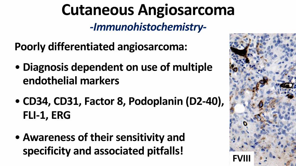
Cutaneous Angiosarcoma-Immunohistochemistry-
Poorly differentiated angiosarcoma:
• Diagnosis dependent on use of multiple endothelial markers
• CD34, CD31, Factor 8, Podoplanin (D2-40), FLI-1, ERG
• Awareness of their sensitivity and specificity and associated pitfalls!
FVIII

CD31 CD34 FLI1 ERG

Cutaneous Angiosarcoma-Prognosis-
• Behaviour largely independent of aetiology
• Overall dismal prognosis–Median survival 15 months– 5 year overall survival = 10-30%– 5 year disease-free survival 10% or less
• Treatment: Radical local surgery adjuvant XRT + ChTx + targeted therapy

Adverse prognostic factors:– Large tumor size >5cm
–Necrosis
– Epithelioid cell change
–Depth of invasion– Status of margins
– Recurrence
–MetastasisDeyrup AT, etal. Am J Surg Pathol. 2008;32(1):72-7.
Morgan M, et al. (2004) J Am Acad Dermatol
Cutaneous Angiosarcoma-Prognosis-

Primary Cutaneous Epithelioid Angiosarcoma-Clinical-
• Rare outside classical settings for AS• Elderly adults, M=F
• Solitary or multiple nodules• Extremities• Poor prognosis with distant metastasis
and associated mortalitySuchak R. et al. Am J Surg Pathol 2011;35(1): 60-69.

37 yo female, 6 months h/o flesh coloured nodule on the left lower calf


70 yo female, left thigh tumour

CD31 CD34
ERG WT1
Immunohistochemistry

AE1/3 MNF116

INI1

Epithelioid Angiosarcoma-DDx-
• Melanoma• Carcinoma• Epithelioid Angiomatous Nodule• Epitheliod Haemangioendothelioma• Epithelioid Sarcoma

Epithelioid Hemangioendothelioma-Clinical-
• Rare epithelioid vascular tumour• Deep soft tissues and visceral organs (lung, liver, bone)
• Skin involvement as part of multicentricity or extension from underlying soft tissue• Primary cutaneous EHE is very rare
Quante M, et al. Am J Dermatopathol. 1998 Dec;20(6):541-6.Mentzel T, et al. Am J Surg Pathol. 1997 Apr;21(4):363-74.
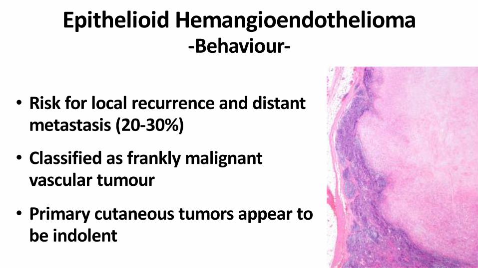
Epithelioid Hemangioendothelioma-Behaviour-
• Risk for local recurrence and distant metastasis (20-30%)• Classified as frankly malignant
vascular tumour
• Primary cutaneous tumors appear to be indolent


CD31 ERG
MNF116 SMA

Epithelioid Hemangioendothelioma-Genetics-
• WWTR1-CAMTA1 gene fusion on chr 1 and 3 in 90%• YAP1-TFE3
• anti CAMTA 1 AB, nuclear staining positive in 90% and reliable diagnostic marker
Doyle LA, Fletcher CD, Hornick JL. Am J Surg Pathol. 2016;40(1):94-102.Flucke U, et al. Diagn Pathol. 2014;9:131. Antonescu CR, et al. Genes Chromosomes Cancer. 2013 Aug;52(8):775-84. Tanas MR, et al. Sci Transl Med. 2011;3(98):98ra82.

Epithelioid Angiomatous Nodule
• Solitary small nodules or papules (0.5 cm)
• Erythematous-bluish discoloration• Trunk and extremities, head and neck area and
mucosa
• Wide age distribution (37 years)• M=F
• Benign clinical behaviorBrenn T and Fletcher CD. Am J Dermatopathol. 2004;26(1):14-21.


CD31 CD34 SMA

CASE 2
• 6 yo Iraqi boy• with a left forearm mass
• 2 years earlier treated with chemotherapy and radiation• Now local recurrence


EMAMNF116
CD31CD34

INI1

Diagnosis:
Epithelioid Sarcoma

Conventional Epithelioid Sarcoma-Clinical-
• Adolescents and young adults (mean 25 years)• Male predominance (2:1)• Enlarging plaque/nodule of few centimeters,
occasionally ulcerated• Distal extremities: fingers, hand, wrist


CD34
CD31
MNF116
EMA

Epithelioid Sarcoma-Immunohistochemistry-
Miettinen M, et al. Am J Surg Pathol. 2013;37:1580-5.Stockman DL, et al. Mod Pathol. 2014;27:496-501.
Also variable positive for:• ERG• FLI1• D2-40

Epithelioid Sarcoma-Genetics-
• Abnormalities of chromosome 22q
• Mutations in tumour suppressor gene SMARCB1 (hsNF5, INI1)
• Similar to paediatric rhabdoid tumours
INI1Hornick JL, et al. Am J Surg Pathol. 2009;33(4):542-50.Modena P, et al. Cancer Res. 2005 15;65(10):4012-9.

Epithelioid Sarcoma-Prognosis-
• Aggressive tumor with protracted clinical course• Extensive spread along fascial and tendinous structures• Clinically underestimated• Local recurrence (70-80%)• Distant metastasis to lymph node and lung (40%)• 5 year survival ~70%• But dismal long term survival ~25% at 20 years• Poor prognostic factor: size > 5 cm

CASE 3
• 78 yo M• 10 cm ill-defined tumour• on the frontal scalp


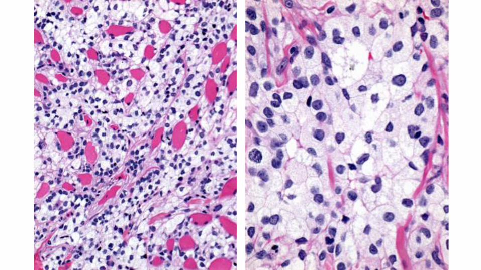


CD31 CD68
ERG

Diagnosis:
Foam cell Angiosarcoma

Morphological Variants of Angiosarcoma-Clinical Presentation-
• Foam-, granular- and signet ring-cell change
• Sun-damaged skin of head and neck
• Elderly men
• Large erythematous-violaceousplaques
• Rarely small nodulesWood A, et al. Histopathology 2015;66(6):856-63.

Granular cell Angiosarcoma


CD31 ERG

Signet ring cell Angiosarcoma

CD31 ERG
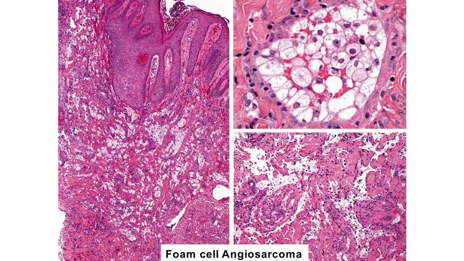
Foam cell Angiosarcoma

CD68 CD163

CD31 ERG

Variants of Epithelioid Angiosarcoma• Awareness and recognition• Misdiagnosis as• histiocytic neoplasm• granular cell tumour• signet ring cell / sebaceous carcinoma• adipocytic neoplasm
• Interpretation in correct clinical setting and appropriate use of immunohistochemical markers

Summary• Short overview of cutaneous epithelioid angiosarcoma• Emphasis on• Clinical and histological spectrum• Differential Diagnosis• Diagnostic pitfalls• Appropriate use of immunohistochemistry

Many Thanks!


