Vol.14,n.2,pp.05-08 (Mar - May 2016) Brazilian Journal of Surgery and Clinical Research- BJSCR BJSCR Openly accessible at http://www.mastereditora.com.br/bjscr DERMOIDE CYST IN FLOOR MOUTH AMANDA MACEDO MENDES 1 , DEISE KERSTEN ALVES FERREIRA 1 , RENAN CARLOS LOPES CAVALCANTE 2 , MARIA APARECIDA ALBUQUERQUE CAVALCANTE 3 , RODOLFO JOSE GOMES DE ARAUJO 4 , ANDRESSA SORAIA BARROS MAYHEW 1 1. Student of dentistry - Higger School of Amazônia; 2. Specialist in Oral and Maxillofacial Surgery at the Federal University of Rio de Janeiro.Rio de Janeiro, RJ, Brazil; 3. Surgery Course Coordinator Oral and Maxillofacial the Federal University of Rio de Janeiro. Rio de Janeiro, RJ, Brazil; 4. PhD Student in biology of infectious agents and Parasitological - Federal University of Para. Rua Boa Ventura da Silva 675, Belém, Pará, Brazil CEP: 66055-090 [email protected]Received: 03/08/2015; Accepted: 03/11/2015 ABSTRACT Dermoid cysts are uncommon cystic lesions in the head and neck region (2% of all desmoid cysts), with a higher preva- lence in young people, with no gender preference. It is con- sidered a form of benign cystic teratoma derived mainly from the embryonic germinal epithelium containing attached structures on your wall, as sebaceous glands, sweat and hair follicles, classifying it as a development cyst. This article aims to inform clinical and histopathological aspects of dermoid cyst and report a clinical case treated surgically in the ante- rior mouth floor in the midline. The patient came in Oral and Maxillofacial Surgery Service of the Federal University of Rio de Janeiro (UFRJ), where he was treated with surgical excision in the midline of the mouth floor under general an- esthesia. Due to the process of evolution of the lesion and the size that can achieve early diagnosis and surgical treatment become imperative to improve the patient's condition. The location of the lesion relative to mylohyoid andgeniohy- oidmuscles is very important to determine the surgical ap- proach that will provide the best treatment. Once the com- plete excision of the lesion, the recurrence becomes rare. KEYWORDS: Dermoid cyst, cysts, oral pathology, odonto- genic. 1. INTRODUCTION The dermoid cyst is a cystic malformation of the unu- sual development. It is lined with epithelium similar to the epidermis, and the wall contains skin appendages. Gener- ally can be considered is a benign cystic teratoma of the way. By definition, a true teratoma is a tumor development comprises tissue derived from the three germ layers: (A) ectoderm, (B) and mesoderm (C) endoderm 1 . In 1955, Meyer updated the concept of dermoid cyst to describe three histological variants, true dermoid cyst, epidermoid and teratoid cyst 2 . Dermoid cysts are simpler in structure than the complex teratomas. Although not contain tissue of the three germ layers, probably represent a failed form of teratoma. In the oral cavity can be ob- served similar cysts coated by epithelium identical to the skin, but skin appendages are not observed in the cyst wall 1 . They are coated by stratified squamous epitheli- umorthokeratinized, with a prominent granular layer. It is common to find abundant keratin inside the cyst light. The cyst wall is composed of fibrous connective tissue that contains one or more skin appendages, such as sebaceous glands, sweat glands or hair follicles 1 . Can be found most often in the midline of the oral floor, a sublingual swelling can move the tongue superi- orly and cause difficulties in feeding, speech or even breathing. The cysts that appear above the genius-hioideo muscle produce, often a submental swelling, with appear- ance of "double chin". They are more common in children and young adults; 15% of the reported cases are congeni- tal 1 . In general, the lesion grows slowly without causing pain, presenting itself as a rubber-mass or paste, which often retains the imprint of fingers under finger pressure. Secondary infection may occur, and the lesion can drain into the oral cavity or the skin 1 . For diagnosis and delineate the extent of the injury, can be used as the tests: Ultrasound, Computed Tomography, Magnetic Resonance, Histopathological and X-rays. Surgical removal is indicated in these cases, those lo- cated above the geniohyoid muscle can be removed by intraoral incision. It has recurrence is unusual. And there are few reported cases where there was a malignant trans- formation into squamous cell carcinoma. 2. CASE REPORT The male patient, 15 years old, leucoderma, came in Oral and Maxillofacial Surgery Service of the Federal University of Rio de Janeiro (UFRJ). Complaining about "a ball under the tongue" (Figure 1). During inspection and palpation of the head and neck showed a swelling on the floor of the mouth, in the anterior region, in the
Transcript
Vol.14,n.2,pp.05-08 (Mar - May 2016) Brazilian Journal of Surgery and Clinical Research- BJSCR
BJSCR Openly accessible at http://www.mastereditora.com.br/bjscr
DERMOIDE CYST IN FLOOR MOUTH
AMANDA MACEDO MENDES1, DEISE KERSTEN ALVES FERREIRA1, RENAN CARLOS LOPESCAVALCANTE2, MARIA APARECIDA ALBUQUERQUE CAVALCANTE3, RODOLFO JOSE GOMES DEARAUJO4, ANDRESSA SORAIA BARROS MAYHEW1
1. Student of dentistry - Higger School of Amazônia; 2. Specialist in Oral and Maxillofacial Surgery at the Federal University of Riode Janeiro.Rio de Janeiro, RJ, Brazil; 3. Surgery Course Coordinator Oral and Maxillofacial the Federal University of Rio de Janeiro.Rio de Janeiro, RJ, Brazil; 4. PhD Student in biology of infectious agents and Parasitological - Federal University of Para.
Rua Boa Ventura da Silva 675, Belém, Pará, Brazil CEP: 66055-090 [email protected]
Received: 03/08/2015; Accepted: 03/11/2015
ABSTRACTDermoid cysts are uncommon cystic lesions in the head andneck region (2% of all desmoid cysts), with a higher preva-lence in young people, with no gender preference. It is con-sidered a form of benign cystic teratoma derived mainly fromthe embryonic germinal epithelium containing attachedstructures on your wall, as sebaceous glands, sweat and hairfollicles, classifying it as a development cyst. This article aimsto inform clinical and histopathological aspects of dermoidcyst and report a clinical case treated surgically in the ante-rior mouth floor in the midline. The patient came in Oral andMaxillofacial Surgery Service of the Federal University ofRio de Janeiro (UFRJ), where he was treated with surgicalexcision in the midline of the mouth floor under general an-esthesia. Due to the process of evolution of the lesion and thesize that can achieve early diagnosis and surgical treatmentbecome imperative to improve the patient's condition. Thelocation of the lesion relative to mylohyoid andgeniohy-oidmuscles is very important to determine the surgical ap-proach that will provide the best treatment. Once the com-plete excision of the lesion, the recurrence becomes rare.
1. INTRODUCTIONThe dermoid cyst is a cystic malformation of the unu-
sual development. It is lined with epithelium similar to theepidermis, and the wall contains skin appendages. Gener-ally can be considered is a benign cystic teratoma of theway. By definition, a true teratoma is a tumor developmentcomprises tissue derived from the three germ layers: (A)ectoderm, (B) and mesoderm (C) endoderm1.
In 1955, Meyer updated the concept of dermoid cyst todescribe three histological variants, true dermoid cyst,epidermoid and teratoid cyst2. Dermoid cysts are simplerin structure than the complex teratomas. Although notcontain tissue of the three germ layers, probably representa failed form of teratoma. In the oral cavity can be ob-served similar cysts coated by epithelium identical to the
skin, but skin appendages are not observed in the cystwall1.
They are coated by stratified squamous epitheli-umorthokeratinized, with a prominent granular layer. It iscommon to find abundant keratin inside the cyst light. Thecyst wall is composed of fibrous connective tissue thatcontains one or more skin appendages, such as sebaceousglands, sweat glands or hair follicles1.
Can be found most often in the midline of the oralfloor, a sublingual swelling can move the tongue superi-orly and cause difficulties in feeding, speech or evenbreathing. The cysts that appear above the genius-hioideomuscle produce, often a submental swelling, with appear-ance of "double chin". They are more common in childrenand young adults; 15% of the reported cases are congeni-tal1.
In general, the lesion grows slowly without causingpain, presenting itself as a rubber-mass or paste, whichoften retains the imprint of fingers under finger pressure.Secondary infection may occur, and the lesion can draininto the oral cavity or the skin1.
For diagnosis and delineate the extent of the injury, canbe used as the tests: Ultrasound, Computed Tomography,Magnetic Resonance, Histopathological and X-rays.
Surgical removal is indicated in these cases, those lo-cated above the geniohyoid muscle can be removed byintraoral incision. It has recurrence is unusual. And thereare few reported cases where there was a malignant trans-formation into squamous cell carcinoma.
2. CASE REPORT
The male patient, 15 years old, leucoderma, came inOral and Maxillofacial Surgery Service of the FederalUniversity of Rio de Janeiro (UFRJ). Complaining about"a ball under the tongue" (Figure 1). During inspectionand palpation of the head and neck showed a swelling onthe floor of the mouth, in the anterior region, in the
Mendes et al. / Braz. J. Surg. Clin. Res. V.14,n.2,pp.05-08 (Mar - May 2016)
BJSCR Openly accessible at http://www.mastereditora.com.br/bjscr
midline, with pinkish-yellowish color and intact mucosa.
Figure 1. Patient has a swelling on the floor of the mouth in the anteriorregion
Figure 2. In the image oral Intra the patient has a sublingual mass.
Figure 3. In the surgical procedure an incision was made in the midlineto access the cyst.
The injury became more evident when the tongue waslifted; to the palpation, showed soft and painlessconsistency (Figure 2). It presents dysphagia anddiscomfort to sleep. The clinical diagnosis was: dermoidcyst. The patient was treated with surgical excision in themidline of the mouth floor under general anesthesia (Fig-ure 3). It was performed total excision of the lesion whichmakes extremely rare the recurrence (Figure 4).
Figure 4. Total cyst removal.
Figure 5. Large volume removed during the surgical procedure.
Histopathological examination confirmed the initialdiagnosis (Figure 5).
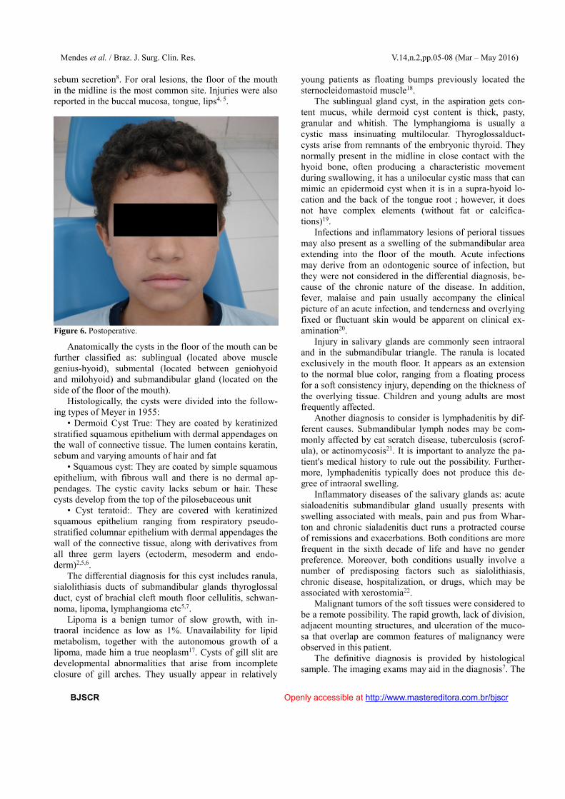
The patient was followed by a two-year period and didnot present relapse (Figure 6).
3. DISCUSSIONThe origin of dermoid cyst of mouth floor, like other
developing cysts is controversial. Two theories have beenproposed: congenital and acquired. Congenital theory isthat these cysts are derived from cellular debris, trappedduring the closing of the average of the first line and sec-ond bilateral brachial arches. The theory proposes that thecysts are acquired, they say they can be derived from atraumatic or iatrogenic including epithelial cells or occlu-sion of the sebaceous gland duct3,4.
The majority of cases occur between the ages of 15and 35, with a slight male predilection. Authors reportedsudden increases in volume at puberty, related to increased
Mendes et al. / Braz. J. Surg. Clin. Res. V.14,n.2,pp.05-08 (Mar – May 2016)
BJSCR Openly accessible at http://www.mastereditora.com.br/bjscr
sebum secretion8. For oral lesions, the floor of the mouthin the midline is the most common site. Injuries were alsoreported in the buccal mucosa, tongue, lips4, 5.
Figure 6. Postoperative.
Anatomically the cysts in the floor of the mouth can befurther classified as: sublingual (located above musclegenius-hyoid), submental (located between geniohyoidand milohyoid) and submandibular gland (located on theside of the floor of the mouth).
Histologically, the cysts were divided into the follow-ing types of Meyer in 1955:
• Dermoid Cyst True: They are coated by keratinizedstratified squamous epithelium with dermal appendages onthe wall of connective tissue. The lumen contains keratin,sebum and varying amounts of hair and fat
• Squamous cyst: They are coated by simple squamousepithelium, with fibrous wall and there is no dermal ap-pendages. The cystic cavity lacks sebum or hair. Thesecysts develop from the top of the pilosebaceous unit
• Cyst teratoid:. They are covered with keratinizedsquamous epithelium ranging from respiratory pseudo-stratified columnar epithelium with dermal appendages thewall of the connective tissue, along with derivatives fromall three germ layers (ectoderm, mesoderm and endo-derm)2,5,6.
The differential diagnosis for this cyst includes ranula,sialolithiasis ducts of submandibular glands thyroglossalduct, cyst of brachial cleft mouth floor cellulitis, schwan-noma, lipoma, lymphangioma etc5,7.
Lipoma is a benign tumor of slow growth, with in-traoral incidence as low as 1%. Unavailability for lipidmetabolism, together with the autonomous growth of alipoma, made him a true neoplasm17. Cysts of gill slit aredevelopmental abnormalities that arise from incompleteclosure of gill arches. They usually appear in relatively
young patients as floating bumps previously located thesternocleidomastoid muscle18.
The sublingual gland cyst, in the aspiration gets con-tent mucus, while dermoid cyst content is thick, pasty,granular and whitish. The lymphangioma is usually acystic mass insinuating multilocular. Thyroglossalduct-cysts arise from remnants of the embryonic thyroid. Theynormally present in the midline in close contact with thehyoid bone, often producing a characteristic movementduring swallowing, it has a unilocular cystic mass that canmimic an epidermoid cyst when it is in a supra-hyoid lo-cation and the back of the tongue root ; however, it doesnot have complex elements (without fat or calcifica-tions)19.
Infections and inflammatory lesions of perioral tissuesmay also present as a swelling of the submandibular areaextending into the floor of the mouth. Acute infectionsmay derive from an odontogenic source of infection, butthey were not considered in the differential diagnosis, be-cause of the chronic nature of the disease. In addition,fever, malaise and pain usually accompany the clinicalpicture of an acute infection, and tenderness and overlyingfixed or fluctuant skin would be apparent on clinical ex-amination20.
Injury in salivary glands are commonly seen intraoraland in the submandibular triangle. The ranula is locatedexclusively in the mouth floor. It appears as an extensionto the normal blue color, ranging from a floating processfor a soft consistency injury, depending on the thickness ofthe overlying tissue. Children and young adults are mostfrequently affected.
Another diagnosis to consider is lymphadenitis by dif-ferent causes. Submandibular lymph nodes may be com-monly affected by cat scratch disease, tuberculosis (scrof-ula), or actinomycosis21. It is important to analyze the pa-tient's medical history to rule out the possibility. Further-more, lymphadenitis typically does not produce this de-gree of intraoral swelling.
Inflammatory diseases of the salivary glands as: acutesialoadenitis submandibular gland usually presents withswelling associated with meals, pain and pus from Whar-ton and chronic sialadenitis duct runs a protracted courseof remissions and exacerbations. Both conditions are morefrequent in the sixth decade of life and have no genderpreference. Moreover, both conditions usually involve anumber of predisposing factors such as sialolithiasis,chronic disease, hospitalization, or drugs, which may beassociated with xerostomia22.
Malignant tumors of the soft tissues were considered tobe a remote possibility. The rapid growth, lack of division,adjacent mounting structures, and ulceration of the muco-sa that overlap are common features of malignancy wereobserved in this patient.
The definitive diagnosis is provided by histologicalsample. The imaging exams may aid in the diagnosis7. The
Mendes et al. / Braz. J. Surg. Clin. Res. V.14,n.2,pp.05-08 (Mar - May 2016)
BJSCR Openly accessible at http://www.mastereditora.com.br/bjscr
ultrasound is the anechogenic cystic mass. A liquid fat-linewith fluid and/or a floating mass are suggestive. Comput-ed tomography is the unilocular mass of thin-walled, withfat content. The low amount of fluid, moving with positionchange is characteristic. Computed tomography (CT) andMagnetic Ressonâninca (RM) are important to identify theagencies involved and guide the choice of surgical ap-proach10.
The treatment includes surgical expected completeenucleation13,14,15 without breaking the cyst as the intralu-minal contents can act as irritants fibrovascular tissue,producing postoperative inflammation. However, marsu-pialization is another alternative for the management oflarge cysts16. Based on the location of the cyst or an intraor extra-oral surgical approach is followed7. The first isthe most suitable for small injuries, reserving the latter forlarge lesions. In a study of mandibular osteotomy and in-traoral approach and combined extra-oral9. An exclusivelyintra-oral approach (as in our case) can be used in largelesions if there is no superinfection or disability; whichimplies a lower risk of postoperative superinfection, whichreduces the length of hospital stay, and provides an excel-lent cosmetic result9.
4. CONCLUSIONDue to the process of evolution of the lesion and the
size that can achieve early diagnosis and surgical treat-ment become imperative to improve the patient's condition.The location of the lesion relative to mylohyoid andgen-iohyoidmuscles is very important to determine the surgicalapproach that will provide the best treatment. Postopera-tive prognosis is good, and malignant transformation isexceptional.
REFERENCES[1] Neville BW, Damm DD, Allen CM, Bouquot
JE. PatologiaBucal. 2ª ed. Philadelphia: Saunders; 2002[2] Meyer I. Dermoid cistos (dermoids) do assoalho da boca. Oral
SurgPathol Oral Oral Med. 1955; 27:1149-64[3] Shylaja MD, Attur K, Mohtta A, Goud S. Teratóides cisto da
língua: Relato de uma variante rara de cisto dermóide e revisãoda literatura sobre dermoid cisto indiano J Stomatol. 2011; 2:267-9
[4] Shear, M. Cistos da região buco-maxilofacial.3a ed. São Paulo:Santos; 1999
[5] Jeyaraj P, Sahoo NK. Um caso de uma invulgarmente grandecisto dermóide sublingual da região maxilofacial. J DentMed-MedSci. 2012; 2: 19-25.
[6] Jadwani S, Misra B, Kallianpur S, Bansod S. Dermoid cisto doassoalho da boca com cabelo abundante: relato de caso. J Ma-xillofac Oral Surg. 2009; 8:. 388-9
[7] Fernandez JL, JL Rojas, JA Fernandez, Quevedo MS. Cistodermóide do assoalho da boca. Acta Otorrinolaringol Esp. 2007;58: 31-3. [PubMed].
[8] 8.GR Seward.Cistosdermóides do assoalho da boca. Br J SurgOral, 3 (1965), pp. 36-47.
[9] F. Longo, P. Maremonti, Mangone GM, et al. Cisto da linhamédia dermoid do assoalho da boca: relato de 16 casos e avali-ação de técnicas cirúrgicas. PlastSurgReconst, 112 (6) (2003),pp. 1.560-1.565.
[10] Y. Ariyosni, M. Shimahara. A ressonância magnética de cistodermóide submental: relato de um caso. J Oral MaxillofacSurg,61 (2003), pp. 507-510.
[11] Yousem D, R. GrossmanNeurorradiologia:. Os requisitos 3rd ed.Philadelphia, PA: Mosby; 2010. ISBN: 978-0323045216.
[12] Harnsberger H, Glastonbury C. Diagnóstico por Imagem:. DeCabeça e Pescoço 2nd ed. Philadelphia, PA: Amirsys; 2011.ISBN: 978-1931884785.
[13] Tsirevelou P, M Papamanthos, Chlopsidis P, Zourou I, Skoula-kis C. Cisto epidermóide do assoalho da boca: relato de doiscasos. Casos J. 2009; 2: 9360 [PMC artigo livre] [PubMed]
[14] Jha AK, Sahoo NK. Um caso incomum de cisto epidermóidesubmental em uma criança de dez anos: relato de caso. Int J CasoRep Imagem. 2011; 2 (10): 10-13.
[15] Chaturvedi J, K Hegde, Ananthamurthy A, Nayar R. Submentalepidermóidecyst- relato de caso. Internet J Head NeckSurg.2009; 4 (2).
[17] El-Momen MH, Gaafar AH, MagdyEA. .Lipomas da cabeça epescoço: variabilidade Apresentação e work-up diagnóstico JLaryngolOtol. 2006; 120: 47-55. [PubMed]
[18] Glosser JW, Pires CA, Feinberg SE. Fissura branquial ou cistoslinfoepiteliais cervicais: Etiologia e gestão.JAmDent Assoc.2003; 134: 81-6. [PubMed].
[19] Ellis PD, van Nostrand AW. A anatomia aplicada de remanes-centes do trato tireoglosso. Laryngoscope.1977; 87: 765-70.
[20] Tosios K, G Rallis, Vallianatou D, tumor VlachodimitropoulosD. Amarelo-branca no assoalho da boca.OralSurg Oral MedOral Pathol Oral RadiolEndod. 2006; 101:. 701-4.
[21] Rei RC, Smith BR, Burk JL. Cisto dermóide no assoalho daboca. A revisão da literatura e relatos de caso. Oral SurgPatholOral Oral Med. 1994; 78:. 567-76.
[22] Huang C, Damrose E, Bhuta S, tumor Abemayor E. Kuttner(sclerosingsialadenitis crónica) Am J Otolaryngol. 2002; 23:.394-7.