CASE AND RESEARCH LETTERS 97 Figure 2 Image of the lesions obtained with a polarized light dermatoscope attached to a Canon Powershot A160 camera using the DermLite Foto attachment (3Gen LLC). Rosettes (indi- cated by a white circle in the lesion on the left; a larger number of rosettes are seen in the central area of the lesion on the right) are in evidence, along with irregularly shaped vessels and a clear rainbow pattern. why the pattern is found in other types of skin lesions with active vascularization, such as our patient’s scars. When first described in actinic keratosis, rosettes were characterized as ‘‘4 white points arranged as a 4-leaf clover.’’ 2 The sign has since been reported in other skin lesions such as squamous cell carcinoma, basal cell carci- noma, melanoma, and lichenoid keratosis. 3---7 Rosettes are believed to be the result of an optical effect caused by interaction between polarized light and follicular openings. 6 The rainbow pattern and rosettes are not considered to be specific dermoscopic features of the lesion. Since it appears that they are secondary effects of the interaction between different skin structures and polarized light, they will likely be observed in various types of skin lesions. References 1. Hu SC, Ke CL, Lee CH, Wu CS, Chen GS, Cheng ST. Dermoscopy of Kaposi’s sarcoma: Areas exhibiting the multicoloured ‘rainbow pattern’. J Eur Acad Dermatol Venereol. 2009;23:1128---32. 2. Cuellar F, Vilalta A, Puig S, Palou J, Salerni G, Malvehy J. New dermoscopic pattern in actinic keratosis and related conditions. Arch Dermatol. 2009;145:732. 3. Garcia-Garcia B, Perez-Oliva N. Dermoscopic rainbow pat- tern in basal cell carcinoma. J Eur Acad Dermatol Venereol. 2010;24:499---500. 4. Vázquez-López F, García-García B, Rajadhyaksha M, Marghoob AA. Dermoscopic rainbow pattern in non-Kaposi sarcoma lesions. Br J Dermatol. 2009;161:474---5. 5. Liebman TN, Jaimes-Lopez N, Balagula Y, Rabinovitz HS, Wang SQ, Dusza SW, et al. Dermoscopic features of basal cell carcinomas: Differences in appearance under non- polarized and polarized light. Dermatol Surg. 2012;38: 392---9. 6. Liebman TN, Scope A, Rabinovitz H, Braun RP, Marghoob AA. Rosettes may be observed in a range of conditions. Arch Der- matol. 2011;147:1468. 7. Liebman TN, Rabinovitz HS, Dusza SW, Marghoob AA. White shiny structures: Dermoscopic features revealed under polarized light. J Eur Acad Dermatol Venereol. 2011;26: 1493---7. 8. Cheng ST, Ke CL, Lee CH, Wu CS, Chen GS, Hu SC. Dermoscopic rainbow pattern in non-Kaposi sarcoma lesions-reply. Br J Der- matol. 2010;162:458---9. 9. Cheng ST, Ke CL, Lee CH, Wu CS, Chen GS, Hu SC. Rainbow pattern in Kaposi’s sarcoma under polarized dermoscopy: A dermoscopic pathological study. Br J Dermatol. 2009;160:801---9. L. Pérez-Pérez, * J. García-Gavín, F. Allegue, A. Zulaica Servicio de Dermatología, Complejo Hospitalario Universitario de Vigo, Vigo, Spain * Correspondig author. E-mail address: [email protected](L. Pérez-Pérez). Dermoscopic Rainbow Pattern in Atypical Fibroxanthoma Patrón dermatoscópico en arcoíris en fibroxantoma atípico We present the case of a 73-year-old man with a his- tory of non-insulin-dependent diabetes mellitus, arterial hypertension, abdominal aortic aneurysm, and hypercholes- terolemia. He was referred to our department for evaluation of a tumor on the scalp that had appeared 6 weeks earlier. The tumor was pink with some reddish and violaceous areas, had a maximum diameter of 18 mm and distinct borders, was nonulcerated, and displayed mild scaling in the center (Fig. 1). Please cite this article as: Pitarch G. Patrón dermatoscópico en arcoíris en fibroxantoma atípico. Actas Dermosifiliogr. 2014;105:97---99. Dermoscopic examination was performed with a polar- ized light contact dermoscope (DermLite Foto, 3Gen LLC) using ultrasound gel as the liquid interface. The dermo- scopic images showed a round, symmetrical lesion with a reddish peripheral area from which atypical, irregularly distributed, out-of-focus blood vessels----mostly linear and unbranched----extended in a vaguely radial pattern (Fig. 2). Most of the tumor surface displayed rainbow-patterned areas, often arranged in parallel to the linear, irregular blood vessels. None of the criteria specific to melanocytic lesions were observed. Shiny whitish areas were observed between the rainbow-patterned structures, and scales were visible on the surface. Complete surgical excision of the lesion was performed. Histologic examination revealed a nodular cell prolifera- tion in the dermis comprising aberrant spindle-shaped cells, epithelioid cells, multinucleated giant cells, and abundant mitotic figures. Hemorrhagic zones were observed in some areas. Immunohistochemistry was positive for vimentin, CD68, and CD10 and negative for CD31, CD34, FVIII, S100, Document downloaded from http://www.elsevier.es, day 29/05/2017. This copy is for personal use. Any transmission of this document by any media or format is strictly prohibited.
Transcript
CASE AND RESEARCH LETTERS 97
Figure 2 Image of the lesions obtained with a polarized lightdermatoscope attached to a Canon Powershot A160 camerausing the DermLite Foto attachment (3Gen LLC). Rosettes (indi-cated by a white circle in the lesion on the left; a larger numberof rosettes are seen in the central area of the lesion on the right)are in evidence, along with irregularly shaped vessels and a clearrainbow pattern.
why the pattern is found in other types of skin lesions withactive vascularization, such as our patient’s scars.
When first described in actinic keratosis, rosettes werecharacterized as ‘‘4 white points arranged as a 4-leafclover.’’2 The sign has since been reported in other skinlesions such as squamous cell carcinoma, basal cell carci-noma, melanoma, and lichenoid keratosis.3---7 Rosettes arebelieved to be the result of an optical effect caused byinteraction between polarized light and follicular openings.6
The rainbow pattern and rosettes are not consideredto be specific dermoscopic features of the lesion. Since itappears that they are secondary effects of the interactionbetween different skin structures and polarized light, theywill likely be observed in various types of skin lesions.
References
1. Hu SC, Ke CL, Lee CH, Wu CS, Chen GS, Cheng ST. Dermoscopyof Kaposi’s sarcoma: Areas exhibiting the multicoloured ‘rainbowpattern’. J Eur Acad Dermatol Venereol. 2009;23:1128---32.
2. Cuellar F, Vilalta A, Puig S, Palou J, Salerni G, Malvehy J. Newdermoscopic pattern in actinic keratosis and related conditions.Arch Dermatol. 2009;145:732.
3. Garcia-Garcia B, Perez-Oliva N. Dermoscopic rainbow pat-tern in basal cell carcinoma. J Eur Acad Dermatol Venereol.2010;24:499---500.
4. Vázquez-López F, García-García B, Rajadhyaksha M, MarghoobAA. Dermoscopic rainbow pattern in non-Kaposi sarcoma lesions.Br J Dermatol. 2009;161:474---5.
5. Liebman TN, Jaimes-Lopez N, Balagula Y, Rabinovitz HS,Wang SQ, Dusza SW, et al. Dermoscopic features of basalcell carcinomas: Differences in appearance under non-polarized and polarized light. Dermatol Surg. 2012;38:392---9.
6. Liebman TN, Scope A, Rabinovitz H, Braun RP, Marghoob AA.Rosettes may be observed in a range of conditions. Arch Der-matol. 2011;147:1468.
Dermoscopic Rainbow Pattern in AtypicalFibroxanthoma�
Patrón dermatoscópico en arcoíris enfibroxantoma atípico
We present the case of a 73-year-old man with a his-tory of non-insulin-dependent diabetes mellitus, arterialhypertension, abdominal aortic aneurysm, and hypercholes-terolemia. He was referred to our department for evaluationof a tumor on the scalp that had appeared 6 weeks earlier.The tumor was pink with some reddish and violaceous areas,had a maximum diameter of 18 mm and distinct borders,was nonulcerated, and displayed mild scaling in the center(Fig. 1).
� Please cite this article as: Pitarch G. Patrón dermatoscópicoen arcoíris en fibroxantoma atípico. Actas Dermosifiliogr.2014;105:97---99.
Dermoscopic examination was performed with a polar-ized light contact dermoscope (DermLite Foto, 3Gen LLC)using ultrasound gel as the liquid interface. The dermo-scopic images showed a round, symmetrical lesion witha reddish peripheral area from which atypical, irregularlydistributed, out-of-focus blood vessels----mostly linear andunbranched----extended in a vaguely radial pattern (Fig. 2).Most of the tumor surface displayed rainbow-patternedareas, often arranged in parallel to the linear, irregularblood vessels. None of the criteria specific to melanocyticlesions were observed. Shiny whitish areas were observedbetween the rainbow-patterned structures, and scales werevisible on the surface.
Complete surgical excision of the lesion was performed.Histologic examination revealed a nodular cell prolifera-tion in the dermis comprising aberrant spindle-shaped cells,epithelioid cells, multinucleated giant cells, and abundantmitotic figures. Hemorrhagic zones were observed in someareas. Immunohistochemistry was positive for vimentin,CD68, and CD10 and negative for CD31, CD34, FVIII, S100,
Document downloaded from http://www.elsevier.es, day 29/05/2017. This copy is for personal use. Any transmission of this document by any media or format is strictly prohibited.
Figure 1 Tumor on the scalp and nearby actinic keratoses.
cytokeratins, epithelial membrane antigen, desmin, andsmooth muscle actin. The proliferative index, measuredusing Ki-67, was between 10% and 20%. A diagnosis of atyp-ical fibroxanthoma was established on the basis of thehistopathologic findings.
Atypical fibroxanthoma is a rare low-grade tumor.It usually develops on chronically sun-damaged skin,especially on the head or neck of elderly patients.1 Clin-ically, it presents as a single, rapidly growing nodule.Differential diagnosis should include basal cell carcinoma,squamous cell carcinoma, Merkel cell carcinoma, andmelanoma. Lymph node metastases are rare and distantmetastases are very rare.
Only 4 dermoscopic observations of this rare tumorhave been described in the literature. An atypical vas-cular pattern with irregularly distributed polymorphicvessels----including linear, punctate, globular, tortuous, and
Figure 2 Dermoscopic image, obtained using a polarized lightcontact dermoscope, showing irregular blood vessels in theperipheral area and a rainbow pattern in the center.
arborizing vessels----was observed in 3 cases.2 Whitish areaswere observed in 2 of these patients and heterogeneoushyperpigmentation in the other. The remaining case was acollision tumor associated with a basal cell carcinoma, inwhich the atypical fibroxanthoma component appeared as ahemorrhagic, ulcerated lesion with small telangiectasias.3
In our patient, the most striking dermoscopic finding wasthe rainbow pattern in the center of the tumor. Although therainbow pattern was initially considered to be a character-istic feature of Kaposi sarcoma, it has since been observedin various conditions, including melanoma, stasis dermatitis,lichen planus, hemosiderotic dermatofibroma, and basal cellcarcinoma.4-7 The rainbow pattern is an optical phenomenonthat can only be observed with a polarized light dermoscope(either contact or noncontact). It occurs when light in dif-ferent states of polarization interacts with the structuresof the lesion.6 Bugatti et al.2 specified that a contact der-moscope was used in the 3 cases they reported but did notstate whether or not the device used polarized light, withoutwhich the rainbow pattern cannot be observed.
The shiny white structures can take the form of shinywhite streaks (also known as chrysalis structures), shinywhite areas, or rosettes. These structures can be observedmuch more clearly with a polarized light dermoscope. Thepresence of shiny white structures has been described invarious benign and malignant tumors----including melanoma,Spitz nevus, dermatofibroma, actinic keratosis, squamouscell carcinoma, and basal cell carcinoma----and is indicativeof an increase in dermal collagen.8,9
All dermoscopic observations of atypical fibroxanthomareported to date have described an atypical vascular pat-tern characterized by polymorphic vessels interspersed withwhitish areas. This dermoscopic pattern is nonspecific andindicative of chaotic neoangiogenesis during growth. It isalso seen in other tumors that are clinically similar to atypi-cal fibroxanthoma, such as squamous cell carcinoma, Merkelcell carcinoma, and amelanotic melanoma.10 Since the rain-bow pattern is also found in a number of these tumors, thisfinding alone is not sufficient to establish a dermoscopicdiagnosis.
In summary, we present the first dermoscopic observationof the rainbow pattern in an atypical fibroxanthoma, therebyadding atypical fibroxanthoma to the list of entities thatcan present this finding. A vascular pattern characterizedby polymorphic vessels is a constant finding in all reportedcases of this tumor.
References
1. Gómez de la Fuente E, Sols M, Pinedo F, Álvarez-Fernández JG,Vicente FJ, Naz E, et al. Fibroxantoma atípico. Estudio clinico-patológico de 10 casos. Actas Dermosifiliogr. 2005;96:153---8.
2. Bugatti L, Filosa G. Dermatoscopic features of cutaneousatypical fibroxanthoma: three cases. Clin Exp Dermatol.2009;34:e898---900.
3. Alves R, Ocana J, Vale E, Correia S, Viana I, Bordalo O. Basalcell carcinoma and atypical fibroxanthoma: an unusual collisiontumor. J Am Acad Dermatol. 2010;63:e74---5.
4. Hu SC, Ke CL, Lee CH, Wu CS, Chen GS, Cheng ST. Dermoscopyof Kaposi’s sarcoma: areas exhibiting the multicoloured‘‘rainbow pattern’’. J Eur Acad Dermatol Venereol. 2009;23:1128---32.
Document downloaded from http://www.elsevier.es, day 29/05/2017. This copy is for personal use. Any transmission of this document by any media or format is strictly prohibited.
5. Cheng ST, Ke CL, Lee CH, Wu CS, Chen GS, Hu SC. Rainbowpattern in Kaposi’s sarcoma under polarized dermoscopy: a der-moscopic pathological study. Br J Dermatol. 2009;160:801---9.
6. Vázquez-López F, García-García B, Rajadhyaksha M, MarghoobAA. Dermoscopic rainbow pattern in non-Kaposi sarcomalesions. Br J Dermatol. 2009;161:474---5.
7. García-García B, Pérez-Oliva N. Dermoscopic rainbow pat-tern in basal cell carcinoma. J Eur Acad Dermatol Venereol.2010;24:499---500.
8. Balagula Y, Braun RP, Rabinovitz HS, Dusza SW, Scope A, LiebmanTN, et al. The significance of crystalline/chrysalis structures inthe diagnosis of melanocytic and nonmelanocytic lesions. J AmAcad Dermatol. 2012;67:194, e1---8.
10. Zalaudek I, Kreusch J, Giacomel J, Ferrara G, Catricalà C,Argenziano G. How to diagnose nonpigmented skin tumors: areview of vascular structures seen with dermoscopy. J Am AcadDermatol. 2010;63:377---86.
G. Pitarch
Sección de Dermatología, Hospital General de Castelló,
Anetodermia primaria asociada a síndrome deSjögren primario y anticuerposanticardiolipina
Anetoderma is a rare acquired chronic skin condition thatinvolves the loss of elastic fibers. There is no specific effec-tive treatment.1,2 We present the case of a patient withprimary anetoderma associated with both primary Sjögrensyndrome and anticardiolipin antibodies. Biopsy showed anacute inflammatory infiltrate observed in the reticular der-mis. The condition was treated with colchicine and dapsone.
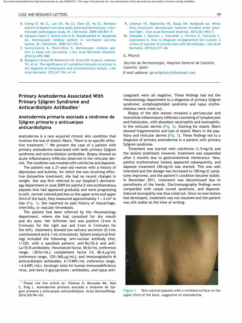
The patient was a 21-year-old woman with a history ofdepression and bulimia, for which she was receiving effec-tive duloxetine treatment; she had no recent changes inweight. She was first referred to our hospital’s dermatol-ogy department in June 2009 for painful 5-mm erythematouspapules that had appeared gradually and were progressingto soft, normal-colored patches on the upper arms and upperthird of the back; they measured approximately 1 × 2 cm2 insize (Fig. 1). She reported no past history of miscarriage,infertility, or vascular thrombosis.
The patient had been referred by the rheumatologydepartment, where she had consulted for dry mouthand dry eyes. Her Schirmer test was positive (3 mm in5 minutes for the right eye and 5 mm in 5 minutes forthe left). Sialometry showed low salivary secretion (0.3 mLunstimulated and 6.1 mL stimulated). Salient analytical find-ings included the following: anti-nuclear antibody titer,1/320, with a speckled pattern; anti-Ro/SS-A and anti-La/SS-B antibodies; rheumatoid factor, 56 IU/mL (referencerange, < 20 IU/mL); complement factor C4, 86.6 �g/mL(reference range, 120---360 �g/mL); and immunoglobulin Manticardiolipin antibodies, 6.73 MPL/mL (reference range,< 4.6 MPL/mL). Serologic tests for human immunodeficiencyvirus, anti-beta-2-glycoprotein i antibodies, and lupus anti-
� Please cite this article as: Yélamos O, Barnadas MA, DíazC, Puig L. Anetodermia primaria asociada a síndrome de Sjö-gren primario y anticuerpos anticardiolipina. Actas Dermosifiliogr.2014;105:99---101.
coagulant were all negative. These findings had led therheumatology department to a diagnosis of primary Sjögrensyndrome; antiphospholipid syndrome and lupus erythe-matosus were ruled out.
Biopsy of the skin lesions revealed a perivascular andinterstitial inflammatory infiltrate consisting of lymphocytesand histiocytes, with abundant neutrophils and eosinophils,in the reticular dermis (Fig. 2). Staining for elastic fibersshowed fragmentation and loss of elastic fibers in the pap-illary and reticular dermis (Fig. 3). These findings led to adiagnosis of primary anetoderma in a patient with primarySjögren syndrome.
Treatment was started with colchicine (1.5 mg/d) andthe lesions stabilized; however, treatment was suspendedafter 2 months due to gastrointestinal intolerance. New,painful erythematous lesions appeared subsequently, anddapsone treatment (50 mg/d) was started. This was welltolerated and the dosage was increased to 100 mg/d; symp-toms improved, and the patient’s condition became stable.In December 2011, treatment was discontinued due toparesthesia of the hands. Electromyography findings werecompatible with carpal tunnel syndrome, and dapsone-induced neuropathy was thus ruled out. Since no new lesionshad developed, treatment was not resumed and the patientwas still stable at the time of writing.
Figure 1 Skin-colored papules with a wrinkled surface on theupper third of the back, suggestive of anetoderma.
Document downloaded from http://www.elsevier.es, day 29/05/2017. This copy is for personal use. Any transmission of this document by any media or format is strictly prohibited.