Page 1
Please cite this article as: Akila RM et al., Design and Characterization of Biodegradable Chitosan
Nanoparticles Loaded With Almotriptan Malate For Migraine Therapy. American Journal of Pharmacy
& Health Research 2018.
Research Article
www.ajphr.com
2018, Volume 6, Issue 07
ISSN: 2321–3647(online)
Design and Characterization of Biodegradable Chitosan
Nanoparticles Loaded With Almotriptan Malate For Migraine
Therapy
Akila RM1*, Sneha Anna Saju1
Department of Pharmaceutics, College of Pharmacy, Sri Ramakrishna Institute of Paramedical
Sciences, Coimbatore, India.
ABSTRACT
Nanoparticles are designed to improve the pharmacological and therapeutic properties of
conventional drugs. In the present research work, almotriptan malate was formulated as
nanoparticle drug delivery system for the effective management of migraine by ion-gelation
technique. Shape and size of the nanoparticles were evaluated by TEM images. FTIR and DSC
studies confirmed that there were no interactions between the drug and the other ingredients.
XRD study was carried out to confirm the crystalline or amorphous nature of the nanoparticles.
The entrapment efficiency of was found to be 74 to 82% and drug loading capacity was found to
be 15 to 16% The in-vitro release studies concluded that the release was sustained after an initial
burst. The in vitro mucoadhesion study using goat nasal mucosa was found to be 54% and 69%.
The kinetic study revealed that the almotriptan malate nanoparticles followed the first order
kinetics.
Keywords: biodegradable chitosan, nanoparticles, almotriptan malate, mucoadhesion
*Corresponding Author Email: [email protected]
Received 10 June 2018, Accepted 26 June 2018
Page 2
Akila et al., Am. J. Pharm Health Res 2018;6(07) ISSN: 2321-3647
www.ajphr.com 2
INTRODUCTION
The method by which the drug is delivered can have a significant effect on its efficacy. Aiming
therapeutic compound to the desired site is a major problem in treatment of many diseases. A
traditional application of drugs is identified by limited action, poor bio- distribution and lack of
acuteness[1]. These limitations and drawback can be overcome by controlling drug delivery. In
controlled drug delivery system the drug is delivered to the place of action, thus its influence on
important tissues and harmful side effect can be reduced. In addition controlled drug delivery
system guards the drug from rapid, degradation or clearance and amplifies drug concentration in
target tissues, therefore lower doses of drug required. Cell-specific targeting can be attained by
attaching drugs to individually designed carriers. Chitosan nanoparticles have acquired more
importance as drug delivery carriers because of their stability, low toxicity, simple and mild
preparation method and yielding versatile routes of administration. Chitosan has the special
possibility of adhering to the mucosal surfaces within the body because of their submicron size
and is suitable for mucosal route i.e. oral, nasal and ocular mucosa which is a non-invasive route
[2]. Diseases of the CNS such as schizophrenia, meningitis, migraine, Parkinsons disease and
Alzheimers disease require delivery of the drug to the brain for treatment. However such
conveyance remains difficult especially for hydrophilic and large molecular weight drugs due to
the impervious nature of the endothelial membrane separating the systemic circulation and
interstitial fluid of the blood brain barrier (BBB). Some of the currently employed invasive
approaches include interstitial delivery, intracerebroventricular delivery, intracerebral delivery
and nasal delivery[3]. A migraine is more than just a headache. It is a complicated neurological
problem which can affect the whole body and can result in many symptoms, sometimes without
a headache at all [4]. The triptan category of drugs is used in the termination of migraine attack
by acting as vasoconstrictors by inhibiting neurogenic inflammation peripherally, inhibiting
nociceptors[5]. As the oral route of delivery triptans has few disadvantages like nausea, the
present research focuses on delivering almotriptan malate using chitosan nanoparticles as
intranasal brain targeted delivery.
MATERIALS AND METHOD
Almotriptan malate was received as a gift sample from Azakem laboratories, Hyderabad, India.
Chitosan (85% deacetylated), sodium tripolyphosphate were obtained from Sigma Aldrich,
Mumbai and tween 80 was purchased from S.D Fine-Chem Pvt. Ltd. (Mumbai, India). All other
reagents were of analytical grade and used as purchased.
Page 3
Akila et al., Am. J. Pharm Health Res 2018;6(07) ISSN: 2321-3647
www.ajphr.com 3
Preparation of drug loaded chitosan nanoparticles [6]
Chitosan nanoparticles were formulated based on the ionic gelation of chitosan and sodium
tripolyphosphate anions. The weighed chitosan (1,2,3mg/ml) was dissolved in 1%v/v of an
aqueous solution of acetic acid, to which 0.5% of Tween 80 and almotriptan malate(2mg) was
added. The sodium tripolyphosphate solution (0.5,1,1.5mg/ml) was prepared in distilled water.
This solution was added to the drug-loaded chitosan nanoparticles and stirred continuously using
magnetic stirrer (REMI-2MLH) at room temperature for 30min. Subsequently, the pH was
adjusted to 5.5 with the help of a required amount of 1N HCl or NaOH and then centrifuged at
10000 rpm using a centrifuge for 30min, washed and dried.
Table 1 Formulation of Chitosan Nanoparticles
Batch code Chitosan
(mg/ml)
Sodium
tripolyphosphate(mg/ml)
Almotriptan
malate(mg)
B0 1 0.5 0
B1 1 0.5 2
B2 2 1 2
B3 3 1.5 2
Characterization of nanoparticles
Invitro drug release study of the prepared formulations [7]
The in vitro drug release study was performed in a USP Type II (Paddle type) apparatus using
the principle of diffusion through the dialysis membrane. Drug-loaded nanoparticles redispersed
in 2 ml phosphate buffer (pH 6.5), were placed in a dialysis bag soaked in distilled water 1hour
prior to its use, and the bag was tied to the paddle of the apparatus. The paddle was rotated at 100
rpm and the dissolution media was 500 ml of phosphate buffer pH 6.5 maintained at 37 ±1◦C.
Sample aliquots of 5 ml were collected at specific time intervals and were replenished with an
equal amount of fresh dissolution medium. The samples were analyzed, spectrophotometrically
at λmax = 228 nm against suitable blank and amount of drug released at various time intervals
were calculated. The results were expressed in terms of average % drug release ± standard
deviation.Invitro mucoadhesion studies [8]
Invitro mucoadhesion studies [8]
A piece of goat nasal mucosa was cleaned and placed in Krebs solution in Petri plate. It was
washed with distilled water, and then accurately weighed almotriptan malate loaded chitosan
nanoparticles equivalent to 100 mg were spread on to it. The mucosa was kept aside for 15 min.
Then surface was washed with phosphate buffer pH 6.5. Nanoparticles retained on the mucosal
surface after first washing was removed by rinsing thoroughly by phosphate buffer 6.5. The
Page 4
Akila et al., Am. J. Pharm Health Res 2018;6(07) ISSN: 2321-3647
www.ajphr.com 4
solution was stirred to dissolve drug, filtered and absorbance was recorded. The amount of
adhered nanoparticles was estimated as the difference between the amount of applied
nanoparticles and the amount of flowed nanoparticles. The percent mucoadhesion was calculated
using the following
𝑝𝑒𝑟𝑐𝑒𝑛𝑡𝑎𝑔𝑒 𝑜𝑓 𝑚𝑢𝑐𝑜𝑎𝑑ℎ𝑠𝑒𝑠𝑖𝑜𝑛 =𝑎𝑚𝑜𝑢𝑛𝑡 𝑜𝑓 𝑑𝑟𝑢𝑔 𝑖𝑛 𝑤𝑎𝑠ℎ 𝑜𝑢𝑡 𝑙𝑖𝑞𝑢𝑖𝑑
𝑎𝑐𝑡𝑢𝑎𝑙 𝑎𝑚𝑜𝑢𝑛𝑡 𝑜𝑓 𝑑𝑟𝑢𝑔 𝑖𝑛 𝑛𝑎𝑛𝑜𝑝𝑎𝑟𝑡𝑖𝑐𝑙𝑒𝑠× 100
In-vitro drug release kinetics study [9]
In order to describe the mode of release of drug from nanoparticles the in-vitro release data of
optimized formulation, B2 was fitted into the following mathematical models: (zero order, first
order, Higuchi equation and Korsmeyer-Peppas equations).
Qt = K0 t (Zero Order Kinetics)
Log (Qt / Q0) = – K1 t / 2.303 (First-order Kinetics)
Qt = KKP tn (Korsmeyer and Peppas equation)
Qt = KH t 1/2 (Higuchi’s equation) Where, Qt is the percent of drug released at a time‘t’, K0, K1,
KKP, and KH are the coefficients of Zero order, First order, Korsmeyer-Peppas and Higuchi’s
equations.
Drug Content [10]
1ml of almotriptan malate loaded chitosan nanoparticles suspension is dissolved in the 10 ml of
distilled water. The amount of almotriptan malate was determined using UV spectrophotometer
at λ max = 228 nm. The placebo formulation prepared similarly to drug-loaded chitosan is used
as a blank. The total drug content was calculated.
TDC= concentration ×dilution factor ×volume of formulation x100
Transmission Electron Microscopy (TEM ) [11]
The nanoparticles of the optimized batch, after suitable dilution with HPLC grade water, were
examined for surface morphology and particle size under a transmission electron microscope
(Jeol/ JEM 2100 STIC, Cochin ), operated at 200 kV. The sample was placed on a carbon-coated
copper grids and was stained with 2% phosphotungstic acid (freshly prepared, pH 7.0).
Entrapment Efficiency and Drug Loading Capacity[12]
The entrapment efficiency can be determined by measuring the concentration of the free drug in
the dispersion. Entrapment efficiency and drug loading capacity of nanoparticles in different
formulations are determined by ultra-centrifugation of samples at 10,000 rpm for 30 min. The
amount of free almotriptan malate was determined in the clear supernatant by UV
spectrophotometry at 227nm using supernatant of non-loaded (blank) nanoparticles as the basic
Page 5
Akila et al., Am. J. Pharm Health Res 2018;6(07) ISSN: 2321-3647
www.ajphr.com 5
correction. The drug loading capacity of nanoparticles and entrapment efficiency (EE) of the
process were calculated from the following equations:
𝐸𝑛𝑡𝑟𝑎𝑝𝑚𝑒𝑛𝑡 𝐸𝑓𝑓𝑖𝑐𝑖𝑒𝑛𝑐𝑦(𝐸𝐸%) =𝑡𝑜𝑡𝑎𝑙 𝑎𝑚𝑜𝑢𝑛𝑡 𝑜𝑓 𝑑𝑟𝑢𝑔 − 𝑓𝑟𝑒𝑒 𝑑𝑟𝑢𝑔
𝑡𝑜𝑡𝑎𝑙 𝑑𝑟𝑢𝑔× 100
𝐷𝑟𝑢𝑔 𝑙𝑜𝑎𝑑𝑖𝑛𝑔 𝑐𝑎𝑝𝑎𝑐𝑖𝑡𝑦 =𝑡𝑜𝑡𝑎𝑙 𝑎𝑚𝑜𝑢𝑛𝑡 𝑜𝑓 𝑑𝑟𝑢𝑔 − 𝑓𝑟𝑒𝑒 𝑑𝑟𝑢𝑔
𝑡𝑜𝑡𝑎𝑙 𝑤𝑒𝑖𝑔ℎ𝑡 𝑜𝑓 𝑛𝑎𝑛𝑜𝑝𝑎𝑟𝑡𝑖𝑐𝑙𝑒𝑠× 100
Infrared (IR) spectrum[13]
The IR spectra of the formulations were carried out for drug-loaded chitosan nanoparticles as
well as for the physical mixture of drug and excipients using the JASCO FTIR 4100 instrument
in the frequency range of 400-4000cm-1 with the resolution of 4cm-1
Differential scanning calorimetry (DSC) [14]
DSC studies were performed to understand the behavior of cross-linked chitosan on the
application of thermal energy. DSC was performed on a DSC-7 (Perkin-Elmer Corp., USA) at a
heating rate of 0°C/min in the temperature range of 0- 350°C using the empty aluminum pan as a
reference standard
Powder X-ray diffraction study [15]
The XRD patterns of pure drug, dummy nanoparticle and optimized nanoparticle are to be
measured on a diffractometer. Monochromatic radiation (λ = 1.5418A˚), at 40 kV and 40 mA,
was used as the X-ray source. The diffractograms were recorded between 20º and 70º with an
increasing step of 0.02º and 2 s as the time for each step.
Stability studies[16]
The stability study was carried out to assess the stability of almotriptan malate loaded chitosan
nanoparticles. For this purpose, samples were kept in borosilicate glass vials and stored at room
temperature, in a refrigerator (5±1°C), and 45 ± 1°C (75% relative humidity) in the stability
chamber. Samples were analyzed at the intervals of 0, 7, 14, 21, 28, 35, and 45 days for their
changes in physical appearance and for the drug content.
RESULTS AND DISCUSSION
In-vitro drug release study
The in-vitro release profile of almotriptan malate from different formulations is shown in
Figure1. The percent drug release was not same for all the formulations, it varies with time.
The release profile for chitosan biodegradable nanoparticles was obtained in phosphate buffer
solution, pH 6.5. Initially, the chitosan nanoparticles showed the immediate release of
almotriptan malate, with about 45% almotriptan malate being released in 10 minutes which may
Page 6
Akila et al., Am. J. Pharm Health Res 2018;6(07) ISSN: 2321-3647
www.ajphr.com 6
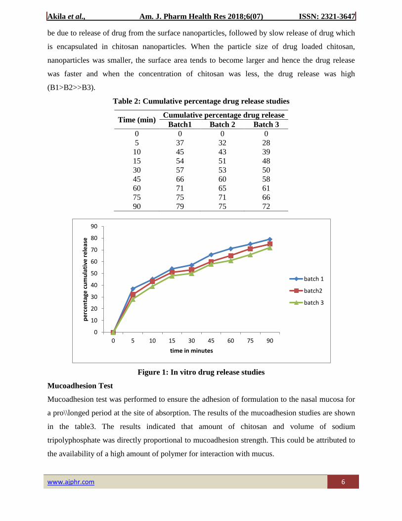
be due to release of drug from the surface nanoparticles, followed by slow release of drug which
is encapsulated in chitosan nanoparticles. When the particle size of drug loaded chitosan,
nanoparticles was smaller, the surface area tends to become larger and hence the drug release
was faster and when the concentration of chitosan was less, the drug release was high
(B1>B2>>B3).
Table 2: Cumulative percentage drug release studies
Time (min) Cumulative percentage drug release
Batch1 Batch 2 Batch 3
0 0 0 0
5 37 32 28
10 45 43 39
15 54 51 48
30 57 53 50
45 66 60 58
60 71 65 61
75 75 71 66
90 79 75 72
Figure 1: In vitro drug release studies
Mucoadhesion Test
Mucoadhesion test was performed to ensure the adhesion of formulation to the nasal mucosa for
a pro\\longed period at the site of absorption. The results of the mucoadhesion studies are shown
in the table3. The results indicated that amount of chitosan and volume of sodium
tripolyphosphate was directly proportional to mucoadhesion strength. This could be attributed to
the availability of a high amount of polymer for interaction with mucus.
0
10
20
30
40
50
60
70
80
90
0 5 10 15 30 45 60 75 90
pe
rce
nta
ge c
um
ula
tive
re
leas
e
time in minutes
batch 1
batch2
batch 3
Page 7
Akila et al., Am. J. Pharm Health Res 2018;6(07) ISSN: 2321-3647
www.ajphr.com 7
Table 3: Percentage Mucoadhesion Of The Drug Loaded Nanoparticles
Sl. No Batch code Percentage mucoadhesion (%)
Of the drug loaded Nanoparticles
1 B1 69
2 B2 61
3 B3 54
Figure 2: Percentage Mucoadhesion Of The Drug Loaded Nanoparticles
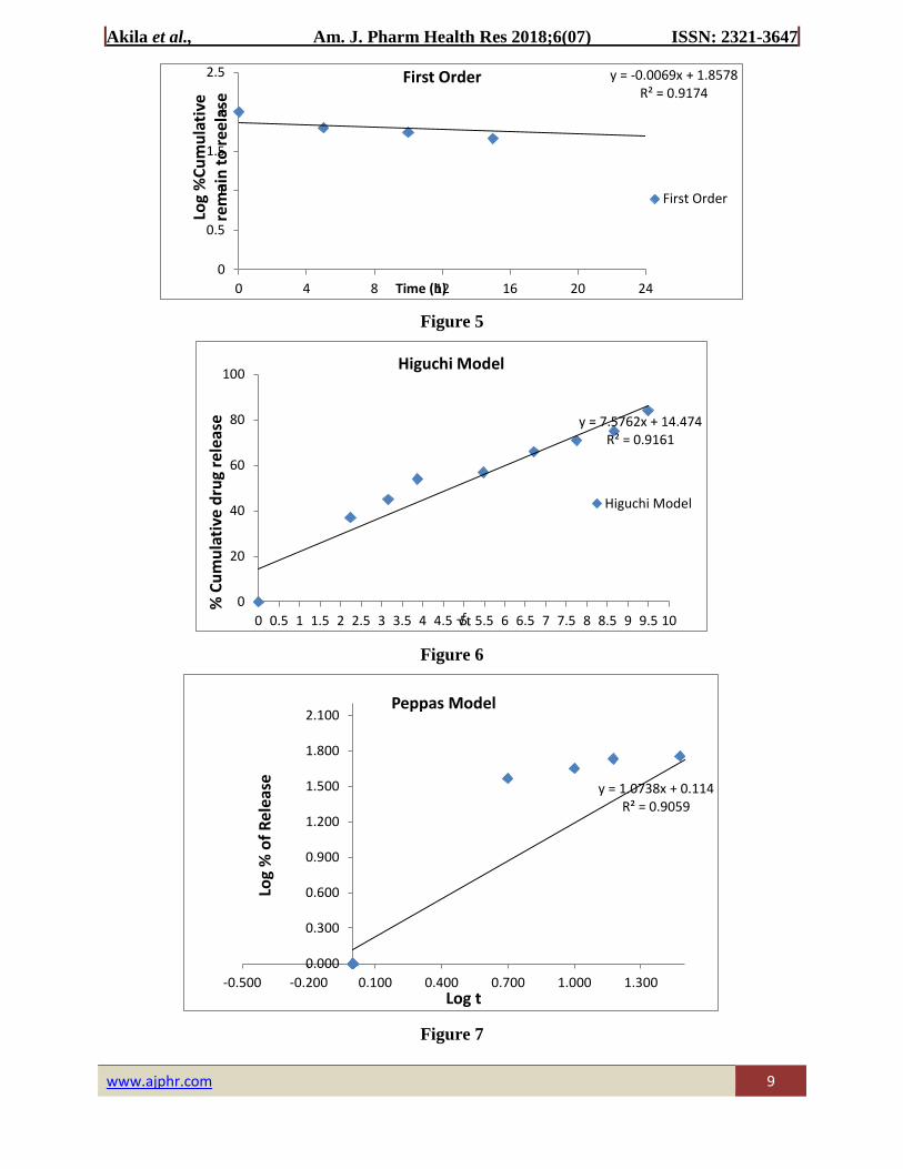
In-vitro drug release kinetic study
To analyze the mechanism for the release and release rate kinetics of the dosage form, the data
obtained from in-vitro drug release from optimized formulation (B1)was fitted to model’s
representation zero order, first order, Higuchi, and Korsmeyer-Peppas. The release data, when
fitted into release kinetic equations, produced correlation coefficients (R2) of 0.917, 0.916 and
0.905 for the first order, Korsmeyer–Peppas, and Higuchi models respectively and non-linearity
when plotted by the zero-order equation (Figure 3-6). Hence, it can be concluded that the major
mechanism of drug release follows first order kinetics. Data based on the first order models
usually provide evidence to the diffusion mechanism of drug release from sustained release
delivery systems.
Drug content
The drug content ranges from 82% to 91% and it is more in B3 as it was trapped in a higher
concentration of chitosan.
Entrapment Efficiency
0
10
20
30
40
50
60
70
80
% M
uco
adh
esio
n
batch 1 batch2 batch 3
Page 8
Akila et al., Am. J. Pharm Health Res 2018;6(07) ISSN: 2321-3647
www.ajphr.com 8
The drug entrapment efficiency was found to be in a range of 73.5% to 81.9% and this indicates
that when chitosan concentration is more the drug is trapped in the chitosan network and are
diffused slowly into the environment. (Figure 4)
Loading capacity
The loading capacity decreases from 16.38% to 14.79%. and this indicates when chitosan
concentration is less, drug is loaded more (Figure 4)
Figure 3: Entrapment Efficiency and Drug Loading Capacity
Figure 4
0
10
20
30
40
50
60
70
80
90
b1 b2 b3
% e
ntr
apm
en
t e
ffic
ien
cy
batch code
% entrapment effciency drug loading capacity
0
20
40
60
80
100
0 4 8 12 16 20 24
% o
f C
um
ula
tive
Dru
g R
elea
se
Time (h)
Zero order Release
Zero order Release
Page 9
Akila et al., Am. J. Pharm Health Res 2018;6(07) ISSN: 2321-3647
www.ajphr.com 9
Figure 5
Figure 6
Figure 7
y = -0.0069x + 1.8578R² = 0.9174
0
0.5
1
1.5
2
2.5
0 4 8 12 16 20 24
Log
%C
um
ula
tive
re
mai
n t
o r
eela
se
Time (h)
First Order
First Order
y = 7.5762x + 14.474R² = 0.9161
0
20
40
60
80
100
0 0.5 1 1.5 2 2.5 3 3.5 4 4.5 5 5.5 6 6.5 7 7.5 8 8.5 9 9.5 10
% C
um
ula
tive
dru
g re
leas
e
√t
Higuchi Model
Higuchi Model
y = 1.0738x + 0.114R² = 0.9059
0.000
0.300
0.600
0.900
1.200
1.500
1.800
2.100
-0.500 -0.200 0.100 0.400 0.700 1.000 1.300
Log
% o
f R
elea
se
Log t
Peppas Model
Page 10
Akila et al., Am. J. Pharm Health Res 2018;6(07) ISSN: 2321-3647
www.ajphr.com 10
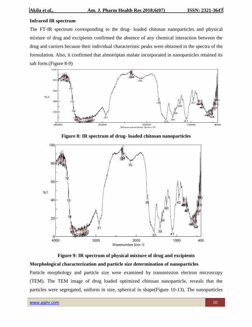
Infrared IR spectrum
The FT-IR spectrum corresponding to the drug- loaded chitosan nanoparticles and physical
mixture of drug and excipients confirmed the absence of any chemical interaction between the
drug and carriers because their individual characteristic peaks were obtained in the spectra of the
formulation. Also, it confirmed that almotriptan malate incorporated in nanoparticles retained its
salt form.(Figure 8-9)
Figure 8: IR spectrum of drug- loaded chitosan nanoparticles
Figure 9: IR spectrum of physical mixture of drug and excipients
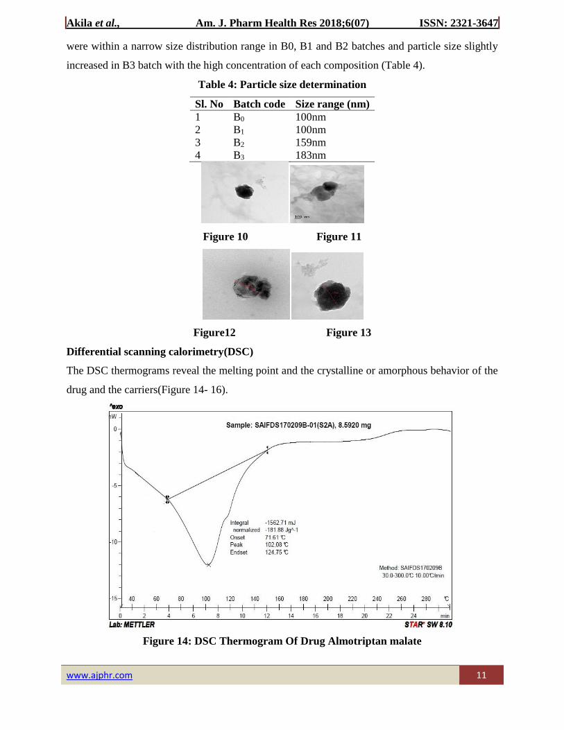
Morphological characterization and particle size determination of nanoparticles
Particle morphology and particle size were examined by transmission electron microscopy
(TEM). The TEM image of drug loaded optimized chitosan nanoparticle, reveals that the
particles were segregated, uniform in size, spherical in shape(Figure 10-13). The nanoparticles
Page 11
Akila et al., Am. J. Pharm Health Res 2018;6(07) ISSN: 2321-3647
www.ajphr.com 11
were within a narrow size distribution range in B0, B1 and B2 batches and particle size slightly
increased in B3 batch with the high concentration of each composition (Table 4).
Table 4: Particle size determination
Sl. No Batch code Size range (nm)
1 B0 100nm
2 B1 100nm
3 B2 159nm
4 B3 183nm
Figure 10 Figure 11
Figure12 Figure 13
Differential scanning calorimetry(DSC)
The DSC thermograms reveal the melting point and the crystalline or amorphous behavior of the
drug and the carriers(Figure 14- 16).
Figure 14: DSC Thermogram Of Drug Almotriptan malate
Page 12
Akila et al., Am. J. Pharm Health Res 2018;6(07) ISSN: 2321-3647
www.ajphr.com 12
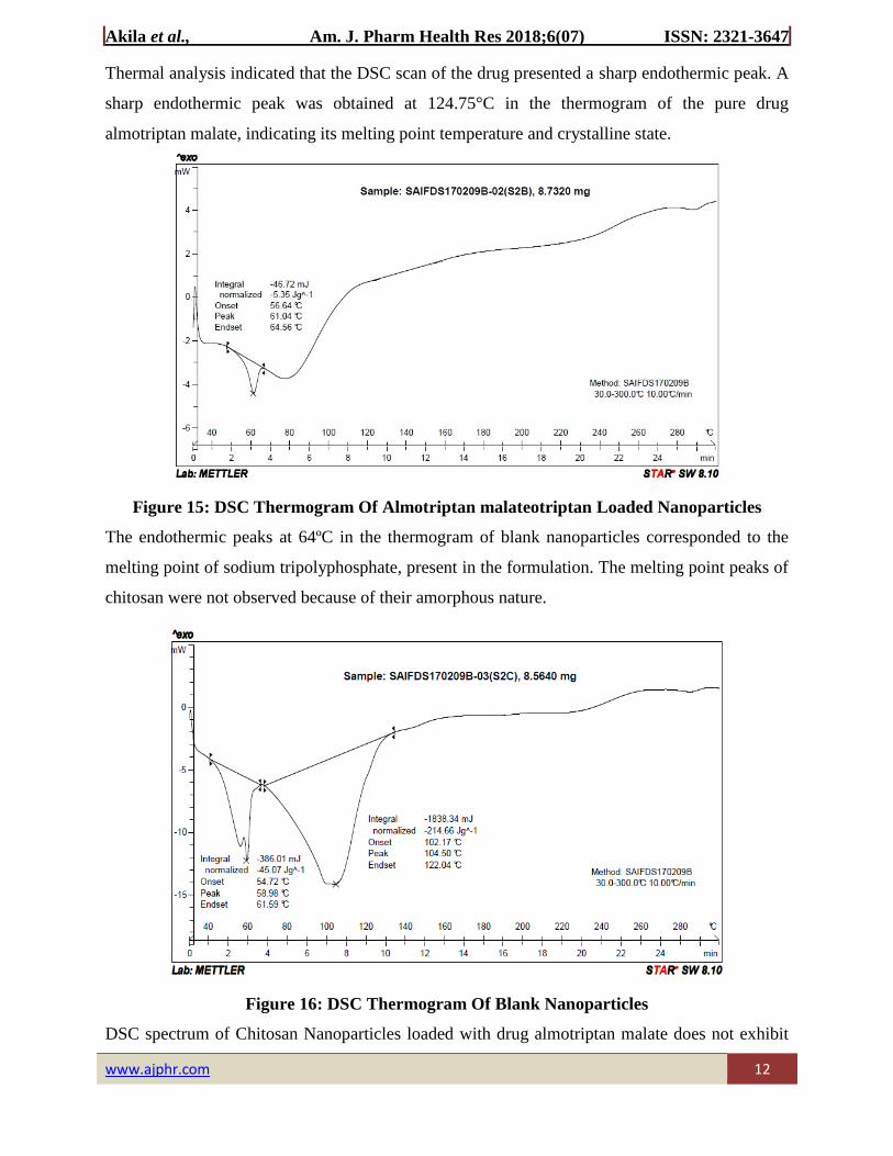
Thermal analysis indicated that the DSC scan of the drug presented a sharp endothermic peak. A
sharp endothermic peak was obtained at 124.75°C in the thermogram of the pure drug
almotriptan malate, indicating its melting point temperature and crystalline state.
Figure 15: DSC Thermogram Of Almotriptan malateotriptan Loaded Nanoparticles
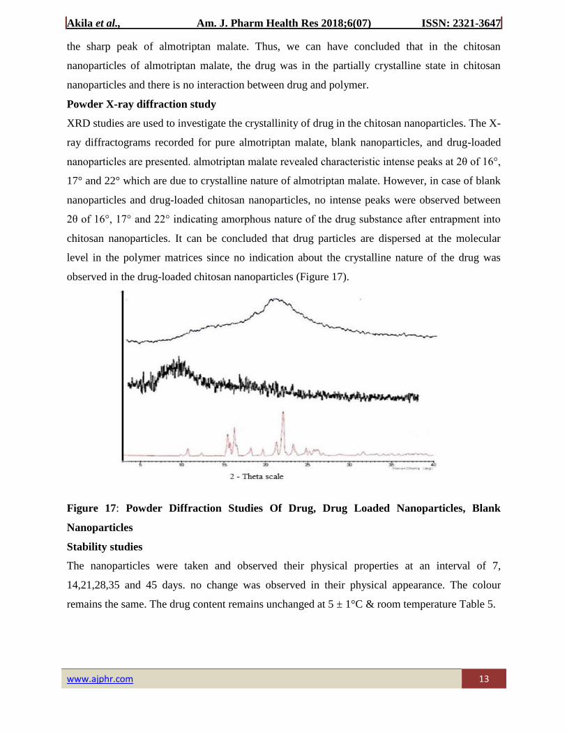
The endothermic peaks at 64ºC in the thermogram of blank nanoparticles corresponded to the
melting point of sodium tripolyphosphate, present in the formulation. The melting point peaks of
chitosan were not observed because of their amorphous nature.
Figure 16: DSC Thermogram Of Blank Nanoparticles
DSC spectrum of Chitosan Nanoparticles loaded with drug almotriptan malate does not exhibit
Page 13
Akila et al., Am. J. Pharm Health Res 2018;6(07) ISSN: 2321-3647
www.ajphr.com 13
the sharp peak of almotriptan malate. Thus, we can have concluded that in the chitosan
nanoparticles of almotriptan malate, the drug was in the partially crystalline state in chitosan
nanoparticles and there is no interaction between drug and polymer.
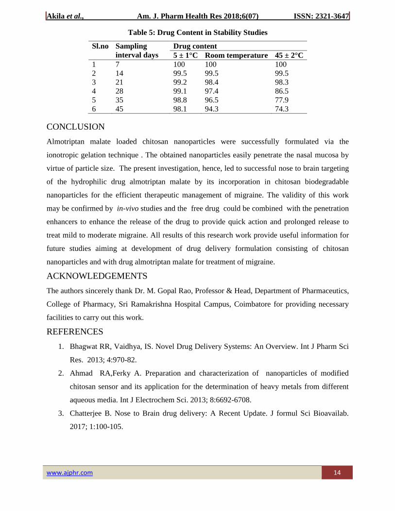
Powder X-ray diffraction study
XRD studies are used to investigate the crystallinity of drug in the chitosan nanoparticles. The X-
ray diffractograms recorded for pure almotriptan malate, blank nanoparticles, and drug-loaded
nanoparticles are presented. almotriptan malate revealed characteristic intense peaks at 2θ of 16°,
17° and 22° which are due to crystalline nature of almotriptan malate. However, in case of blank
nanoparticles and drug-loaded chitosan nanoparticles, no intense peaks were observed between
2θ of 16°, 17° and 22° indicating amorphous nature of the drug substance after entrapment into
chitosan nanoparticles. It can be concluded that drug particles are dispersed at the molecular
level in the polymer matrices since no indication about the crystalline nature of the drug was
observed in the drug-loaded chitosan nanoparticles (Figure 17).
Figure 17: Powder Diffraction Studies Of Drug, Drug Loaded Nanoparticles, Blank
Nanoparticles
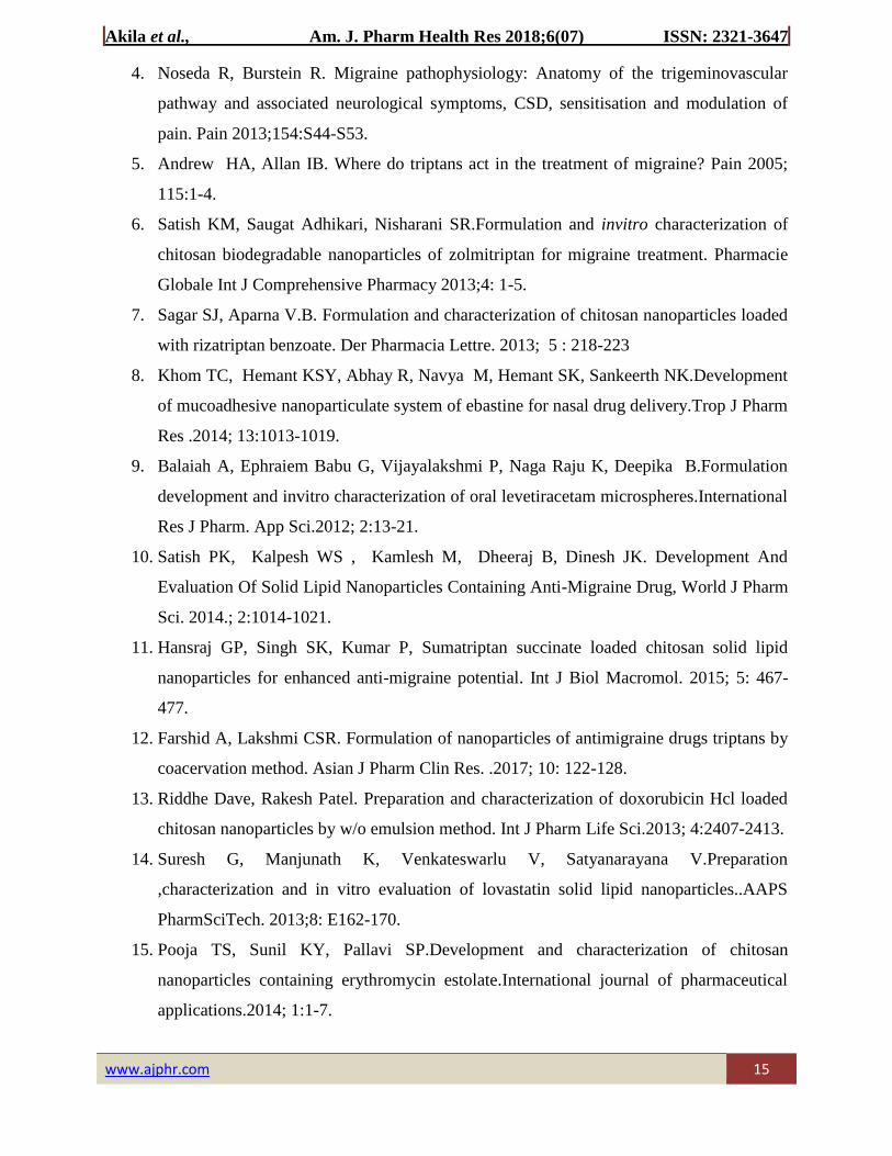
Stability studies
The nanoparticles were taken and observed their physical properties at an interval of 7,
14,21,28,35 and 45 days. no change was observed in their physical appearance. The colour
remains the same. The drug content remains unchanged at 5 ± 1°C & room temperature Table 5.
Page 14
Akila et al., Am. J. Pharm Health Res 2018;6(07) ISSN: 2321-3647
www.ajphr.com 14
Table 5: Drug Content in Stability Studies
Sl.no Sampling
interval days
Drug content
5 ± 1°C Room temperature 45 ± 2°C
1 7 100 100 100
2 14 99.5 99.5 99.5
3 21 99.2 98.4 98.3
4 28 99.1 97.4 86.5
5 35 98.8 96.5 77.9
6 45 98.1 94.3 74.3
CONCLUSION
Almotriptan malate loaded chitosan nanoparticles were successfully formulated via the
ionotropic gelation technique . The obtained nanoparticles easily penetrate the nasal mucosa by
virtue of particle size. The present investigation, hence, led to successful nose to brain targeting
of the hydrophilic drug almotriptan malate by its incorporation in chitosan biodegradable
nanoparticles for the efficient therapeutic management of migraine. The validity of this work
may be confirmed by in-vivo studies and the free drug could be combined with the penetration
enhancers to enhance the release of the drug to provide quick action and prolonged release to
treat mild to moderate migraine. All results of this research work provide useful information for
future studies aiming at development of drug delivery formulation consisting of chitosan
nanoparticles and with drug almotriptan malate for treatment of migraine.
ACKNOWLEDGEMENTS
The authors sincerely thank Dr. M. Gopal Rao, Professor & Head, Department of Pharmaceutics,
College of Pharmacy, Sri Ramakrishna Hospital Campus, Coimbatore for providing necessary
facilities to carry out this work.
REFERENCES
1. Bhagwat RR, Vaidhya, IS. Novel Drug Delivery Systems: An Overview. Int J Pharm Sci
Res. 2013; 4:970-82.
2. Ahmad RA,Ferky A. Preparation and characterization of nanoparticles of modified
chitosan sensor and its application for the determination of heavy metals from different
aqueous media. Int J Electrochem Sci. 2013; 8:6692-6708.
3. Chatterjee B. Nose to Brain drug delivery: A Recent Update. J formul Sci Bioavailab.
2017; 1:100-105.
Page 15
Akila et al., Am. J. Pharm Health Res 2018;6(07) ISSN: 2321-3647
www.ajphr.com 15
4. Noseda R, Burstein R. Migraine pathophysiology: Anatomy of the trigeminovascular
pathway and associated neurological symptoms, CSD, sensitisation and modulation of
pain. Pain 2013;154:S44-S53.
5. Andrew HA, Allan IB. Where do triptans act in the treatment of migraine? Pain 2005;
115:1-4.
6. Satish KM, Saugat Adhikari, Nisharani SR.Formulation and invitro characterization of
chitosan biodegradable nanoparticles of zolmitriptan for migraine treatment. Pharmacie
Globale Int J Comprehensive Pharmacy 2013;4: 1-5.
7. Sagar SJ, Aparna V.B. Formulation and characterization of chitosan nanoparticles loaded
with rizatriptan benzoate. Der Pharmacia Lettre. 2013; 5 : 218-223
8. Khom TC, Hemant KSY, Abhay R, Navya M, Hemant SK, Sankeerth NK.Development
of mucoadhesive nanoparticulate system of ebastine for nasal drug delivery.Trop J Pharm
Res .2014; 13:1013-1019.
9. Balaiah A, Ephraiem Babu G, Vijayalakshmi P, Naga Raju K, Deepika B.Formulation
development and invitro characterization of oral levetiracetam microspheres.International
Res J Pharm. App Sci.2012; 2:13-21.
10. Satish PK, Kalpesh WS , Kamlesh M, Dheeraj B, Dinesh JK. Development And
Evaluation Of Solid Lipid Nanoparticles Containing Anti-Migraine Drug, World J Pharm
Sci. 2014.; 2:1014-1021.
11. Hansraj GP, Singh SK, Kumar P, Sumatriptan succinate loaded chitosan solid lipid
nanoparticles for enhanced anti-migraine potential. Int J Biol Macromol. 2015; 5: 467-
477.
12. Farshid A, Lakshmi CSR. Formulation of nanoparticles of antimigraine drugs triptans by
coacervation method. Asian J Pharm Clin Res. .2017; 10: 122-128.
13. Riddhe Dave, Rakesh Patel. Preparation and characterization of doxorubicin Hcl loaded
chitosan nanoparticles by w/o emulsion method. Int J Pharm Life Sci.2013; 4:2407-2413.
14. Suresh G, Manjunath K, Venkateswarlu V, Satyanarayana V.Preparation
,characterization and in vitro evaluation of lovastatin solid lipid nanoparticles..AAPS
PharmSciTech. 2013;8: E162-170.
15. Pooja TS, Sunil KY, Pallavi SP.Development and characterization of chitosan
nanoparticles containing erythromycin estolate.International journal of pharmaceutical
applications.2014; 1:1-7.
Page 16
Akila et al., Am. J. Pharm Health Res 2018;6(07) ISSN: 2321-3647
www.ajphr.com 16
16. Neha G, Upendra N, Shubhini AS, Intranasal delivery of chitosan nanoparticles for
migraine therapy. Sci Pharm.2013;3:843-854.
AJPHR is
Peer-reviewed
monthly
Rapid publication
Submit your next manuscript at
[email protected] / [email protected]