Detailed of Course Structure Name of the Department: Chemistry Name of the Progrmme (U.G./P.G./Ph.D.): Post Graduate (M. Sc II and M. Sc. IV); Under Graduate: B Sc VI Semester students may also go through it) Semester: M Sc. II-Semester/M. Sc. IV (molecular Spectroscopy-one unit) Name of the paper/course (unit or title if there are multiple files of the same paper): Principle of Organic Synthesis and Organic Spectroscopy/Molecular Spectroscopy/Organic Spectroscopy Name of the teacher: Dr. Biswajit Maji Topic enclosed herewith on “Structure Determination of Organic Compounds using Spectroscopic Techniques (UV-Visible/IR/1H- NMR/13C-NMR, Mass Spectrometry) Disclaimer: There is no claim of the originality of the material and it is given only for the students to study References: I. Clayden Organic Chemistry
Transcript
Detailed of Course Structure
Name of the Department: Chemistry Name of the Progrmme (U.G./P.G./Ph.D.): Post Graduate (M. Sc II and M. Sc. IV); Under Graduate: B Sc VI Semester students may also go through it) Semester: M Sc. II-Semester/M. Sc. IV (molecular Spectroscopy-one unit) Name of the paper/course (unit or title if there are multiple files of the same paper): Principle of Organic Synthesis and Organic Spectroscopy/Molecular Spectroscopy/Organic Spectroscopy Name of the teacher: Dr. Biswajit Maji
Topic enclosed herewith on “Structure Determination of Organic Compounds using Spectroscopic Techniques (UV-Visible/IR/1H-
NMR/13C-NMR, Mass Spectrometry)
Disclaimer: There is no claim of the originality of the material and it is given only for the students to study
References:
I. Clayden Organic Chemistry
M. Sc II-Semester
Organic Spectroscopy
Unit V: Structure Determination
(Course Teacher: Dr B Maji)
1. Steps to deduce the structure of any unknown organic compound having known molecular formula and other supportive spectroscopic data and other clues: Step 1: To determine the unknown molecular structure of any unknown compound with empirical molecular formula (CxHyOz), first step is the identification of Double Bond Equivalent (DBEs) Step 2: Draw all the possible structures including different isomers, different compounds those exactly match with the numbers of C, H, O and other atoms present in a formula. At the same time count the Double Bond Equivalent also. Step 3: Try to correlate all the spectroscopic data (UV-Vis, IR, 1H-NMR, 13C-NMR, 2D NMR etc, including Mass Spectrometry data etc) with all the possible structures. Finally, the best fit data to the structure will be the correct one. Step 4: For complex molecule, the depth knowledge of spectroscopic techniques and ability to analysis of spectral data is highly fundamental in order to resolve the structural issues such as stereochemical relationship (cis-trans or syn-anti related problem), positional isomers, atom-connectivity etc. Mostly, the presence of functional groups (alcohol or carbonyl or any other FGs) could be detected by IR spectra. Moreover, a depth knowledge of chemical shift (δ in ppm) value, multiplicity (spin-spin multiplicity), coupling constants (J values) in 1H-NMR, 13C-NMR, 19F-NMR, 31P-NMR is very much significant. Finally, various 2D NMR (COSY, HMBC, HMQC, NOESY, ROESY etc) spectroscopy helps a lot to determine a very complicated structure.
Step I: DBEs calculation Let us consider an organic molecule having molecular formula C7H12O i) Maximum number of H atoms for 7Cs 2n + 2 = 16 ii) Subtract the actual number of H atoms (12) i.e. 16-12 = 4 iii) Divide by 2 to give the DBEs 4/2 = 2
Equation: DBEs = 1/2 [2n4 – n1 + 2], where n4 is the number of tetravalent atoms (C), n1 is the number of hydrogen
Now apply for C7H12O, DBEs = 1/2 [7x2 – 12 + 2] = 1/2 [16 – 12] = 1/2 [4] = 2 Other examples:
H3C
O
HO
C7H10O
DBEs = 3One for carbonyl (C=O)One for double bondOne for ringTotal = 3
OMe
C7H8O
DBEs = 4Three double bondsOne ring structureTotal = 4
If, N is present in unknown organic compound, the double bond equivalent identification is not following the same as above. DBEs identification for nitrogen containing compounds follows as below:
Equation: DBEs = 1/2 [2n4 – n1 + n3 + 2], where n4 is the number of tetravalent atoms (C), n1 is the number of hydrogen (H), n3 is the number of trivalent atoms (N). Applying the equation for the following nitrogen containing organic compounds and let see the identification of DBEs;
O
C7H12O
DBEs = Two (2)
NH2
C7H17N
DBEs = 0
NO2
C7H15NO2
DBEs = 1One for N=O
N Me
O
C7H13NO
DBEs = 2One for C=OOne for ring
N
NMeMe
DMAP
C7H10N2
DBEs = 4Three double bondsOne for ring
2. A compound having molecular formula C14H12O2 obtained by a following reaction and
shows the following proton NMR. Suggest a structure of an organic compound A.
O
H
NHC-salt
baseC14H12O2
A 1H NMR: δ 7.91 (d, J = 8.0 Hz, 2H), 7.50 (t, J = 7.6 Hz, 1H), 7.46-7.20 (m, 7H), 5.96 (s, 1H), 4.59 (bs, 1H). Disappear the peak at 4.59 on addition of D2O. Answer:
3. An organic compound A having molecular formula C6H12O shows the following proton NMR. Suggest a structure of an organic compound A. IR (cm-1): 1715 1H NMR: δ 2.1 (s) and 1.2 (s) Answer:
5. A compound of molecular formula C4H8O gave the following 13C and IR data. IR: 1730 cm-1 13C: δ 201.6, 45.8, 15.7 and 13.7. Deduce the structure. Answer:
7. An organic compound (molecular formula C8H8O2) shows a strong signals in IR spectrum at 1684cm–1 while in 1H NMR spectrum the protons appeared as follows: 1H NMR (CDCl3, 400 MHz): δ 9.9 (1H, s), 7.85 (2H, d), 7.00 (2H, d), 3.90 (s, 3H). Deduce the structure.
Answer: DBE = 5
Me
O
O
H3CCH3
O
H CH3
O
12
34
1: 202.82: 45.83: 15.74: 13.71
H3CCH3
O1
23
4
13C NMR 13C NMR
1: 29.42: 209.33: 36.94: 7.9
MeOO
H δ 9.9 (s, 1H)
H H δ 7.85 (d, J = 8.0 Hz, 2H)δ 7.00 (d, J = 8.0 Hz, 2H)
δ 3.90 (s, 3H)
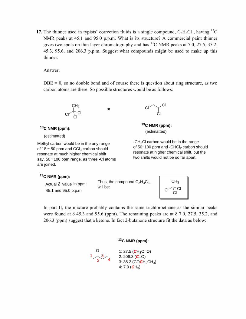
8. Three compounds of molecular formula C4H8O gave the following 13C and IR data. Compound A: IR (3200 broad cm-1) : 13C NMR (δ 134.7, 117.2, 61.3 and 36.9) Compound B: IR (No peaks except CH and fingerprint) : 13C NMR (δ 67.9 and 25.8) Compound A reacts with hydrogen over a palladium catalyst to give the product C of molecular formula C4H10O, IR 3200 (broad) cm-1 and 13C NMR δ 62.9, 36.0, 20.3 and 15.2. Deduce the structures for Compound A, B & C.
Answer: P. T. O.
Structure A Could be:
OH OH OH
13C NMR 13C NMR 13C NMR
1
2
3
4
1
2
3
4
1
2
3
4
Expected value in ppm:δ
1: 61.32: 36.93: 134.74: 117.2
1: 62.92: 132.73: 128.54: 18.3
The above structure is enoland enol carbon (1) appear in the range of 160-170. So, It could not be the compound
Match with given 13C-dataSo the compound would bebut-3-ene-1-ol
Compound B: No characteristic IR data, except CH-stretching frequency and fingerprint region. In 13C-NMR, only two peaks, 67.9 and 25.8, Therefore,the compound could be THF i.e Tetrahydrofuran.
Compound A
O11
2 2
13C NMR δ value:
1: 67.92: 25.8
Compound C:
OHPd/C, H2
OH
1
2
3
4
13C NMR δ value:
1: 62.92: 36.03:20.34:15.2
BC
9. When acetone is treated with base, a higher boiling liquid is obtained from the reaction mixture. The spectroscopic data of the liquid are: infrared, 1620 cm-1, 1695 cm-1; 1H-NMR spectra: δ 1.9 (3H, s), 2.1 (6H, s), 6.15 (1H, s); Mass: m/z (RA), 55 (100), 83 (90); 13C NMR: δ 20, 27, 31, 124, 154 and 197. Deduce the structure. Answer:
13C NMR
Me Me
O NaOH
H2O
O ContinuedO
Me Me
O O
aldol reaction
Proposed Spectroscopic data:IR : 1620 and 1695 cm-1
12. An organic compound having molecular formula C5H8O gives positive iodoform test. It shows the following spectral data: UV: λmax 277 nm εmax 4600 IR: Among other peaks a more prominent band at 1685 cm-1 NMR: Four nonequivalent proton signals. Deduce the structure of the compound. Answer:
Iodoform reaction is given by a compound having either CH3-CO- group or CH3-CH(OH)- group
i.e.H3C
O
Keto methyl group
orH3C
OH [O]
H3C
O
1 alcohol° Only ethanol
ethanal, acetone, 2-pentanone etc give the iodoform test as shown below:
13. C8H10O: 4 degrees of unsaturation. • Infrared Spectrum: 3500, 3028, 1800-1950 cm-1. • 1 H NMR Spectrum: δ 7.35-7.15 (m, 5H), two 2H-protons triplets, ~2.0 (broad singlet, 1H), 13C NMR Spectrum: 138.7, 129.1, 128.5, 126.4, 63.5, and 39.2. Deduce the structure. Answer:
As suggested the spectroscopic data,IR: 3500, 3028, 1800-1950 cm-1
1H-NMR: 5 ArH, 2 triplet and one broad singletat ~2.0 ppm
C8H10O; DBE = 4
i.e. The probable compound could be:
Estimated:
If, substituted phenol then,four aromatic proton must be there.two methylene proton resonates as quartet and methyl protons appearas triplet.Estimated:
14. A compound C9H10O reacts with aq. KMnO4. The compound shows the following 1H NMR spectroscopic data:
1H NMR: two doublets at δ 5.2 and 6.1 and each doublet appeared in J value 17.6 Hz; five aromatic protons at the range δ 7.12-7.58 ppm; three protons appeared at δ 3.7 ppm.
IR: No characteristic peaks at 3400 cm-1 and 1700 cm-1.
Answer:
Therefore the compound is :
OMe
(2-Methoxy-vinyl)-benzene
C9H10O; DBE = 5, as IR data suggests that no presence of carbonyl and free OH group presentin the compound structure; thus, the possible compound structure might be as follows:
OMe
(1-Methoxy-vinyl)-benzene
O
Propenyloxy-benzene
O
Isopropenyloxy-benzene
1H NMR (in ppm): 2 doublets at 5.2 and 6.1; J = 17.6 Hz i.e indicates trans-olefin5 ArH at 7.12-7.58 3 H at 3.70
H
H
d, 3J = 14-18 Hz
HH
d, 3J = 10-12 Hz
HH
d, 2J = 5-6 Hz
OMe
OMeArδ 7.58-7.12 (m, 5H)
H
H δ 6.1 (d, J =17.1 Hz, 1H)
δ 5.20 (d, J =17.1 Hz, 1H)
δ 3.70 (s, 3H)
No characteristics carbonyl and OH-stretching frequency
15. Follow the below reaction scheme and other clues and determine the structure of X (Clayden)
H
O
OHHO HBrC5H9BrO2
IR: No OH, no carbonyl, no alkene related stretching frequency were observed; 1128 cm–1
13C NMR: See the spectrum as belowm/z = 181, 73 (100%)
(X)
C3H4O
C2H6O2
C5H11BrO3 C5H9BrO2
H2O
Deduce the structure of X.
Answer: The 13C-NMR spectrum of CH2=CH–CHO i.e. propenal clearly shows one carbonyl group and two carbons on a double bond. These have all disappeared in the product and for the five carbon atoms we are left with four signals, two saturated, one next to oxygen, and one at 102.6 ppm. just creeping into the double bond region. It can’t be an alkene as an alkene is impossible with only one carbon atom! The IR spectrum gives us another puzzle—there appear to be no functional groups at all! No OH, no carbonyl, no alkene—what else can we have? Anyway, the DBEs identified from molecular formula of the product (X) is 1 (One).
Compound having halogen (F, Cl, Br) the DBE or IHD (Index Hydrogen Deficiency) can be calculated as follows, For C5H9BrO2 IHD or DBE = 1/2 [2n4 – nH – nX + 2]; nH = number of hydrogen, nX = number of halogen. IHD/DBE = 1/2 [2x5 – 9 – 1 + 2] = 1/2 [12-10] = 2/2 = 1 The index Hydrogen Deficiency value clearly indicates that in the structure X has one ring structure. The possible structures with the molecular formula C5H9BrO2 may represent as follows: (NB: IR and 13C data indicates there is no chances of double bond in the X).
O O
Me
Br
OO
MeBr
O O
Br
O
O
Br
O
O
Br
Applying basic knowledge of organic chemistry, one may propose a mechanism for the reaction scheme which is shown below,
H
O
HBr H
OH
Br
Br H
OH
H+
enol
Br H
O
HOOH
HBr
acetal synthesis
Br HO O
Mechanism
A decision can easily be reached from the base peak in the mass spectrum at 73. This is a fragment corresponding to the five-membered ring and not to the six-membered ring. Therefore the X is:
Br HO OX
Br HO O
m/z = 181, 73 (100%)
C5H9BrO2Mol. Wt.: 181.0278
IR (cm-1): 1128 which represent C-O stretching frequency13C NMR (ppm): As assigned in below spectrum
1
22
3
4
Mass spectra analysis is finally reached at decision (see the fragmentation)
O
O
Br
m/z = 181
+e-1
O
O
Br
Br
Radical
O O
O O
m/z = 73
Thus, the mass peak at m/z 73 clearly indicates the preence of five membered ring, not six membered ring
16. The 13C NMR spectrum for ethyl benzoate contains these peaks: 17.3, 61.1, 100–150 p.p.m. (four peaks), and 166.8 p.p.m. Which peak belongs to which carbon atom? Answer:
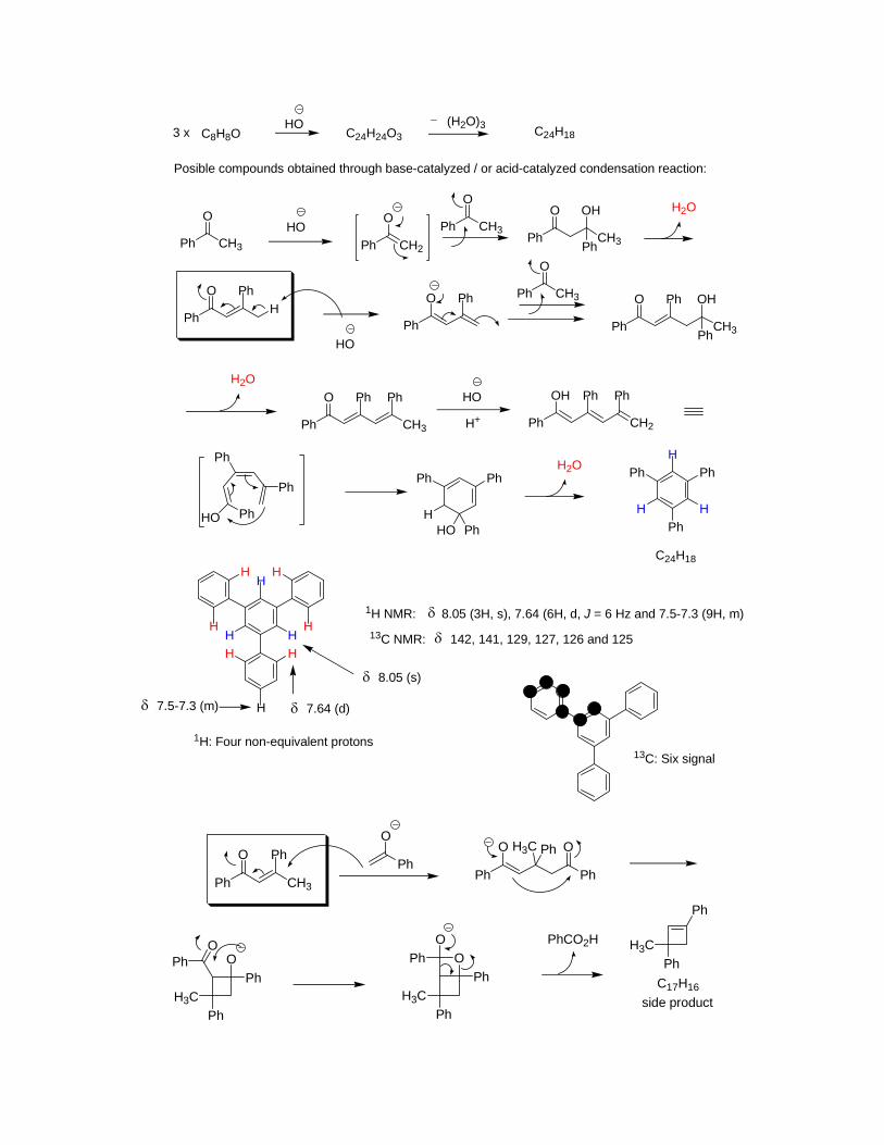
17. The thinner used in typists’ correction fluids is a single compound, C2H3Cl3, having 13C NMR peaks at 45.1 and 95.0 p.p.m. What is its structure? A commercial paint thinner gives two spots on thin layer chromatography and has 13C NMR peaks at 7.0, 27.5, 35.2, 45.3, 95.6, and 206.3 p.p.m. Suggest what compounds might be used to make up this thinner. Answer: DBE = 0, so no double bond and of course there is question about ring structure, as two carbon atoms are there. So possible structures would be as follows:
Cl
CH3
ClCl
or ClCl
Cl
13C NMR (ppm):13C NMR (ppm):
(estimatted)(estimatted)
Methyl carbon would be in the any range of 18 50 ppm and CCl3 carbon should resonate at much higher chemical shiftsay, 50 100 ppm range, as three -Cl atomsare joined.
-CH2Cl carbon would be in the range of 50 100 ppm and -CHCl2 carbon shouldresonate at higher chemical shift, but thetwo shifts would not be so far apart.
13C NMR (ppm):
Actual valueδ in ppm:45.1 and 95.0 p.p.m
Thus, the compound C2H3Cl3will be:
Cl
CH3
ClCl
In part II, the mixture probably contains the same trichloroethane as the similar peaks were found at δ 45.3 and 95.6 (ppm). The remaining peaks are at δ 7.0, 27.5, 35.2, and 206.3 (ppm) suggest that a ketone. In fact 2-butanone structure fit the data as below:
18. Four compounds, each having the formula C3H5NO, have the IR spectra summarized here. What are their structures? Without 13C NMR data, it may be easier to tackle this problem by first writing down all the possible structures for C3H5NO. In what specific ways would 13C NMR data help? A) One sharp band above 3000 cm–1; one strong band at about 1700 cm–1 B) Two sharp bands above 3000 cm–1; two bands between 1600 and 1700 cm–1 C) One strong broad band above 3000 cm–1; a band at about 2200 cm–1
Answer:
“N”-atom is here, so DBEs = 2 A. The possible structures would be as follows:
NH
O
HN
O
vs
As depicted in question part A, the A) One sharp band above 3000 cm-1, might be NH stretching frequency; one strong band at about 1700 cm-1, which would be of course carbonylstretching frequency. In the above two probable structures, lactam carbonyl should appeareat higher value than 1700 cm-1. Thus, the best fit the structure II, although, 13C-NMR spectraldata give the confirmed structure.Because, in 13C NMR amide carbonyl carbon and ketone carbonyl carbon chemical shifts have two different values.
β−
I II
B. The possible structure would be as follows:
As depicted in question part B, Two sharp band above 3000 cm-1, might be NH2 stretching frequency(one for symmetrical stretching and another for antisymmetrical stretching. Other strong bands at 1600 to 1700 cm-1, which would be of course one for carbonyl (C=O) and other for olefin (C=C) stretching frequency. Therefore, the possible structures are III or IV. 13C-NMR definitely could help to solve which one is the correct one, as amide carbonyl (C=O) and aldehydic carbonyl carbon have differentchemical shift.
β−
III IV
H2N
O
H
O
NH2vs
C. One strong broad band above 3000 cm–1; a band at about 2200 cm–1 Thus, the probable structures would be as follows:
As depicted in question part C, one broad band at above 3000 cm-1, might be -OHstretching frequency. Other strong bands at 2300 cm-1, which would be, of course, for one triple bond. Therefore, the possible structures are V or VI. 13C-NMR definitely could help to solve which one is the correct one, as shown in str. VI, the carbon attahedwith one -OH and one -CN (EWG) groups resonates at higher chemical shift.
V VI
vsHO
N N
OH
19. Three compounds of molecular formula C4H8O have the IR and 13C NMR spectra given below. Suggest a structure for each compound, explaining how you make your deductions. Compound A: IR: 1730 cm–1; 13C NMR: 13.3, 15.7, 45.7, and 201.6 p.p.m. Compound B: IR: 3200 (broad) cm–1; 13C NMR: 36.9, 61.3, 117.2, and 134.7 p.p.m. Compound C: IR: no peaks except CH and fingerprint; 13C NMR: 25.8 and 67.9 p.p.m. Compound D: IR: 3200 (broad) cm–1; 13C NMR: 15.2, 20.3, 36.0, and 62.9 p.p.m. Compound A reacts with NaBH4 to give compound D. Compound B reacts with hydrogen gas over a palladium catalyst to give the same compound D. Compound C reacts with neither reagent. Suggest a structure for compound D from the data given and explain the reactions. (Note. H2 reduces alkenes to alkanes in the presence of a palladium catalyst.) Answer: The DBE for the molecular formula C4H8O is 1 (ONE). Compound A: IR: 1730 cm-1, that implies that the compound A contain carbonyl group and another clue is the reduction by NaBH4 suggests the presence of aldehyde functionality which further again confirmed by 13C-NMR at δ 201.6 ppm. Thus, the compound could be butanal.
Compound B appears a broad peak at 3200 (broad) cm–1 in IR spectrum, suggest a free –OH functional group. In 13C NMR, it is clear one unsaturation double bond i.e. olefin is present in compound B and the olefin carbons resonate at δ 134.7 and 117.2 ppm. The probable structures would be as follows:
OH OHOH OH
I II III IV
Among the above structures, compound III and IV would not be as on treatment with Pd/H2 affords secondary alcohol which is not the same as the compound D. Between the compound I and II, compound I would be as the olefin carbons resonate at unequal δ 134.7 and 117.2 ppm value. In compound II, equal substitution pattern might be resulted the alkene carbons resonate at a very close δ values.
Compound C: IR: no peaks except CH and fingerprint; 13C NMR: 25.8 and 67.9 p.p.m. In IR spectrum, there are no characteristic functional groups peaks were observed. In 13C NMR spectrum, only two peaks at δ 25.8 and 67.9 suggest the compound C would be symmetric. One important another clue is that it has one DBE without any unsaturation which only possible to think THF i.e. tetrahydrofuran.
1
2
13C NMR (ppm):
1: 67.9 (OCH2)2: 28.9 (-CH2CH2-)
IR: NO FG peaks
C1
2C
20. You have dissolved t-BuOH (Me3COH) in MeCN with an acid catalyst, left the solution overnight, and found crystals with the following characteristics there in the morning. What are they? IR: 3435 and 1686 cm–1 13C NMR: 169, 50, 29, and 25 p.p.m. Mass spectrum (%): 115 (7), 100 (10), 64 (5), 60 (21), 59 (17), 58 (100), and 56 (7). (Don’t try to assign all of these!) Answer: The peaks at 3435 and 1686 cm–1 in IR spectrum suggest that the compound having characteristic amide NH (-CONHR) and carbonyl (-CONHR) stretching frequency. If you dissolved t-BuOH (Me3COH) in MeCN with an acid catalyst, left the solution overnight, the following compound would be formed. Mechanism:
22. How would mass spectra help you distinguish these structures?
O O O
C5H10OMol. Wt.: 86.1323
C5H10OMol. Wt.: 86.1323
C5H10OMol. Wt.: 86.1323
Answer: Above three ketones are isomers of same molecular formula C5H10O. These three isomers can be easily distinguished by the Mass spectra. The fragmented mass peaks for each ketone appeared at m/z value which is follows:
O+ e-1
Oa b a
Om/z = 86 m/z = 71
CO
m/z = 43
b
H3C Om/z = 43
COCH3
+
m/z = 15
O
m/z = 86
α
βγ + e-1 O
Hγ
McLafferty rearrangement
OH
m/z = 58
O+ e-1
Oa a a
O
m/z = 86 m/z = 57
CO
m/z = 29
β
γO
m/z = 86
α
No carbonNo McLafferty rearrangement
O+ e-1 Oa b a
m/z = 86
O
m/z = 71
CO
m/z = 43b
H3C O
m/z = 43
COCH3
+
m/z = 15
β
γO
m/z = 86
αNo carbon
No McLafferty rearrangement
23. The following hydroxyketone shows no peaks in its infrared spectrum between 1600 and 1800 cm–1 but it does show a broad absorption at 3000 to 3400 cm–1. In the 13C NMR spectrum, there are no peaks above 150 p.p.m. but there is a peak at 110 p.p.m. Suggest an explanation.
OHO
Hydroxyketone Answer: In the hydroxyketone structure, there are one ketone (C=O) functional group and one –OH functional group. In a solution, in fact, the hydroxyketone exists in a cyclic hemiacetal form (see below scheme) and as a result, no characteristic ketone carbonyl stretching frequency at the range of ~1700 cm-1 was observed. The equilibrium favors towards right side and which is again ascertained by spectral analysis (13C-NMR). In the 13C NMR spectrum, there are no peaks above 150 p.p.m. but there is a peak at 110 p.p.m. No peaks at above δ 150 p.p.m. indicate no carbonyl functional group exists in a solution of hydroxyketone substrate. A peak at 110 p.p.m. suggests an alkene or a carbon bonded with two oxygen (hemiacetal/acetal type compound). Dehydration step for this substrate is not a easy task, in fact, in the presence of trace acid (H+), the substrate hydroxyketone rather easily from hemiacetal (Scheme below). The hemiacetal fits all the spectroscopic data in a correct manner.
OHO
Hydroxyketone
H+(trace)
OHO
HO
OH
O
OH
H
H+
solution
H+
100 ppmδ
((hemiacetal)
24. At room temperature, DMF shows three signals and at higher temperature the DMF shows only two signals in 13C-NMR. Explain the fact. Answer: (From clayden) The C–N bond length to the carbonyl group is closer to that of a standard C–N double bond (127 pm) than to that of a single bond (149 pm). This partial double bond
character is responsible for the restricted rotation about this C–N bond. We must supply 88 kJ mol–1 if we want to rotate the C–N bond in DMF (remember a full C–C double bond takes about 260 kJ mol–1). This amount of energy is not available at room temperature and so, for all intents and purposes, the amide C–N bond is locked at room temperature as if it were a double bond. This is shown in the carbon NMR spectrum of DMF. How many carbon signals would you expect to see? There are three carbon atoms altogether and three signals appear—the two methyl groups on the nitrogen are different. If free rotation were possible about the C–N bond, we would expect to see only two signals. In fact, if we record the spectrum at higher temperatures, we do indeed only see two signals since now there is sufficient energy available to overcome the rotational barrier and allow the two methyl groups to interchange
Look at protein structure????
25. How many signals will there be in the 1H NMR spectrum of each of these compounds? Estimate the chemical shifts of the signals.
N
N
N
NH
O
Me2N
OMeMeOF3C N
SiO
Me
O
O
H
Answer:
N
N
NNH
O
Me2N Me
OMeMeO
F3C NSi
O
Me
O
O
H
1
11
One signal
δ 9.31δ 2.29
11
2 2 δ 1.12
Two signals
12
3
δ 1.20δ 2.27
δ 3.20
Three signals
1
2
3δ 1.05δ 3.10
δ 0.45
1δ 1.50
δ 8.502
Three signals Two signals
26. One isomer of dimethoxybenzoic acid has the 1H NMR spectrum 3.85 (6H, s), 6.63 (1H, t, J = 2 Hz), 7.17 (2H, d, J = 2 Hz) and one isomer of coumalic acid has the 1H NMR spectrum 6.41 (1H, d, J = 10 Hz), 7.82 (1H, dd, J = 2, 10 Hz), 8.51 (1H, d, J = 2 Hz). In each case, which isomer is it? The substituents in black can be on any carbon atoms.
CO2H
OMeMeOO O
HO2C
Answer:
δ
CO2HMeO
MeO
CO2H
MeO OMe
CO2H
OMeOMe
estimated:3 signals in aromatic region
estimated:2 signals in aromatic region
estimated:3 signals in aromatic region
As observed in the 1H-NMR spectrum, two methoxy protons resonate at 3.85 ppm i.e. the position of the methoxy groups in a ring such a way the molecule would be symmetric.In aromatic region, one proton resonates at 6.63 ppm and appeared as triplet with J value 2.0 Hz which implies meta-coupling. Two another aromatic protons appeared as doublet with the same J value (2.0 Hz) that clearly indicate again meta-coupling.Thus, among above three isomeric dimethoxybenzoic acids, the compound could be 3,5-dimethoxybenzoic acid.
HH
H
6.63 (d, J = 2 Hz)δ 6.63 (d, J = 2 Hz)
δ 7.17 (t, J = 2 Hz)
m-coupling
Similarly,
O O
HO2C
O O
CO2H
O O
CO2H
O OHO2C
δ
O O
CO2HHH
H
8.51 (1H, d, J = 2 Hz)(m-couplinng)
δ 7.82 (1H, dd, J = 2, 10 Hz)(o-/m-couplinng)
δ 6.41 (1H, d, J = 10 Hz)(o-couplinng)
27. The reaction below was expected to give product A and did indeed give a product
with the correct molecular formula by mass spectrometry. The 1H NMR spectrum of the product was however: δ H (p.p.m.) 1.27 (6H, s), 1.70 (4H, m), 2.88 (2H, m), 5.4–6.1 (2H, broad s, exchanges with D2O), 7.0–7.5 (3H, m). Though the detail is missing from this spectrum, how can you already tell that this is not the compound expected?
Answer: If you clinically analyze the product obtained through expected Beckmann rearrangement reaction condition, none of the protons resonate in lactams ring are fit. If we consider the expected ring expanded product as in the above scheme shown the lactams product should contain four aromatic protons, six methylene protons and one amide proton which might be exchangeable with D2O. Actually, the 1H NMR spectral data is completely mismatching with the expected one. Three aromatic protons resonate at [7.0–7.5 (3H, m)], two protons appeared as broad singlet [5.4–6.1 (2H, broad s, exchanges with D2O)] which implies two amide [CONH2]-protons. Therefore, one may consider to some other unexpected reaction occurred and give substituted tetralin product with amide functionality. The plausible reaction mechanism is depicted as follows: Mechanism
HONPPA
H+
HN
O
(expected)Beckmann product
H+
NHO
H
CNCN
F-C
alkylation
CONH2
H+
H+
Unexpectedproduct obtained
NOT FORMED
Now, you can correlate the 1H NMR spectral data easily.
28. A nitration product (C8H11N3O2) of this pyridine has been isolated which has a nitro (NO2) group somewhere on the molecule. From the 90 MHz 1H NMR spectrum, deduce whether the nitro group is (a) on the ring, (b) on the NH nitrogen atom, or (c) on the aliphatic side chain and then exactly where it is. Give a full analysis of the spectrum.
Answer:
N NH
CH3
O2N
III
H
H
H
δ 8.1 (d, J = 8.0 Hz, 1H)
δ 6.3 (d, J = 8.0 Hz, 1H)
δ 9.1 (d, J = 2.4 Hz, 1H)
δ 5.9 (bs, 1H)
a
b
c
a: δ 3.2 (t, 2H) δ 1.8 (q, 2H) δ 1.1 (t, 3H)b: c:
Third Assumption (c):If NO2-group is at propyl chain
N NH
CH3
NO2
N NH
CH3
NO2
N NH
NO2
diastereotopic protons
dd
* *
diastereotopic protons
d, 3H t, 2H
(quintet, 2H)
t, 2H
Second Assumption (b)If NO2-group is attached with N
N NCH3
NO2
In 1H NMR spectra as shown above the chemical shift value 5.9 ppm is typical for NH proton so it is free NH.δ
All the above estimated spectral analysis is not exactly matching with the above spectral data. Thus -NO2 group is not attached with side propyl chain, it is attached with aromatic carbon.
First Assumption (a)If NO2-group at aromatic region:
N NH
CH3
NO2
N NH
CH3
NO2
N NH
CH3
O2N
N NH
CH3O2N
I II III IV
The most significant feature of the aromatic region is that one prton appeared at a very high downfieldposition about 9.1 ppm with m-coupling (J = 2~3 Hz) which rulles out the isomer II and IV as no neighbours protons are there. Between I and III, all the protons most probably fit well with the isomer III and the value is designated for all the protons as below:
N NH
CH3
O2N
III
H
H
H
δ 8.1 (d, J = 8.0 Hz, 1H)
δ 6.3 (d, J = 8.0 Hz, 1H)
δ 9.1 (d, J = 2.4 Hz, 1H)
29. The natural product bullatenone was isolated in the 1950s from a New Zealand myrtle and assigned the structure A. Then compound A was synthesized and found not to be identical with natural bullatenone. Predict the expected 1H NMR spectrum of A. Given the full spectroscopic data available nowadays, but not in the 1950s, say why A is definitely wrong and suggest a better structure for bullatenone. Spectra of bullatenone: Mass spectrum: m/z 188 (10%) (High resolution confirms C12H12O2), 105 (20%), 102 (100%), and 77 (20%) Infrared: 1604 and 1705 cm–1. 1H NMR: 1.45 (6H, s), 5.82 (1H, s), 7.35 (3H, m), and 7.68 (2H, m).
OPh
OCH3 C12H12O2
Mol. Wt.: 188.2225
A Answer: Mid 20th century, the chemists were faced a tremendous problem to deduce the structure of any unknown compound that could be extracted from natural plants. The IR and mass spectra of above structure (A) are possibly matching. Later, after synthesizing the proposed compound and once the NMR spectra had taken, it was found that the proposed structure of A, natural bullatenone is completely wrong. Thus later the structure is revisited and it’s true that simple using NMR techniques this above problem can be solved. In the 1H NMR spectral analysis it is found that the compound has five aromatic protons (ArH), six methyl protons (CMe2) and one olefin proton at normal olefin region. Again, the analysis of IR spectra clearly indicates one carbonyl group (C=O) so if we add together the molecular formula would be C12H12O. Oxygen is short. It is expected that one more oxygen might be in the ring. So the probable structure would be as follows:
O
O
PhO
OPh
O
O
PhO
O
Ph
O
O
Ph
O
O
Ph
I II III IV V VI
Lactone (I to IV) could not be:i) Lactone carbonyl carbon stretching frequency would be ~1745-1780 cm-1, although in the strucrtures I and II, there is a double bond in conjugation with carbonyl group still it never came at 1705 cm-1.ii) In the structure IV, the alkene proton must be appeared at higher chemical shift value than normal olefin region.
Between V and VI, the correctedstructure could be VI. All the spectroscopic data is fit.
O
O
Ph
H
δ 7.35 (3H, m) and 7.68 (2H, m).5 ArH
δ 5.82 (1H,s)δ
1.45 (6H, s)
C=O str: 1705 cm-1
30. Suggest structures for the products of these reactions, interpreting the spectroscopic data.
Answer:
δOBr MeOH
OBr
O
OMe
O
OMe
O
Favroski rearrangementreaction
OMe
O
1.20 (9H, s)
δ 3.67 (3H, s)
(179; C=O)
(52; OCH3)
(39, CMe3)
(27, CH3)
MeO
MeO
MeO
OO
H+
EtOH
H+
OO H
EtOH
O
OH
OEt OH OEtO
O
O O
O
O O
δ 1.28 (t, J = 7.0 Hz, 3H)
δ 4.2 (q, J = 7.0 Hz, 2H)
δ 3.24 (s, 2H)
δ 2.21 (s, 3H)
1H NMR
13C NMR
O
O O
(203)(170)
(62)
(15)(22)
(39)
C=O str: 1705 cm-1
δ 9.8 (s, 1H)
1H NMR
13C NMR
OMeHO
SMe
i) SOCl2, Et3N
ii) H2OMeS
MeMe
O
H
(i)
OMeCl
SMeS
OMe
Me
MeSOMe
H2O(ii)
MeS
MeMe
O
H
δ 1.12 (s, 6H)
δ 2.8 (s, 3H)
MeS
MeMe
O
H
(202)
(15)
(45)
(22)
31. Precocene is an organic compound that causes insect larvae to pupate and can also be found in some plants (Ageratum spp.) where it may act as an insecticide. It was isolated in minute amounts and has the following spectroscopic details. Propose a structure for precocene. Spectra of precocene: Mass spectrum: m/z (high resolution gives C13H16O3), M—15 (100%) and M—30 (weak). Infrared: CH and fingerprint only. 1H NMR: 1.34 (6H, s), 3.80 (3H, s), 3.82 (3H, s), 5.54 (1H, d, J = 10 Hz), 6.37 (1H, d, J = 10 Hz), 6.42 (1H, s), and 6.58 (1H, s).
Answer: DBEs = 6 Mass spectrum: M—15 (100%); Methyl group easily fragmented from the molecule. Infra red spectra: Only the peaks of CH and fingerprint only which clearly suggest that all the three oxygens are in ether linkages. No carbonyl functionality is present in the molecule. 1H NMR: 1.34 (6H, s) —Two set of methyl protons (Chemically equivalent; probably –CMe2) 3.80 (3H, s), 3.82 (3H, s) —Two set of methoxy protons (-OMe) attached 5.54 (1H, d, J = 10 Hz), 6.37 (1H, d, J = 10 Hz) —Suggest two olefin protons and their relationship is cis-to each other. 6.42 (1H, s), and 6.58 (1H, s) —clarify two aromatic protons attached in a 1,4-position and appeared as a singlet. There are no neighbouring protons are there. Thus the fragments are as follows:
Me Me
??
OMe
OMe?
? HH
??
H
H
?
?
?
?
If we connect all these above fragments, the following structures would be possible:
H
H
MeO
MeO O
H
H
H
HMeO O
H
HMeO C13H16O3
O
MeO
MeO
H
H
less no of Hs Benzofuran ring, twoHs coupling constant(J) value would be less than 10
C13H16O3 O
MeO
MeO
H
H
I II
III
C13H16O3
J = <10 Hz
IV
O
MeO
MeO
H
H
C13H16O3
V
As suggested before the possible structures, structure I and II would not be possible as five membered ring systems, the protons coupling constants would be small, never would be 10 Hz. Thus these structures rule out. The other six-membered heterocycles (III, IV and V), the structures IV and V again would not be possible as the olefin hydrogen connected with oxygen resonate at higher chemical shift value and of course, there would be a substantial chemical shift differences were observed between two olefin Hs. Thus only left the structure no III, which fits all the spectroscopic data which is represented as follows:
O
MeO
MeO
H
H
III
DBEs = 6 (4 double bonds and two rings)Mass: M-15 is possibleIR: only C-H and fingerprint region, as no characteristic functional groups are present.1H NMR: Best fit as observed.
δ 6.42 or 6.58 (1H, s)
δ 6.42 or 6.58 (1H, s)
HH
δ 6.37 (1H, d, J = 10 Hz))
δ 5.54 (1H, d, J = 10 Hz))
δ 1.34 (6H, s)
δ 3.80 or 3.82 two 3H singlet
32. Suggest structures for the products of these reactions, interpreting the spectroscopic data.
Answer: (Solution I)
OMe Br
AlCl3Friedel-Crfats alkylation
OMe
This is a traditional Friedel-Crafts alkylation reaction. In the IR spectral analysis, it is observed that no as such characteristic functional groups are present except C-H stretching frequency and the peaks at fingerprint region. In 1H-NMR spectrum, two methyl protons resonate at δ value 1.21 ppm as a singlet which implies –CHMe2, one proton appeared as septet at δ value 2.83 ppm i.e. presence of methane proton and attached with a sic neighbouring protons. One methoxy group is there (at 3.72 ppm) and four aromatic protons are present. Two pair of doublet at δ values 6.74 ppm and 7.18 with equal ortho-coupling pattern indicating the substitution pattern is 1,4-disubstituted.