POSITION STATEMENT DETECTION AND MANAGEMENT OF WOMEN WITH FETAL GROWTH RESTRICTION IN SINGLETON PREGNANCIES Fetal growth restriction (FGR) is associated with stillbirth, neonatal death and perinatal morbidity and an increased risk of adverse health outcomes into adulthood. Improving the detection and care of pregnancies with FGR is an important strategy to reduce adverse outcome and is relevant to all maternity care providers. Contents 1 Purpose 2 Definitions 3 Risk factor assessment 4 Symphyseal fundal height (SFH) measurement 5 Diagnosis and management 6 Placenta 7 Neonatal care 8 Subsequent pregnancy care 9 Education and clinical audit 10 Evidence gaps 11 Working Group 12 References
Transcript
POSITION
STATEMENT
DETECTION AND MANAGEMENT OF WOMEN WITH FETAL GROWTH RESTRICTION IN SINGLETON PREGNANCIES
Fetal growth restriction (FGR) is associated with stillbirth, neonatal death and perinatal morbidity and an increased risk of adverse health outcomes into adulthood. Improving the detection and care of pregnancies with FGR is an important strategy to reduce adverse outcome and is relevant to all maternity care providers.
Contents
1 Purpose
2 Definitions
3 Risk factor assessment
4 Symphyseal fundal height (SFH) measurement
5 Diagnosis and management
6 Placenta
7 Neonatal care
8 Subsequent pregnancy care
9 Education and clinical audit
10 Evidence gaps
11 Working Group
12 References
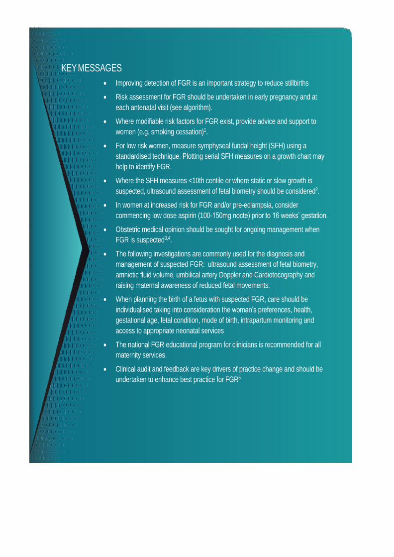
KEY MESSAGES
• Improving detection of FGR is an important strategy to reduce stillbirths
• Risk assessment for FGR should be undertaken in early pregnancy and at
each antenatal visit (see algorithm).
• Where modifiable risk factors for FGR exist, provide advice and support to
women (e.g. smoking cessation)1.
• For low risk women, measure symphyseal fundal height (SFH) using a
standardised technique. Plotting serial SFH measures on a growth chart may
help to identify FGR.
• Where the SFH measures <10th centile or where static or slow growth is
suspected, ultrasound assessment of fetal biometry should be considered2.
• In women at increased risk for FGR and/or pre-eclampsia, consider
• Obstetric medical opinion should be sought for ongoing management when
FGR is suspected3,4.
• The following investigations are commonly used for the diagnosis and
management of suspected FGR: ultrasound assessment of fetal biometry,
amniotic fluid volume, umbilical artery Doppler and Cardiotocography and
raising maternal awareness of reduced fetal movements.
• When planning the birth of a fetus with suspected FGR, care should be
individualised taking into consideration the woman’s preferences, health,
gestational age, fetal condition, mode of birth, intrapartum monitoring and
access to appropriate neonatal services
• The national FGR educational program for clinicians is recommended for all
maternity services.
• Clinical audit and feedback are key drivers of practice change and should be
undertaken to enhance best practice for FGR5
1 Purpose of the position statement
The purpose of this position statement is to improve perinatal outcomes through better antenatal detection and management of pregnancies with FGR. These recommendations have been derived from a literature review including multiple international SGA/FGR guidelines5-10.
Definitions
FGR is best defined as a fetus that has not reached its growth potential. In practice, small for gestational age (SGA) is often used as a proxy for FGR (see Table 1). However, not all SGA fetuses are growth restricted, and some growth restricted fetuses are not SGA11. There are also differences between early and late FGR12, which are detailed in Table 2.
Table 1: Definitions relating to FGR
Fetal Growth Restriction (FGR) A fetus that has not reached its growth potential.
(in practice, small for gestational age (SGA) is often used as a proxy for FGR)
Small for gestational age (SGA) Estimated fetal weight/birthweight <10th centile
Severe FGR SGA <3rd centile is often used as a proxy for severe FGR
Early FGR FGR <32 weeks gestation
Late FGR FGR >32 weeks gestation
Table 2: Early vs Late FGR, Adapted from Figueras et al12.
Early FGR Late FGR
Gestation <32 weeks ≥32 weeks
Prevalence13 0.5 – 1% 5 – 10%
Pre-eclampsia Strong association Weak association
Placental pathology Strong association Weak association
Relation to SGA Often SGA <10th centile Not always SGA
Umbilical artery Dopplers Often Abnormal Normal or abnormal
Detection14 Often are readily detectable Challenging to detect
Clinical consequences14 Risks of prematurity, high mortality
and morbidity
Associated with increased mortality
and morbidity
2
Risk factor assessment
Risk assessment for FGR can be undertaken by healthcare providers prior to conception, in early pregnancy, and at each antenatal visit 5,15 through inquiry about:
1. maternal characteristics and medical history 2. previous obstetric history 3. risk factors that may arise in pregnancy
It is good practice to inform women about FGR1 at their booking visit and, where there is a diagnosis of FGR, ongoing communication on the management of FGR throughout the pregnancy. Where modifiable risk factors for FGR exist, provide advice and support to women (e.g. smoking and drug/alcohol cessation)1.
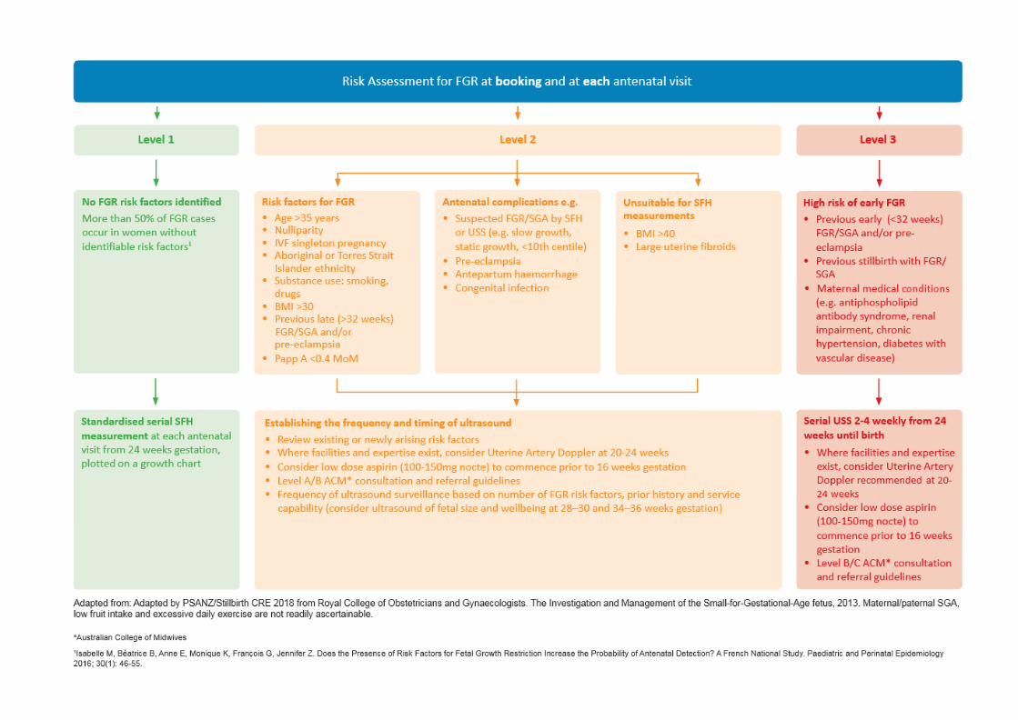
Antenatal surveillance for FGR may be modified according to a woman’s individual risk factors and this is detailed in the Risk Assessment Algorithm for FGR (Figure 1) at each antenatal visit.
Women can be stratified into three groups depending on their existing or newly arising risk factors for FGR. Consider low dose aspirin (100-150mg nocte) to commence prior to 16 weeks’ gestation for women at increased risk of FGR. Frequency of ultrasound surveillance for suspected FGR should be based on FGR risk factors, prior history and the woman’s preferences.
Symphyseal fundal height (SFH) measurement
Measurement of symphyseal fundal height (SFH) can be undertaken every 2-4 weeks starting from 24 weeks gestation1,11. In women with high BMI, or who have uterine fibroids that are unsuitable for SFH measurement, serial ultrasound can be considered for assessment of fetal growth5.
The limitations of SFH measurement in the detection of FGR are well described16. A standardised approach to SFH measurement may reduce inter and intra-observer error1,2. One widely accepted approach to SFH measurement includes measuring from the fundus to the superior margin of the symphysis pubis, using a non-elastic tape measure with numbers facing downwards
Serially plotting SFH measurements on a growth chart may assist in the detection of FGR. Consider ultrasound assessment when a SFH measurement is <10th centile, or if there is clinical suspicion of static or slowing growth on serial SFH measurments2.
Diagnosis and management of FGR
Accurate gestational age dating is important in the assessment of fetal size17,18.
The following investigations are commonly used for the diagnosis and management of suspected FGR.
Table 3: Common investigations for diagnosis and management of suspected FGR
Investigation Description Suggestive of FGR
Fetal biometry by ultrasound • Abdominal circumference (AC)
• Head circumference (HC)
• Biparietal diameter (BPD)
• Femur length (FL)
• Estimated fetal weight (EFW)
EFW or AC <10th centile
and/or reduced growth velocity of EFW or AC
Amniotic fluid volume (AFV) Measured by the single deepest vertical pocket (DVP) of amniotic fluid
DVP <2cm
4
5
3
Umbilical artery Doppler (UAD) Measures resistance to blood flow in the umbilical artery and placenta
UAD Pulsatility or Resistance Index (PI or RI) >95th centile, absent or reverse end diastolic flow (AREDF)
Cardiotocography (CTG)
Continuous recording of fetal heart rate and uterine activity
Abnormal CTG trace
Enquiry about fetal movements Ask each woman to identify her baby’s normal pattern of movements19.
Maternal concern about decreased fetal movements (strength and/or frequency).
Seek obstetric medical opinion for ongoing management when FGR is suspected1.
Birth planning
When planning the birth of a baby with suspected SGA/FGR, the aim is to achieve the maximum maturity possible where it is safe to do so. Benefits of early birth to reduce stillbirth need to be carefully weighed against the risk of intervention for the baby at a given gestation. Care should be individualised and woman-centred, using decision aids where possible. The following points should be considered and discussed:
• Woman/family preferences
• Maternal condition
• Gestational age, EFW and fetal condition
• Mode of birth
• Intrapartum monitoring
• Access to appropriate neonatal services
Placenta
The major underlying cause of FGR is placental in origin20. Early onset FGR is often associated with maternal vascular malperfusion (MVM) of the placenta resulting in placental infarction or poor early placentation20.
Rarer causes of placental pathology associated with FGR include: massive perivillous fibrin deposition (maternal floor infarction) and chronic intervillositis, both of which have high recurrence rates in subsequent pregnancies20.
Compared to early onset FGR, the incidence of placental pathology in late onset FGR occurs less often12.
It is recommended that the placentae of suspected SGA/FGR babies be sent for histopathology. The findings of which may support the clinical findings and influence subsequent pregnancy care5.
Neonatal management
The clinical diagnosis of FGR in the neonate can be as challenging as it is antenatally. Care of the newborn with SGA/FGR should include monitoring and maintenance of oxygenation, temperature and blood glucose levels.
Paired cord blood gases can be undertaken to assess acid base status at birth.
In the care of the preterm growth restricted neonate, consider specific issues relating to prematurity such as lung disease, increased risk of infection, neurological complications and necrotising enterocolitis.
6
7
8
Subsequent pregnancy care
The birth of a baby with FGR is a major risk factor for FGR in a subsequent pregnancy 5. Where possible, the underlying cause for FGR should be investigated to assess for recurrence risk. This includes review of placental histopathology and any investigations undertaken for FGR before and after birth20.
Where SGA/FGR has been associated with stillbirth or severe long term adverse outcomes, consider additional parental psychosocial support in a subsequent pregnancy21.
Prior to a subsequent pregnancy is an opportunity to address modifiable risk factors for FGR e.g. smoking cessation, optimising pre-existing medical conditions and weight reduction if obese1.
Consider low dose aspirin (100-150mg nocte) in addition to serial ultrasound assessment in a subsequent pregnancy for women who have had previous FGR5.
Education and clinical audit
Improving the detection and management of SGA/FGR is an opportunity to improve health outcomes1,22.
Educational programs for maternity care providers have been shown to improve the detection of SGA/FGR and reduce stillbirth rates in the UK2.
Clinical audit and feedback is a key driver of practice change5. Clinical case audit of best practice recommendations for SGA/FGR enables monitoring of practice change and evaluation of the impact on health outcomes including false positive and false negative findings23.
Benchmarking practice across services identifies variation upon which to focus to improve outcomes. In Australia, the national core maternity indicator for SGA/FGR is the proportion of babies born at or after 40 weeks gestation who weighed less than 2750g at birth24.
Evidence gaps
Further high-quality studies are required to improve practice and health outcomes.
Current evidence gaps in FGR research include:
• Placental biomarker and ultrasound screening for FGR
• Routine third trimester ultrasound to detect FGR
• Population vs customised growth charts in predicting FGR morbidity and mortality
• Interventions to reduce FGR
• Optimal frequency of fetal surveillance in suspected FGR
• Screening and management using a risk factor-based approach
• Systematic review of neonatal growth charts
Working group
Glenn Gardener, Megan Weller, Euan Wallace, Christine East, Jeremy Oats, David Ellwood, Alison Kent, Adrienne Gordon, Caroline Homer, Philippa Middleton, Sue McDonald, Farah Sethna, Lynn Sinclair, Claire Foord, Christine Andrews, Lisa Oro, Tracy Firth, Jonathan Morris, Vicki Flenady
9
10
11
12
References
1. Department of Health. Clinical Practice Guidelines: Pregnancy Care. Canberra: Australian Government Department of Health, 2018.
2. Gardosi J, Giddings S, Clifford S, Wood L, Francis A. Association between reduced stillbirth rates in England and regional uptake of accreditation training in customised fetal growth assessment. BMJ open 2013; 3(12): e003942.
3. Australian College of Midwives. National Midwifery Guidelines for Consultation and Referral, 2014. 4. RANZCOG. Maternal suitability for models of care, and indicators for referral within and between models of care, 2015. 5. Ivers N, Jamtvedt G, Flottorp S, et al. Audit and feedback: effects on professional practice and healthcare outcomes. Cochrane
Database of Systematic Reviews 2012; (6). 6. Royal College of Obstetricians and Gynaecologists. The Investigation and Management of the Small-for- Gestational-Age
fetus, 2013. 7. New Zealand Maternal Fetal Medicine Network. Guideline for the management of suspected small for gestational age
singleton pregnancies and infants after 34 weeks’ gestation, 2014. 8. Lausman A, Kingdom J. Intrauterine Growth Restriction: Screening, Diagnosis, and Management. Journal of Obstetrics and
Gynaecology Canada 2013; 35(8): 741-8. 9. Institute of Obstetricians and Gynaecologists, Royal College of Physicians of Ireland and Directorate of Clinical Strategy and
Programmes, Health Service Executive. Clinical practice guideline: Fetal growth restriction - recognition, diagnosis and management, 2017.
10. American College of Obstetricians and Gynaecologists. Fetal growth restriction. ACOG Practice bulletin no. 134. Obstetrics and Gynaecology 2013; 121: 1122-33.
11. Berkley E, Chauhan SP, Abuhamad A. Doppler assessment of the fetus with intrauterine growth restriction. American journal of obstetrics and gynecology 2012; 206(4): 300-8.
12. McCowan LM, Figueras F, Anderson NH. Evidence-based national guidelines for the management of suspected fetal growth restriction: comparison, consensus, and controversy. American journal of obstetrics and gynecology 2018; 218(2s): S855-s68.
13. Figueras F, Caradeux J, Crispi F, Eixarch E, Peguero A, Gratacos E. Diagnosis and surveillance of late-onset fetal growth restriction. American journal of obstetrics and gynecology 2018; 218(2s): S790-S802.e1.
14. Crovetto F, Triunfo S, Crispi F, et al. First-trimester screening with specific algorithms for early- and late- onset fetal growth restriction. Ultrasound in Obstetrics & Gynecology 2016; 48(3): 340-8.
15. Figueras F, Gardosi J. Intrauterine growth restriction: new concepts in antenatal surveillance, diagnosis, and management. American journal of obstetrics and gynecology 2011; 204(4): 288-300.
16. Isabelle M, Béatrice B, Anne E, Monique K, François G, Jennifer Z. Does the Presence of Risk Factors for Fetal Growth Restriction Increase the Probability of Antenatal Detection? A French National Study. Paediatric and Perinatal Epidemiology 2016; 30(1): 46-55.
17. Pay A, Wiik J, Backe B, Jacobsson B, Strandell A, Klovning A. Symphysis-fundus height measurement to predict small-for-gestational-age status at birth: a systematic review. BMC pregnancy and childbirth 2015;15(1): 22.
18. Butt K, Lim K, Lim K, et al. Determination of Gestational Age by Ultrasound. Journal of Obstetrics and Gynaecology Canada 2014; 36(2): 171-81.
19. NHMRC Centre for Research Excellence in Stillbirth. Decreased fetal movements eLearning guide for health care providers. 2017. https://www.stillbirthcre.org.au/resources/bundle-of-care/decreased-fetal-movements. (accessed 30 July 2018).
20. Whitworth M, Bricker L, Mullan C. Ultrasound for fetal assessment in early pregnancy. Cochrane Database of Systematic Reviews 2015; (7).
21. Kingdom JC, Audette MC, Hobson SR, Windrim RC, Morgen E. A placenta clinic approach to the diagnosis and management of fetal growth restriction. American journal of obstetrics and gynecology 2018; 218(2s): S803-s17.
22. Mills TA, Ricklesford C, Cooke A, Heazell AE, Whitworth M, Lavender T. Parents’ experiences and expectations of care in pregnancy after stillbirth or neonatal death: a metasynthesis. BJOG 2014; 121(8):943-50.
23. Diksha P, Permezel M, Pritchard N. Why we miss fetal growth restriction: Identification of risk factors for severely growth-restricted fetuses remaining undelivered by 40 weeks gestation. The Australian & New Zealand journal of obstetrics & gynaecology 2018.
24. AIHW Perinatal Epidemiology and Statistics Unit. National core maternity indicators. Canberra: AIHW, 2013.
13
Recommended Citation:
Gardener G, Weller M, Wallace E, East C, Oats J, Ellwood D, Kent A, Gordon A, Homer C, Middleton P, McDonald S, Sethna F, Sinclair L, Foord C, Andrews, C, Oro L, Firth T, Morris J, Flenady V. Position Statement: Detection and Management of Fetal Growth Restriction in Singleton Pregnancies. Perinatal Society of Australia and New Zealand/Stillbirth Centre of Research Excellence, 2018.