Detection of Epstein-Barr Virus Genomes in Normal HumanLacrimal Glands
CECELIA A. CROUSE,~12t* STEPHEN C. PFLUGFELDER,2 TIMOTHY CLEARY,"13STEPHEN M. DEMICK,2 AND SALLY S. ATHERTON"2
Department of Microbiology and Immunology, University of Miami Medical School,' and Departments ofOphthalmology2 and of Pathology and Microbiology,3 Jackson Memorial Hospital, Miami, Florida 33101
Received 30 August 1989/Accepted 30 January 1990
Epstein-Barr virus (EBV) has been implicated in several ocular diseases; however, detection of the EBVgenome in ocular tissues has not been documented. We report the detection of amplified EBV genomicsequences in 11 of 26 normal lacrimal gland DNA samples by using the polymerase chain reaction. Serum wasavailable for 19 of the lacrimal gland donors. All 19 were EBV seropositive, although of the 19 lacrimalgland-seropositive patients, EBV sequences were detected in only 10 of the samples. Further, amplified EBVsequences were not detected in circulating lymphocyte DNA from normal seropositive volunteers, most likelybecause of the low frequency of circulating EBV-infected B cells. Amplification of EBV from cadaver lacrimalgland DNA was possible with minute quantities of DNA, whereas peripheral blood mononuclear cell DNA fromnormal volunteers did not amplify EBV sequences. Interestingly, the peripheral blood mononuclear cellpolymerase chain reactions contained approximately 100 times more DNA than the lacrimal gland polymerasechain reactions. We conclude that the lacrimal gland may be a site for EBV persistence and that positive EBVserology is not an indicator of which individuals may have EBV harbored within their lacrimal glands.
Epstein-Barr virus (EBV) is a member of the gammaherpes group of viruses. The majority of adults worldwidehave antibodies to EBV, indicating prior infection with thisvirus. EBV is the agent responsible for infectious mononu-cleosis, and this virus has been associated with severalhuman malignancies, including nasopharyngeal carcinoma,Burkitt's lymphoma, thymoma, and B-cell lymphomas inimmunosuppressed individuals (3, 8, 15, 17, 21, 30, 44). EBVnucleic acid sequences have been detected in these neoplas-tic tissues as well as in oropharyngeal epithelial cells, parotidgland ducts, cervical epithelium, and circulating B lympho-cytes of previously infected individuals (7, 10, 14, 19, 35, 40).The EBV genome within latently infected cells may be
present as an extrachromosomal circular DNA molecule ormay be integrated into the host genome (18, 26). Specificlatency-associated proteins (EBV nuclear antigens [EBNAs])are frequently detected in latently infected cells (16, 29).Serum antibodies to EBNA appear weeks to months afterprimary infection and are used as serologic markers of pastinfections (26).Normal individuals periodically shed EBV into their saliva
after primary infection (2, 27, 28, 37, 42). This virus mostlikely originates from epithelial and/or lymphoid cells liningthe oropharynx (1, 12, 35, 40). The amount of viral reacti-vation in these cells appears to be influenced by the state ofthe immune system, because patients with acquired immu-nodeficiency syndrome or those receiving immunosuppres-sive medications shed more virus than immunocompetentindividuals (6, 20, 45).Other mucosal tissues are possible sites of EBV latency.
The human conjunctival sac and oropharynx have many
similarities. Both are epithelium-lined cavities lubricated bysecretions from exocrine glands; secretions from the lacri-mal glands (LG) lubricate the eyes, and secretions from the
salivary glands lubricate the mouth. Both the LG andsalivary glands are components of the mucosal-associatedlymphoid tissue (11). These tissues contain secretory immu-noglobulin A (IgA)-producing plasma cells in close approx-imation to acinar epithelial cells (39). Furthermore, theliterature cites several ocular diseases associated with acuteor chronic EBV infections, such as bilateral uveitis, kerati-tis, and follicular conjunctivitis (23, 24, 36, 41) as well asautoimmune dysfunctions, such as Sjogren's syndrome (9).As a result of similar reports of LG and conjunctival inflam-mation during infectious mononucleosis and LG lymphopro-liferative disorders in acquired immunodeficiency syndromepatients (4, 25), we hypothesized that the normal human LGmay also be a site of EBV latency.We tested this hypothesis by examining LG tissue and
peripheral blood mononuclear (PBMN) cells from asympto-matic normal donors for the presence of EBV genomes.Southern hybridization will detect approximately one EBV-infected cell per 10 noninfected cells (26). EBV genomeshave been detected in Burkitt's lymphoma and nasopharyn-geal carcinoma biopsies by using this technique; however,this technique lacks sufficient sensitivity to detect viralgenomes in PBMN cells of infectious mononucleosis pa-tients, in which there is approximately one EBV-infectedcell per 106 B cells (38). Because of the minute quantities ofLG tissue typically available for analysis, we elected to usethe polymerase chain reaction (PCR), a recently developedin vitro DNA amplification technique, to evaluate biopsies ofLG cells from normal cadavers and PBMN cells fromvolunteers for EBV sequences. The PCR method, describedby Saiki et al. (32), allows the logarithmic amplification of aspecific gene sequence by a thermostable DNA polymeraseby using designated primers flanking the desired gene se-
quence. The sensitivity of this technique is remarkable inthat it is theoretically able to amplify a single-copy genepresent in a sample containing the DNA from a single cell.We report herein the detection ofEBV genomes within the
normal human LG. We conclude that the LG may be a site
of latency for EBV. EBV sequences were not detected inPBMN DNA from normal seropositive volunteers.
MATERIALS AND METHODS
DNA preparation. Positive control cell lines containingEBV genomes included three Burkitt's lymphoma cell lines(BL-8, P3HR-1, and Ly-67) and the marmoset cell line B95-8. The negative EBV control was the Burkitt's lymphomacell line BL-3. Approximately 2 x 10⁶ cells were placed inthree volumes of lysis buffer (100 mM NaCI, 10 mM Trischloride, pH 8.0, 25 mM EDTA, 0.5% sodium dodecylsulfate [SDS], 0.1 mg of proteinase K per ml), brieflyvortexed, and incubated at 37°C for 14 to 18 h. DNA was
isolated by phenol-chloroform-isoamyl alcohol extractionsfollowed by ethanol precipitations. Pellets were suspendedin 10 mM Tris hydrochloride-1 mM EDTA, pH 7.5. Wholeblood (5 ml) from normal volunteers from the University ofMiami School of Medicine was placed on a Leukoprepgradient (Becton Dickinson, Mountainview, Calif.) and cen-
trifuged as per the manufacturer's recommended protocol.The buffy coat was removed and frozen immediately at-70°C. Lymphocyte DNA preparation was as describedabove, with 0.5 ml of buffy coat cells (2 x 10⁶ cells). LGtissues were obtained from cadavers shortly after death.Only donors serologically negative for human immunodefi-ciency virus and hepatitis B virus at the time of death wereused in these studies. These donors ranged from 17 to 60years old and did not have a history of ocular disorders or
systemic diseases with LG involvement, i.e., sarcoidosis,lymphoma, or connective tissue disorders. Histological sec-
tions of the LG from most cadaver specimens showed a
typical LG morphology, including discernible acini struc-tures. Biopsies were placed in individual sterile foil packetsand frozen at -76°C. For tissue preparation, the foil packetswere placed in liquid nitrogen and pulverized with a ham-mer, and the powdered tissue was added to three volumes oflysis buffer. DNA extraction was as described for the celllines. The DNA was quantitated by using a TKO Fluorom-eter (Hoeffer, San Francisco, Calif.)PCR, agarose gel electrophoresis, slot blots, and hybridiza-
tion of labeled probe. The PCR protocol was based on themethod of Saiki et al. (32). The EBV primers and probespecific for a 240-base-pair region in the BamHI-K region ofthe EBV genome (Fig. 1) were purchased from SyntheticGenetics (San Diego, Calif.). The EBV DNA sequenceswere 5'-GACGAGGGGCCAGGTACA-3' and 5'-GCAGCCAATGGCAACTTGGACGTTTTTGG-3' for the 5' and 3'primers, respectively, and 5'-CGTCCTCGTCCTCTTCCCCGTCCTCGTCCATGGTTATCACC-3' for the probe. TheP-globin primers were specific for the delta region of theP-globin gene (31, 32).LG pre-PCR reactions contained various concentrations
ofDNA (5 ng to 1.0 ,ug). Lymphocyte pre-PCRs contained 1,ug ofDNA. Samples were boiled for 5 min, and then 50 mMKCI, 10 mM Tris chloride (pH 8.3), 2.7 mM MgCl2, 1 ,uM ofeach primer, 200 ,uM deoxynucleoside triphosphates, 200 ,ugof gelatin per ml, and 2.5 U of Taq polymerase (Cetus,Emeryville, Calif.) were added. Amplifications were carriedout by using the DNA Thermocycler (Perkin-Elmer Cetus,Norwalk, Conn.) for 40 cycles. A single cycle consisted of (i)94°C for 90 min, (ii) 45°C for 120 min, and (iii) 66°C for 120min with a 1-min autoextension at 66°C. After amplification,30 ptI of the sample was added to 170 pul of Tris-EDTA and 20pi of 3 M NaOH and incubated at 60°C for 1 h followed bythe addition of 220 pul of 2 M ammonium acetate. Samples
0 10 20 30 40 50 60 70 80 90 100 1 10_ I
IR3;i,
BnmH K
3gm*r 32 P Probe
120x 106
3.3.s3' ~~~ -5*
3 primer2240 be puir3 -
FIG. 1. EBV genomic map (120 x 106 daltons) denoting thelocation of the 5.2 x 10⁶-dalton BamHI K fragment. The syntheticoligonucleotides used for amplification in this region produce a 240-base-pair fragment. Terminal tandem direct repeats (RT), uniqueshort (US), unique long (UL), and internal repeats (IR) are indi-cated.
were slot blotted onto Hybond (Amersham Corp., ArlingtonHeights, Iii.) filter paper as per the Schleicher & Schuell,Inc. (Keene, N.H.) slot blot protocol. Filters were vacuumbaked at 80°C for 30 min, prewashed in 6x SSC (lx SSC is0.15 M NaCi plus 0.015 M sodium citrate)-0.5% SDS-0.5%sodium pyrophosphate-100 ,ug of salmon testis DNA per mlfor 4 h, and then hybridized (6x SSC, 0.5% sodium pyro-phosphate, 100 ,ug of tRNA per ml) with -32P-end-labeledoligonucleotide probe (specific activity, 2 x 10⁵ cpm/ng) at42°C for 18 h. The filters were washed with shaking fourtimes at 37°C for 5 min each in 6x SSC-0.5% SDS-0.5%sodium pyrophosphate and then for 30 min in 3 x SSC-0.5%SDS{).5% sodium pyrophosphate at 60°C. Autoradiographywas for 5 h at -76°C on Kodak XAR film with an intensifyingscreen. Three observers who did not have previous knowl-edge of the experimental details rated the signals by visuallycomparing the negative control background to the experi-mental samples. All DNA samples were initially amplifiedwith primers to the delta P-globin gene region to ensure thatthe DNA was suitable for amplification. The remaining DNAsample was used for PCR amplification with EBV primers.Gel electrophoresis of the control samples was performed byadding 10 ,ul of amplified sample and 2 pt1 of running dye (22)to a 1.4% agarose gel, and the DNA was electrophoresed at35 V for 15 h. Low-molecular-weight DNA (Bio-Rad Labo-ratories, Richmond, Calif.) standards were used to deter-mine the size of the amplified fragment.EBV serology. An indirect fluorescence assay (Gull Labo-
ratories, Salt Lake City, Utah) was used for the detection ofantibody to EBV viral capsid antigen (EBV-VCA) and EBVearly antigen (EBV-EA). An anti-complement immunofluo-rescence test (Gull Laboratories) was used for the detectionof antibody to EBNA.The assays for IgG and IgM antibodies against EBV-VCA
were performed by using P3HR-1 infectious mononucleosiscells. Fluorescein isothiocyanate-labeled caprine anti-humanIgM (heavy-chain specific) was used in the IgM assay. Acaprine anti-human globulin conjugate was used for thedetection of IgG antibody (Clinical Sciences Inc., Whap-pany, N.J.). An enzyme-linked immunosorbent assay (Clin-ical Sciences) was performed on five random normal humanLG serum samples, as was the immunofluorescence assay.Both tests showed that all five samples were positive. Theassays for antibodies against EBV-EA and EBNA used Raji
cells from a Burkitt's lymphoma. The cells were chemicallyinduced in the presence of inhibitors to DNA synthesis toensure expression of EBV-EA.A screening dilution of 1:10 was used to detect IgM
antibody to EBV-VCA and specific antibody to EBNA. Allassays for EBV antibodies were read independently by twoobservers. Each test included a positive serum control ofknown titer and a negative serum control. The negativeserum control for the IgM antibody assay contained IgGantibody to EBV-VCA.
RESULTS
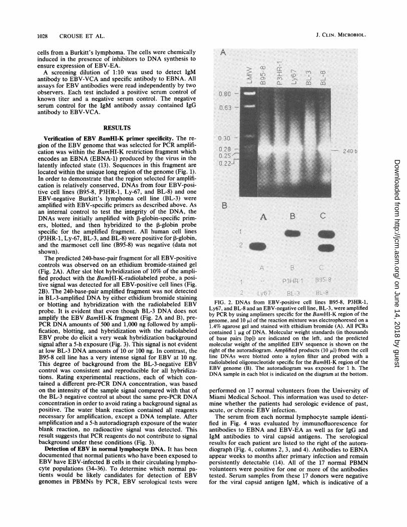
Verification of EBV BamHI-K primer specificity. The re-gion of the EBV genome that was selected for PCR amplifi-cation was within the BamHI-K restriction fragment whichencodes an EBNA (EBNA-1) produced by the virus in thelatently infected state (13). Sequences in this fragment arelocated within the unique long region of the genome (Fig. 1).In order to demonstrate that the region selected for amplifi-cation is relatively conserved, DNAs from four EBV-posi-tive cell lines (B95-8, P3HR-1, Ly-67, and BL-8) and oneEBV-negative Burkitt's lymphoma cell line (BL-3) wereamplified with EBV-specific primers as described above. Asan internal control to test the integrity of the DNA, theDNAs were initially amplified with P-globin-specific prim-ers, blotted, and then hybridized to the ,B-globin probespecific for the amplified fragment. All human cell lines(P3HR-1, Ly-67, BL-3, and BL-8) were positive for ,3-globin,and the marmoset cell line (B95-8) was negative (data notshown).The predicted 240-base-pair fragment for all EBV-positive
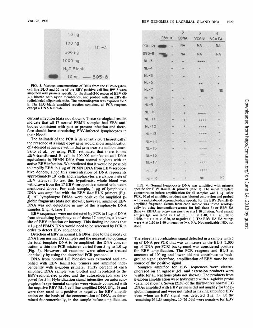
controls was observed on an ethidium bromide-stained gel(Fig. 2A). After slot blot hybridization of 10% of the ampli-fied product with the BamHI-K-radiolabeled probe, a posi-tive signal was detected for all EBV-positive cell lines (Fig.2B). The 240-base-pair amplified fragment was not detectedin BL-3-amplified DNA by either ethidium bromide stainingor blotting and hybridization with the radiolabeled EBVprobe. It is evident that even though BL-3 DNA does notamplify the EBV BamHI-K fragment (Fig. 2A and B), pre-PCR DNA amounts of 500 and 1,000 ng followed by ampli-fication, blotting, and hybridization with the radiolabeledEBV probe do elicit a very weak hybridization backgroundsignal after a 5-h exposure (Fig. 3). This signal is not evidentat low BL-3 DNA amounts of 10 or 100 ng. In contrast, theB95-8 cell line has a very intense signal for EBV at 10 ng.This degree of background from the BL-3-negative EBVcontrol was consistent and reproducible for all hybridiza-tions. Rating experimental reactions, each of which con-
tained a different pre-PCR DNA concentration, was basedon the intensity of the sample signal compared with that ofthe BL-3 negative control at about the same pre-PCR DNAconcentration in order to avoid rating a background signal as
positive. The water blank reaction contained all reagentsnecessary for amplification, except a DNA template. Afteramplification and a 5-h autoradiograph exposure of the waterblank reaction, no radioactive signal was detected. Thisresult suggests that PCR reagents do not contribute to signalbackground under these conditions (Fig. 3).
Detection of EBV in normal lymphocyte DNA. It has beendocumented that normal patients who have been exposed toEBV have EBV-infected B cells in their circulating lympho-cyte populations (34-36). To determine which normal pa-
tients would be likely candidates for detection of EBVgenomes in PBMNs by PCR, EBV serological tests were
0 30 -
0 28 -
0 2 5 "
Ori.........
b
B
A B C
~~~~~~~~~~~~~~~~~~~
FIG. 2. DNAs from EBV-positive cell lines B95-8, P3HR-1,Ly67, and BL-8 and an EBV-negative cell line, BL-3, were amplifiedby PCR by using amplimers specific for the BamHI-K region of thegenome, and 10 Fd of the reaction mixture was electrophoresed on a1.4% agarose gel and stained with ethidium bromide (A). All PCRscontained 1 tg of DNA. Molecular weight standards (in thousandsof base pairs [bp]) are indicated on the left, and the predictedmolecular weight of the amplified EBV sequence is shown on theright of the autoradiogram. Amplified products (10 ,ul) from the cellline DNAs were blotted onto a nylon filter and probed with aradiolabeled oligonucleotide specific for the BamHI-K region of theEBV genome (B). The autoradiogram was exposed for 1 h. TheDNA sample in each blot is indicated on the diagram at the bottom.
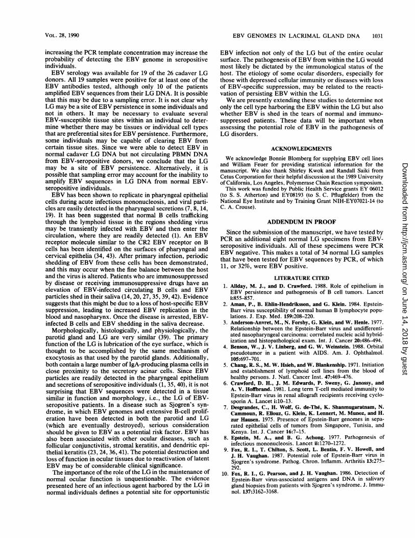
performed on 17 normal volunteers from the University ofMiami Medical School. This information was used to deter-mine whether the patients had serologic evidence of past,acute, or chronic EBV infection.The serum from each normal lymphocyte sample identi-
fied in Fig. 4 was evaluated by immunofluorescence forantibodies to EBNA and EBV-EA as well as for IgG andIgM antibodies to viral capsid antigens. The serologicalresults for each patient are listed to the right of the autora-diograph (Fig. 4, columns 2, 3, and 4). Antibodies to EBNAappear weeks to months after primary infection and remainpersistently detectable (14). All of the 17 normal PBMNvolunteers were positive for one or more of the antibodiestested. Serum samples from these 17 donors were negativefor the viral capsid antigen IgM, which is indicative of a
FIG. 3. Various concentrations of DNA from the EBV-negativecell line BL-3 and 10 ng of the EBV-positive cell line B95-8 were
amplified with primers specific for the BamHI-K region of EBV (30,ul), blotted onto nylon membranes, and probed with an EBV-K-radiolabeled oligonucleotide. The autoradiogram was exposed for 5
h. The H20 blank amplified reaction contained all PCR reagentsexcept a DNA template.
current infection (data not shown). These serological resultsindicate that all 17 normal PBMN samples had EBV anti-bodies consistent with past or present infection and there-fore should have circulating EBV-infected lymphocytes intheir blood.The hallmark of the PCR is its sensitivity. Theoretically,
the presence of a single-copy gene would allow amplificationof a desired sequence within that gene nearly a million times.Saito et al., by using PCR, estimated that there is one
EBV-transformed B cell in 100,000 uninfected-cell DNAequivalents in PBMN DNA from normal subjects with anactive EBV infection. We predicted that it would be possibleto amplify EBV in 1 ,ug ofPBMN DNA from EBV-seropos-itive donors, since this concentration of DNA representsapproximately 105 cells and lymphocytes are a known site ofEBV latency. To test this hypothesis, whole blood was
withdrawn from the 17 EBV-seropositive normal volunteersmentioned above. For each sample, 1 Ftg of lymphocyteDNA was amplified with the EBV BamHI-K primers (Fig.4). All lymphocyte DNAs were positive for amplified P-globin fragments (data not shown); however, amplified EBVDNA was not detectable in any of the lymphocyte DNAsamples (Fig. 4, lane 1).EBV sequences were not detected by PCR in 1 ,ug ofDNA
from circulating lymphocytes of these 17 samples, a knownsite of EBV infection or latency. This finding indicates that>1 p.g of PBMN DNA would need to be screened by PCR inorder to detect EBV sequences.
Detection ofEBV in normal LG DNA. Due to the paucity ofDNA from normal LG samples and the necessity to optimizethe total template DNA to be amplified, the DNA concen-tration within the PCR mixtures varied from 5 ng to 1.0 ,ug(Fig. 5). However, all reactions were otherwise treatedidentically by using the described PCR protocol.DNA from normal LG biopsies was extracted and am-
plified with EBV BamHI-K primers and amplified inde-pendently with P-globin primers. Thirty percent of eachamplified DNA sample was blotted and hybridized to theEBV-radiolabeled probe, and the autoradiograph was ex-posed for 5 h. Hybridization signal intensities on autoradio-graphs of experimental samples were visually compared withthe negative EBV BL-3 cell line amplified DNA (Fig. 3) andwere then rated as a positive or negative for EBV amplifi-cation on the basis of the concentration of DNA, as deter-mined fluorometrically, in the sample before amplification.
NL-4
NL-5
NL-6
NL-7
NL-5
NL-9
_+
- ND
- + ND ND
NL- 1o
NL-1 1
NL-12
NL- 13
NL- 14
NL- 1 5
NL- 16
NL- 1 7
NL- 18
.. ..
+ .+4...
4. +..+
__+ ++.4
+ +.+..
Nl - l 9 - + ND NEt
FIG. 4. Normal lymphocyte DNA was amplified with primersspecific for EBV BamHI-K primers (lane 1). The initial templateconcentration before amplification for all samples was 1 ,ug. AfterPCR, 30 ,tI of amplified product was blotted onto nylon and probedwith a radiolabeled oligonucleotide specific for the EBV BamHI-K-amplified fragment. Serum from each sample was tested serologi-cally by using immunofluorescence for IgG (lane 3) or EBV-EA(lane 4). EBNA serology was positive at a 1:10 dilution. Viral capsidantigen IgG was rated as + at 1:10, ++ at 1:40, +++ at 1:80 to1:160, + + + + at >1:320, or negative (-). The EBV-EA EA ratingswere + at 1:10 to 1:40 or negative (-). NA, Not applicable; ND, notdone.
Therefore, a hybridization signal detected in a sample with 5ng of DNA pre-PCR that was as intense as the BL-3 (1,000ng of DNA pre-PCR) background was considered positivefor EBV amplification. The PCR reagents and BL-3 atamounts of 100 ng and lower did not contribute to back-ground signal; therefore, amplification of EBV must be thesource of the positive signal.Samples amplified for EBV sequences were electro-
phoresed on an agarose gel, and extension products werevisible for all reactions (data not shown). The products from,-globin amplification were hybridized with a P-globin probe(data not shown). Seven (21%) of the thirty-three normal LGDNAs amplified with EBV primers did not amplify for the I-
globin fragment and were not rated as having amplified EBVeven when an EBV signal was detected (Fig. 5). Of theremaining 26 LG samples, 15 (61.5%) were negative for EBV
FIG. 5. DNA from normal LG was amplified with primers spe-cific for the BamHI-K region of the EBV genome, and 30 jtl of thereaction was blotted onto nylon and probed with the EBV-K-radiolabeled oligonucleotide. Interpretation of this blot is providedon the right. Signal intensity ratings, positive (+) or negative (-),were based on visual comparisons of the BL-3 negative control forbackground (Fig. 3). Samples labeled NA were not rated for EBVpositivity, since they were negative for ,-globin amplification. Theamount of DNA in the reaction before amplification is shown to theright of the rating (in nanograms).
and 11 (42.3%) were positive. Although quantitation of EBVcopy number within a sample was impossible, we observedan intense hybridization signal for EBV when amplifyingonly 30 ng ofLG DNA and a moderate signal from as little as5 ng of LG DNA.EBV serology was performed on serum samples from 19
of the 26 cadaver LG donors. All of these donors wereseropositive for viral capsid antigen IgG, EBV-EA, and/or
TABLE 1. EBV amplification in LG DNA and EBV serologicaldata for normal LG donors
Fig. 5.b Grading of EBV antibody titers is as described in the Iegend to Fig. 4.
EBNA antibodies (Table 1), and all were negative for IgM.Positive EBV serology does not appear to be a reliableindicator for the presence of EBV sequences in LG tissues,since EBV sequences could be amplified in only 10 of the 19seropositive LG donors. Unfortunately, only serum sampleswere available from these normal cadaver LG donors, and asa consequence PCR analysis for EBV genomes ofDNA fromcadaver lymphocytes could not be performed. Our resultsindicate that there is a significantly higher probability ofdetecting EBV genomes in the DNA of normal LG tissuesthan in blood cells (P < 0.002; Fisher two-tailed exact test).The results of using the PCR to detect EBV in normal
lacrimal tissue are summarized in Fig. 5. Using the mostextreme case as an example, when amplifying EBV in 5 ng ofnormal LG DNA (approximately 104 cells), or 99.5% lessDNA (5 ng versus 1 Fag) than was used in the lymphocyteamplification study, moderate EBV positivity was evident inthe DNA sample. The inability to amplify EBV sequencesfrom 1 tg of PBMN DNA from normal seropositive patientssuggests that the detection of EBV sequences in the normalLG results from cells containing latent EBV permanentlyresiding within the gland rather than the amplification of theEBV sequences in an incidental transient EBV-infected Bcell circulating through this gland.
DISCUSSION
We have presented evidence for the presence of EBVsequences in normal LG DNA with PCR. The studiesreported herein describe the detection of PCR-amplifiedEBV sequences in normal LG DNA in 11 (42.3%) of 26biopsies. By slot blot hybridization and ethidium bromide-stained agarose gels, it was possible to detect amplified EBVsequences from three samples containing less than 10 ng(pre-PCR) of LG DNA and seven samples with 10 to 100 ngof DNA. In contrast, amplification of 1 p.g of PBMN DNAfrom 17 EBV-seropositive donors was negative for EBVsequences. Serological analysis of LG donors for EBVantibodies did not provide a means to predict whether EBVsequences in LG DNA could be detected by PCR. We havepreviously been unsuccessful in detecting EBV genomicsequences or EBV antigens in LG tissues of normal individ-uals by using techniques less sensitive than PCR. Detectionof EBV sequences within the LG by PCR does not clarify theinfected cell type nor determine whether the amplified EBVsequences are latent EBV genomes or replicating EBVparticles, although the inability to detect the virus by less-sensitive techniques would favor the hypothesis that thevirus is latent in the LG.
It was hypothesized that the sensitivity of the PCR tech-nique could detect EBV sequences in circulating lymphocyteDNA from EBV-seropositive patients, since circulating Bcells are a known site of latency. A previous report of PCRresults estimated that approximately 1 in 105 circulating Bcells is infected with EBV in seropositive individuals (32).The PCR amplification for EBV sequences in each PBMNDNA contained about 105 B cells, whereas in the LG DNAPCR the average number of cells was approximately 104.Our inability to detect EBV genomes in 1 ,ug ofPBMN DNAis most likely due to the low frequency of EBV-infected cellsin our samples. Saito et al., using PCR to estimate thenumber of infected B cells in normal PBMN DNA (1EBV-infected cell per 105 uninfected cells), could detectEBV in 1 ,ug of normal PBMN DNA in only 3 of 50 samples(33). The report did not state whether the donors were EBVseropositive. Amplification of B-cell-enriched samples or
increasing the PCR template concentration may increase theprobability of detecting the EBV genome in seropositiveindividuals.EBV serology was available for 19 of the 26 cadaver LG
donors. All 19 samples were positive for at least one of theEBV antibodies tested, although only 10 of the patientsamplified EBV sequences from their LG DNA. It is possiblethat this may be due to a sampling error. It is not clear whyLG may be a site of EBV persistence in some individuals andnot in others. It may be necessary to evaluate severalEBV-susceptible tissue sites within an individual to deter-mine whether there may be tissues or individual cell typesthat are preferential sites for EBV persistence. Furthermore,some individuals may be capable of clearing EBV fromcertain tissue sites. Since we were able to detect EBV innormal cadaver LG DNA but not circulating PBMN DNAfrom EBV-seropositive donors, we conclude that the LGmay be a site of EBV persistence. Alternatively, it ispossible that sampling error may account for the inability toamplify EBV sequences in LG DNA from normal EBV-seropositive individuals.EBV has been shown to replicate in pharyngeal epithelial
cells during acute infectious mononucleosis, and viral parti-cles are easily detected in the pharyngeal secretions (7, 8, 14,19). It has been suggested that normal B cells traffickingthrough the lymphoid tissue in the regions shedding virusmay be transiently infected with EBV and then enter thecirculation, where they are readily detected (1). An EBVreceptor molecule similar to the CR2 EBV receptor on Bcells has been identified on the surfaces of pharyngeal andcervical epithelia (34, 43). After primary infection, periodicshedding of EBV from these cells has been demonstrated,and this may occur when the fine balance between the hostand the virus is altered. Patients who are immunosuppressedby disease or receiving immunosuppressive drugs have anelevation of EBV-infected circulating B cells and EBVparticles shed in their saliva (14, 20, 27, 35, 39, 42). Evidencesuggests that this might be due to a loss of host-specific EBVsuppression, leading to increased EBV replication in theblood and nasopharynx. Once the disease is arrested, EBV-infected B cells and EBV shedding in the saliva decrease.
Morphologically, histologically, and physiologically, theparotid gland and LG are very similar (39). The primaryfunction of the LG is lubrication of the eye surface, which isthought to be accomplished by the same mechanism ofexocytosis as that used by the parotid glands. Additionally,both contain a large number of IgA-producing plasma cells inclose proximity to the secretory acinar cells. Since EBVparticles are readily detected in the pharyngeal epitheliumand secretions of seropositive individuals (1, 35, 40), it is notsurprising that EBV sequences were detected in a tissuesimilar in function and morphology, i.e., the LG of EBV-seropositive patients. In a disease such as Sjogren's syn-drome, in which EBV genomes and extensive B-cell prolif-eration have been detected in both the parotid and LG(which are eventually destroyed), serious considerationshould be given to EBV as a potential risk factor. EBV hasalso been associated with other ocular diseases, such asfollicular conjunctivitis, stromal keratitis, and dendritic epi-thelial keratitis (23, 24, 36, 41). The potential destruction andloss of function in ocular tissues due to reactivation of latentEBV may be of considerable clinical significance.The importance of the role of the LG in the maintenance of
normal ocular function is unquestionable. The evidencepresented here of an infectious agent harbored by the LG innormal individuals defines a potential site for opportunistic
EBV infection not only of the LG but of the entire ocularsurface. The pathogenesis ofEBV from within the LG wouldmost likely be dictated by the immunological status of thehost. The etiology of some ocular disorders, especially forthose with depressed cellular immunity or diseases with lossof EBV-specific suppression, may be related to the reacti-vation of persisting EBV within the LG.We are presently extending these studies to determine not
only the cell type harboring the EBV within the LG but alsowhether EBV is shed in the tears of normal and immuno-suppressed patients. These data will be important whenassessing the potential role of EBV in the pathogenesis ofLG disorders.
ACKNOWLEDGMENTSWe acknowledge Bonnie Blomberg for supplying EBV cell fines
and William Feuer for providing statistical information for themanuscript. We also thank Shirley Kwok and Randall Saiki fromCetus Corporation for their helpful discussion at the 1989 Universityof California, Los Angeles, Polymerase Chain Reaction symposium.
This work was funded by Public Health Service grants EY 06012(to S. S. Atherton) and EY08193 (to S. C. Pflugfelder) from theNational Eye Institute and by Training Grant NIH-EY07021-14 (toC. A. Crouse).
ADDENDUM IN PROOFSince the submission of the manuscript, we have tested by
PCR an additional eight normal LG specimens from EBV-seropositive individuals. All of these specimens were PCREBV negative. This makes a total of 34 normal LG samplesthat have been tested for EBV sequences by PCR, of which11, or 32%, were EBV positive.
LITERATURE CITED1. Allday, M. J., and D. Crawford. 1988. Role of epithelium inEBV persistence and pathogenesis of B cell tumors. Lanceti:855-857.
2. Aman, P., B. Ehlin-Hendriksson, and G. Klein. 1984. Epstein-Barr virus susceptibility of normal human B lymphocyte popu-lations. J. Exp. Med. 159:208-220.
3. Anderson-Anvret, M., N. Forsby, G. Klein, and W. Henle. 1977.Relationship between the Epstein-Barr virus and undifferenti-ated nasopharyngeal carcinoma: correlated nucleic acid hybrid-ization and histopathological exam. Int. J. Cancer 20:486-494.
4. Benson, W., J. V. Linberg, and G. W. Weinstein. 1988. Orbitalpseudotumor in a patient with AIDS. Am. J. Ophthalmol.105:697-701.
5. Chang, R. S., M. W. Hsieh, and W. Blankenship. 1971. Initiationand establishment of lymphoid cell lines from the blood ofhealthy persons. J. Natl. Cancer Inst. 47:469-476.
6. Crawford, D. H., J. M. Edwards, P. Sweny, G. Janossy, andA. V. Hoffbrand. 1981. Long term T-cell mediated immunity toEpstein-Barr virus in renal allograft recipients receiving cyclo-sporin A. Lancet i:10-13.
7. Desgrandes, C., H. Wolf, G. de-Thé, K. Shanmugaratnam, N.Cammoun, R. Ellouz, G. Klein, K. Lennert, M. Munoz, and H.zur Hausen. 1975. Presence of Epstein-Barr genomes in sepa-rated epithelial cells of tumors from Singapore, Tunisia, andKenya. Int. J. Cancer 16:7-15.
8. Epstein, M. A., and B. G. Achong. 1977. Pathogenesis ofinfectious mononucleosis. Lancet ii:1270-1272.
9. Fox, R. I., T. Chilton, S. Scott, L. Bentin, F. V. Howell, andJ. H. Vaughan. 1987. Potential role of Epstein-Barr virus inSjogren's syndrome. Pathog. Chron. Inflamm. Arthritis 13:275-292.
10. Fox, R. I., G. Pearson, and J. H. Vaughan. 1986. DetectionofEpstein-Barr virus-associated antigens and DNA in salivarygland biopsies from patients with Sjogren's syndrome. J. Immu-nol. 137:3162-3168.
11. Franklin, R. M., and L. E. Remus. 1984. Conjunctival-associ-ated lymphoid tissue: evidence for a role in the secretoryimmune system. Invest. Ophthalmol. Vis. Sci. 25:181-187.
12. Gerber, P., J. Whang-Peng, and J. H. Moore. 1969. Transfor-mation and chromosomal changes induced by Epstein-Barrvirus in normal human leukocyte cultures. Proc. Natl. Acad.Sci. USA 63:740-747.
13. Glaser, R., A. Boyd, J. Stoerker, and J. Holliday. 1983. Func-tional mapping of the Epstein-Barr virus genome: identificationof sites coding for the restricted early antigen, the diffuse earlyantigen, and the nuclear antigen. Virology 129:188-198.
14. Golden, H. D., R. S. Chang, W. Prescott, E. Simpson, and T. Y.Cooper. 1973. Leukocyte-transforming agent: prolonged excre-tion by patients with mononucleosis and excretion by normalindividuals. J. Infect. Dis. 127:471-473.
15. Hanto, D. W., G. Frizzera, G. Purtilo, K. Sakamoto, J. L.Sullivan, A. K. Saemundsen, G. Klein, R. L. Simmons, and J. S.Najarian. 1981. Clinical spectrum of lymphoproliferative disor-ders in renal transplant recipients and evidence for the role ofEpstein-Barr virus. Cancer Res. 41:1253-1261.
16. Henle, W. G., and C. Horowitz. 1974. Epstein-Barr virus diag-nostic tests in infectious mononucleosis. Hum. Pathol. 5:551-565.
17. Huang, D. P., H. C. Ho, W. Henle, D. Saw, and M. Lui. 1978.Presence of EBNA in nasopharyngeal carcinoma and controlpatient tissues related to EBV serology. Int. J. Cancer 22:266-274.
18. Lawrence, J. B., C. A. Villnave, and R. H. Singer. 1988.Sensitive, high resolution chromatin and chromosome mappingin situ: presence and orientation of two closely integrated copiesof EBV in a lymphoma line. Cell 52:51-61.
19. Lemon, S. M., L. M. Hutt, J. E. Shaw, J.-L. Li, X. Nonoyama,and J. S. Pagano. 1977. Replication of Epstein-Barr virus inepithelial cells during infectious mononucleosis. Nature (Lon-don) 268:268-270.
20. Leoni, P., R. C. Garcia, and A. C. Allison. 1978. Effects ofcyclosporin A on human lymphocytes in culture. J. Lab. Immu-nol. 1:67-72.
21. Leyvraz, S., W. Henle, A. P. Chahinian, C. Perlmann, G. Klein,R. E. Gordon, M. Rosenblum, and J. F. Holland. 1985. Associ-ation of Epstein-Barr virus with thymic carcinoma. N. Engl. J.Med. 312:1296-1299.
22. Maniatis, T., E. F. Fritsch, and J. Sambrook. 1982. Molecularcloning: a laboratory manual. Cold Spring Harbor Laboratory,Cold Spring Harbor, N.Y.
23. Matoba, A. Y., and S. P. McCulley. 1985. Epstein-Barr virus andits ocular manifestations, p. 112-117. In W. V. Darrell (ed.),Viral diseases of the eye. Lea & Febiger, Philadelphia.
24. Matoba, A. Y., K. R. Wilhelmus, and D. B. Jones. 1986.Epstein-Barr viral stromal keratitis. Ophthalmology 93:746-751.
25. Meisler, D. O., D. E. Bosworth, and J. H. Krachmer. 1981.Ocular infectious mononucleosis manifested as Parinaud's ocu-loglandular syndrome. Am. J. Ophthalmol. 92:722-726.
26. Miller, G. 1985. Epstein-Barr virus, p. 563-588. In B. N. Fields(ed.), Virology. Raven Press, New York.
27. Miller, G., J. C. Niederman, and L. L. Andrews. 1973. Pro-longed oropharyngeal excretions of Epstein-Barr virus afterinfectious mononucleosis. N. Engl. J. Med. 288:229-232.
28. Morgan, D. G., J. C. Niederman, G. Miller, H. W. Smith, and
J. M. Dowalibi. 1979. Site of Epstein-Barr virus replication inthe oropharynx. Lancet ii:1154-1157.
29. Moss, D. J., A. B. Rickinson, L. E. Wallace, and M. A. Epstein.1981. Sequential appearance of Epstein-Barr virus nuclear andlymphocyte-detected membrane antigens in B-cell transforma-tion. Nature (London) 291:664-666.
30. Nilsson, K. 1971. Elevated-frequency establishment of humanimmunoglobulin-producing lymphoblastoid lines from normaland malignant lymphoid tissue and peripheral blood. Int. J.Cancer 8:432-441.
31. Poncz, M., E. Schwartz, M. Ballantine, and S. Surrey. 1983.Nucleotide sequence analysis of the delta ,-globin gene regionin humans. J. Biol. Chem. 258:11599-11609.
32. Saiki, R. K., S. Scharf, F. Faloona, K. B. Mullis, G. T. Horn,H. A. Erlich, and N. Arnheim. 1985. Enzymatic amplification ofP-globin genomic sequences and restriction site analysis fordiagnosis of sickle cell anemia. Science 230:1350-1354.
33. Saito, I. B., T. Compton, and R. Fox. 1989. Detection ofEpstein-Barr virus DNA by polymerase chain reaction in bloodand tissue biopsies from patients with Sjogren's syndrome. J.Exp. Med. 169:2191-2198.
34. Sixbey, J. W., D. S. Davis, L. S. Young, L. Hutt-Fletcher, T. F.Tedder, and A. B. Rickinson. 1987. Human epithelial cellexpression of an Epstein-Barr virus receptor. J. Gen. Virol.68:805-811.
35. Sixbey, J. W., J. G. Nedrud, N. Raab-Traub, R. A. Hanes, andJ. S. Pagano. 1984. Epstein-Barr virus replication in oropharyn-geal epithelial cells. N. Engl. J. Med. 310:1225-1230.
36. Tanner, O. R. 1954. Ocular manifestations of infectious mono-nucleosis. Arch. Ophthalmol. 51:229-241.
37. Tobi, M., Z. Ravid, V. Feldman-Weis, E. Ben-Chetrit, A. Morag,I. Chowers, Y. Michaeli, M. Shalit, and H. Knobler. 1982.Prolonged atypical illness associated with serological evidenceof persistent Epstein-Barr virus infection. Lancet i:61-63.
38. Tosato, G., and R. M. Blaese. 1985. Epstein-Barr virus andimmunoregulation in man. Adv. Immunol. 37:102-129.
39. Weiczorek, R., F. A. Jakobiec, E. H. Sacks, and D. M. Knowles.The immunoarchitecture of the normal human lacrimal gland.Ophthalmology 95:100-109.
40. Wolf, H., and E. Wilmes. 1984. Persistence of Epstein-Barrvirus in the parotid gland. J. Virol. 51:795-798.
41. Wong, K. W., D. J. D'Amico, T. R. Hedges III, H. K. Soong,R. T. Schooley, and K. R. Kenyon. 1987. Ocular involvementassociated with chronic Epstein-Barr virus disease. Arch. Oph-thalmol. 105:788-792.
42. Yao, Q. Y., A. B. Rickinson, and M. A. Epstein. 1985. Are-examination of the Epstein-Barr virus carrier state in healthyseropositive individuals. Int. J. Cancer 35:35-42.
43. Young, L. S., J. W. Sixbey, D. Clark, and A. B. Rickinson. 1966.Epstein-Barr virus receptor on human pharyngeal epithelia.Lancet i:240-242.
44. zur Hausen, H., G. Klein, W. Henle, P. Clifford, and L.Santesson. 1970. Epstein-Barr virus DNA in biopsies of Burkitttumors and anaplastic carcinomas of the nasopharynx. Nature(London) 228:1056-1058.
45. Zutter, M. P., J. Martin, G. E. Sale, H. M. Shulman, L. Fischer,E. D. Thomas, and D. M. Durnam. 1988. Epstein-Barr viruslymphoproliferation after bone marrow transplantation. Blood72:520-529.