Gill-associated virus (GAV) is a rod-shaped, envel-oped (+) RNA nidovirus that has been associatedwith sporadic outbreaks of disease and mortalities infarmed Penaeus monodon in Australia (Spann et al.1997, Cowley et al. 2000b, Walker et al. 2001). Dis-eased P. monodon display lethargy, lack of appetite,red colouration of the cephalothorax and tail, and
pinkish colouration of the gills. Similar signs of diseaseand rapidly progressing mortalities follow experimen-tal injection with infected tissue extracts or by expo-sure to diseased shrimp tissue by ingestion or immer-sion (Spann et al. 2000, Spann et al. unpubl.). Indiseased shrimp, GAV causes extensive necrosis of thelymphoid organ and there is evidence of a dissemi-nated infection in the gills and other tissues (Spann etal. 1997). However, as has been reported for several
Detection of gill-associated virus (GAV) by in situhybridization during acute and chronic infections
of Penaeus monodon and P. esculentus
Kirsten M. Spann1, 2, Russell J. McCulloch1, Jeff A. Cowley, Iain J. East1, 3, Peter J. Walker1,*
1Cooperative Research Centre for Aquaculture, CSIRO Livestock Industries, Queensland Bioscience Precinct, 306 Carmody Road, St Lucia, Queensland 4067, Australia
2Present address: Laboratory of Infectious Diseases, National Institutes of Allergy and Infectious Diseases, Bethesda, Maryland 20892-0720, USA
3Present address: Office of the Chief Veterinary Officer, Agriculture Fisheries and Forestry Australia, GPO Box 858, Canberra, ACT 2601, Australia
ABSTRACT: Chronic and acute gill-associated virus (GAV) infections were examined by in situhybridization (ISH) using a DNA probe targeting a 779 nucleotide region of the ORF1b-gene.Chronic GAV infections were observed in healthy Penaeus monodon collected from farms andhealthy P. esculentus surviving experimental infection. During chronic-phase infections in both spe-cies, GAV was detected only in partitioned foci of cells with hypertrophied nuclei (spheroids) withinthe lymphoid organ. Acute-phase infections were observed in moribund P. monodon and P. esculen-tus infected experimentally with a high dose of GAV, and in moribund P. monodon collected fromfarms during outbreaks of disease. During acute experimental infections in P. monodon, ISH detectedGAV throughout the lymphoid organ, in gills and in connective tissues throughout the cephalothorax.In moribund P. monodon collected from natural outbreaks of disease, GAV was also detected in thegills and in connective tissues of the cephalothorax, but the distribution of virus within the lymphoidorgan varied. In acutely infected P. esculentus, GAV was detected in connective tissues, but wasrestricted to the inner stromal matrix cells and endothelial cells of intact lymphoid organ tubules. Thetissue distribution of GAV identified by ISH suggests that shrimp are able to control and maintainchronic asymptomatic infection by a process involving lymphoid organ spheroids. Acute phase infec-tions and the development of disease appear to be dose-related and involve the systemic distributionof virus in connective tissues throughout the cephalothorax.
KEY WORDS: Penaeid shrimp · GAV · DNA probe · In situ hybridization
Resale or republication not permitted without written consent of the publisher
Dis Aquat Org 56: 1–10, 2003
other shrimp viruses (Bonami et al. 1992, Flegel et al.1992, Hasson et al. 1995, Tsai et al. 1999), GAV doesnot always cause disease. Chronic, persistent GAVinfection occurs commonly in healthy P. monodonfarmed in Australia. A survey by reverse transcriptase(RT)-nested PCR of 148 broodstock captured from sitesin NE Queensland during 1997 to 1999 has indicated aprevalence of chronic infection approaching 100%(Walker et al. 2001). A similar prevalence of infectionwas detected in healthy cultured postlarvae and juve-nile P. monodon spawned from broodstock collected atthese sites. Examination of healthy shrimp by standardhistological methods and by transmission electronmicroscopy has indicated that GAV is restricted pri-marily to partitioned areas of vacuolated cells withhypertrophied nuclei (‘spheroid bodies’) within thelymphoid organ (Spann et al. 1995). There is also evi-dence that GAV is present in the gonads of healthymale and female broodstock and that chronic infectionis transmitted vertically during spawning (Walker et al.2001, Cowley et al. 2002).
In this paper, we apply in situ hybridization (ISH) tocompare the tissue distribution of GAV in naturally orexperimentally infected Penaeus monodon with acutedisease, and in healthy P. monodon with naturalchronic GAV infections. We also show that experi-mental infection in Penaeus esculentus, which is not anatural host of GAV, may be chronic or acute, depend-ing on the dose of infection.
MATERIALS AND METHODS
Source of shrimp and experimental conditions. Threegroups of shrimp were collected. Group 1: healthy,sub-adult Penaeus monodon (11 to 17 g) were obtainedfrom a farm in SE Queensland in November 1998;Group 2: moribund sub-adult P. monodon (14 to 18 g)with signs of acute GAV infection were collected from1 farm in northern Queensland and 1 farm in SEQueensland during disease outbreaks in January andNovember 1996; Group 3: healthy, sub-adult P. escu-lentus (10 to 23 g) were obtained either from a com-mercial farm in SE Queensland in March 1997 or froma research facility at CSIRO Marine Research, Cleve-land, in November 1997. Some of these shrimp weresacrificed and processed for histology and ISH asdescribed below; the remaining shrimp were main-tained in the aquarium facility at CSIRO Long PocketLaboratories for experimental infections and subse-quent histological analyses.
Experimental infections. A filtered GAV inoculumwas prepared as described previously from Penaeusmonodon collected during an outbreak of disease on afarm in northern Queensland in 1996 (Spann et al.
1997, 2000). Briefly, virus was extracted from the cepha-lothorax by homogenization in lobster haemolymphmedium (LHM; Paterson & Stewart 1974), clarification,and passage through 0.45 and 0.20 µm filters. Forexperimental infections, 50 µl of inoculum was injectedinto muscle tissue at the second abdominal segmentusing a 26-gauge needle. Prior to and following infec-tion, shrimp were maintained in 100 l circular plastictanks in seawater at 26°C and 27 ppt salinity. Waterwas partially exchanged and the shrimp fed pelletedcommercial food daily. Uninfected control shrimp wereinjected with 50 µl of LHM and maintained similarly in100 l tanks in a separate room.
Preparation of fixed tissue sections. Cephalotho-raxes were bisected longitudinally, fixed in Davidson’sfixative for 48 h, dehydrated through a graded seriesof ethanol, wax-infiltrated and embedded using stan-dard histological procedures. Sections (5 µm) werecut, mounted on silanized slides and either stainedwith haematoxylin and eosin (H&E) or used for ISH.Abdomens were not examined in this study.
Preparation of DIG-labeled DNA probe. The originof the GAV cDNA clone pG12 has been reported pre-viously (Cowley et al. 1999, 2000a). Clone pG12 con-tains a 779 nucleotide region of the GAV genome over-lapping the C-terminus of the conserved helicasedomain in the ORF1b gene. To prepare a DIG-labelledpG12 probe, plasmid DNA was prepared using thePERFECTprep™ plasmid DNA kit (5 Prime-3 Prime),the insert was excised with Eco RI, fractionated byelectrophoresis in a 1% low melting point agarosegel containing 0.5 µg ml–1 ethidium bromide and puri-fied using the BRESAclean™ DNA purification kit(GeneWorks). The DNA probe was prepared from thepurified 779 bp insert by random priming and incorpo-ration of DIG (Digoxigenin) using DIG-High Prime(Roche Molecular Biochemicals). The yield of labeledprobe was determined by dot-blot of a dilution series ofDIG-pG12 DNA onto Hybond N+ membrane (Amer-sham Pharmacia) followed by detection with anti-DIG-alkaline phosphatase Fab fragment (Roche MolecularBiochemicals) according to the manufacturer’s instruc-tions.
In situ hybridization (ISH). For ISH, slides preparedas described above were deparaffinized by heating at65°C for 30 min and immersion in toluene for 16 min,then rehydrated through a graded series of ethanolconcentrations. Sections were permeabilized by diges-tion with 25 µg ml–1 Proteinase K in TNE (50 mM Tris-HCl pH 7.4, 10 mM NaCl, 1 mM EDTA) for 10 min at37°C and post-fixed with ice-cold 0.4% formaldehydefor 5 min. Sections were washed briefly in 2 ¥ SSC(saline sodium citrate; 1 ¥ SSC = 0.15 M NaCl, 0.015 Mtri-sodium citrate, pH 7.0) and incubated in hybridiza-tion buffer (4 ¥ SSC, 50% formamide, 1 ¥ Denhardt’s
2
Spann et al.: Acute and chronic GAV infections
reagent, 5% dextran sulphate, 0.5 mg ml–1 salmonsperm DNA) without probe in a humid chamber at42°C for 1 h. The DIG-labeled DNA probe was dena-tured by boiling for 10 min. Sections were then incu-bated in hybridization buffer containing approxi-mately 30 ng denatured DIG-labeled probe in a humidchamber at 42°C for at least 15 h.
Following hybridization, sections were washed inSSC of increasing stringency (2 ¥ SSC for 20 min at25°C, 1 ¥ SSC for 20 min at 42°C, 0.5 ¥ SSC for 20 minat 58°C) and blocked with Buffer I (0.1 M Tris-HClpH 7.5, 0.15 M NaCl) containing 0.5% BlockingReagent (Roche Molecular Biochemicals; Buffer II) at37°C for 30 min. Sections were then incubated in anti-DIG-alkaline-phosphatase Fab fragments (Roche Mol-ecular Biochemicals) diluted 1:2000 in Buffer II at 37°Cfor 30 min. Slides were washed in Buffer III (100 mMTris-HCl pH 9.5, 100 mM NaCl, 50 mM MgCl2)for 20 min at room temperature and incubated incolour developer (38 µl nitro-blue tetrazolium [100 mgml–1] and 38 µl 5-bromo-4-chloro-3-indoyl phosphate[150 mg ml–1] in 10 ml Buffer III) in a dark, humidchamber for 15 to 60 min. Colour development wasstopped by immersion in 1 ¥ TN (1 mM Tris-HClpH 8.0, 0.1 mM NaCl) for 20 min. Slides were thencounter-stained in 0.5% Bismarck brown for 2.5 min,dehydrated through a graded series of ethanol, clearedwith toluene and mounted under cover slips.
RESULTS
Chronic GAV infection in healthy sub-adultPenaeus monodon
Healthy sub-adult Penaeus monodon collected froma farm in SE Queensland and infected naturally with
GAV were sacrificed, fixed and examined by histologyand ISH. By histology, the lymphoid organs appearednormal except for the presence of distinct partitionedareas of vacuolated cells containing hypertrophiednuclei (i.e. spheroid bodies). By ISH, GAV was de-tected only in the cytoplasm of cells within in thespheroid bodies (Fig. 1a). There was no evidence ofinfection other than in lymphoid organ cells. Othertissue types including connective, cardiac, muscularand intestinal tissues were histologically normal anddisplayed no evidence of GAV infection by ISH.
Acute GAV infection in experimentally infectedPenaeus monodon
Healthy Penaeus monodon collected from a farm inSE Queensland were infected experimentally byinjection with an undiluted standard inoculum pre-pared from an extract of shrimp from an outbreak ofacute GAV disease. Progressively accumulating mor-talities were observed from 5 d post-injection. Mori-bund shrimp displayed gross signs of disease charac-teristic of acute GAV infection (Spann et al. 1997).Histological examination of lymphoid organsrevealed extensive cellular necrosis, loss of definedtubule structure and tubule lumen occlusion. How-ever, the degree to which this occurred variedbetween individuals and, in most cases, some tubulesremained intact. Spheroid bodies were either absentor comprised less than 10% of the lymphoid organsection. By ISH, GAV was detected throughout thelymphoid organ, both in areas where tubule structurehad been lost (Fig. 1b) and within intact tubules (Fig.1c). When spheroids were present, cells within thesestructures reacted with the ISH probe (Fig. 1b).Within intact tubules, GAV was either detected
3
Fig. 1. Penaeus monodon. In situ hybridization using a DIG-labeled gill-associated virus (GAV) probe with the lymphoid organ ofGAV-infected P. monodon. Tissues are counter stained with Bismarck brown. (a) Natural, chronic GAV infection showing spher-oids, scale bar = 50 µm; (b,c) experimental GAV infection in which the virus is distributed throughout areas of tubule necrosis (b)
and within intact tubules (c), scale bars = 20 µm. S: spheroid body
Dis Aquat Org 56: 1–10, 2003
throughout the stromal matrix (Fig. 1c) or restricted tocells at the periphery of the tubule and adjacent tothe central lumen.
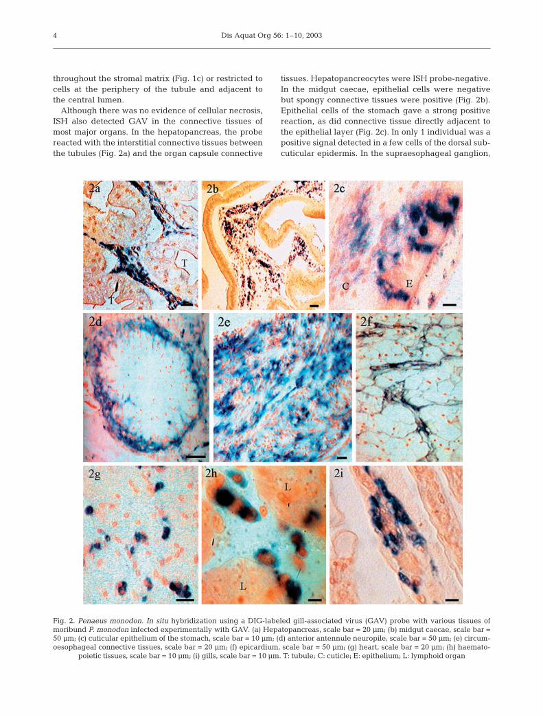
Although there was no evidence of cellular necrosis,ISH also detected GAV in the connective tissues ofmost major organs. In the hepatopancreas, the probereacted with the interstitial connective tissues betweenthe tubules (Fig. 2a) and the organ capsule connective
tissues. Hepatopancreocytes were ISH probe-negative.In the midgut caecae, epithelial cells were negativebut spongy connective tissues were positive (Fig. 2b).Epithelial cells of the stomach gave a strong positivereaction, as did connective tissue directly adjacent tothe epithelial layer (Fig. 2c). In only 1 individual was apositive signal detected in a few cells of the dorsal sub-cuticular epidermis. In the supraesophageal ganglion,
4
Fig. 2. Penaeus monodon. In situ hybridization using a DIG-labeled gill-associated virus (GAV) probe with various tissues ofmoribund P. monodon infected experimentally with GAV. (a) Hepatopancreas, scale bar = 20 µm; (b) midgut caecae, scale bar =50 µm; (c) cuticular epithelium of the stomach, scale bar = 10 µm; (d) anterior antennule neuropile, scale bar = 50 µm; (e) circum-oesophageal connective tissues, scale bar = 20 µm; (f) epicardium, scale bar = 50 µm; (g) heart, scale bar = 20 µm; (h) haemato-
poietic tissues, scale bar = 10 µm; (i) gills, scale bar = 10 µm. T: tubule; C: cuticle; E: epithelium; L: lymphoid organ
Spann et al.: Acute and chronic GAV infections
the probe reacted with glial cells around the anteriorantennule neuropile (Fig. 2d), within the cortical glia,and glial cells of the circumoesophageal connectives(Fig. 2e). Glial cells of the ventral nerve cord were alsopositive. The neuro-secretory, globuli and giant cells ofthe supraesophageal ganglion and segmental gangliawere negative. In the heart, the connective tissues ofthe epicardium gave a positive signal (Fig. 2f). Myocar-dial cells were negative but, in some individuals, thefixed phagocytes were positive (Fig. 2g). The connec-tive tissues of the antennal gland and the muscle bun-dles in the head region gave a positive reaction (notshown). In haematopoietic tissues, the parenchymalcells of the lobules were negative but the develop-ing haemocytes between the lobules were positive(Fig. 2h). A positive signal was observed in the centralaxes and the epithelial pillar cells of the primary andsecondary filaments of the gills (Fig 2i) and was, insome cases, associated with necrosis and fusion of thefilament tips.
Acute GAV infection in Penaeus monodon collectedfrom disease outbreaks on farms
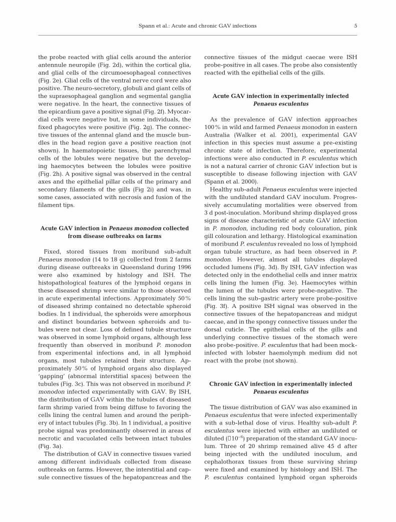
Fixed, stored tissues from moribund sub-adultPenaeus monodon (14 to 18 g) collected from 2 farmsduring disease outbreaks in Queensland during 1996were also examined by histology and ISH. Thehistopathological features of the lymphoid organs inthese diseased shrimp were similar to those observedin acute experimental infections. Approximately 50%of diseased shrimp contained no detectable spheroidbodies. In 1 individual, the spheroids were amorphousand distinct boundaries between spheroids and tu-bules were not clear. Loss of defined tubule structurewas observed in some lymphoid organs, although lessfrequently than observed in moribund P. monodonfrom experimental infections and, in all lymphoidorgans, most tubules retained their structure. Ap-proximately 50% of lymphoid organs also displayed‘gapping’ (abnormal interstitial spaces) between thetubules (Fig. 3c). This was not observed in moribund P.monodon infected experimentally with GAV. By ISH,the distribution of GAV within the tubules of diseasedfarm shrimp varied from being diffuse to favoring thecells lining the central lumen and around the periph-ery of intact tubules (Fig. 3b). In 1 individual, a positiveprobe signal was predominantly observed in areas ofnecrotic and vacuolated cells between intact tubules(Fig. 3a).
The distribution of GAV in connective tissues variedamong different individuals collected from diseaseoutbreaks on farms. However, the interstitial and cap-sule connective tissues of the hepatopancreas and the
connective tissues of the midgut caecae were ISHprobe-positive in all cases. The probe also consistentlyreacted with the epithelial cells of the gills.
Acute GAV infection in experimentally infectedPenaeus esculentus
As the prevalence of GAV infection approaches100% in wild and farmed Penaeus monodon in easternAustralia (Walker et al. 2001), experimental GAVinfection in this species must assume a pre-existingchronic state of infection. Therefore, experimentalinfections were also conducted in P. esculentus whichis not a natural carrier of chronic GAV infection but issusceptible to disease following injection with GAV(Spann et al. 2000).
Healthy sub-adult Penaeus esculentus were injectedwith the undiluted standard GAV inoculum. Progres-sively accumulating mortalities were observed from3 d post-inoculation. Moribund shrimp displayed grosssigns of disease characteristic of acute GAV infectionin P. monodon, including red body colouration, pinkgill colouration and lethargy. Histological examinationof moribund P. esculentus revealed no loss of lymphoidorgan tubule structure, as had been observed in P.monodon. However, almost all tubules displayedoccluded lumens (Fig. 3d). By ISH, GAV infection wasdetected only in the endothelial cells and inner matrixcells lining the lumen (Fig. 3e). Haemocytes withinthe lumen of the tubules were probe-negative. Thecells lining the sub-gastric artery were probe-positive(Fig. 3f). A positive ISH signal was observed in theconnective tissues of the hepatopancreas and midgutcaecae, and in the spongy connective tissues under thedorsal cuticle. The epithelial cells of the gills andunderlying connective tissues of the stomach werealso probe-positive. P. esculentus that had been mock-infected with lobster haemolymph medium did notreact with the probe (not shown).
Chronic GAV infection in experimentally infectedPenaeus esculentus
The tissue distribution of GAV was also examined inPenaeus esculentus that were infected experimentallywith a sub-lethal dose of virus. Healthy sub-adult P.esculentus were injected with either an undiluted ordiluted (¥10–6) preparation of the standard GAV inocu-lum. Three of 20 shrimp remained alive 45 d afterbeing injected with the undiluted inoculum, andcephalothorax tissues from these surviving shrimpwere fixed and examined by histology and ISH. TheP. esculentus contained lymphoid organ spheroids
5
Dis Aquat Org 56: 1–10, 20036
Spann et al.: Acute and chronic GAV infections
typical of natural, chronic GAV infection in Penaeusmonodon. By ISH, the spheroids reacted with theGAV probe but the surrounding healthy tubules wereprobe-negative (Fig. 3g). GAV infection was not de-tected in any other tissue examined by ISH. P. esculen-tus administered a 1 ¥ 10–6 diluted dose of the standardinoculum also displayed histopathology characteristicof chronic GAV infection from 14 d post-injection. Thelymphoid organs of all shrimp examined containedprobe-positive spheroids or zones of necrotic andvacuolated cells between normal tubules (Fig. 3h). Thehistology of all other organs appeared normal andGAV was not detected by ISH. However, in 1 indi-vidual, probe-positive ectopic spheroids were identi-fied in the connective tissues of the hepatopancreasadjacent to the lymphoid organ (Fig. 3i). P. esculentusmock-infected with lobster haemolymph medium dis-played normal histology and no ISH reactions occurredwith the GAV probe.
DISCUSSION
This paper describes the use of ISH to characterizethe chronic and acute phases of GAV infection in natu-rally and experimentally infected shrimp. The study hasexamined healthy Penaeus monodon carrying a nat-ural, persistent GAV infection, moribund P. monodoncollected from ponds during disease outbreaks, and P.monodon super-infected experimentally with a highdose of GAV. The study has also examined dose-relatedstates of infection in P. esculentus which, although notnatural carriers of GAV, are susceptible to experimentalinfection and disease (Spann et al. 2000). Aspects of thehistopathology of GAV infections in P. monodon havebeen reported previously (Spann et al. 1995, 1997). Thespecificity of ISH has allowed a more definitive investi-gation of the distribution of virus during the chronicand acute infection states and has shown that chronicinfections can be established without the onset of dis-ease by exposure to a low dose of virus.
In healthy, juvenile Penaeus monodon infected natu-rally with GAV, ISH detected virus only in the cyto-plasm of hypertrophied cells within lymphoid organ
spheroids (Fig. 1a). This supports previous observa-tions by TEM and histology on the distribution of GAV(previously also named lymphoid organ virus, LOV)during chronic states of infection (Spann et al. 1995).We have also reported previously that low levels ofGAV can be detected in the gills and haemocytes ofhealthy juveniles by RT-PCR (Cowley et al. 2000a),and higher levels of virus have been observed in thegonads of healthy P. monodon broodstock (Walker etal. 2001, Cowley et al. 2002). Failure to detect GAV byISH in tissues other than the lymphoid organ is likely toreflect the abundance of virus in spheroids and therelative insensitivity of these probes compared to RT-nested PCR. The application of in situ RT-PCR (Brodieet al. 1998) may reveal whether PCR targets detectedin other tissues are indicative of low levels of activeviral replication.
During the acute phase of infection in moribund,experimentally infected Penaeus monodon, GAV wasdetected by ISH throughout the lymphoid organ inwhich tubule structure was no longer differentiateddue to extensive tissue necrosis (Fig 1b). GAV was alsocommonly detected in many connective tissues, as wellas haemocytes, glial cells and epithelial cells of thestomach and gill filaments (Fig. 2). Tang et al. (2002)have recently reported a similar distribution of GAV inP. monodon and Litopenaeus vannamei during acuteexperimental infection using a cross-reacting YHV(yellow head virus) DNA probe.
In moribund Penaeus monodon infected naturallywith GAV, the extent of loss of tubule structure variedbetween individuals (Fig, 3a–c). However, naturallyinfected shrimp in acute phase generally displayedless necrosis of the lymphoid organ and more variableprobe signal within the lymphoid organ and othertissues than shrimp infected experimentally. Shrimpinjected with undiluted inocula clearly receive a largedose of virus and the associated lymphoid organhistopathology may well reflect an extreme situationthat rarely occurs in nature. We have estimated bytitration in vivo that undiluted GAV inocula contain upto 108 infectious doses and can cause mortalities injuvenile P. monodon when diluted to 10–5 (I.J.E. &P.J.W. unpubl. data). As almost all farmed P. monodon
7
Fig. 3. Penaeus monodon and P. esculentus. In situ hybridization using a DIG-labeled gill-associated virus (GAV) probe with var-ious tissues of GAV-infected P. monodon and P. esculentus. (a–c) Moribund P. monodon infected naturally with GAV; (a) infectionin areas of necrotic, vacuolated cells between lymphoid organ tubules (T), scale bar = 20 µm; (b) infection in cells surroundingthe lumen (L) of a lymphoid organ tubule, scale bar = 50 µm; (c) gapping (arrow) in tissues between tubules, scale bar = 50 µm.(d–f) Moribund P. esculentus infected experimentally with a high dose of GAV; (d) lymphoid organ tubules with occluded lumens(arrows), scale bar = 50 µm; (e) infected cells lining the lumen (L) of lymphoid organ tubules, scale bar = 20 µm; (f) infection incells lining the sub-gastric artery (arrows), scale bar = 50 µm; (g) P. esculentus surviving infection with a high dose of GAV. Infec-tion confined to lymphoid organ spheroids, scale bar = 50 µm. (h–i) P. esculentus infected with a sublethal dose of GAV; (h) zones
of infected lymphoid organ cells, scale bar = 50 µm; (i) ectopic spheroids in the hepatopancreas, scale bar = 50 µm
Dis Aquat Org 56: 1–10, 2003
in Australia carry a pre-existing chronic GAV infec-tion, acute-phase infections and associated mortalitiesmay occur when environmental stress factors such astemperature or low dissolved oxygen levels aggravatethe chronic condition (Flegel et al. 1997, Tsai et al.1999, Vidal et al. 2001). There is also evidence thatboth chronic and acute GAV infections may be trans-mitted horizontally by ingestion or immersion (Spannet al. unpubl.). The variability in histopathology of thelymphoid organ in moribund farmed shrimp with acuteGAV infections may reflect variations in the initialroute and dose of infection and the likelihood thatacute infections in ponds derive from pre-existingchronic infections.
Penaeus esculentus injected with a high dose ofGAV also developed an acute infection and displayeda similar distribution of virus as P. monodon, but therewas no loss of tubule structure in the lymphoid organand the virus did not appear as widespread within thelymphoid organ or connective tissues (Fig. 3d–f). GAVwas detected in cells lining the sub-gastric artery andtubule lumen within the lymphoid organ. As haemo-lymph first enters the lymphoid organ via the sub-gastric artery and must pass through the thin layer ofendothelial cells lining the lumen before entering thestromal matrix of the tubules (Oka 1969, Martin et al.1987, Anggraeni & Owens 2000), this may be indica-tive of an early stage of infection in the lymphoidorgan. Tang et al. (2002) also reported no loss ofnormal tubule structure in GAV-infected Litopenaeusvannamei which, like P. esculentus, is not a naturalhost of GAV.
We have reported previously that acute GAV infec-tion and associated mortalities develop more slowly inPenaeus esculentus than in P. monodon and some indi-viduals survive an acute GAV infection, even when thedose of virus is relatively high (Spann et al. 2000). Insurvivors of high dose infection and in P. esculentusinfected with a very low dose of GAV, lymphoid organspheroids containing probe-positive cells typical ofchronic infection in P. monodon were formed and GAVwas not detected by ISH in any other tissue (Fig. 3g–h).In some P. esculentus infected with a low dose of GAV,‘zones’ of infected cells were observed between nor-mal tubules. Similar areas of infection between intacttubules were also observed in the lymphoid organs ofP. monodon with natural, acute infection. However, inhealthy P. esculentus the infected zones were clearlydifferentiated from normal tubules and not as wide-spread. These zones may be distended haemal sinusesin which phagocytes and other haemocytes containingsequestered GAV have accumulated (Anggraeni &Owens 2000). The ‘gapping’ observed in some shrimp(Fig. 3i) suggests that the haemal sinuses and connec-tive tissues become necrotic as a result of the heavy
accumulation of infected cells and contribute to thedevelopment of disease.
Lymphoid organ spheroids have been observed dur-ing infection with several other shrimp viruses, and ithas been proposed that spheroid formation is indica-tive of a non-specific defensive response (Owens et al.1991, Bonami et al. 1992, Flegel et al. 1992, Hassonet al. 1995). Hasson et al. (1999a) and Anggraeni &Owens (2000) have suggested that spheroids areformed when resident phagocytes, or haemocytesmigrating from the tubule lumen through the stromalmatrix, sequester virus and accumulate in the haemalsinuses between the tubules. Based on cytopathologyand ISH probe reactions during recovery from TSVinfections, Hasson et al. (1999a) classified spheroids inLitopenaeus vannamei as 3 distinct and serial morpho-types. The Type A morphotype, the earliest detected,appeared to originate from lymphoid organ phago-cytes that had sequestered TSV, and was characterisedby a lightly basophilic, homogeneous cell mass con-taining few necrotic cells. Type B spheroids, whichappeared to evolve from Type A, contained increasednumbers of necrotic cells with pyknotic nuclei andmoderate vacuolization and remained persistently in-fected with TSV. Type C spheroids, in which there wasno evidence of TSV infection by ISH, developed laterand contained condensed, highly basophilic nuclei andincreased cytoplsamic vacuolization characteristic ofapoptosis (Hasson et al. 1999b). The spheroids we haveobserved in healthy Penaeus monodon with natural,chronic GAV infections, and in P. esculentus survivingexperimental infection, resemble Type B spheroids,displaying a diffuse probe signal, vacuolization, pyk-notic nuclei and some cell necrosis. Type B spheroidswere also reported to be the most common morphotypeduring the chronic phase of TSV infection (6 to 16 wkpost-infection) (Hasson et al. 1999b). The focal concen-tration of GAV in Type B spheroids during naturalchronic infection in P. monodon and in survivors ofexperimental infection in P. esculentus suggests activesequestration of virus does occur and this may modu-late the spread and replication of virus in other tissues.The capacity of shrimp to contain GAV infectionappears to be dose-related. As chronic GAV infec-tions are also transmitted vertically during spawning(Cowley et al. 2002), the route of infection and age ofthe shrimp may also influence their capacity to containthe infection and avoid disease.
For TSV, Hasson et al. (1999b) have proposed amodel in which spheroids have a central role in themodulation of infection. According to this model, TSVmay continue to replicate within Type B spheroids,escape and enter the circulatory system, and return tothe lymphoid organ where it is sequestered by phago-cytes and new spheroids are formed. Alternatively,
8
Spann et al.: Acute and chronic GAV infections
spheroids eventually eliminate the virus through apop-tosis of infected cells, clearing the infection and result-ing in a normal lymphoid organ. Hasson et al. (1999b)suggest that these processes probably occur concur-rently in chronic infection. Depending on the balanceof the outcomes, infection may remain persistent or becleared from the shrimp. However, our observationssuggest that the former process dominates in chronicGAV infection. The very high prevalence of chronicGAV infection in Penaeus monodon postlarvae, juve-niles and broodstock in eastern Australia (Walker et al.2001) indicates that the virus is rarely cleared frominfected shrimp, and the presence of GAV in haemo-lymph suggests a cyclic systemic infection (Cowley etal. 2000a). We have also observed that followingexperimental infection at low dose, P. esculentus lym-phoid organs remain PCR-positive for at least 50 d(I.J.E. & P.J.W. unpubl. data). Indeed, life-long, persis-tent, chronic infections and vertical transmissionappear to be key elements of the GAV infection cycle,sustaining the virus in shrimp populations in the ab-sence of disease (Walker et al. 2001, Cowley et al. 2002).
Despite the high prevalence of chronic GAV infec-tion in wild and farmed Penaeus monodon, diseaseoutbreaks are not common in Australia and little is yetknown of the process by which ubiquitous chronic,persistent infections result in disease and mortalities.Understanding the transition from chronic to acuteinfection may identify environmental stress as a factorthat can compromise the capacity of the host to main-tain the chronic infection state and allow the identifi-cation of functional indicators of impending disease. Atpresent, the detection of virus outside lymphoid organspheroids is the most useful indicator of a transition todisease. ISH provides a reliable means of detectingGAV in lymphoid organ tubules, gill and connectivetissues that appear normal using standard histologicalstains and may prove to be a useful tool with which toidentify early, asymptomatic stages of disease develop-ment.
Acknowledgements. We would like to thank Gold CoastMarine Aquaculture and CSIRO Marine Research, Cleveland,for supplying experimental shrimp and Mr. Sam Johnson,CSIRO Livestock Industries, for assistance with digitalimaging.
LITERATURE CITED
Anggraeni MS, Owens L (2000) The haemocytic origin of lym-phoid organ spheroid cells in the penaeid prawn Penaeusmonodon. Dis Aquat Org 40:85–92
Bonami JR, Lightner DV, Redman RM, Poulos BT (1992)Partial characterisation of a togavirus (LOVV) associatedwith histopathological changes of the lymphoid organ of
penaeid shrimps. Dis Aquat Org 14:145–152Brodie SJ, Bardsley KD, Diem K, Mecham JO, Norelius SE,
Wilson WC (1998) Epizootic haemorrhagic disease: analy-sis of tissues by amplification and in situ hybridisationreveals widespread orbivirus infection in low copy num-bers. J Virol 72:3863–3871
Cowley JA, Dimmock CM, Wongteerasupaya C, Boonsaeng V,Panyam S, Walker PJ (1999) Yellow head virus from Thai-land and gill-associated virus from Australia are closely re-lated but distinct viruses. Dis Aquat Org 36:153–157
Cowley JA, Dimmock CM, Spann KM, Walker PJ (2000a)Detection of Australian gill-associated virus (GAV) andlymphoid organ virus (LOV) of Penaeus monodon by RT-nested PCR. Dis Aquat Org 39:159–167
Cowley JA, Dimmock CM, Spann KM, Walker PJ (2000b)Gill-associated virus of Penaeus monodon prawns: aninvertebrate virus with ORF1a and ORF1b genes relatedto arteri- and coronaviruses. J Gen Virol 81:1473–1484
Cowley JA, Hall MR, Cadogan LC, Spann KM, Walker PJ(2002) Vertical transmission of gill-associated virus (GAV)in the black tiger prawn Penaeus monodon. Dis Aquat Org50:95–104
Flegel TW, Fegan DF, Kongsom S, Vuthikornudomkit S and5 others (1992) Occurrence, diagnosis and treatment ofshrimp diseases in Thailand. In: Fulks W, Main KL (eds)Diseases of cultured penaeid shrimp in Asia and theUnited States. The Oceanic Institute, Honolulu, p 57–112
Flegel TW, Boonyaratpalin S, Withychumnarnkul B (1997)Progress in research on yellow-head virus and white spotvirus in Thailand. In: Flegel TW, McCrae IH (eds) Diseasesin Asian aquaculture III. Fish Health Section, Asian Fish-eries Society, Manila, p 285–295
Hasson KW, Lightner DV, Poulos BT, Redman RM, White BL,Brock JA, Bonami JR (1995) Taura syndrome in Penaeusvannamei: demonstration of a viral etiology. Dis AquatOrg 23:115–126
Hasson KW, Lightner DV, Mohney LL, Redman RM, PoulosBT, White BM (1999a) Taura syndrome virus (TSV) lesiondevelopment and the disease cycle in Pacific white shrimpPenaeus vannamei. Dis Aquat Org 36:81–93
Hasson KW, Lightner DV, Mohney LL, Redman RM, WhiteBM (1999b) Role of lymphoid organ spheroids in chronicTaura syndrome virus (TSV) infections in Penaeus van-namei. Dis Aquat Org 38:93–105
Martin GG, Hose LE, Kim JJ (1987) Structure of hematopoi-etic nodules in the ridgeback prawn, Sicyonia ingentis:light and electron microscopic observations. J Morphol192:193–204
Oka M (1969) Studies on Penaeus orientalis Kishinouye. VIII.Structure of the newly found lymphoid organ. Bull Jpn SocSci Fish 35:245–250
Owens L, De Beer S, Smith J (1991) Lymphoid parvo-likeparticles in Australian penaeid prawns. Dis Aquat Org11:129–134
Paterson WD, Stewart JE (1974) In vitro phagocytosis byhemocytes of American lobster (Homarus americanus).J Fish Res Board Can 31:1051–1056
Spann KM, Vickers JE, Lester RJG (1995) Lymphoid organvirus of Penaeus monodon from Australia. Dis Aquat Org23:127–134
Spann KM, Cowley JA, Walker PJ, Lester RJG (1997) A yel-low-head-like virus from Penaeus monodon cultured inAustralia. Dis Aquat Org 31:169–179
Spann KM, East IJ, Donaldson RA, Cowley JA, Walker PJ(2000) Differences in the susceptibility of some penaeidprawn species to gill-associated virus (GAV) infection. DisAquat Org 42:221–225
9
Dis Aquat Org 56: 1–10, 2003
Tang KFJ, Spann KM, Owens L, Lightner DV (2002) In situdetection of Australian gill-associated virus with a yellowhead virus gene probe. Aquaculture 205:1–5
Tsai MF, Kou GH, Liu HC, Liu KF and 5 others (1999) Long-term persistence of white spot syndrome virus (WSSV) in acultivated shrimp population without disease outbreaks.Dis Aquat Org 38:107–114
Vidal OM, Granja CB, Aranguren F, Brock JA, Salazar M(2001) A profound effect of hyperthermia on survival of
Litopenaeus vannamei juveniles infected with white spotsyndrome virus. J World Aquacult Soc 32:364–372
Walker PJ, Cowley JA, Spann KM, Hodgson RAJ, Hall MA,Withyachumnarnkul B (2001) Yellow head complexviruses: transmission cycles and topographical distributionin the asia-pacific region. In: Browdy CL, Jory DE (eds)The new wave. Proc Spec Session Sustainable ShrimpCulture, Aquaculture 2001. The World Aquaculture Soci-ety, Baton Rouge, FL, p 227–237