Determination of the Parameters of the Skin-Electrode Impedance Model for ECG Measurement C. Assambo, A. Baba, R. Dozio, and M. J. Burke Department of Electronic and Electrical Engineering Trinity College Dublin REPUBLIC OF IRELAND http://www.mee.tcd.ie Abstract: - Medical practitioners rely heavily upon the information obtained from electrocardiography. Some seriously ill hospital patients have their ECG constantly monitored throughout their stay, where accuracy in monitoring is essential to maintaining a high level of patient care. Fidelity in the measurement of the electrical activity of the heart is of primary importance for accurate diagnosis and treatment. This requires the ability of a monitoring system to reproduce the morphology of the measured signal with as high a degree of accuracy as possible. It is well known that the skin-electrode interface can be the cause of a large degree of distortion in the ECG signal that can lead to misinterpretation of the signal if the amplifier is not suitably matched to it. To allow this it is important to be able to model the properties of this interface. This has become more difficult for dry, pasteless electrodes, which are becoming increasingly popular for long-term ECG monitoring. We present a technique for measuring the model parameters of the skin-electrode interface based on dc and ac measurements, which yields values for the resistive and capacitive elements. Key-Words: ECG, Impedance Measurement, Skin-Electrode Interface, Electrode Modeling. 1 Introduction The Electrocardiogram (ECG) measures the electrical activity of the heart and provides the physiological information necessary to evaluate the condition and performance of the cardiovascular system and to detect cardiac abnormalities. In addition, the ECG can provide valuable information on the regularity of the heart rate as well as the size and the position of the chambers, the presence of any damage and the effect of drugs or devices used to regulate its function. The contraction of the heart muscle is associated with electrical changes called ‘depolarisation’ that can be detected by electrodes attached to the surface of the body. Surface electrodes convert the ionic current within the body into electronic current in metal connecting leads. In practice, skin, tissue, electrodes and recording amplifiers limit the capacity of the measurement system to reproduce the physiological signal with perfect fidelity [1, 2]. The nature of the skin- electrode interface and its mismatch with the amplifier input impedance can introduce serious distortion in the recording of physiological signals. The impedance of the skin may be reduced by abrasion with sand paper [3]. However, such aggressive techniques are not applicable to patient populations such as elderly, allergenic and paediatric subjects. Experience of measurements during bioelectric signal recording using Nasicon ceramic semi-dry electrodes proved the existence of significant noise allocated to electrochemical features of the skin- electrode interface [4]. The noise in the bandwidth 1Hz-100Hz was found to be more intense than the thermal noise of the resistive part of the electrode plus the amplifier voltage and current noise combined. Therefore, knowledge of the skin- electrode impedance becomes essential in order to ensure correct amplifier matching to avoid distortion. Previous studies have demonstrated that measurement of the dc skin-electrode impedance does not provide sufficient information. Because of the capacitive components, corresponding to the epidermal layer and the electrode’s permittivity, ac measurement is needed to obtain an accurate estimate of the skin-electrode impedance [5]. The authors have attempted to measure the resistive and capacitive properties of wet and dry electrodes using the step response of the electrode system to a small dc current and an analysis of the magnitude and phase of the frequency response in the range 0.015 – 10 Hz. Measurement results provide valuable information for the design of the input stage of the ECG amplifier according to criteria published by The American Heart Association. Proceedings of the 6th WSEAS Int. Conf. on Electronics, Hardware, Wireless and Optical Communications, Corfu Island, Greece, February 16-19, 2007 90
Transcript
Determination of the Parameters of the Skin-Electrode Impedance Model for ECG Measurement
C. Assambo, A. Baba, R. Dozio, and M. J. Burke
Department of Electronic and Electrical Engineering Trinity College Dublin
REPUBLIC OF IRELAND http://www.mee.tcd.ie
Abstract: - Medical practitioners rely heavily upon the information obtained from electrocardiography. Some seriously ill hospital patients have their ECG constantly monitored throughout their stay, where accuracy in monitoring is essential to maintaining a high level of patient care. Fidelity in the measurement of the electrical activity of the heart is of primary importance for accurate diagnosis and treatment. This requires the ability of a monitoring system to reproduce the morphology of the measured signal with as high a degree of accuracy as possible. It is well known that the skin-electrode interface can be the cause of a large degree of distortion in the ECG signal that can lead to misinterpretation of the signal if the amplifier is not suitably matched to it. To allow this it is important to be able to model the properties of this interface. This has become more difficult for dry, pasteless electrodes, which are becoming increasingly popular for long-term ECG monitoring. We present a technique for measuring the model parameters of the skin-electrode interface based on dc and ac measurements, which yields values for the resistive and capacitive elements. Key-Words: ECG, Impedance Measurement, Skin-Electrode Interface, Electrode Modeling.
1 Introduction
The Electrocardiogram (ECG) measures the electrical activity of the heart and provides the physiological information necessary to evaluate the condition and performance of the cardiovascular system and to detect cardiac abnormalities. In addition, the ECG can provide valuable information on the regularity of the heart rate as well as the size and the position of the chambers, the presence of any damage and the effect of drugs or devices used to regulate its function. The contraction of the heart muscle is associated with electrical changes called ‘depolarisation’ that can be detected by electrodes attached to the surface of the body. Surface electrodes convert the ionic current within the body into electronic current in metal connecting leads. In practice, skin, tissue, electrodes and recording amplifiers limit the capacity of the measurement system to reproduce the physiological signal with perfect fidelity [1, 2]. The nature of the skin-electrode interface and its mismatch with the amplifier input impedance can introduce serious distortion in the recording of physiological signals. The impedance of the skin may be reduced by abrasion with sand paper [3]. However, such aggressive techniques are not applicable to patient populations such as elderly, allergenic and paediatric subjects.
Experience of measurements during bioelectric signal recording using Nasicon ceramic semi-dry electrodes proved the existence of significant noise allocated to electrochemical features of the skin-electrode interface [4]. The noise in the bandwidth 1Hz-100Hz was found to be more intense than the thermal noise of the resistive part of the electrode plus the amplifier voltage and current noise combined. Therefore, knowledge of the skin-electrode impedance becomes essential in order to ensure correct amplifier matching to avoid distortion.
Previous studies have demonstrated that measurement of the dc skin-electrode impedance does not provide sufficient information. Because of the capacitive components, corresponding to the epidermal layer and the electrode’s permittivity, ac measurement is needed to obtain an accurate estimate of the skin-electrode impedance [5]. The authors have attempted to measure the resistive and capacitive properties of wet and dry electrodes using the step response of the electrode system to a small dc current and an analysis of the magnitude and phase of the frequency response in the range 0.015 – 10 Hz. Measurement results provide valuable information for the design of the input stage of the ECG amplifier according to criteria published by The American Heart Association.
Proceedings of the 6th WSEAS Int. Conf. on Electronics, Hardware, Wireless and Optical Communications, Corfu Island, Greece, February 16-19, 2007 90
2 Models for the skin-electrode interface
2.1 The single time constant model
Experiments of Swanson & Webster have demonstrated a dominant contribution of the skin layer to the skin-electrode impedance [6]. They originally proposed a model of the skin-electrode interface as a parallel combination of a resistor and a capacitor in series with a second resistor. Using this model, the authors attempted to measure the skin-electrode impedance using a pair of Ag/AgCI self adhesive electrodes with a diameter of 5cm.
We considered a dual electrode configuration connected to a resistive load impedance. One electrode is fed with a sinusoidal voltage from a signal analyser (Agilent 35670A) and connected to the body. A second electrode is used to detect the resulting signal from the skin and feeds it to the input of the analyser. Fig.1. illustrates the instrumentation set up for measuring the frequency response of the skin-electrode interface.
Fig. 1 Experimental set-up for electrode impedance measurement.
Extracting the magnitude at the lowest and the
highest frequencies (0.1Hz and 30Hz respectively), we estimated the series resistors at 5kΩ and the shunt resistance at 70kΩ. The plot of the phase vs. frequency indicated a peak at 2.6Hz. The capacitive element was then evaluated at just under 1µF. Fig. 2 compares the measured phase response with a curve fit obtained using MatLab for a three-element RC model.
The fitted curve matches the measurements in the region of 1 to 5Hz, centred on the peak but outside of this frequency range the match is unacceptably poor. This indicates that the three-element model is not adequate for use in
establishing the requirements of a recording amplifier of high performance.
Frequency (Hz)
phase (deg)
Frequency (Hz)
phase (deg)
Fig. 2 Plot (a) represents the measured phase response of the skin-electrode interface. Plot (b) shows the phase of the three element RC circuit fit to the data. 2.2 The double time constant model
Kaczmarek & Webster presented a more
accurate model which describes the skin-electrode interface as a double time constant system with probably time-varying parameters [7]. Because of complex current dependent voltage sources, capacitances, and ohmic resistors, the skin electrode interface is seen as a non-linear second-order filter. The electrical equivalent circuit of a six-element skin-electrode interface model is shown in Fig. 3. Wet and dry electrodes are expected to have the same electrical model but the parameter values will be significantly different.
Fig. 3 Skin-electrode interface and electrical equivalent circuit [8].
Vo
Channel1
Skin / Electrodes Tissue
Buffer
Channel2
Vi
Signal Analyser
C4
C4
R4 R3
Voff
Ro
Voff
R3 R4
Rtissue
source
Electrode
Epidermis
Dermis subcutaneous Rsubcutaneous
Eelectrode/sweat
Rsweat
RelectrodeCelectrode
Electrolyte/ Sweat
Rstrat cor
Esweat/strat corn
Cstrat cor
Stratum
Proceedings of the 6th WSEAS Int. Conf. on Electronics, Hardware, Wireless and Optical Communications, Corfu Island, Greece, February 16-19, 2007 91
The experimental set-up used to evaluate the electrode parameters of the six-element model is shown in Fig. 4.
Fig. 4. Schematic representation of the measurement set up assuming a double time constant model. 3 Determination of the parameters of a double time constant model 3.1 Information extracted from the phase
The phase of the system measured at the load in the set-up of Fig. 4 can be shown to be equal to:
ω1, ω2 and ω3 (as well as -ω1, -ω2 and -ω3). These solutions correspond to the frequencies that make the first derivative of the phase equal to zero. From this the following system of equations can be built:
( )
( )
( )
−=
++=
−−−=
⇔
=+++
=+++
=+++
1523
22
21
13a
1423
22
21
23
22
21
2a
1323
12
2
12
1
11a
06
33a4
32a2
31a1
06
23a4
22a2
21a1
06
13a4
12a2
11a1
ωωω
ωωω
ωωω
ωωω
ωωω
ωωω
ωωω
The phase response contains three local extrema
having corresponding frequencies which can be extracted. In this case ω1, ω2 and ω3 are three distinguished real and positive frequencies as shown in Fig. 5.
frequency
phase
ω1 ω2 ω3
frequency
phase
ω1 ω2 ω3
Fig. 5. Plot of the phase vs. frequency for τ2=120ms and τ4=9ms. 3.2 The step response
The step response was measured by switching a dc current I0 through the skin-electrode interface at a very low frequency. The transient voltage V0 was used to determine the series resistance and the value of the overall resistive component was evaluated using the steady state voltage Vs as shown in Fig. 6.
( )16IVV
R0
offsetos
−=
( ) ( )17IVV
RR2R0
offsets42s
−=++
time (s)
Voltage (V)
Voffset V0
Vs
0 5 10 15 20 25
1
2
3
4
5
6
0
time (s)
Voltage (V)
Voffset V0
Vs
0 5 10 15 20 25
1
2
3
4
5
6
0
Fig. 6 Plot of the voltage across the skin-electrode interface in response to an impulse current I0=0.8µA.
Vo
Channel1
Electrode 1
Electrode 2 Skin
Skin
Tissue Buffer
Channel 2
Vi
Signal Analyser
C2
C2 C4
C4
R2
R2 R4
R3
R3
R1
R1
Rtissue
Ess
Ess Ees
Ees
Ro
R4
Proceedings of the 6th WSEAS Int. Conf. on Electronics, Hardware, Wireless and Optical Communications, Corfu Island, Greece, February 16-19, 2007 92
3.3 Identification
We introduce the following variables, computed from the step response:
( )184T
o
RRa =
( )19RR
Raso
o5 +=
The combination and rearrangement of equations (10), (11), (12), (18) and (19) yield the following system of equations:
( )200242R2
5aR2
24R25a
R2
42
22
4a5a3a
R34a
R1a oo
oo =+++++
ττ
ττ
( )2105a
24
223
4a24
22
5a3a2
42
22
4a5a3a
34a1a
4a2a
=++−
ττ
ττττ
( ) ( ) ( )22024R42R34
324a
44R22R5a3a=++
+ττ
ττ
ττ
( )2305a4a4a5a
R4R22R2 o =−
−+
( )2405a
5a1RsR o =
−−
We then introduce q, the product of the two time constants and its square Q, so that:
( )25q 42ττ=
( )262qQ = Equation (21) can then be reduced to that of a single variable. Q is obtained by computing the roots of the following third order polynomial:
( )270Qa3Q
aa
Qa
aaaa
aa3 3
5
2
4
22
4
5313
4
25
23 =++−−
Equations (20) and (22) are combined with the solutions of equation (27) to obtain an expression for R4 in terms of q and τ22:
( ) ( )28qaa
aaqaqaaa
aa2R
2254
2235
44
3453
45o4R
−+
−−
=τ
τ
This expression is then substituted into equation (18) to obtain τ2 as a function of the input parameters.
( )290422
2210 =++ τρτρρ
( )30aq
qaaaaa
aq
5
5
3453
54
4
2
0
−−
−=ρ
( ) ( )31aa
aaqaqqaaa
aaqaaa3
aa
54
353
43
453
5422
4
53
4
11
+
−−
−
+=ρ
( )32aa
qaaaaa
a1
4
33
453
54
52
−−
−=ρ
Once τ2 is obtained, the other unknown parameters are easily deduced.
( )33Raa2aaRR 4
54
45in2 −
−=
( )34RC 222 τ= ( )35q 24 ττ = ( )36R/C 444 τ=
3.4 Simulation results
An algorithm was written which, given the five parameters a4, a5, ω1, ω2 and ω3 as input obtained from the experimental data, computes the values of all of the components of the six-element model to a reasonable degree of accuracy. In the case of measurements carried out on a constructed hardware electrical model the component values were reproduced with a maximum error less than 10%. The error is due to the limited accuracy in determining the frequencies of the peaks and trough in the phase response, which can be difficult due to the low values of absolute phase.
3.5 Limitation of the method
It was observed that very often the phase response did may not display two distinct peaks but only a single peak. This means that the third order polynomial represented in equation (9) has one distinct, real and positive solution. This situation occurs when the two time constants τ2 and τ4 are relatively close to each other. Fig. 7 illustrates the correlation between the plot of the phase and the ratio τ2 over τ4.
α=1
α=2
α=5
α=10α=20
Phase (degree)
Frequency (Hz)
10mHz 100mHz 1Hz 10Hz 100Hz
50md
100md
150md
200md
0d
α=1
α=2
α=5
α=10α=20
Phase (degree)
Frequency (Hz)
10mHz 100mHz 1Hz 10Hz 100Hz
50md
100md
150md
200md
0d
Fig. 7 Plot of the phase against frequency for
different values of the ratio α=τ2/τ4: Ro=10MΩ, Rseries=10kΩ, R2=35kΩ and R4=30kΩ.
The load resistor needs to be much larger than the
impedance of the electrodes to obtain two adequate peaks in the phase response. Consequently, measurements provide very small values of absolute phase and only minute differences in phase at the two peaks when present. These can be extremely difficult to resolve.
Proceedings of the 6th WSEAS Int. Conf. on Electronics, Hardware, Wireless and Optical Communications, Corfu Island, Greece, February 16-19, 2007 93
4 Fitting magnitude and phase
Improved results have been obtained by using both the magnitude and phase response in a skin-electrode impedance fitting algorithm. For the purpose, a least squares error minimization program has been developed in MATLAB, based on the function lsqcurvefit.
The procedure has been applied to measurements on a pair of adhesive electrodes, 5cm in diameter (Wandy, E-50mm Hydrogel). The algorithm was first used to fit a single time constant model, as in the measurement set up in Fig. 1. The result of the curve fitting is depicted in Fig. 8 for the phase data and again underlines how a simple 3-parameter model is not suitable for describing the skin-electrode impedance.
Fig. 8 Comparison between the measured phase and that obtained as a result of the single time constant fitting.
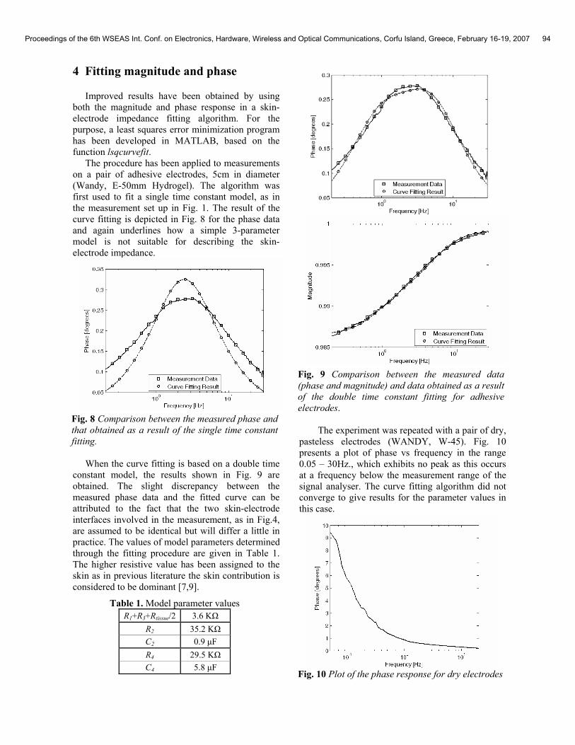
When the curve fitting is based on a double time
constant model, the results shown in Fig. 9 are obtained. The slight discrepancy between the measured phase data and the fitted curve can be attributed to the fact that the two skin-electrode interfaces involved in the measurement, as in Fig.4, are assumed to be identical but will differ a little in practice. The values of model parameters determined through the fitting procedure are given in Table 1. The higher resistive value has been assigned to the skin as in previous literature the skin contribution is considered to be dominant [7,9].
Table 1. Model parameter values R1+R3+Rtissue/2 3.6 KΩ
R2 35.2 KΩ C2 0.9 µF R4 29.5 KΩ C4 5.8 µF
Fig. 9 Comparison between the measured data (phase and magnitude) and data obtained as a result of the double time constant fitting for adhesive electrodes.
The experiment was repeated with a pair of dry,
pasteless electrodes (WANDY, W-45). Fig. 10 presents a plot of phase vs frequency in the range 0.05 – 30Hz., which exhibits no peak as this occurs at a frequency below the measurement range of the signal analyser. The curve fitting algorithm did not converge to give results for the parameter values in this case.
Fig. 10 Plot of the phase response for dry electrodes
Proceedings of the 6th WSEAS Int. Conf. on Electronics, Hardware, Wireless and Optical Communications, Corfu Island, Greece, February 16-19, 2007 94
5 Discussion
Different approaches have been undertaken to model the skin-electrode interface. Experiences with self-adhesive electrodes show that an early model that describes the interface as a single time constant RC network is inadequate. A model involving two time constants proves more accurate. Based on this model, an algorithm has been implemented to identify the parameters of any double time constant system whose phase displays a double peak. Simulations returned highly accurate results when the two time constants forming the system are in a ratio greater than 10 to 1. The method reaches its limits, however, when the time constants are close to each other and the difference in the phase of the two peaks in the response is too small to be accurately measured by the instruments available.
A technique involving simultaneous fitting of the magnitude and phase responses returned satisfactory results when the double time constant model was used. However, this technique did not converge to give practical results when used with dry electrodes. Work is continuing to try to employ time-domain measurements to obtain parameter values for pasteless electrodes.
The information obtained with self-adhesive electrodes can be used to design the front end of the ECG amplifier. Recommendations published by the American Heart Association require a phase shift less than the phase introduced by an analog 0.05Hz single-pole high pass filter in order to preserve the T-wave and ST segments of the ECG waveform [10]. With 1µF input capacitors added to block the dc polarization voltage, the minimum differential input impedance necessary to fulfill this criterion can be shown to be given by:
( )37inC1
2
4R
4
2R2*
05.0*21
Rin
++→
ττπ
For the model parameter values given in Table 1, a minimum input impedance of 14MΩ is required to fulfil the requirements.
[2] Toazza A.L., Mendes de Azevedo F., Neto, J.M.; Microcontrolled system for measuring skin/electrode impedance in biomedical recordings; Proceedings of the 2nd IEEE International Conference on Devices, Circuits and Systems, pp. 278-281, 1998.
[3] A. Rood, A. Mudigonda, K. Sparks and D.
Rosenbaum; A Novel Dry Electrode for ECG Applications; Orbital Research Inc., 2005.
[4] Yacoub S., Novakov E., Gumery P.-Y., Gondran
C., Siebert E.; Noise analysis of NASICON ceramic dry electrodes; Engineering in Medicine and Biology Society, IEEE 17th Annual Conference , Vol. 2, pp.1553-1554, 1995.
[5] I. Zepeda-Carapia, A. Marquez-espionaza, C.
Alvarado-Serrano; Measurement of the skin-electrode impedance for a 12-lead electrocardiogram; 2nd International Conference on Electronics Engineering and XI Conference on Electrical Engineering, 2005.
[6] Swanson D.K., Webster J.G.; A model for skin-
electrode impedance, In: Biomedical Electrode Technology - Theory and Practice, pp.117-128, Academic Press, 1974.
[7] Kaczmarek K.A., Webster J.G.; Voltage-current
characteristics of the electrotactile skin-electrode interface; Proceedings of Annual International Conference of the IEEE Engineering in Medicine and Biology Society, Vol. 5, pp.1526-1527, 1989.
[8] Neuman M. R.; Biopotential Electrodes, In:
Medical Instrumentation - Application and Design, 3rd ed., pp. 183-232, John Wiley & Sons, 1998.
[9] Gatzke R.D.; The electrode: A measurement
system viewpoint, In: Biomedical Electrode Technology-Theory and Practice, pp.99-116, Academic Press, 1974.
[10] Bailey J.J., Berson A.S., Garson A. J. R., Horan
L.G., Macfarlane P.W., Mortarad W., Zywietz C.; Recommendations for standardization and specifications in automated electrocardiography: bandwidth and digital signal processing; Circulation, Vol. 81, No. 2, pp. 730-739, 1990.
Acknowledgment: This work was partially funded by an IRCSET Award, Gov. of Rep. of Ireland.
Proceedings of the 6th WSEAS Int. Conf. on Electronics, Hardware, Wireless and Optical Communications, Corfu Island, Greece, February 16-19, 2007 95