+ All Categories
Home > Documents > Development of a national knee ligament registry

Development of a national knee ligament registry

Date post: 22-Oct-2021
Category:
Upload: others
View: 2 times
Download: 0 times
Share this document with a friend
137
Orthopaedic Center Division of Neuroscience and Musculoskeletal Medicine, Ullevaal university Hospital Faculty of Medicine University of Oslo Development of a national knee ligament registry Lars-Petter Granan Oslo, June 15 th , 2009
Transcript

Orthopaedic Center Division of Neuroscience and Musculoskeletal

Medicine, Ullevaal university Hospital Faculty of Medicine University of Oslo

Development of a

national knee ligament registry

Lars-Petter Granan

Oslo, June 15th, 2009

2

This thesis is dedicated to my girlfriend and best friend Maren,

and our three children Frida, Lukas and Aksel.

“Those of you who say it can’t be done should not interrupt those of us who are doing it.”

George Bernard Shaw

3

Acknowledgements The path from no or only a vague idea to a substantiated paper is amazingly long. Many

have helped me along the way. I am particularly grateful for the help I have received

from my two advisors Lars Engebretsen and Roald Bahr. Lars, thank you for your

generosity, your inexplicable believe in me, your seemingly unlimited working capacity

and your high ambitions. You have been an invaluable inspiration for me. Roald, thank

you for sharing your never failing ability to draw the important distinctions, and your

impeccable corrections of my manuscripts.

I have had the good fortune of working at the Oslo Sports Trauma Center, Norwegian

School of Sport Sciences. Gunnar Breivik, Arne Ekeland, Anders Hauge Engebretsen,

Tonje Wåle Flørenes, Lena Klasson Heggebø, Ingar Holme, Unni Lund (deceased), Lene

Røe, Kristoffer Solumshengslet, Kathrin Steffen, Truls Martin Straume-Næsheim, Steinar

Sulheim and Karianne Ytterstad – you have all been a pleasure to work with, and you

have all contributed to this project. Thanks also to Jarle Breivik, Marie Eikrem, John-

Arne Røttingen and Else-Marie Siebke at the Medical Student Research Program for

making it possible to realize my unsteady dream about research.

I would also like to thank my wonderful colleagues at the NKLR and NAR in Bergen

Birgitte Espehaug, Knut Fjeldsgård, Ove Nord Furnes, Merete Husøy, Stein Atle Lie,

Stein Håkon Låstad Lygre, Torbjørn Strand, Ruth Gunvor Wasmuth and Marianne

Wiese. Without you the NKLR would have been worthless.

I would also like to pay a special tribute to Tron Krosshaug (OSTRC), Kjersti Steindal

(NKLR) and Tone Øritsland (OSTRC). Without you this project would have failed due to

my incompetence with computers and administrative work.

Magnus Forssblad and Martin Lind, my co-authors, I give you my sincere gratitude for

being patient in my, always delayed, work on the Scandinavian ACL registries, and for

your crucial contributions on the statistics and writing.

Robert Magnussen and the entire MOON staff11

, thank you for helping me out with the

last article and thus making it possible to finish my thesis.

Alan Chan and Stine S. Westby, thank you for giving me feedback on the entire

manuscript in my writing process.

Ragnhild Ollendorf, thank you for nourishing my first love to the musculoskeletal

medicine.

Finally I would like to express my sincere gratitude to everyone who has made this thesis

possible and to friends, fellow students and colleagues who apparently find my work

interesting, and thus inspire me further.

1 Annunziato Amendola, Jack T Andrish, Robert Brophy, James L Carey, Warren R Dunn, David Flanigan,

Laura J Huston, Morgan Jones, Christopher C Kaeding, Eric C McCarty, Robert G Marx, Matthew J

Matava, Richard D Parker, Kurt P Spindler, Armando Vidal, Michelle Wolcott, Brian R Wolf, Rick W

Wright.

4

This project was carried out at the Oslo Sports Trauma Center, Norwegian School of

Sport Sciences, Norway, in the period of 2003-2009. The support and working facilities

provided for this project at this institution are greatly appreciated.

This project was mainly financed by the Oslo Sports Trauma Research Center, which has

been established at the Norwegian School of Sport Sciences through generous grants

from the Royal Norwegian Ministry of Culture, the South-Eastern Norway Regional

Health Authority, the Norwegian Olympic Committee & Confederation of Sport, and

Norsk Tipping AS.

Parts of this project were financed through a grant from the Norwegian Medical

Association's Fund for Quality Improvement.

π

5

Contents Acknowledgements p. 3

Contents p. 5

Abbreviations and Glossary p. 7

Papers included p. 8

Introduction part 1 – The ACL p. 9

Epidemiology p. 9

Embryology p. 9

Anatomy p. 9

Biomechanics p. 12

Injury mechanisms p. 13

Diagnosis p. 13

Prognosis p. 14

Indications for treatment p. 16

Non-operative treatment of ACL tears in adults p. 17

Timing of ACL reconstruction p. 18

Introduction part 2 – The NKLR p. 19

Background p. 19

Objectives p. 20

Design p. 20

Classification, coding and data systems p. 21

End points p. 21

KOOS p. 22

Selection of a minimal set of necessary data p. 22

Collection of data p. 23

Research and information p. 24

Data protection, patient identification and ethics p. 24

Staff and budget p. 25

Scandinavian cooperation p. 25

Aims of this thesis p. 26

Subjects, Materials and Methods p. 27

Statistical methods p. 28

Results and Discussions p. 30

Results p. 30

General discussion p. 32

RCTs versus observational studies p. 32

Why establish a registry p. 33

Purposes of a registry p. 34

Limitations and Strengths of the NKLR p. 36

Subjective end points p. 39

Specific discussion p. 40

Future perspectives p. 44

Summary of thesis p. 45

References p. 46

Appendix p. 60

6

A1. Pre operative KOOS form p. 61

A2. Post operative KOOS form p. 65

A3. Registration form p. 70

Papers p. 72

Paper I p. 73

Paper II p. 82

Paper III p. 92

Paper IV p. 108

7

Abbreviations and Glossary

ACL Anterior Cruciate Ligament

ACLR Anterior Cruciate Ligament Reconstruction

ACL-RSI ACL Return to Sport after Injury scale

ADL Activities of Daily Living

AM Anteromedial

CI Confidence Interval

EMG Electromyography

ICRS International Cartilage Repair Society

IKDC 2000 International Knee Documentation Committee

KOOS Knee injury and Osteoarthritis Outcome Score

KOS-ADLS Knee Outcome Survey – Activities of Daily Living Scale

LCL Lateral Collateral Ligament

LFC Lateral Femoral Condyle

MCL Medial Collateral Ligament

MOON Multicenter Orthopaedic Outcomes Network (United States)

MRI Magnetic Resonance Imaging

NAR Norwegian Arthroplasty Register

NKLR National Knee Ligament Registry

NOA Norwegian Orthopaedic Association

NPR Norwegian Patient Register (Norsk pasientregister)

OA Osteoarthritis

OSTRC Oslo Sports Trauma Research Center

PCL Posterior Cruciate Ligament

PL Posterolateral

PLC Posterolateral Corner

QOL Quality of Life

RCT Randomized Controlled Trial

WOMAC Western Ontario and McMaster Universities Osteoarthritis Index

8

Papers included This dissertation is based on the following original research papers, which are referred to

in the text by their Roman numerals:

Paper I

Granan LP, Bahr R, Steindal K, Furnes O, Engebretsen L. Development of a national

cruciate ligament surgery registry: the Norwegian National Knee Ligament Registry. Am

J Sports Med 2008 Feb;36(2):308-15.

Paper II

Granan LP, Bahr R, Lie SA, Engebretsen L. Timing of anterior cruciate ligament

reconstructive surgery and risk of cartilage lesions and meniscal tears: a cohort study

based on the Norwegian National Knee Ligament Registry. Am J Sports Med 2009

May;37(5):955-61.

Paper III

Granan LP, Lind M, Forssblad M, Engebretsen L. The Scandinavian ACL registries

2004-2007: baseline epidemiology. (Accepted to Acta Orthopaedica)

Paper IV

Magnussen RA, Granan LP, Dunn WR, Amendola A, Andrish JT, Brophy R, Carey JL,

Flanigan D, Huston LJ, Jones M, Kaeding CC, McCarty EC, Marx RG, Matava MJ,

Parker RD, Vidal A, Wolcott M, Wolf BR, Wright RW, Spindler K, Engebretsen L.

Cross-cultural Comparison of Patients Undergoing ACL Reconstruction in the United

States and Norway. (Submitted to Knee Surgery, Sports Traumatology, Arthroscopy

(KSSTA))

9

Introduction part 1 – The ACL

Epidemiology

From epidemiological studies we know that 10-19% of acute injuries seen by a

Scandinavian doctor in the emergency room are sustained during sporting activity

(Arendt and Dick 1995, Bahr et al 2003). Sports participation is the cause in one out of

three hospital treated injuries among children (Bahr et al 2003), and in general three out

of four acute knee injuries are sports related (Frobell et al 2007). Serious knee injuries

(e.g. ACL injuries) are of particular concern (de Loes 1990, Ytterstad 1996). Pivoting

sport athletes (e.g. football, basketball, soccer, handball) aged 15-25 years have the

highest incidence of ACL injuries, and the incidence is at least tripled in females

(Myklebust et al 1997, Myklebust et al 1998). A recent publication by Frobell et al

(2007) described an incidence of ACL injuries of 81 per 100,000 inhabitants aged 10-64

years. Data from the NKLR revealed that the annual population incidence of primary

ACL reconstruction surgeries was 34 per 100,000 citizens, while the incidence in the 16-

39 year age group was 85 per 100,000 citizens (Granan et al 2004).

Embryology

The knee starts to form from a mesenchymal concentration in the fourth week of

gestation. This formation process is rapid, and within the sixth week a recognizable knee

joint is apparent (Reiman and Jackson 1987). The ACL appears as a condensation in the

blastoma at approximately 6.5 weeks (Ellison and Berg 1985), and is well developed by

the ninth week (Merida-Velasco et al 1997). After that the ACL will continue to grow,

but no major organizational or compositional changes occur after this point (Gardner and

O’Rahilly 1968). It begins as a ventral ligament and gradually invaginates with the

formation of the intercondylar space. It appears well before joint cavitation and remains

extra synovial at all times. Although it changes very little to achieve its final form, it does

migrate posteriorly.

Anatomy

The ACL is an intra articular structure that traverses the knee joint attaching to the tibia

and femur. The ACL is composed of collagen tissue arranged as longitudinally oriented

fibrils ranging from 20 to 170 µm in diameter (Baek et al 1998). Both the cross sectional

area and collagen fibrils’ diameters are largest in the distal region and decreases as it

moves more proximally (Baek et al 1998). The relative amount of collagen in relation to

the cross sectional area does not vary significantly between the regions in the ligament

(Baek 1998). Collagen fibrils are organized into bundles which make up subfascicular

units. These units are surrounded by a thin sheath of loose connective tissue, the

endotenon. Bundles of subfasciculi make up a collagen fasciculus, which again is

surrounded by an epitenon – a much denser connective tissue than the endotenon. The

entire ligament is surrounded by a paratenon, which blends in with the epitenon

(Danylchuk et al 1978, Arnoczky 1983). On a histological level the ligament is composed

of fibroblasts which are surrounded by a matrix consisting mainly of type I collagen, and

near the insertion sites with additions of small amounts of type III and type VI collagen

(Amiel et al 1984, Baek et al 1998).

10

The ACL is usually described as consisting of two discrete bundles (Duthon et al 2006,

Giron et al 2006), the AM and PL, the nomenclature corresponds to their anatomical

insertion site on the tibia. On the femoral side, the AM bundle originates more proximally

and the PL bundle originates more distally with the leg in extension (Chhabra et al 2006,

Steckel et al 2007). The divisions are based on the fiber orientation and tensioning

characteristics during flexion and extension (Girgis et al 1975, Furman et al 1976,

Arnoczky 1983, Harner et al 1999). Norwood and Cross (1977) suggested the presence of

a third bundle, the intermediate, this has later been supported by Amis and Dawkins

(1991), and Hollis et al (1991). Other authors describe the ACL as one continuous

structure (Welsh 1980, Odensten and Gillquist 1985). The conflicting results of studies of

the (adult) ACL can be at least partially explained by the variation in bundle orientation

in different degrees of knee flexion, making it difficult to visualize both bundles in any

single transverse histological section (Feretti et al 2007). In addition, the vascular

connective tissue flows into and out of the ligament, further complicating the anatomic

picture (Feretti et al 2007).

The bundles relative size have varied across various studies, but Girgis et al (1975),

Harner et al (1999) and Palmer (1938) found no significant difference between the two

bundles. Disregarding the discrepancies in anatomic description, and considering a view

from a functional standpoint, the ACL demonstrates varying tensile characteristics across

its width. Edwards et al (1999) demonstrated the following elongation behavior of the

ACL bundles: The strain pattern of the intact ACL’s AM bundle exhibited an initial

shortening from 0° to approximately 30° of flexion, followed by lengthening from 30°-

120° of flexion, and thus reaching its maximal strain at 120°. Net lengthening from the

baseline length at 0° was only observed in the range of approximately 70°-120° of

flexion. The strain pattern of the intact ACL’s PL bundle exhibited shortening over the

entire range (0°-120°) of flexion, and reaching minimal strain at full flexion. The PL

bundle shortened approximately two thirds of its maximal length from 0°-50° of flexion.

The relative position of the AM bundle and the PL bundle varies with the flexion of the

knee. In extension, the two bundles are parallel (Chhabra et al 2006). In flexion, the

femoral insertion site of the PL bundle moves anteriorly, and the two bundles are crossed

(Chhabra et al 2006). In flexion, the AM bundle tightens as the PL bundle loosens (Amis

and Dawkins 1991). In extension, the PL bundle tightens and the AM bundle loosens

(Amis and Dawkins 1991). The PL bundle tightens during internal and external rotation

of the knee (Chhabra et al 2006).

The insertion sites of the ACL are marked by a gradual transition, within a depth of less

than one millimeter, of relatively flexible ligament tissue that merges into rigid bone.

Electron and light microscopic evaluation of this region performed by Cooper and Misol

(1970) described four morphologic zones, each with clearly defined characteristics. Zone

1 is the ligament tissue, primarily made up of collagen; zone 2 represents a mix of

collagen blended with unmineralized fibrocartilage; zone 3 contains mineralized

fibrocartilage; and zone 4 represents the subchondral bone. This anatomy is thought to be

mechanically advantageous, allowing for force dissipation and having implications with

regard to ligament failure modes (Cooper and Misol 1970, Noyes et al 1974).

The ACL fascicles course in a spiral rotation and are described as fanning out as they

approach their tibial insertion (Smith et al 1993). They traverse from the femoral

11

attatchment in a distal, anterior, and medial direction (Girgis et al 1975, Arnoczky 1983).

Although the bundles were indeed parallel in extension, flexion of the joint resulted in a

crossing of the PL bundle over the AM bundle (Girgis et al 1975, Arnoczky 1983). The

reported average length of the ACL ranges from 22 mm to 41 mm, with a mean of 32 mm

(Dienst et al 2002).

The ACL has attachment sites on both the femur and tibia. The femoral insertion site, a

circular area encompassing averagely 113 mm² (Harner 1999), originates from the

posteromedial aspect of the intercondylar notch on the lateral femoral condyle. Steiner et

al (2008) published a review were he consolidated the findings of Girgis et al (1975),

Odensten and Gillquist (1985), Colombet et al (2006), Mochizuki et al (2006) and

Heming et al (2007) and emphasizing the more recent data, found that the ACL femoral

insertion has an approximate average length of 18 mm, width of 10 mm, and a separation

of up to 4 mm from the articular cartilage. The tibial insertion site, an oval area

encompassing averagely 136 mm² (Harner et al 1999), is on the intercondylar eminence

of the tibia but does not attach to either the medial or lateral tubercles of the intercondylar

eminence (Girgis et al 1975, Morgan et al 1995, Heming et al 2007). From more recent

studies there have been reported that the tibial insertion lengths varies from 15 to 18.5

mm and widths from 10 to 13 mm (Odensten and Gillquist 1985, Morgan et al 1995,

Heming et al 2007), giving an oval attachment with an approximate length of 18 mm and

approximate width of 10 mm as a consolidated overall result (Steiner et al 2008).

Although general agreement exists on the size and shape of the tibial insertion, there is

still debate over best method to identify its anterior and posterior boundaries (Steiner et al

2008). Both insertion sites are more than 3.5 times larger than the cross sectional area of

the ligament midsubstance, which measure averagely just below 40 mm² (Harner et al

1999).

Both the cruciate ligaments are covered by a synovial fold that originates at the

intercondylar notch’s posterior inlet and extends to the ACL’s anterior tibial attachment

site, resulting in the cruciate ligaments being both intra articular and extra synovial at the

same time (Arnoczky 1983). The ACL is mainly supplied by vessels originating from the

middle genicular artery which leaves the popliteal artery and directly pierces the posterior

capsule (Arnoczky 1985). Branches, that form a periligamentous plexus, enter the

synovial membrane at the junction of the joint capsule distal to the infrapatellar fat pad

(Ellison and Berg 1985, Kennedy et al 1974). The ACL is surrounded by a synovial

plexus along its entire length. Smaller, connecting branches penetrate the ligament and

anastomose with a network of endoligamentous vessels that are oriented in a longitudinal

direction and lie parallel to the collagen bundles within the ligament (Arnoczky 1983,

Ellison and Berg 1985).

The majority of neural structures have been found in the subsynovial layer and near the

insertions of the ACL (Reiman and Jackson 1987). The posterior articular nerve, a branch

of the tibial nerve, is the major neurobundle (Kennedy et al 1974); it arises from the tibial

nerve in the popliteal fossa, wraps around the popliteal artery and vein, penetrates the

posterior capsule, and forms the popliteal plexus. Branches from this plexus course

through the synovial lining of the cruciate ligaments, follow the course of the blood

vessels, and extend as anterior as the infrapatellar fat pad. Neurotracer studies performed

by Hogervorst and Brand (1998) revealed the existence of very few receptors in the ACL.

12

Further studies by Krauspe et al (1995) narrowed it down to a maximum of 17. These

numbers decrease with age and disease. The receptors found are primarily Ruffini

receptors and free nerve endings that are thought to function as stretch receptors and

nociceptors, respectively (Hogervorst and Brand 1998). Free nerve endings may also

serve as local effectors by releasing neuropeptides with vasoactive function, thus having

modulator effect in normal tissue homeostasis or in remodeling of grafts (Hogervorst and

Brand 1998). In 1998 Hogervorst and Brand (1998) stated that convincing evidence on

the direct effects of mechanoreceptors of the ACL on electromyographic activity of

muscles surrounding the knee was missing. Later it has been demonstrated that low

intensity electrical stimulation of the intact ACL in humans could induce clear cut

excitations or inhibitions of the isometrically contracting quadriceps and semitendinosus

muscles (Dyhre-Poulsen and Krogsgaard 2000). Even so, Sjölander et al (2002)

concluded in a review that to what extent the EMG effects are mediated through

interneuronal pathways directly to the skeletomotoneurones, or indirectly via reflex

actions on the γ-muscle spindle system, is not possible to elucidate using only EMG

registration technique.

Biomechanics

The ACL functions as the primary restraint to anterior translation of the tibia in relation

to the femur (Butler et al 1980, Fukubayashi et al 1982) and as a secondary restraint to

internal-external rotation, varus-valgus angulation and combinations thereof (Markolf et

al 1976, Markolf et al 1990, Marder et al 1991, Markolf et al 1995, Fleming et al 2001,

Kanamori et al 2002). The ACL has an average cross sectional area of 44 mm². The

ultimate tensile load measured in young ACLs (22 to 35 years) is 2160 ± 157 N and a

stiffness of 242 ± 28 N/mm, while for older ACLs (60 to 97 years) the numbers are 658 ±

129 N and 180 ± 25 N/mm, respectively (Woo et al 1991). Normal daily loads on the

ACL are estimated to approximately 20% of its load capacity (Frank and Jackson 1997,

Martelli et al 1998). Age is an important factor in the strength aspect of the ACL; older

ACLs fail under lower loads than younger ACLs do (Woo et al 1991). The forces in the

intact ACL range from approximately 100 N during passive knee extension (Markolf et al

1996) to about 400 N with walking and reach, and up to 1700 N with cutting and

acceleration-deceleration activities (Butler et al 1985, Nogalski and Bach 1994).

Beynnon and Fleming (1998) presented a review of in vivo strain patterns in the intact

ACL during a variety of activities and exercises. The most crucial variables influencing

ACL strain were the knee position and the dynamic interaction of muscle activity.

Increased strain was seen with increasing knee extension. Activities that produced

isolated quadriceps activity led to the highest ACL strain, contrary to the isolated

hamstring activity that produced the lowest levels of ACL strain. Co contraction of the

hamstrings during closed chain extension activities provided a moderating effect to the

strain produced by isolated quadriceps activity seen in open chain activities.

Nevertheless, in the real patient, the ACL probably carries loads approaching its failure

capacity only during relatively unusual combinations of loading of the knee by external

forces or muscles (Frank and Jackson 1997).

13

Injury mechanisms

According to Bahr and Krosshaug (2005) injury mechanisms is poorly defined in the

literature. They propose a comprehensive model that includes information on different

levels when an injury occurs. Their model includes a full description of the mechanisms

for a particular injury type in a given sport. In such a model the inciting event of an injury

could be grouped into four categories and described as: 1) vital aspects of the

playing/sports situation; 2) athlete and opponent behavior; 3) gross biomechanical

characteristics (whole body) and 4) detailed biomechanical characteristics (joint/tissue).

Further more they conclude that until a complete description is available which includes

information on all causative factors, it may be difficult to predict which factors are the

most suitable to influence through interventions.

Independent of the model proposed by Bahr and Krosshaug (2005) one may divide injury

mechanisms into contact and non contact maneuvers. Contact type mechanisms are fairly

easy to understand and interpret since the blow to the knee (often from the side or front)

usually is obvious. These mechanisms often produce injuries to multiple structures in the

knee joint. The motion sequence of non-contact injury is usually a sharp deceleration

associated with or without a change of direction; landing on one or two legs;

hyperextension of the knee; sharp, pivoting motion of the body around a planted leg or

varus collapse of the knee (Boden et al 2000). A review by Quatman and Hewett (2009)

concluded that non-contact ACL injuries almost certainly occur during complex,

multiplanar knee joint load states during multiplanar sports movements.

Diagnosis

The Lachman test (Torg et al 1976) is the most sensitive and specific clinical

investigation to diagnose pathological anterior tibial translation in ACL instability

(Scholten et al 2003). In a recent meta-analysis by Crawford et al (2007) the pooled

sensitivity and specificity of the Lachman test were 85% and 94%, respectively. For the

Pivot shift test the specificity was 98%, but the sensitivity only 24% (Crawford et al

2007). But a recent study by Frobell et al (2007) stated that the lack of agreement

between clinical instability – measured as anterior-posterior knee laxity by the Lachman

test and/or the drawer sign – and MRI findings indicates that a large group of patients

with severe knee injuries may not be diagnosed correctly, and thus not receives optimal

treatment. MRI is accurate for discriminating complete ACL graft tear from partial

thickness tear and intact graft (Horton et al 2000). According to a review by Sandberg et

al (1987), MRI had a sensitivity of 86%, a specificity of 95%, and an accuracy of 93% for

an ACL tear, as confirmed by arthroscopy. Thus, it is important to remember that the

MRI is inaccurate in nearly 10% of the cases, and for these patients the examiners’

clinical experience is crucial After all, ACL rupture is a clinical diagnosis and experience

indicates that the Pivot shift test is the best, performed by an experienced examiner. The

pivot shift motion is the major symptom in ACL insufficiency. It represents a sub-

luxation of the lateral femoral condyle behind the lateral tibial plateau, caused by an

anterior rotation of the lateral tibia (i.e. internal rotation of the tibia). Finally, it is claimed

by Frobell et al (2007) that every second patient with an acute ACL injury risks being

sent home from the orthopaedic emergency unit diagnosed as an uncomplicated knee

sprain, if not further assessed by MRI in the sub acute phase.

14

ACL tears rarely occur in isolation but are in at least 50% of the cases associated with

other ligament sprains, meniscus tears, articular cartilage injuries, bone bruises, and

sometimes intra-articular fractures (Beynnon et al 2005a, Beynnon et al 2005b). In

addition to the acute events associated with a joint trauma, the lack of a functionally

normal ACL or meniscus will change the static and dynamic loading of the knee,

generating increased forces on the cartilage and other joint structures (Dye 1996,

Andriacchi et al 2004, Song et al 2006). As a result, additional lesions commonly occur

(or become symptomatic) with time in the ACL injured knee and, in particular, in the

meniscus (Dunn et al 2004, Fithian et al 2005, Meunier et al 2007).

To prevent missing posterior rotational instability of the lateral compartment, due to

failure of the posterolateral corner, which may result in recurrent instability after ACL

reconstruction (Carson et al 2004) the following standard knee investigation is

recommended (Krogsgaard 2007):

• Tests for varus and valgus stability (with 0º and 30º of flexion)

• The Lachman test

• The anterior and posterior drawer test

• The Pivot shift test (not in acute cases)

• The posterior sag test

• The external rotation test (in 30º, 60º and 90º of flexion)

Prognosis

The natural history of the ACL deficient knee has not been characterized by a well

designed prospective cohort study, leaving us with uncertainty when describing the

complete natural history of this injury. Although the majority of patients cannot return to

high level activities after ACL injury because of continual episodes of knee giving way

(non copers) (Eastlack et al 1999), a small percentage make a full, asymptomatic return to

all pre injury activities (copers) without surgery (Daniel et al 1994, Eastlack et al 1999).

Nevertheless, a common consensus exist that ACL injury can result in long term absence

from work and sports, and dramatically increases the risk of long term sequelae such as

abnormal joint dynamics and early onset of degenerative joint disease, i.e. OA (Beynnon

et al 2005a, Myklebust and Bahr 2005, Roos 2005, Thelin et al 2006). Despite massive

research efforts, there is still lacking evidence to suggest that reconstructive surgery of

either menisci or cruciate ligaments decreases the rate of post traumatic OA (Beynnon et

al 2005a, Myklebust and Bahr 2005). The reported rates of radiographic signs of OA after

an ACL injury vary between 10% and 90% at 10 to 20 years after the ACL injury,

regardless of treatment choice (Gillquist and Messner 1999, Lohmander and Roos 2004,

Lohmander et al 2004, Beynnon et al 2005a, Myklebust and Bahr 2005, Shelbourne and

Gray 2009). Stating a mean OA rate is difficult because of the great variability of the

reported results, but an overall long-term mean of more than 50% may be suggested

(Lohmander et al 2007). A recent systematic review by Øiestad et al (2009) found that

previously reported prevalence rates of knee OA at a minimum of 10 years after ACLR

have been too high. They found a 0-13% prevalence rate of knee OA in individuals with

isolated ACL injuries, and a prevalence rate of 21-48% in individuals with combined

15

injuries. Roos et al (1995) have previously shown that subjects with ACL injury and

posttraumatic OA are, on average, 15 to 20 years younger than patients with primary OA

when they seek medical advice for their symptoms and when their joints show

radiographic evidence of OA.

In line with previous studies (Butler et al 1980, DeHaven 1980, Noyes et al 1980) Frobell

et al (2007) found that at least every second ACL injury is associated with a meniscal

injury. Despite this a recent paper by Drogset et al (2006) suggested that early surgical

intervention would be beneficial since the knees at an early stage had less cartilage

damage compared to knees with late surgery. At 16 years follow up only 11% of the

patients that had early surgical intervention had developed OA, while 4% had developed

OA in the contra lateral knee. It is important to emphasize that only 50% of these patients

had been through radiographic investigations. Furthermore, they had a low activity level

compared to those with high activity level and pivoting activities which had

approximately 50% OA. Nevertheless, the systematic review by Øiestad et al (2009)

found that meniscal injuries and meniscectomy are well-documented risk factors for the

development of knee OA at a minimum of 10 years after ACL injury. They also found

that factors such as ACLR, age, obesity, knee joint laxity, performance on hop tests, and

loss of knee extension are still not sufficiently documented to be considered as risk

factors. Eitzen and colleagues (2009a) have in a prospective cohort study documented

that two years after surgery, individuals with preoperative quadriceps muscle strength

deficits on the injured side above 20% still have abnormal muscular asymmetry. A recent

study by Meuffels et al (2009) found similar performance between conservatively treated

and surgically treated high level athletes at 10 years after ACL injury. Except for higher

objectively measurable instability for the conservative group, there were no statistical

difference with respect to OA, meniscal lesions, activity level, and objective and

subjective functional outcome. Neuman et al (2008) found that in patients with ACL

injury willing to moderate their activity level, initial treatment without ACLR should be

considered due to favorable long-term outcome regarding incidence of radiographic knee

OA, knee function and symptoms, and need for ACLR. Chaudhari et al (2009) found that

there are anthropometric differences between the knees of subjects with a non contact

ACL injury and those without an ACL injury, and suggests that ACL volume may play a

direct role in non contact ACL injury. Posthumus et al (2009) found an association

between ACL ruptures in an independent population and a specific genetic

polymorphism. And propose that this sequence variant be the first specific genetic

element to be included in multifactorial models developed to understand the etiology and

risk factors for ACL rupture. A recent study by Lohmander et al (2007) found that the

KOOS (see Part 2 for details) subscales Function in Sport and Recreation and knee

related QOL showed marked changes over time, suggesting that monitored by valid

patient-administered outcome measures, results of ACL rehabilitation and/or surgery are

at best at 1 to 2 years of follow-up and then gradually deteriorate over time.

Recent studies show that it may be possible to reduce the incidence of knee injuries by

using various training programs (Hewett et al 1999, Wedderkopp et al 1999, Heidt et al

2000, Junge et al 2002, Myklebust et al 2003, Mandelbaum et al 2005, Olsen et al 2005,

Gilchrist et al 2008, Pasanen et al 2008, Soligard et al 2008). However it is not known

which program component is the key ingredient in preventing knee injuries or how they

work (Bahr and Krosshaug 2005). An interesting finding is presented by Liederbach et al

16

(2008), they found a low incidence of ACL injuries among elite ballet and modern

dancers compared with published literature on athletes from other sports that involve

jumping movements, and no significant difference was found between genders. This may

be due to emphasize on lower extremity alignment, and jump and balance training

(Liederbach et al 2008). At least in part, our ability to target and improve current

prevention programmes is limited by an incomplete understanding of the causes of

injuries (Bahr and Krosshaug 2005). Nevertheless, Quatman and Hewett (2009) states

that according to the biomechanics of non-contact ACL injuries, preventive multiplanar

training exercises should focus on lowering risky biomechanics in multiple planes such as

large knee valgus, internal/external knee rotations and shallow knee flexion angles. They

also propose that sex-specific mechanisms of ACL injury may occur.

Indications for treatment

Beynnon et al (2005a) recently stated that because the true natural history of the ACL

deficient knee and consequently the ultimate outcome of ACL reconstruction are

unknown, rigid criteria for patient selection for surgical versus non surgical

reconstruction have not been established. The rationale for non surgical treatment

assumes that the ACL deficient knee may function reasonably well under certain

circumstances and that reconstruction does not necessarily prevent the untoward sequela

of OA. Patients with a complete ACL tear may be treated satisfactorily, even with

advanced OA of the involved knee. The rationale for surgical treatment is based on the

observation that the ACL is vital for knee function, that ACL deficient knees frequently

degenerate, and that surgical reconstruction of the ACL can succeed in restoring normal

function. A nearly universally accepted indication for ACL reconstruction is a high risk

lifestyle requiring heavy work, sports, or recreational activity and repeated episodes of

giving way (pivot shift episodes) despite proper rehabilitation. Nevertheless, Spindler and

Wright (2008) categorically states in a clinical practice article in the New England

Journal of Medicine in November 2008 that “surgical treatment is indicated if the patient

has a sensation of instability in normal activities of daily living or wants to resume

activities that involve cutting and pivoting, (…) [or] occupations (…) [that] require an

ACL-stabilized knee.” In 2008 Kostogiannis et al (2008) published a cohort study with

15 years follow up regarding clinically assessed knee joint laxity as a predictor for

ACLR, with patient age ranging from 14 to 41 at the time of injury. They found that a

positive Pivot shift test at 3 months after injury in an awake patient is the strongest

predictor for the future need for ACLR, and that a normal Pivot shift test at 3 months

indicates a low risk for later ACLR and is characteristic for copers. Clinical tests for knee

laxity in the acute phase were not a good predictor of the need for later ACLR. The latter

is also supported by Eitzen et al (2009b). Patients may be classified as rehabilitation

versus early surgical candidates using a screening examination developed at the

University of Delaware (Fitzgerald et al 2000). This classification algorithm was used in

a study (Hurd et al 2008) where patients were categorized as non copers or potential

copers based on giving-way episodes, timed hop, global rating of knee function, and

KOS-ADLS (Irrgang et al 1998) scores. The algorithm uses a group of variables that

collectively capture neuromuscular function and predict patient outcomes, and such

clinical tests may therefore be useful in guiding in individualized patient management

after ACL injury (Hurd et al 2008). A recent article by Swirtun et al (2006) revealed that

17

patients who chose early reconstruction (< 6 months post injury), chose surgical

treatment for reasons based on assumptions of future problems associated with the knee

injury, whereas patients who chose late reconstruction, chose surgical treatment for

reasons based on experience of knee function.

Non operative treatment of ACL tears in adults

When considering the scientific basis for non operative treatment of ACL tears there is

considerable amounts of information based on case series, comparisons of case series and

cross sectional studies, but also the additional problems with bias (i.e. selection bias,

information bias and confounders). Four Swedish RCTs (Odensten et al 1985, Sandberg

et al 1987, Andersson et al 1989, Andersson et al 1991) are available concerning non

operative ACL treatment versus ACL repair or repair with augmentation. Repair is no

longer a preferred surgical treatment due to inferior surgical outcomes. One of the studies

above (Sandberg et al 1987) found no differences in outcomes compared with non

operative treatment. This may be attributed to the use of cast rather than active

rehabilitation. The other three (Odensten et al 1985, Andersson et al 1989, Andersson et

al 1991) found that superior results were achieved in patients with ACL repairs with

augmentation using the iliotibial band, compared with those with repair alone or non

operative treatment. A 15-year follow-up of the Andersson and colleagues 1989-study

revealed rather similar results in both groups regarding OA, knee function, and activity

level, but 1/3 of the non surgically treated subjects underwent later ACL reconstruction

for instability (Meunier et al 2007).

There is general agreement that over the course of years after the trauma, the injured

knee, reconstructed or not, will be submitted to abnormal loading patterns in everyday

activities as well as in sports, significantly increasing the risk of OA (Lohmander et al

2007). A report based on review of an administrative database (The US Army Total

Army Injury and Health Outcomes Database (TAIHOD)) suggested that ACLR in a

young and active population provided some protection against additional procedures,

compared with those not reconstructed (Dunn et al 2004).

To date no RCTs that compare bone-patellar-tendon-bone or multi-strand hamstring

autograft ACL reconstructions with non operative treatment has been published.

Therefore, ultimately the doctor and/or surgeon must evaluate each patient individually

and inform them about all possible advantages and risks of any treatment method

proposed. Some guiding in this process is obtained in a review by Lewek et al (2003),

where an algorithm that includes screening tests to determine which patients with an

ACL injury that may be candidates to non operative treatment is described. Nevertheless,

patients classified as non-copers at the initial examination have substantial potential to

regain dynamic knee stability at one year follow-up, and should not be excluded as

rehabilitation candidates (Moksnes et al 2008b) since it is likely that patients receiving

non-operative treatment have at least as good results as the ACLR one year post-injury

(Moksnes and Risberg 2009). On the other hand we do not know if the copers also have

less cartilage lesions and meniscal tears, only that they are pivoting less.

18

Timing of ACL reconstruction

No consensus exists on the ideal timing for ACL reconstruction. Beynnon et al (2005a)

claim that after reviewing the literature on this subject, it appears that the time interval

from ACL injury to reconstruction is not as important as the condition of the knee at the

time of surgery (i.e. the knee should have a full range of motion with minimal effusion;

the patient should have minimal pain and be mentally prepared for the reconstruction and

rehabilitation after surgery.). And they conclude that there are no absolutes as to when

ACL reconstruction should be performed. Despite this, a retrospective study that claims

that ACL reconstructions should be carried out within 12 months of injury to minimize

the risk of meniscal tears and degenerative change has been published (Church and

Keating 2005). Even so it is important to emphasize, as mentioned above, that Drogset et

al (2006) found only 11% of patients with developed OA at 16 years follow up when

ACL surgery were performed no more than 10 days after injury, even when accompanied

with long post operative immobilization and slow rehabilitation. Papastergiou et al (2007)

recommend that ACLR should be carried out within three months after injury to

minimize the risk of developing secondary meniscal tears. Yoo et al (2009) found in a

case series that ACL deficient knees with or without a medial meniscal lesion can suffer

subsequent damage to the medial meniscus, and this should be expected if ACLR is

delayed beyond six months after initial MRI. In the present thesis, paper II concludes that

early surgery may be recommended, as a consequence of its association with fewer

meniscal tears and cartilage lesions.

19

Introduction part 2 – The NKLR

Background

National quality registries have been used in several medical specialties to improve health

care in Scandinavia (Kjaerheim 1999, Lichtenstein et al 2002, Akesson 2003, Heaf 2004,

Sokka 2004, Kallen 2005, Pahlman et al 2005, Ohm and Derom 2006), including Norway

(Kjaerheim 1999, Irgens 2000, Bergem 2002, Kvien 2003). Because of the inferior

clinical results associated with some hip prosthesis designs in the early 1980s (Havelin

1995), the nationwide Norwegian Hip Arthroplasty Register (NAR) was established in

1987 with implant revision as the main endpoint (Havelin et al 1993). The aim was the

early detection of inferior results caused by implants, cements or surgical techniques

(Havelin et al 2000, Furnes et al 2002). In 1994, the registry was expanded to include all

joint replacements (Havelin et al 2000). In 1995, two papers (Havelin et al 1995a,

Havelin et al 1995b) described the detection of inferior implants at an early stage, a

finding only possible through registry studies.

The NAR is based on a simple reporting system (approximately one minute is required to

complete a single-page registration form) and the hospitals are provided with continuous

feedback from the registry (Havelin et al 2000). These two factors are believed to explain

why the compliance rate of nearly 100% has not declined during 20 years of operation

(Havelin et al 2000, Espehaug et al 2006). Immediately after each operation, the surgeon

completes the registration form, which is mailed to the NAR office (Havelin et al 1993).

Patient identification and the different procedures, including the type of implant and

cement used, are specified on the registration form. Feedback is given as annual national

reports. In addition, each hospital receives a report on its own activities and results,

which can be compared with the national average. A wide range of studies have been

published based on the NAR database (Havelin et al 2000).

In contrast to joint replacement surgery, where national registries have been established

in Norway, Sweden (1975), Finland (1980), Denmark (1995), Australia (1998), Hungary

(1998), New Zealand (1998), Scotland (1999), Canada (2000), Italy (2000), Romania

(2001), England and Wales (2003), Slovakia (2003), and Spain (2004), no national

prospective surveillance system exists for monitoring the outcome of knee ligament

surgery in a predefined population. Evidence from the Scandinavian joint replacement

registries indicates that a national knee ligament registry could be highly beneficial

(Havelin et al 1995a, Havelin et al 1995b, Herberts and Malchau 1997, Malchau et al

2002). First, treatment outcome can be improved through feedback to the hospitals and

surgeons from the registries. Second, there are still several unresolved issues related to

cruciate ligament surgery and postoperative rehabilitation methods. Some of these can

and should be addressed by conducting properly designed RCTs. However, because of

practical, financial or other restraints, such studies are often not possible. Also, some

questions can only be answered by large cohort studies. This can include the detection of

procedures and devices that result in premature failure. Third, a large cohort study can be

used to identify prognostic factors associated with good and poor outcomes.

This background served as the impetus for designing the Norwegian NKLR. A working

group was established with members from NAR and the OSTRC in 2002. The group

designed the registry, constructed forms, performed a pilot study, planned the logistics,

20

and contacted the hospitals. The NKLR is owned by the NOA, and a steering committee

with six members is appointed jointly by NOA and OSTRC. Since the official start on

June 7, 2004, the steering committee has been responsible for the budget, planning,

continuous evaluation of the data set, and for reviewing the results on a regular basis.

Objectives

It is the NKLR’s main intention to contribute to quality control and improvement of the

surgical cruciate ligament procedures. This may be done through establishing evidence

based national guidelines and protocols for surgical procedures and rehabilitation. To

understand the importance of reported failures, we need to know the actual number of

reconstruction and revision surgeries that are performed. NAR has previously provided

accurate data of sufficient quality. The NKLR has calculated that if 14 patients with one

specific fixation device fail, this may be considered a failure of that specific device. This

will enable the NKLR to give early warnings on procedures and devices as well as

identify prognostic factors.

The NKLR is providing information for the orthopaedic community at regular intervals

on the outcomes of surgical treatment of the cruciate ligaments with different methods.

The hard end points are clear and unequivocal, i.e. revision reconstruction and total knee

replacement. Causality of failure may not be sufficiently and accurately documented in

the NKLR, but it will provide information as to where there may be potential problems

and direct future analysis and studies toward these areas. Since the NKLR will provide

real time information and thus can be analyzed on an ongoing basis, it has the potential to

reveal problems long before they would be reported by traditional methods (e.g. RCTs).

This will undoubtedly benefit all interested parties, not at least the patients.

Design

The NKLR is designed to collect information prospectively on all cases of cruciate

ligament reconstruction surgery, and subsequent knee joint surgeries. To be included in

the cohort, a patient should be a resident of Norway undergoing primary or revision

reconstruction surgery for an ACL and/or PCL injury at a Norwegian hospital. In

addition, the NKLR also records all surgical procedures to a knee joint that has

previously undergone primary or revision ACL and/or PCL reconstruction surgery.

Participation is voluntary, and all patients are asked to sign an informed consent form

before surgery. The consent form contains information about the NKLR, the type of

information recorded, data protection, the procedure for follow ups and informs the

patient that he or she may be invited to participate in research projects at a later stage.

The patients are also asked to complete a validated knee outcome score form (Roos et al

1998a, Roos et al 1998b, Roos and Lohmander 2003), the KOOS (appendix 1-4). At any

time, any patient can withdraw their contribution to the NKLR without stating a reason;

previous, present and/or future contributions. Confidentiality is assured for patients and

individual surgeons.

Orthopaedic surgeons report the patient’s social security number on the paper-based

registration form (appendix 5 and 6) to identify the patient. By using this identification,

information on outcomes (revision reconstructions and other knee surgeries) can be

linked to the baseline information (primary cruciate ligament reconstruction surgery)

21

even if later procedures are performed elsewhere. With this kind of system, and when all

hospitals, clinics and orthopaedic surgeons are contributing and where data on patients’

knee arthroplasties, deaths or emigrations are available, the follow up of patients can be

nearly complete.

Classification, coding and data systems

Index side, date of surgery, performed procedures, choice of graft, fixation devices, and

systemic antibiotic prophylaxis are examples of reported information. Cartilage lesions

are graded according to the ICRS (Newsletter ICRS 1998). The NKLR collects separate

information on fixation devices both for grafts and synthetic fixation of meniscal lesions.

All fixation devices have been classified according to the three different variables: inside

or outside of channel; material used (metal, plastic or bioabsorbable); type (e.g.

interference screws, pins). These variables are registered along with the name of the

manufacturer and the products reference number (not the LOT number). As a result of

this detailed information, the results for the different implants can be calculated

separately. To ensure accurate information on the implant, the orthopaedic surgeons use

the unique bar code stickers with the catalogue numbers of the implants supplied by the

manufacturers. The orthopaedic surgeons Torbjørn Strand and Knut Fjeldsgård have

coded the implants in the registry. As of today all coding updates are performed by Knut

Fjeldsgård.

The NKLR’s server is located at Helse Vest IKT AS and is run from there. They are also

responsible for the back ups.

End points

The registry makes use of both objective and subjective end points. Similar to NAR, the

hard end points are revision surgery after cruciate ligament surgery and total knee

replacement. Unlike NAR, the NKLR has included routine follow-ups on all patients at

two, five and ten years postoperatively using KOOS as a soft end point. The follow-up

forms include three additional questions to increase the detection rate of patients with

unreported poor outcomes (see appendix 3 and 4). The KOOS form is completed by the

patients preoperatively and used as the patients individual baseline score. Some surgeons

claim that the patients’ mental status differs on the day of surgery, and that this may be

due to them exaggerating complaints too convince themselves that surgery is the best

choice of treatment. This argument is not supported by Bryant and coworkers (2008),

they found that patients undergoing surgery for the treatment of a chronic knee injury

(e.g. ACL injury) can provide an accurate self-assessment of their quality of life, general

health, and functional status on the day of surgery (Bryant et al 2008). Further on, they

also found that investigators can improve the efficiency of data collection for clinical

studies for chronic knee injured patients, with no expected loss of statistical power, by

obtaining self-assessments on the day of surgery. This finding supports the suggested

routine from the NKLR, to let the patients complete the KOOS on the day of surgery or

the day before.

22

KOOS

The KOOS form is a knee specific instrument, developed to assess patients’ opinion

about their knee and associated problems, and is intended to be used for knee injuries that

can result in post traumatic OA; i.e. ACL injury, meniscus injury, chondral injury, etc. It

is meant to be used over short and long time intervals; to assess the magnitude of change

over time. KOOS can be used to assess groups and to monitor individuals. The

questionnaire, validated for several languages, and a scoring manual are available at the

KOOS website http://www.koos.nu. It includes 42 items in 5 separately scored subscales:

pain (9 items), other symptoms (7 items), function in ADL (17 items), function in Sport

and Recreation (5 items), and knee related QOL (4 items). Each item is responded to by

marking one of five response options on a Likert scale. The WOMAC OA Index LK 3.0

(Bellamy et al 1988) items are included in the first three KOOS subscales. The KOOS

form will at the time for follow-ups be dispatched from the NKLR secretariat.

The KOOS form did not exist in a Norwegian translation when the preliminary work on

the registry was performed. Since the KOOS form already existed in English and

Swedish two separate translations into Norwegian were arranged; one of the authors did

one translation from English to Norwegian, and the translation from Swedish were done

by a former researcher at the Norwegian School of Sport Sciences, who is bilingual in

both Norwegian and Swedish. The translations were compared, and due to only minor

differences in the use of synonyms the NKLR chose a wording as close to the Swedish

translation as possible. This is due to the fact that the creators of the KOOS form are

Swedes, even though the first form was made in English. This procedure is not satisfying

as a validation process (Guillemin 1993).

Later on the KOOS form has been validated by researchers at NAR. KOOS was

translated from the Swedish version by two orthopaedic researchers. The choice of using

the Swedish version was based on the assumption that cultural differences between the

two neighbor countries would be minimal due to similarities in language and lifestyle.

The translation was checked by two bilingual orthopaedic surgeons (Swedes with

permanent address in Norway). The form was tested on knee arthroplasty patients to

clarify potential misinterpretations. Then the NAR and the NKLR versions were

compared, minor adjustments were done, and the translators agreed upon a common

translation. The final validated Norwegian version is named KOOS Norwegian version

LK 1.0, and is available from the KOOS website.

Selection of a minimal set of necessary data

The NKLR is built on experiences from the NAR. When NAR was started, a high

number of different procedures were utilized, a situation fairly identical to what the

NKLR experienced when it was established. Little knowledge was available on the

epidemiology of pre operative, surgical and post operative procedures for cruciate

ligament injuries in Norway, and this led us to perform an epidemiological study in

February 2003. A questionnaire, which contained questions regarding surgical

frequencies, surgical procedures and rehabilitation methods in relation to ACL surgery

conducted in 2001 and 2002, was mailed to every Norwegian hospital and clinic with a

surgical division, and we were able to obtain answers from all of them. The results from

this study were published in 2004 (Granan et al 2004), and concluded that the incidence

23

of ACL injuries was higher than previously estimated; that there is a probable increase in

the number of surgical interventions due to ACL ruptures in Norway; and that there still

is considerable variation in surgical procedures and choice of post operative rehabilitation

methods among the different hospitals in Norway.

These findings and experiences combined with our original intention that collection of

data should be motivating for the orthopaedic surgeon, rather than exhausting. Although

it is desirable to collect as much information as possible, in large multi centre registries,

there appears to be an inverse relationship between the amounts of information asked for

and the quality of data delivered (Robertsson 2007). The real value of information

depends on the completeness and accuracy of the data (Robertsson 2007). Consequently,

the data sets were minimal and after pilot testing at three hospitals, agreed limited to the

variables seen on the registration form. The data items recorded are a minimal set suited

for paper-based or web-based reporting system not to exceed one page. The items were

chosen based on the following three criteria: Can the question addressed be clearly

specified and justified? Is the question clinically relevant? Can the item be completed

post operatively while dictating the surgery notes, not needing to seek information from

other sources?

Since the real value of information from the registration form is depending on the

completeness and accuracy of the reported data it is important not to get too ambitious on

behalf of the research and thus make the data set too extensive. This will only return

incomplete data and make the information useless. Additionally, if the data are to be

compared with other registries it is essential to use a core minimum data set that has been

agreed upon. The already performed compliance studies (paper I) suggest that the

reported number of primary ACLR to the NKLR is reliable. However, regarding

completeness (i.e. if the same information is reported to the NKLR as written in the

surgical logbooks), no studies have been carried out so far.

Collection of data

The registration form collects information on the details of surgery. One form is

completed for each knee joint undergoing surgical treatment. Similar to NAR, the form is

completed by the surgeon immediately after the surgery has been performed.

One copy of the registration form is sent to NKLR and the original is retained in the

patient’s hospital chart. On arrival at the NKLR, the KOOS and registration forms are

checked for completeness and entered into a computerized data management system. This

is developed, by Kjersti Steindal at the NKLR and NAR, as an Oracle database (Oracle

Corporation, Redwood Shores, California, US) with clerical and electronic data checks,

as well as automated coding and reporting facilities. After registration, the data is further

checked to secure the quality, eliminate possible duplicates and illogical combinations in

the form, and ensure conformity between registration and KOOS forms.

A copy of the registration form is returned to the hospital if the form is incomplete (e.g. if

essential data such as the date of operation or the social security number is missing). If

the form is not returned after one reminder or the data cannot be found, the form is

marked as incomplete and labeled “missing” for the missing data; thus retaining the

possibility of using incomplete forms in the analysis. To limit the amount of missing data

24

there are printed directions on how to fill in the form on the back of the registration form,

in addition to explanations to the questions on the front page. This additional information

is based on the pilot study and our experience on what usually is misunderstood when

filling in the form.

The KOOS form is not returned to the patient if incomplete. Missing data are treated

according to the guidelines for KOOS calculation (Roos et al 1998a). Patients that do not

respond will receive a reminder after three months, also explaining the importance of

their reply to the NKLR and validity of the registry’s database. In addition the NKLR will

offer different ways of returning the completed KOOS forms, such as regular mail and

internet, as an attempt to ensure a high compliance rate.

Research and information

Requests for data from the NKLR for research and clinical use are encouraged, and data

files are returned to the surgeon or hospital in question after approval of a written request

addressed to the steering committee. Only the official hospital contact can ask for patient-

identifiable information from his/her own hospital. Some legal restrictions exist,

primarily the combination of the NKLR with other population based registries in

Norway. Requests for more extensive data for research projects also require a written

application to the steering committee. If external researchers wish to combine data from

the NKLR with their own data files, specific approval is required from the Data

Inspectorate and the appropriate Regional Committee for Medical Research Ethics.

Descriptive national data are provided in an annual report, which is sent to all members

of the NOA, all hospitals performing cruciate ligament surgery, and to the health

authorities. This report is also published on the joint website of NAR and NKLR

(http://www.haukeland.no/nrl). In addition, each participating hospital will receive

descriptive statistics and outcome data for their own hospital, which they can compare

with the national report.

The data collected through the KOOS and registration forms are sufficient for routine

information needs regarding descriptive statistics and annual reporting. Nevertheless

there will be research questions that will require further information and the need of

specialized study designs to supplement the existing information available from the

NKLR.

Data protection, patient identification and ethics

Due to the anonymity the NAR has promised their contributing orthopaedic surgeons, the

NKLR has decided to follow the same line of policy in this matter. Therefore the quality

control of surgical procedures and devices will be controlled by the registry, while the

quality of the orthopaedic surgeons must be monitored locally in the hospital. The

surgeon signs the form, but the surgeon’s identity is not recorded, and thus cannot be

traced in the registry. This is expected to ensure that results at hospital level are treated

confidentially.

The patients are identified by their unique social security number (including date of

birth), which is assigned to all Norwegian residents. The social security number is used to

link the KOOS and registration forms, and to update the registry annually with data about

25

knee arthroplasties from NAR, and death and emigration data before extracting data files

for analysis. The NKLR has been approved by the Data Inspectorate as an expansion of

the NAR concession.

Since the establishing of the registry, it has been fully financed through the OSTRC. It is

expected that with time the NKLR will receive government grants. To keep it’s neutrality

in respect of research questions and towards the orthopaedic community, it is important

to the NKLR that it is continued without commercial sponsorship.

Staff and budget

The NKLR employs a secretary (50% position), a computer engineer (50%), and an

orthopaedic surgeon (20%) as the administrative head of the NKLR. In addition, each

hospital provides secretarial assistance amounting to approximately 10% of a full

position. Due to the extensive collaboration with NAR the NKLR also has access to

statisticians experienced in conducting registry studies. The total operating budget for

2009 for the central NKLR office is 704,100 NOK (approx. 79,000 €). This cost does not

include salary for additional staff involved in various research projects based on the

NKLR. It is expected that the basic operating costs will increase somewhat as the cohort

and number of follow-ups increase year by year.

Scandinavian cooperation

Corresponding registries to the NKLR were started in Sweden and Denmark in January

and July 2005, respectively. Their objectives, end points and minimal set of necessary

data are almost identical to the NKLR. Main differences are that both the Swedish and

Danish registries use web-based registration of data. The registries are organized and

financed differently. In Norway and Denmark they are financed by the public health care

system, whereas in Sweden one major private clinic provides financing. The majority of

hospitals and clinics are reporting to all three registries. This will generate an expected

annual average of 6600 patients. This pooled number of patients will within few years

enable us to generate important data on the clinical performance of different surgical

techniques, poor performing implants and epidemiologic aspects of knee ligament

reconstructions.

26

Aims of this thesis 1. Establish epidemiologically data on ACL injuries before reconstruction, both

descriptive and subjective outcome scores.

2. Demonstrate that a national population-based knee ligament registry can be

developed, implemented, and maintained in Norway.

3. Establish a registry with sufficient compliance from both surgeons and patients.

4. Demonstrate that the NKLR will work as a warning against aggregation of

inferior results.

5. Demonstrate that clinical useful tools can be directly developed from the registry.

6. Provide reliable KOOS values both pre-operative and post-operative.

7. Compare the Scandinavian registries in respect of epidemiology.

8. Compare the NKLR with the MOON registry to demonstrate cross-cultural

differences and similarities.

27

Subjects, Materials and Methods

All patients included in papers I-IV are extracted from the cumulative Norwegian NKLR-

cohort. In addition patient data were extracted patients from the Swedish, Danish and

MOON cohorts for comparative analysis in papers III and IV.

In paper I the process of establishing the NKLR is outlined. Details considering this

process are described in the Introduction part 2. Arguments and benefits of establishing

such a registry are described. Descriptive statistics as of May 24, 2006 were described,

and the minimum number of incidents needed to be reported to the NKLR – to detect

inferior results and failures – was calculated.

In paper II all patients registered in the NKLR with primary ACL reconstruction surgery

in Norway between June 7, 2004, and December 31, 2006 was reviewed. Details about

age at time of surgery, sex, date of injury and date of surgery, location of any associated

meniscal tears, and location and grading (ICRS) of any associated cartilage lesions were

obtained. Patients were divided into 3 different age groups according to age at time of

surgery: children, 16 years and younger; young adults, 17 to 40 years; and older adults,

41 years and older.

Because of logistic and diagnostic issues, patients not receiving surgical treatment for

their ACL injuries are currently not included in the NKLR cohort. Thus, no control group

was included in this study.

In paper III the Scandinavian (Denmark, Norway and Sweden) ACL registries with their

main function, similarities and preliminary baseline results as of December 31, 2007 are

described. The registries were established in 2004 (Norway) and 2005 (Denmark and

Sweden). In Denmark all hospitals and clinics are legally bound to report to an approved

national database, while the Norwegian and Swedish registries are based on surgeons

voluntarily reporting. Reporting to the Danish and Swedish registries is organized

through a secured internet portal, while the Norwegian registry relies on a paper-based

reporting system.

In Denmark 90% of the orthopaedic departments have been contributing to the registry

with an average compliance of 85% of the performed primary ACLR. In Norway all

hospitals performing ACL surgeries have contributed with a total compliance of 97%. In

Sweden some of the smaller hospitals with small volumes of ACL surgery have not been

included in the registry, yet more than 71% of the hospitals have contributed to the

registry. Follow-up with KOOS are carried out by all three registries.

Data regarding common and comparable variables (activities causing injury; age at injury

and surgery; choice of graft; duration of surgery; frequency of cartilage and meniscal

injuries, meniscal resections, and cartilage treatments; number of reconstructions and

hospitals; graft fixation devices; outpatient surgery, pre operative and post operative

KOOS; prophylactic antibiotics and anticoagulation; sex; and time to surgery) in relation

to primary ACL reconstructions were extracted.

In paper IV the MOON cohort, established in 2002, and the NKLR are compared to

identify similarities and differences in patient demographics (age and sex), activity

associated with injury, time from injury to reconstruction, preoperative KOOS, meniscal

and articular cartilage findings and treatments at reconstruction, and graft choice for

28

reconstruction regarding primary isolated ACLR. These data were compiled from two

different years of MOON data (between January 1 and December 31, 2002; and between

June 1, 2007 and May 31, 2008), and from three and one half years of NKLR data

between June 7, 2004, and December 31, 2007. Approximately 500 primary ACLR are

included in the MOON cohort annually.

We hypothesize that there are statistically and clinically relevant differences between the

cohorts as well as important similarities that should be noted by surgeons attempting to

extrapolate results from such databases to their own patients.

Statistical methods

In paper I we calculated proportion, mean and median values, range and standard

deviation to describe data. The incidences were calculated as the annual number of

primary ACLR in the different age groups and divided by the total number of Norwegian

citizens, in the respective age groups, at the end of 2005. Preoperative KOOS for all

patients were analyzed as groups.

In paper II logistic regression analyses (Cox 1972) were used to estimate the relationship

between time from injury until primary reconstructive ACL surgery and the risk of

meniscal tears or cartilage lesions. First, unadjusted analyses were performed to identify

potential confounders. The relationships between time from injury until surgery and risk

factors and between potential confounders and the risk of cartilage lesions or meniscal

tears were calculated. Risk factors with a significant relationship (using P < 0.20) with

time from injury until surgery and potential confounders with a significant relationship

(using P < 0.20) to either cartilage lesion or meniscal tear prevalence were used as

adjustment factors for potential confounding in the adjusted logistic regression models.

The factors identified were age, sex, previous knee joint surgery (i.e. surgery to MCL,

LCL, PLC, cartilage, medial meniscus, lateral meniscus, or other specified structure),

current knee ligament injury (i.e. LCL, MCL, and/or PLC), meniscal tears, and cartilage

lesions. The analyses were stratified by age groups and adjusted for time to surgery, sex,

age (as a continuous variable), previous knee joint surgery, current knee ligament injury,

and the presence of cartilage lesions or meniscal tears at the time of surgery.

Unadjusted analysis was performed to estimate the mean difference in months from

injury until surgery between risk factors and confounding factors. P values less than 0.05

were considered to be statistically significant. Odds ratios are presented with 95% CIs.

In paper III we calculated proportion, median value and range to describe data. The

incidences were calculated as the annual number of primary ACLR, during the

registration period, in the different age groups and divided by the total number of each

country’s citizens, in the respective age groups. Preoperative KOOS for all patients were

analyzed as groups.

In Paper IV Pearson’s chi-square test was utilized to compare the proportion of men and

women and the incidence of meniscal pathology in each cohort. Nonparametric methods

(Mann-Whitney U test) were utilized to compare patient age and time from injury to

reconstruction between the two groups as the data did not fit a normal distribution. A

score in each of the five KOOS subscales was calculated for each patient utilizing the

KOOS scoring sheet as published online. Mean and standard deviations for each subscale

29

were calculated for all patients for whom data was available in the respective databases

and compared using a t-test as the data fit a normal distribution.

For all statistical analyses in papers I-IV the statistical software package SPSS version

13.0 and 15.0 (SPSS Inc 2001, Chicago, Illinois, US) was used.

30

Results and Discussions

This part of the thesis summarizes the results and discussion in each included paper.

Details are found in each separate paper (I-IV).

Results

Paper I found that the annual population incidence of primary ACLR was 34 per 100

000 citizens, while the incidence in the 16 to 39 years age group was 85 per 100 000

citizens, both higher than previously published figures. Since we do not know the ratio of

surgically treated versus conservatively treated cases, the population incidence of ACL

injuries is not known.

Less than two years after the NKLR had started the compliance rate in relation to the

hospital protocols and the NPR were 97% and 98%, respectively. A compliance of more

than 95% is in line with what can be expected from a well established Norwegian hip or

knee arthroplasty registry.

Furthermore, descriptive statistics are presented for the primary ACLR, primary PCL

reconstructions, combined primary ACL and PCL reconstructions, and revision ACL

and/or PCL reconstructions. These data are presented as baseline data after two years of

running to demonstrate the epidemiology of cruciate ligament surgery in Norway.

The findings in paper II are based on 3475 primary ACLR with known date of injury

and without additional PCL injury or surgery, previous or current. The median time from

injury to surgery was 7 months, median age at time of surgery was 27 years, and 57% of

the patients were males.

Children did not experience a significant increase in odds for either cartilage or meniscal

tears with increase in time from injury. Among the adults there were significantly

increased odds for cartilage lesions for each month that elapsed from the injury date until

the surgery date, for the presence of previous surgery to knee ligaments, and for the

presence of a meniscal tear. But an additional current knee ligament injury or increasing

age of the patient only increased the odds for cartilage lesions among the young adults.

Presence of cartilage lesions increased the odds for meniscal tears among the adults.

While being female or the presence of previous surgery to knee ligaments decreased the

odds for meniscal tears. Increasing age among the young adults also decreased the odds

for meniscal tears. But by each month that elapsed from the injury date until the surgery

date the odds for a meniscal tear increased in the young adult group.

The findings in paper III were based on 4972 primary ACLR registered in Denmark,

5329 in Norway, and 7331 in Sweden. In Norway 57% were males, in Sweden 58%, and

in Denmark 60%. Most often soccer was the cause of injury (Norway 40%; Sweden41 %;

Denmark 50%). Of the Danish patients 39% had simultaneous meniscal injuries and 17%

had cartilage injuries. In the Norwegian patients the corresponding figures were 55% and

27%, and in Sweden 35% and 27%. The median age of the patients at the time of injury

varied between 23 (Sweden) and 27 years (Denmark), while the median age at the time of

surgery varied between 25 (Sweden) and 30 years (Denmark). The median time, in

months from injury to surgery varied between 7 (Norway) and 10 (Sweden).

31

The annual incidence of primary ACLR varied between 32 per 100 000 citizens in

Sweden, and 38/100 000 in Denmark. The real population at risk had an incidence of

85/100 000 in Norway (16-39 year age group), 91/100 000 in Denmark (15-39 year age

group), and 71/100 000 in Sweden (20-39 year age group). Detailed annual incidence

rates for both genders and various age groups are provided in paper III.

In paper IV 713 patients from the MOON cohort were compared with 4928 patients

from the NKLR. A higher percentage of males (NKLR 57%, MOON 52%; p < 0.01) and

increased patient age (NKLR 27 years, MOON 23 years; p < 0.001) were noted in the

NKLR population. ACL injuries were associated with a sport in 89% of those for whom

an injury mechanism was known in the MOON cohort, and 87% in the NKLR. The most

common sports associated with injury in the MOON cohort were basketball (20%),

soccer (17%), and American football (14%); while soccer (42%), handball (16%), and

downhill skiing (10%) were most common in the NKLR. Median time to reconstruction

was 2.4 months in the MOON cohort and 7.9 months in the NKLR cohort (p < 0.001).

Statistically significant differences between the two databases were noted in each KOOS

subscale except QOL; however, only the difference in the “other symptoms” subscale

was clinically significant. Both meniscal tears (MOON 65%, NKLR 48%; p < 0.001) and

articular cartilage defects (MOON 46%, NKLR 26%; p < 0.001) were more common in

the MOON cohort. Hamstring autografts (MOON 44%, NKLR 63%) and patellar tendon

autografts (MOON 42%, NKLR 37%) were commonly utilized in both cohorts.

Allografts were much more frequently utilized in the MOON cohort (MOON 13%,

NKLR 0.04%; p < 0.001).

32

General discussion

RCTs versus observational studies

In December 2005 three editorials (Carr 2005, Horan 2005, Tovey and Bognolo 2005)

were published on the subject of evidence in orthopaedic surgery. They all claimed that

RCTs are far from enough to serve the well being of orthopaedic patients and the

orthopaedic community with sufficient and enough evidence to pursue a qualitatively and

optimally state of the art in surgery. Horan (2005) states that carrying out high quality

RCTs in (orthopaedic) surgery may turn out to be impossible due to ethical

considerations, individual adherence to protocols, sufficient power and the interpersonal

variations in skills and techniques. Pocock and Elbourne (2000) put unwillingness as an

addendum to the list of troublesome considerations. While Carr (2005) claims that

“without sponsorship and financial support surgical trials are simply impractical. An

alternative to trial based research is a register, but these have proved difficult to establish

even for high profile treatments such as hip replacement.” A claim supported by the lack

of success of the National Joint Register in the UK. There is sufficient evidence that other

national Arthroplasty registries have proven differently (Havelin et al 1995a, Havelin et

al 1995b, Herberts and Malchau 1997, Malchau et al 2002).

It is important to emphasize the fact that the results of a well designed observational

study (e.g. a cohort study) are not qualitatively different from a RCT on the same topic in

respect of treatment effects (Benson and Hartz 2000, Concato et al 2000). “Our results

suggest that observational studies usually do provide valid information. They could be

used to exploit the many recently developed, clinically rich databases. Only with a

greater willingness to analyze these databases is it possible to achieve a realistic

understanding of how observational studies can best be used (Benson and Hartz 2000).”

And finally observational studies are used primarily to identify risk factors, prognostic

indicators and in situations in where RCTs are impossible or unethical (Naylor and

Guyatt 1996).

Jahn and Razum (2001) have pointed out that observational studies test real life when

treatment depends on the individual performance of a health worker (i.e. an orthopaedic

surgeon in this setting). Coomber and Perry (2001) suggested that an experiment (a trial)

is fundamentally different from an observation, in that an experiment is designed to test a

hypothesis. Observation is to view the ‘real world’, and the latter should follow the

former to test the experiment’s applicability – the two processes are complementary.

These authors also emphasize that most observational studies are poorly supported

(Coomber and Perry 2001). Jahn and Razum (2001) do, in agreement with the former

authors, conclude their argument by stating: “From a client’s perspective, what matters is

the health benefit conferred by an intervention done in clinical routine, and not its

efficacy in a RCT. Therefore, observational studies are indispensable.”

Finally it is suggested by Pocock and Elbourne (2000) that “observational databases can

be useful adjuncts to RCTs, to see whether efficacy under controlled conditions in

specialist centers translates into effective treatment in routine practice.” And he adds that

observational studies may turn out useful in generating ideas for new controlled trials.

33

In general, it may be argued that RCTs are better than cohort studies to assess the

outcome of cruciate ligament surgery. Although RCTs are preferable to address specific

research questions, such as comparing one surgical procedure to another, they are

difficult to organize, time-consuming, and costly. Therefore, it is often not possible or

even justified to conduct a RCT to address anything but major differences in procedures

or devices. One example may be minor changes in screw design or materials. A national

registry can be used to assess results with minimal additional work or cost. However, it

should be noted that in a nonrandomized cohort study, confounding factors must be

adjusted for, either by selection of homogeneous subgroups or by use of a multiple

regression model when analyzing the results (Havelin et al 2000).

An important addendum regarding registries versus focused longitudinal studies. The

essence is that the latter studies are not likely to provide information that can readily be

generalized. This is due to the fact that studies from these kinds of centers usually omit

the orthopaedic surgeons’ learning curve that may affect early results. Also, these

orthopaedic surgeons tend to be more experienced and interested in this type of surgery

than the general orthopaedic surgeon and therefore their results may prove better.

Why establish a registry

It is important to remember that a medical registry – such as the NKLR – is the only

systematic assessment of quality regarding treatment protocols, devices and outcomes for

patients receiving cruciate ligament surgery in Norway.

In October 2001 Maloney (2001) demonstrated the rationale for the establishment of a

national joint replacement registry in the United States. Although that article dealt with

joint replacement, most of it is also true for cruciate ligament surgery. The following

paragraph is based on his reasoning. All new fixation and other devices used in cruciate

ligament surgery are expected to be at least equivalent to the existing devices. And there

are – regardless the amount of testing prior to release of new devices – only in our

patients that all the known and unknown variables that possibly can affect the outcome

will come into play. As a result premature failures and undesirable outcomes will occur.

Consequently, one must consider if the magnitude of premature failures and undesirable

outcomes are serious enough to warrant the expenditure of resources. If you consider the

incidence of ACL injuries, surgery and the inevitably development of OA, the answer is

yes. Therefore the next question to address is what is the optimal way to disseminate

information in the orthopaedic community in order to have a timely and positive effect on

the problem? Three potential basic methodologies for studying outcomes of performance

are presented as alternatives: RCTs, meta analyses and retrospective case series. RCTs

are rejected amongst others due to impracticalities. Meta analyses are rejected since there

is not enough RCTs examining the relative efficacy of different cruciate ligament surgery

designs carried out in large numbers. Retrospective case series are rejected due to

limitations in the design: these studies often represent the experience of a single surgeon

or center and they are often recognized as sub specialized experts, and do not reflect what

is going on in the general orthopaedic community, where cruciate ligament surgeries are

performed in large measures. The most important limitation is perhaps the fact that the

time delay that is implicit in the design does make it unattractive as an early warning

system for procedures with problems. Since these alternatives fail to answer our question,

34

we are left with the question of how it can be done better. The most likely answer is a

national registry.

In addition the NKLR consider the four following aspects as important when establishing

a national medical registry. Primarily, all data should be collected for research use. The

average number of patients treated with cruciate ligament surgeries in the different

participating hospitals is small, so pooling of data will in the future produce a larger

resource for research. It will also decrease potential problems with bias. Therefore,

information and research provided through the NKLR is a method for assessing the

quality and results of cruciate ligament surgery in Norway (Sachs and Synnerman 1999),

and is considered an important tool to monitor and improve the quality of treatment

(Havelin 1999).

Secondly there are audit purposes; the recording of consecutive cases of the different

surgical procedures helps in preparing patients with similar injuries and allows

comparison for audit purposes, in order to asses the effectiveness and safety of treatment

and set standards – and eventually national guidelines – in the future.

Thirdly it is to study whether centralization of certain procedures yields improved overall

results. The NKLR’s database contains information on parameters such as volume and

treatment outcomes, and may provide facts in the discussion about centralization – and

thus hospital volumes – of cruciate ligament surgeries (Katz et al 2004). Today there is a

need for additional studies in the various orthopaedic subspecialties to establish definitive

conclusions in respect of hospital or surgeon volume and treatment outcomes (Shervin et

al 2007). However, it is also important to give the hospitals with poorer results time to

improve by following the examples of better practices.

And finally, a reason introduced by the health authorities is that a registry may be able to

identify hospitals with general poor surgical outcomes and terminate cruciate ligament

surgery in these hospitals or clinics; a concern that may cause serious compliance

difficulties among the surgeons. Experiences gained from existing registries (e.g. NAR)

indicate that the health authorities intend for registries to serve as tools for continuous

surveillance and quality control rather than as means of regulating the surgeons’ practice.

In Norway, both NAR and the NKLR are owned by NOA, and only summary results are

released to the health authorities. The NKLR set up makes it impossible to track the

individual surgeon, but the hospital may be identified.

Swiontkowski (2003) once wrote that “in order to continue to serve our patients in the

best way possible, we need to understand the results of our treatment so that, as new

treatments and approaches are developed, we may continually offer our patients the best

treatment options possible. This requires a detailed knowledge of the end results or

outcomes of our care. It is our responsibility and is an important component of our efforts

to maintain our competence in caring for patients.” These words may stand as the

NKLR’s superior conscience as we start to serve to orthopaedic community.

Purposes of a registry

Patient registries are established to improve the standards of health care. Specifically,

they are meant to serve three purposes: to improve treatment outcomes through feedback

to the hospitals and surgeons, to detect procedures and devices that result in premature

35

failure, and to identify prognostic factors associated with good and poor outcomes. To

serve these purposes, the accuracy of the outcome measures used is critical. The joint

registries, including NAR, only use revision surgery as an end point. Thus, patients may

have a poor result (e.g. knee laxity, knee instability, knee pain) without this being

registered. In contrast, in addition to revision surgery, NKLR also includes routine

follow-ups with patient-reported KOOS as the primary end point. KOOS are collected

preoperatively from the patients, as well as after 2, 5 and 10 years postoperatively. The

intention is to detect inferior results and early failures, regardless of whether patients with

a failed graft decide to go through revision surgery or not. Also, at a later stage, data from

NKLR can be combined with data from NAR on knee arthroplasties, thus using

surgically verified severe OA as an additional end point.

To serve its first purpose, to improve treatment outcomes through continuous feedback to

the participating hospitals, hospitals are each year provided with results on their own

patients and national data. This is based on the idea that hospitals able to compare their

outcomes with national averages will improve by following the better examples. An

annual report is sent to all the members of the NOA, to all hospitals performing cruciate

ligament surgery, and to the health authorities, and also published on the joint website of

NAR and NKLR (http://www.haukeland.no/nrl). The NKLR depends on participation

from all orthopaedic surgeons performing cruciate ligament surgery, including those

normally not involved in research. Feedback is therefore also important to maintain

motivation and interest in the registry and we believe the reporting procedure explains the

high compliance with the registry. Based on our previous experience with NAR, it may

be expected that compliance will remain high.

The second purpose, to detect procedures and devices that result in premature failure, can

be achieved based on revision surgery or, if a revision has not been performed,

deterioration of the KOOS (Paradowski et al 2006). The following example illustrates

this point. A score of at least 60 points in the QOL subscale may be expected with a

successful outcome after surgery (Roos et al 1998a). Age- and sex-specific general

population reference values are also available for all 5 KOOS subscales (Paradowski et al

2006). A change in the KOOS of 10 points can be considered a clinically significant

difference – as an improvement after surgery or deterioration after graft failure

(Paradowski et al 2006). Thus, the number of patients needed to detect failure in a cohort

study may be calculated. Assuming a more conservative estimate, that a difference of 20

points is sufficient to predict an inferior device or procedure, as few as 14 failures are

needed, using standard statistical values. These estimates also apply if the purpose is to

discover prognostic factors that are associated with good or poor outcomes. For example,

there are many patients with large cartilage lesions (>2 cm2) and lesions graded 3 or 4

that are of special interest as their treatment outcome may be less predictable. Thus,

because it may be estimated that the registry will include 2-year outcome data on

approximately 8000 patients with isolated ACLR after seven years of operation, it seems

reasonable to assume that the registry will be able to provide relevant data on inadequate

procedures and devices. However, less common procedures and devices will be difficult

to assess, and it should be noted that the frequency of devices in use varies considerably

(paper I). Also, isolated PCL reconstructions and combined ACL/PCL reconstructions are

much less frequent than isolated ACLR, and for these procedures it will be difficult to

36

study subgroups, even with a national registry (paper I). However, this may be achieved

when the registries of Sweden, Denmark, and Norway are combined.

Norway is the first country in the world to have a national population based registry of

cruciate ligament surgeries. The costs of establishing and maintaining (budget on 704,100

NOK in 2009) such a registry as the NKLR, is fairly small in relation to the community

costs (conservatively estimated to 616.525 millions NOK) of untreated cruciate ligament

injuries, and these costs should be regarded as an investment.

Limitations and Strengths of the NKLR

A general limitation due to ACL surgery, and not specific to the NKLR, is the influence

psychological factors might have on the patients outcomes following ACLR (Brand and

Nyland 2009). Langford et al (2009) found that only 51% had returned to full

competition 12 months following ACLR. There were no clinical differences between

those who returned and those who did not return to competition. They concluded that

during rehabilitation there are significant psychological differences regarding sport

resumption between athletes who do, and do not, resume competitive sport 12 months

following ACLR. These differences occur already at 6 months postoperatively and

highlight the importance of addressing all aspects of an athlete’s recovery in order to help

facilitate the timely return of athletes to competitive sport. This psychological response

regarding sport resumption during rehabilitation may be identified using the ACL-RSI

(Webster et al 2008). The magnitude of this problem is not sufficiently mapped yet. This

might bias our findings and for now we have no way to counteract this problem.

One might argue that it is inadequate to launch a registry today and make it paper-based,

not web-based. Experiences from NAR reveal a high compliance rate (i.e. a compliance

rate of nearly 100% that has not declined during 20 years of operation) with the paper-

based data collection, and there is an understandable reluctance in changing a well

established practice. A transition to web-based registration will be executed

simultaneously for the two registries. Secondly, there is sound and safe procedure to

introduce one new element (the registration form) for the orthopaedic community and not

two (addition of web-based registration) elements at once. Thirdly, the degree of access

to the external internet in the local hospitals still vary considerably due to data security

reasons, and therefore there would imply unintended extra work for the orthopaedic

surgeon to fill in a web-based registration form. This also includes the problem of not

having a uniform, nationwide standard on surgical and patients’ hospital records, and thus

uniform drafts from these records would either imply extra work for the hospitals or the

same amount of overload to NKLR’s secretariat. This challenge will be taken into

account when a prospective web-based solution is launched. The same argument is also

valid for the amount of administration and data registration that the NKLR’s secretariat

would be subjected to. The paper-based NKLR have also revealed better compliance than

the web-based Swedish and Danish registries, at least for the first few years of running

(paper III).

It is important to emphasize what the NKLR will not be able to demonstrate. There is no

radiographical follow-up of the ACLR patients. Consequently, data regarding the

development of radiographically verified OA will not be obtainable. The choice of not

doing radiographic follow-ups is due to both financial restraints and the intention to not

37

put additional demands on the hospitals that are beyond their own follow-up routines.

More advanced investigations (e.g. gait analysis and muscle strength) are also omitted,

due to the same arguments.

An important limitation in the NKLR is bias due to limitation in follow-ups. We know

that baseline compliance is high both in respect to registration forms and KOOS forms.

Every year the preoperative KOOS form has been completed by more than 80% of the

patients, recognized as a very good compliance rate in an epidemiologic setting. But the

two year follow-up compliance rate is down to 60%, prior to any reminders to the

patients. This raises the question about how much work should NKLR put into attempts

to increase the response rate? Is a 70% response rate instead of 50% sufficiently more

beneficial? Is it cost effective?

Only in recent years have there been conducted well designed studies to document the

consequences of lower response rates, and these studies challenge the presumption that a

lower response rate means lower survey accuracy. Curtin et al (2000) tested the effect of

lower response rates on estimates of the Index of Consumer Sentiment (i.e. degree of

optimism that consumers feel about the overall state of the economy and their personal

financial situation). They assessed the impact of excluding respondents who initially

refused to cooperate (reduction in response rate of 5 to 10 percentage points), respondents

who required more than five calls to complete the interview (reduction of approximately

25 percentage points), and those who required more than two calls (reduction of

approximately 50 percentage points). They found no effect of excluding these respondent

groups on estimates of monthly samples, and only minor effect on the yearly estimates.

Experimental comparisons have also revealed few significant differences between

estimates from surveys with low response rates and short field periods and surveys with

high response rates and long field periods, some studies even show that the least bias

comes from surveys with less than optimal response rates (Visser et al 1996, Holbrook et

al 2005, Keeter et al 2006). Thus it seems that a low response rate does not guarantee

lower survey accuracy and instead simply indicates a potential risk of lower accuracy.

Therefore response rates should be treated with skepticism, and instead one should pay

attention to other indicators of quality, such as insignificant levels of bias, low levels of

missing data, and conformity with other research findings (American Association for

Public Opinion Research 2009). Nevertheless, it is essential to distinguish between

surveys that are meant to be generalized to a representative national average and true

national data.

In Denmark a notification on improvement of nationwide and regional clinical quality

database made data registration to the National clinical databases approved by the

National Board of Health compulsory for all public and private clinics. Furthermore, data

registration in both the Swedish and Danish databases can take place without the patients’

consent. This might be an important condition to maintain a high compliance rate, but yet

enough time to evaluate this approach is still to come. The patients’ unique social

security number makes it easy to reach every patient, and thus increase the response rate

in the follow-ups. Even though compliance is of importance, so is the completeness of the

registered data. Completeness – in respect to the orthopaedic surgeons reporting the same

information to the NKLR as they do to the hospital records – has not been investigated.

38

There still are issues where the NKLR has no solution. Due to logistic and diagnostic

issues, patients not receiving surgical treatment for their ACL injury are currently not

included in the registry. Thus, no data on the outcome of non-operatively treated ACL

injuries are obtained. Nevertheless, some studies have shown that most cruciate ligament

injured patients will see medical care and thus could be entered into the registry

(Grontvedt et al 1999). To be able to retrieve a sufficiently large material on a national

scale, in respect of the conservatively treated ACL injuries, the NKLR should turn to the

Norwegian research center for Active Rehabilitation to conduct a prospective joined

multi center cohort study. The timing of such a study is essential since it will occupy

considerable resources, both in terms of manpower and finances.

Another limitation in these registries is the use of revision as a primary end point. This is

suboptimal since an unknown number of patients accept to live with an inferior clinical

outcome to avoid more surgery. However, if they undergo surgery for debridement or

arthroscopic surgery for other indications, they will be detected in the registry. Knee

arthroplasty has limitations as an endpoint because it can take several decades before a

patient with a poorly functioning knee is accepted as a knee arthroplasty candidate.

Neither do all patients with ACL insufficiencies develop OA to a degree where total knee

arthroplasty is indicated (Lohmander et al 2007, Øiestad et al 2009).

The registration of potential risk factors other than type of surgical procedure may be

subject to selection bias. The data items recorded are a minimal set suited for a paper-

based or web-based reporting system, not to exceed one page. As such there has to be a

careful, ongoing selection of what is expected to be the most important risk factors. Thus,

there is no way of knowing the influence of the omitted variables. Finally there might be

limitations due to differences between Scandinavia and other countries in respect of

indications for surgery and patient success criteria.

Prospective national registries have several advantages. Inclusion of cases from an entire

nation generates a high volume of data. This in turn, will lead to the possibilities of

drawing early decisive conclusions. When the inclusion of an entire nation continues over

several years, there will be an additional benefit of large variation in the population,

which one will be able to study through follow-ups. Another advantage is due to the

nature of cohort studies, an ongoing accumulation of short term and long term follow-up

data. Finally there is the advantage of monitoring development, implementation and

evolution of new – and old – techniques, implants, prophylactic medications and so forth.

Although RCTs are the gold standard in research methods and are immensely valuable

for detailed testing, they are insufficient when assessing techniques. A RCT aiming to

demonstrate a 5 % difference in revision rates after ACL surgery would need nearly 500

patients in each group, far more than usually included in a typical RCT in knee ligament

surgery. The most important benefit of the NKLR is probably that one is allowed to

study several end points and exposures at the same time. For instance one may describe

all the different health consequences attached to one exposing factor, or describe several

different exposures at the same time and attach them to one outcome. By including a high

number of participants the results will be statistically reliable, also when studying rare

cases.

39

Hopefully the NKLR can contribute to monitor if ACL injury prevention efforts, amongst

others initiated and developed by researchers at the OSTRC, is effective also after the

intervention ceases.

Subjective end points

The choice of the KOOS form over other alternatives took a number of elements into

consideration: The form should be patient-based to allow for non-biased outcome data.

The form should be self-explanatory, and time required to complete the form should be

kept to a maximum of 10 minutes to ensure good compliance at follow-ups. Finally, the

form had to be validated for cruciate ligament surgery. These requirements left us with

two choices (Johnson and Smith 2001): KOOS (Roos et al 1998a) or IKDC 2000 (Irrgang

et al 2001). We chose the KOOS form because, in our opinion, it is more user-friendly

from a patient’s perspective than the IKDC 2000. However, it remains to be seen how

well patients will comply with the long-term follow-up procedures.

Like most questionnaires, the KOOS has been substantially validated using analyses

based on classical test theory (e.g. face validity, construct validity, test-retest reliability,

and responsiveness) (DiFabio and Boissonnault 1998, Roos et al 1998a, Roos et al 1998b,

Roos and Lohmander 2003, Roos and Toksvig-Larsen 2003, Comins et al 2008). As

such, it is valid and reliable for short- and long-term follow-up studies of knee injury and

OA in patients aged 14 to 78 years, with both high and low physical activity levels (Roos

et al 1998a, Roos et al 1998b, Roos and Lohmander 2003). In addition the KOOS were

considered reliable and responsive for assessment of knee complaints in a comparative

review of knee specific outcome measures (Garratt et al 2004). Even though this might be

considered as sufficient scientific testing of a questionnaire, others (Wright and Mok

2000) claim that classical test theory is not sufficient to establish unidimensionality (i.e. a

scale’s capacity to measure the specific attribute or dimension of interest, which in

practical terms entails the summation of raw item scores into a single overall score

(Comins et al 2008)). According to Tennant et al (2004) that it is increasingly recognized

that scores generated from questionnaire subscales are more valid if analyses based on

item response theory (e.g. Rasch analysis) have been conducted. Analyses using the

Rasch model of item response theory have previously been used to assess the validity of

WOMAC (Ryser et al 1999, Wolfe and Kong 1999). The study by Comins and coworkers

(2008) is the first to validate any instrument used for subjective assessment of ACL-

deficient persons by using Rasch analysis. This study revealed that only the two subscales

that were added to the WOMAC (i.e. function in Sport and Recreation, and knee related

QOL) exhibited unidimensionality. Thus, the three sub-domains in KOOS extracted from

WOMAC may be appropriate for patients with OA, but not for ACL-deficient persons

that have not yet developed degenerative disease of the knee (Comins et al 2008). To the

best of my knowledge, KOOS is still the only instrument used for subjective assessment

of ACL-deficient persons validated by using Rasch analysis.

The idea behind doing routine follow-ups at two, five and ten years, in addition to the

pre-operative baseline scoring, were diverse. Primarily we wanted to do a screening of all

the patients with the intention to detect inferior results and early failures which is known

to happen within two years. Secondly we wanted to perform a crude screening of the

patient pool to check for soft end points (e.g. patients with a failed graft that have decided

40

to not go through revision surgery or cases that have failed to register subsequent knee

surgery procedures in the NKLR). Thirdly we wanted to know the long term subjective

outcomes of the knee joint after cruciate ligament surgery. Finally we wanted to make the

registration continuous. The relationship between compliance and quality has already

been discussed. But in relation to the soft end points it is essential to distinguish between

research on groups and on individuals. There is impossible to keep track of the

individualized outcome and development over time without feedback. Likewise, when it

comes to rare cases or small population subgroups a high compliance is preferable.

Otherwise the detection of inferior results would be delayed or even possibly ruined.

The complete assessment of the benefits of an intervention must include evidence of the

effect on the patient’s health status and QOL, end points that are of genuine importance

to patients (Garrat 2009). The NKLR has routine follow-ups with the KOOS at two, five

and ten years postoperatively. As such the disease specific patient status is sufficiently

assessed. In addition there should be done an evaluation of the patients’ (general) health-

related quality of life and health status. This is probably best done through adopting the

Swedish registry’s procedure on using the EQ-5D (Brooks et al 1991) in addition to the

KOOS. EQ-5D is available in a validated Norwegian translation (Rabin and de Charro

2001).

Specific discussion

Paper I demonstrates that a national population-based cruciate ligament registry can be

developed, implemented, and maintained in Norway. Such a registry provides data on

more than 95% of all patients undergoing primary ACLR. It may be expected that the

NKLR can enable us to identify inadequate procedures and devices, as well as prognostic

factors associated with good and poor outcomes, at least for the most frequent categories.

There is expected that the registry each year (based on data from 2008) will enroll

approximately 1600 primary ACLR cases, 15 primary PCL reconstruction cases, and 30

cases of primary reconstruction of both cruciate ligaments.

The detection of procedures and devices that result in premature failure can be achieved

based on revision surgery or, if a revision has not been performed, deterioration of the

KOOS. Based on conservative estimates and using standard statistical values, as few as

14 failures are needed, in a given subgroup, to detect failure in a cohort study. This

estimate also applies if the purpose is to discover prognostic factors that are associated

with good or poor outcomes. Less common procedures and devices will be more difficult

to assess. The same applies for the substantially less frequent isolated PCL

reconstructions and combined ACL/PCL reconstructions. For these procedures it will be

difficult to study subgroups, even with a national registry. However, this may be achieved

when the registries of Sweden, Denmark, and Norway are combined.

The main strengths in paper II are the large number of patients included, and that they

originated from a national and general population of ACL-injured patients. The main

weakness is that all details regarding the patients’ cartilage and menisci findings and

descriptions are solely based on the individual orthopaedic surgeon’s arthroscopic

examination and subsequent reporting to the NKLR. On the other hand, the conclusions

41

relate to if meniscal tears and cartilage lesions exist or not, something most orthopaedic

surgeons would agree on.

Patients who had asymptomatic cartilage or meniscal injury before their ACL injuries

represent a potential source of bias. One cannot be entirely sure that the cartilage and

meniscal tears reported to the NKLR had been sustained at or after the index ligament

injury. Another potential limitation is that patients who expect instability to be a problem

or cannot afford instability problems (e.g., manual laborers, professional athletes, those

who perform pivoting leisure-time activities) are more likely to undergo surgery early in

contrast to patients who receive surgery after having experienced at least one episode of

instability or giving way of the knee. Also older patients are more likely to try non

operative treatment before undergoing surgery. On the other hand the chance of having

surgery increases if you sustain further injuries to the knee as time goes by. The

consequence is that we might overestimate the importance of time as a risk factor for

developing degenerative lesions.

Patient’s weight and activity level might also bias the results. Either one of these factors

is considered to increase the incidence of cartilage lesions and/or meniscal tears. The

former were included in the pre operatively and post operatively KOOS forms after the

study period of paper II. The latter is still a limitation in the registration process. The

Tegner Activity Scale (Tegner and Lysholm 1985, Briggs et al 2009) is currently being

considered for inclusion in both pre-operative and post-operative KOOS forms.

Children neither experienced significant increase in odds for either cartilage nor meniscal

tears with increase in time from injury. These data are considered indicating that the

Norwegian approach (Moksnes et al 2008a), to delay ACLR until skeletal maturity, does

not lead to increased incidence of meniscal tears and cartilage lesions. This leads to a

long time period between injury and stabilizing surgery for children with an early ACL

tear. The protocol for these children consists of activity modification and use of a brace

when performing knee-demanding activities. Children with severe knee injuries still

undergo early surgery.

An unexpected finding is that while ageing seems to increase the odds for cartilage

lesions among the early adults, aging decreases the odds for meniscal tears. While the

increase in cartilage lesions cannot be ascribed to the natural development of OA, the

combination of these two findings do not coincide with the common belief that meniscal

tears predispose the knee joint for cartilage lesions and the development of premature

OA. It is more likely that the mechanics behind the ACL rupture also damaged the intra

articular cartilage, but spared the menisci.

On the basis of the results on adults, early surgery may be recommended. Nevertheless, it

must be emphasized that paper II only consider when surgery should be done in

accordance with increased risk of developing cartilage lesions and meniscal tears. It does

not consider if surgery should be done to reduce the development of post traumatic OA.

A reasonable cutoff, tailored to the orthopaedic surgeon’s individual recommendations on

when to perform primary ACLR, can be calculated for each patient based on the data

presented in paper II. Detailed examples on how this cutoff can be calculated are

available in the appendix to paper II. Thus, providing individualized risk profiles for

cartilage lesions and meniscal tears based on the logistic regression model.

42

Paper II concludes that the odds for a cartilage lesion in the adult knee increased by

nearly 1% for each month that elapsed from the injury date until the surgery date, and the

presence of cartilage lesions was associated with a nearly two-fold increase in the risk of

having meniscal tears, and vice versa, independent of patient age. The data suggest that

early surgery is associated with fewer meniscal tears and cartilage injuries.

Paper III found that a similar approach to the patients exist among the Scandinavian

surgeons. Variations do however exist regarding choice of grafts (61% hamstring

autografts in Norway and 86% in Sweden), implants, treatment of simultaneous meniscal

and cartilage injuries, and use of prophylactic anticoagulation (17% in Denmark and 78%

in Norway). This probably reflects cultural variations, while the proportion of ACL

reconstructions performed as outpatient surgery (38% in Norway, 56% in Sweden and

79% in Denmark) probably reflects the variation in the Scandinavian structure of the

health care systems.

In respect of choice of autografts and fixation, the implants used in more than 2/3 of the

cases varied between one and three different implants in the different registries. This

gives an overall total of four to six different implants when looking at various grafts and

their different fixation sites. This variation in the Scandinavian countries might be due to

personal preferences, skill of medical company sales team or local financial decisions, or

a combination.

The only national clinically significant differences in KOOS were that the Danes score

poorer on the KOOS symptom subscale, both pre-operatively and post-operatively.

Furthermore, the Danish and Swedish baseline KOOS reveals an unsatisfactory

compliance rate, both for unknown reasons. The baseline KOOS presented in paper III

are the most comprehensive data set published to date, and should be regarded as the

reference values for preoperative KOOS in ACL injured patients.

As already mentioned, the compulsory reporting to the Danish ACL registry, and the

Danish and Swedish exemption from obtaining patients’ consent might prove beneficial,

given time. The Swedish hospitals and clinics with smaller volumes are not included in

the registry. This is likely to bias the results regarding volume and outcomes, but not

necessarily on an aggregated national level. Both Sweden and Denmark have poorer

compliance in respect of registration forms and KOOS forms. There are only two major

differences between these registries. Norway is the only registry that uses paper-based

reporting, and the only that exclusively report national averages to the public. Sweden

and Denmark both rely solely on web-based solutions and the reports also include data on

individual or department specific levels. One might question if the extra gain in analyzing

data on individual or department level is lost on behalf of compliance rates.

Paper III concludes that the Scandinavian national ACL registries will generate new data

about ACLR. They will contribute important knowledge regarding ACL epidemiology.

They will be the only source for data on performance of a wide range of different

implants and techniques. They will influence the selection of methods for ACLR in

Scandinavia and hopefully elsewhere.

In paper IV there were found that important differences exist between the MOON and

NKLR populations related to patient demographics, activity leading to injury, time to

reconstruction, presence and treatment of intra articular pathology, and graft selection.

43

However, similarities also exist, including the almost identical percentage of injuries due

to sport as well as similarities in pre-operative KOOS.

A key question in the analysis and interpretation of outcomes from prospective databases

is their applicability to geographically and culturally diverse populations. Attempts to

generalize results from one specific population to another could lead to inaccurate

conclusions unless the similarities and clinically relevant differences are known. Even

though several differences exist there are limitations that must be considered before

acting upon the findings in this paper.

The MOON patient group is not a complete cross section of patients with ACL tears in

the United States, as the majority of ACL reconstructions in the country are performed by

surgeons in private practice and MOON recruits their patients from a university

population.

The NKLR is a national registry with reports from all patients in Norway undergoing

ACL surgery, whereas the MOON cohort is recruited exclusively by surgeons at seven

academic medical centers in the United States. Similarly, healthcare system differences

may introduce biases into which patients present to surgeons for reconstruction, given

that not all Americans have insurance and easy access to providers. The argument

regarding the MOON cohort not being representative for the United States’ ACLR

patients is emphasized in the following comparison. According to the responders (57%)

of a survey mailed to physician members of the American Orthopaedic Society for Sports

Medicine in 2006, patellar tendon autograft was preferred most often (46%), followed by

hamstring autograft (32%) and allograft (22%) (Duquin et al 2009). This is in contrast to

the corresponding figures from the MOON cohort, which is 42%, 44% and 13%,

respectively.

Finally, treatment algorithms for ACL injuries differ between the two countries, with

non-operative management of ACL injuries attempted much more frequently in Norway

(50% according to Granan et al 2004) than in the MOON cohort (5-10%).

Regarding the increased amount of associated intra articular injuries at the time of ACLR

and the significantly lower median time to reconstructive surgery in the MOON cohort,

drawing reliable conclusions is difficult. In 14% of the cases in the MOON database there

is lacking information about the injury mechanism. Also, 35% of patients in the MOON

database were unable to identify a specific injury date. These factors are likely to lead to

an underestimation of median time to surgery, misinterpretation of the impact of time on

development of intra articular injuries, and uncertain conclusions on which injuries, and

the severity of them, may be anticipated based on injury-causing activity and injury

mechanisms. Since the MOON cohort is not likely to be representative for the United

States and the American health care system differs in essential ways from the Norwegian,

we must expect to encounter problems with bias in this case as well.

Similar differences potentially exist between other databases from various locations

around the world. Surgeons should investigate the patient and surgical characteristics of

such databases when applying knowledge from these groups to their own patient

populations.

44

Future perspectives

National cruciate ligament registries are an important and irreplaceable contribution to

the scientific solution of the challenges ACL injuries represent. Important future

perspectives have recently been outlined by international ACL authorities:

“The optimal treatment of patients who have a partial ACL tear, who are skeletally

immature and have an ACL tear, or whose ACL graft has failed remains unclear;

multicenter observational studies of such patients are ongoing. The risk of future OA

associated with ACL tears and potential modifiers of this risk (including meniscus and

articular cartilage injuries and their treatments) remain incompletely understood, and it

remains unclear how to best minimize this risk. Further studies are needed to define

appropriate non operative treatment of ACL tears, the optimal time to return to sports,

and the influence of hormones on the risk of such injuries. The potential role of tissue

engineering to enable successful repair of associated injuries (including avascular-zone

meniscus tear and articular cartilage injuries) is unclear.” Spindler and Wright (2008)

“Patient registries are established to improve the standard of health care and should be

used in as many countries as possible. One vision is to have a common international

registry for knee ligament surgery supported by, for example, ESSKA and ISAKOS. For

countries that need a separate database for legal reasons, the software could be the same

for all countries. In a very short time, a huge amount of data could be obtained, and

fruitful international comparisons would be possible.” (Engebretsen and Forssblad 2009)

45

Summary of thesis 1. Paper I provides reliable descriptive baseline data for the general ACL

epidemiology, as well as subjective outcome scores. Further epidemiologically

data are provided in paper II, and further subjective outcome scores are provided

in paper III.

2. Paper I demonstrates that a national registry, such as NKLR, can be developed,

implemented, and maintained in Norway at a reasonable cost.

3. Paper I demonstrates an excellent response rate from both surgeons (97%) and

patients (>80%).

4. Paper I displays that there has been calculated detection limits for reporting

inferior results (≥14 failures), and these limits are integrated in the NKLR

database.

5. Paper II provides a newly developed individualized tool to help in the decision

making in respect of patients’ risk for developing degenerative changes.

6. Paper III provides reliable KOOS values for ACL injured individuals, both pre-

operative and post-operative. New reference values for all three Scandinavian

countries are displayed.

7. Paper III demonstrates that the three Scandinavian ACL registries are comparable

and thus will be the best source for data on performance of a wide range of

different implants and techniques.

8. Paper IV demonstrates the benefits of having a reliable national registry (NKLR)

versus an American registry (MOON) that is not representative for the American

population. Significant diversity in patient, injury, and surgical factors exist

among large prospective cohorts collected in different locations. Surgeons should

investigate the patient and surgical characteristics of such databases before

applying knowledge from these groups to their own patient populations.

46

References Akesson K. Bone and joint diseases around the world. Sweden: a brief update on

burden and priority. J Rheumatol Suppl 2003; 67:38-40.

American Association for Public Opinion Research (AAPOR). Response Rates – An

Overview. http://www.aapor.org/responseratesanoverview (06.06.09)

Amiel D, Frank C, Harwood F, Fronek J, Akeson W. Tendons and ligaments: a

morphological and biochemical comparison. J Orthop Res 1984;1(3):257-65.

Amis AA, Dawkins GP. Functional anatomy of the anterior cruciate ligament. Fibre

bundle actions related to ligament replacements and injuries. J Bone Joint Surg Br

1991 Mar;73(2):260-7.

Andersson C, Odensten M, Good L, Gillquist J. Surgical or non-surgical treatment of

acute rupture of the anterior cruciate ligament. A randomized study with long-term

follow-up. J Bone Joint Surg Am 1989 Aug;71(7):965-74.

Andersson C, Odensten M, Gillquist J. Knee function after surgical or nonsurgical

treatment of acute rupture of the anterior cruciate ligament: a randomized study with

a long-term follow-up period. Clin Orthop Relat Res 1991 Mar;(264):255-63.

Andriacchi TP, Mundermann A, Smith RL, Alexander EJ, Dyrby CO, Koo S. A

framework for the in vivo pathomechanics of osteoarthritis at the knee. Ann Biomed

Eng 2004 Mar;32(3):447-57.

Arendt E, Dick R. Knee injury patterns among men and women in collegiate

basketball and soccer. NCAA data and review of literature. Am J Sports Med 1995

Nov;23(6):694-701.

Arnoczky SP. Anatomy of the anterior cruciate ligament. Clin Orthop Relat Res 1983

Jan;(172):19-25.

Arnoczky SP. Blood supply to the anterior cruciate ligament and supporting

structures. Orthop Clin North Am 1985 Jan;16(1):15-28.

Baek GH, Carlin GJ, Vogrin TM, Woo SL, Harner CD. Quantitative analysis of

collagen fibrils of human cruciate and meniscofemoral ligaments. Clin Orthop Relat

Res 1998 Dec;(357):205-11.

Bahr R, van Mechelen W, Kannus P. Prevention of sports injuries. In: Kjær M,

Krogsgaard M, Magnusson P, et al Textbook of sports medicine. Basic science and

clinical aspects of sports injury and physical activity. Oxford: Blackwell Science,

2003. 299-314.

Bahr R, Krosshaug T. Understanding injury mechanisms: a key component of

preventing injuries in sport. Br J Sports Med 2005 Jun;39(6):324-9.

Bellamy N, Buchanan WW, Goldsmith CH, Campbell J, Stitt LW. Validation study

of WOMAC: a health status instrument for measuring clinically important patient

relevant outcomes to antirheumatic drug therapy in patients with osteoarthritis of the

hip or knee. J Rheumatol 1988 Dec;15(12):1833-40.

47

Benson K, Hartz AJ. A comparison of observational studies and randomized,

controlled trials. N Engl J Med 2000 Jun 22;342(25):1878-86.

Bergem AL Norwegian Twin Registers and Norwegian twin studies--an overview.

Twin Res 2002; 5(5):407-414.

Beynnon BD, Fleming BC. Anterior cruciate ligament strain in-vivo: a review of

previous work. J Biomech 1998 Jun;31(6):519-25.

Beynnon BD, Johnson RJ, Abate JA, Fleming BC, Nichols CE. Treatment of anterior

cruciate ligament injuries, part 1. Am J Sports Med 2005 Oct;33(10):1579-602.

Beynnon BD, Johnson RJ, Abate JA, Fleming BC, Nichols CE. Treatment of anterior

cruciate ligament injuries, part 2. Am J Sports Med 2005 Nov;33(11):1751-67.

Boden BP, Dean GS, Feagin JA, Jr., Garrett WE, Jr. Mechanisms of anterior cruciate

ligament injury. Orthopedics 2000 Jun;23(6):573-8.

Brand E, Nyland J. Patient outcomes following anterior cruciate ligament

reconstruction: the influence of psychological factors. Orthopedics 2009

May;32(5):335.

Briggs KK, Lysholm J, Tegner Y, Rodkey WG, Kocher MS, Steadman JR. The

reliability, validity, and responsiveness of the Lysholm score and Tegner activity

scale for anterior cruciate ligament injuries of the knee: 25 years later. Am J Sports

Med 2009 May;37(5):890-7.

Brooks RG, Jendteg S, Lindgren B, Persson U, Bjork S. EuroQol: health-related

quality of life measurement. Results of the Swedish questionnaire exercise. Health

Policy 1991 Jun;18(1):37-48.

Bryant D, Stratford P, Marx R, Walter S, Guyatt G. Patients can provide a valid

assessment of quality of life, functional status, and general health on the day they

undergo knee surgery. J Bone Joint Surg Am 2008 Feb;90(2):264-70.

Butler DL, Noyes FR, Grood ES. Ligamentous restraints to anterior-posterior drawer

in the human knee. A biomechanical study. J Bone Joint Surg Am 1980

Mar;62(2):259-70.

Butler DL, Grood ES, Noyes FR, Sodd AN. On the interpretation of our anterior

cruciate ligament data. Clin Orthop Relat Res 1985 Jun;(196):26-34.

Carr AJ. Evidence-based orthopaedic surgery: what type of research will best

improve clinical practice? J Bone Joint Surg Br 2005 Dec;87(12):1593-4.

Carson EW, Anisko EM, Restrepo C, Panariello RA, O'Brien SJ, Warren RF.

Revision anterior cruciate ligament reconstruction: etiology of failures and clinical

results. J Knee Surg 2004 Jul;17(3):127-32.

Chaudhari AM, Zelman EA, Flanigan DC, Kaeding CC, Nagaraja HN. Anterior

Cruciate Ligament-Injured Subjects Have Smaller Anterior Cruciate Ligaments Than

Matched Controls: A Magnetic Resonance Imaging Study. Am J Sports Med 2009

Mar 23.

48

Chhabra A, Starman JS, Ferretti M, Vidal AF, Zantop T, Fu FH. Anatomic,

radiographic, biomechanical, and kinematic evaluation of the anterior cruciate

ligament and its two functional bundles. J Bone Joint Surg Am 2006 Dec;88 Suppl

4:2-10.

Church S, Keating JF. Reconstruction of the anterior cruciate ligament: timing of

surgery and the incidence of meniscal tears and degenerative change. J Bone Joint

Surg Br 2005 Dec;87(12):1639-42.

Colombet P, Robinson J, Christel P, Franceschi JP, Djian P, Bellier G, et al

Morphology of anterior cruciate ligament attachments for anatomic reconstruction: a

cadaveric dissection and radiographic study. Arthroscopy 2006 Sep;22(9):984-92.

Comins J, Brodersen J, Krogsgaard M, Beyer N. Rasch analysis of the Knee injury

and Osteoarthritis Outcome Score (KOOS): a statistical re-evaluation. Scand J Med

Sci Sports 2008 Jun;18(3):336-45.

Concato J, Shah N, Horwitz RI. Randomized, controlled trials, observational studies,

and the hierarchy of research designs. N Engl J Med 2000 Jun 22;342(25):1887-92.

Coomber H, Perry I. Observational studies for intervention assessment. Lancet 2001

Jun 30;357(9274):2140-1.

Cooper RR, Misol S. Tendon and ligament insertion. A light and electron

microscopic study. J Bone Joint Surg Am 1970 Jan;52(1):1-20.

Cox DR. Regression models and life tables (with discussion). J Royal Statist Soc

1972;34B:187-220.

Crawford R, Walley G, Bridgman S, Maffulli N. Magnetic resonance imaging versus

arthroscopy in the diagnosis of knee pathology, concentrating on meniscal lesions and

ACL tears: a systematic review. Br Med Bull 2007;84:5-23.

Curtin R, Presser S, Singer E. The Effects of Response Rate Changes on the Index of

Consumer Sentiment. Public Opinion Quarterly 2000;64(4): 413-428.

Daniel DM, Stone ML, Dobson BE, Fithian DC, Rossman DJ, Kaufman KR. Fate of

the ACL-injured patient. A prospective outcome study. Am J Sports Med 1994

Sep;22(5):632-44.

Danylchuk KD, Finlay JB, Krcek JP. Microstructural organization of human and

bovine cruciate ligaments. Clin Orthop Relat Res 1978 Mar;(131):294-8.

DeHaven KE. Diagnosis of acute knee injuries with hemarthrosis. Am J Sports Med

1980 Jan;8(1):9-14.

de Loes M. Medical treatment and costs of sports-related injuries in a total

population. Int J Sports Med 1990 Feb;11(1):66-72.

Dienst M, Burks RT, Greis PE. Anatomy and biomechanics of the anterior cruciate

ligament. Orthop Clin North Am 2002 Oct;33(4):605-20, v.

Di Fabio RP, Boissonnault W. Physical therapy and health-related outcomes for

patients with common orthopaedic diagnoses. J Orthop Sports Phys Ther 1998

Mar;27(3):219-30.

49

Drogset JO, Grontvedt T, Robak OR, Molster A, Viset AT, Engebretsen L. A sixteen-

year follow-up of three operative techniques for the treatment of acute ruptures of the

anterior cruciate ligament. J Bone Joint Surg Am 2006 May;88(5):944-52.

Duquin TR, Wind WM, Fineberg MS, Smolinski RJ, Buyea CM. Current trends in

anterior cruciate ligament reconstruction. J Knee Surg 2009 Jan;22(1):7-12.

Dunn WR, Lyman S, Lincoln AE, Amoroso PJ, Wickiewicz T, Marx RG. The effect

of anterior cruciate ligament reconstruction on the risk of knee reinjury. Am J Sports

Med 2004 Dec;32(8):1906-14.

Duthon VB, Barea C, Abrassart S, Fasel JH, Fritschy D, Menetrey J. Anatomy of the

anterior cruciate ligament. Knee Surg Sports Traumatol Arthrosc 2006

Mar;14(3):204-13.

Dye SF. The knee as a biologic transmission with an envelope of function: a theory.

Clin Orthop Relat Res 1996 Apr;(325):10-8.

Dyhre-Poulsen P, Krogsgaard MR. Muscular reflexes elicited by electrical

stimulation of the anterior cruciate ligament in humans. J Appl Physiol 2000

Dec;89(6):2191-5.

Eastlack ME, Axe MJ, Snyder-Mackler L. Laxity, instability, and functional outcome

after ACL injury: copers versus noncopers. Med Sci Sports Exerc 1999

Feb;31(2):210-5.

Edwards TB, Guanche CA, Petrie SG, Thomas KA. In vitro comparison of elongation

of the anterior cruciate ligament and single- and dual-tunnel anterior cruciate

ligament reconstructions. Orthopedics 1999 Jun;22(6):577-84.

Eitzen I, Holm I, Risberg MA. Preoperative quadriceps strength is a significant

predictor of knee function two years after anterior cruciate ligament reconstruction.

Br J Sports Med 2009a May;43(5):371-6.

Eitzen I, Snyder-Mackler L, Moksnes H, Engebretsen L, Risberg MA. ACL injury:

Who gets surgery?. 2009b. Manuscript submitted for publication.

Ellison AE, Berg EE. Embryology, anatomy, and function of the anterior cruciate

ligament. Orthop Clin North Am 1985 Jan;16(1):3-14.

Engebretsen L, Forssblad M. Why knee ligament registries are important.. Knee Surg

Sports Traumatol Arthrosc 2009 Feb;17(2):115-6.

Espehaug B, Furnes O, Havelin LI, Engesaeter LB, Vollset SE, Kindseth O.

Registration completeness in the Norwegian Arthroplasty Register. Acta Orthop

2006; 77(1):49-56.

Ferretti M, Levicoff EA, Macpherson TA, Moreland MS, Cohen M, Fu FH. The fetal

anterior cruciate ligament: an anatomic and histologic study. Arthroscopy 2007

Mar;23(3):278-83.

Fithian DC, Paxton EW, Stone ML, Luetzow WF, Csintalan RP, Phelan D, et al

Prospective trial of a treatment algorithm for the management of the anterior cruciate

ligament-injured knee. Am J Sports Med 2005 Mar;33(3):335-46.

50

Fitzgerald GK, Axe MJ, Snyder-Mackler L. A decision-making scheme for returning

patients to high-level activity with nonoperative treatment after anterior cruciate

ligament rupture. Knee Surg Sports Traumatol Arthrosc 2000;8(2):76-82.

Fleming BC, Renstrom PA, Beynnon BD, Engstrom B, Peura GD, Badger GJ, et al

The effect of weightbearing and external loading on anterior cruciate ligament strain.

J Biomech 2001 Feb;34(2):163-70.

Frank CB, Jackson DW. The science of reconstruction of the anterior cruciate

ligament. J Bone Joint Surg Am 1997 Oct;79(10):1556-76.

Frobell RB, Lohmander LS, Roos HP. Acute rotational trauma to the knee: poor

agreement between clinical assessment and magnetic resonance imaging findings.

Scand J Med Sci Sports 2007 Apr;17(2):109-14.

Fukubayashi T, Torzilli PA, Sherman MF, Warren RF. An in vitro biomechanical

evaluation of anterior-posterior motion of the knee. Tibial displacement, rotation, and

torque. J Bone Joint Surg Am 1982 Feb;64(2):258-64.

Furman W, Marshall JL, Girgis FG. The anterior cruciate ligament. A functional

analysis based on postmortem studies. J Bone Joint Surg Am 1976 Mar;58(2):179-85.

Furnes O, Espehaug B, Lie SA, Vollset SE, Engesaeter LB, Havelin LI. Early failures

among 7,174 primary total knee replacements: a follow-up study from the Norwegian

Arthroplasty Register 1994-2000. Acta Orthop Scand 2002; 73(2):117-129.

Gardner E, O'Rahilly R. The early development of the knee joint in staged human

embryos. J Anat 1968 Jan;102(Pt 2):289-99.

Garratt AM, Brealey S, Gillespie WJ. Patient-assessed health instruments for the

knee: a structured review. Rheumatology (Oxford) 2004 Nov;43(11):1414-23.

Garratt A. Patient reported outcome measures in trials. BMJ 2009;338:a2597.

Gilchrist J, Mandelbaum BR, Melancon H, Ryan GW, Silvers HJ, Griffin LY, et al. A

randomized controlled trial to prevent noncontact anterior cruciate ligament injury in

female collegiate soccer players. Am J Sports Med 2008 Aug;36(8):1476-83.

Gillquist J, Messner K. Anterior cruciate ligament reconstruction and the long-term

incidence of gonarthrosis. Sports Med 1999 Mar;27(3):143-56.

Giron F, Cuomo P, Aglietti P, Bull AM, Amis AA. Femoral attachment of the

anterior cruciate ligament. Knee Surg Sports Traumatol Arthrosc 2006

Mar;14(3):250-6.

Girgis FG, Marshall JL, Monajem A. The cruciate ligaments of the knee joint.

Anatomical, functional and experimental analysis. Clin Orthop Relat Res 1975

Jan;(106):216-31.

Granan LP, Engebretsen L, Bahr R. [Surgery for anterior cruciate ligament injuries in

Norway]. Tidsskr Nor Laegeforen 2004 Apr 1;124(7):928-30.

Grontvedt T, Heir S, Rossvoll I, Engebretsen L. Five-year outcome of 13 patients

with an initially undiagnosed anterior cruciate ligament rupture. Scand J Med Sci

Sports 1999; 9(1):62-64.

51

Guillemin F, Bombardier C, Beaton D. Cross-cultural adaptation of health-related

quality of life measures: literature review and proposed guidelines. J Clin Epidemiol

1993 Dec;46(12):1417-32.

Harner CD, Baek GH, Vogrin TM, Carlin GJ, Kashiwaguchi S, Woo SL. Quantitative

analysis of human cruciate ligament insertions. Arthroscopy 1999 Oct;15(7):741-9.

Havelin LI, Espehaug B, Vollset SE, Engesaeter LB, Langeland N. The Norwegian

arthroplasty register. A survey of 17,444 hip replacements 1987-1990. Acta Orthop

Scand 1993; 64(3):245-251.

Havelin LI. Hip arthroplasty in Norway 1987-1994. The Norwegian Arthroplasty

Register. Thesis, University of Bergen 1995.

Havelin LI, Espehaug B, Vollset SE, Engesaeter LB. Early aseptic loosening of

uncemented femoral components in primary total hip replacement. A review based on

the Norwegian Arthroplasty Register. J Bone Joint Surg Br 1995a; 77(1):11-17.

Havelin LI, Espehaug B, Vollset SE, Engesaeter LB. The effect of the type of cement

on early revision of Charnley total hip prostheses. A review of eight thousand five

hundred and seventy-nine primary arthroplasties from the Norwegian Arthroplasty

Register. J Bone Joint Surg Am 1995b; 77(10):1543-1550.

Havelin LI. The Norwegian Joint Registry. Bull Hosp Jt Dis 1999;58(3):139-47.

Havelin LI, Engesaeter LB, Espehaug B, Furnes O, Lie SA, Vollset SE. The

Norwegian Arthroplasty Register: 11 years and 73,000 arthroplasties. Acta Orthop

Scand 2000; 71(4):337-353.

Heaf J. The Danish Renal Biopsy Register. Kidney Int 2004; 66(3):895-897.

Heidt RS, Jr., Sweeterman LM, Carlonas RL, Traub JA, Tekulve FX. Avoidance of

soccer injuries with preseason conditioning. Am J Sports Med 2000 Sep;28(5):659-

62.

Heming JF, Rand J, Steiner ME. Anatomical limitations of transtibial drilling in

anterior cruciate ligament reconstruction. Am J Sports Med 2007 Oct;35(10):1708-

15.

Herberts P, Malchau H. How outcome studies have changed total hip arthroplasty

practices in Sweden. Clin Orthop Relat Res 1997;(344):44-60.

Hewett TE, Lindenfeld TN, Riccobene JV, Noyes FR. The effect of neuromuscular

training on the incidence of knee injury in female athletes. A prospective study. Am J

Sports Med 1999 Nov;27(6):699-706.

Hogervorst T, Brand RA. Mechanoreceptors in joint function. J Bone Joint Surg Am

1998 Sep;80(9):1365-78.

Holbrook A, Krosnick J, Pfent A. The Causes and Consequences of Response Rates

in Surveys by the News Media and Government Contractor Survey Research Firms.

In: Lepkowski JM, Tucker CN, Brick JM, et al. Advances in telephone survey

methodology. New York: Wiley. 2007.

52

Hollis JM, Takai S, Adams DJ, Horibe S, Woo SL. The effects of knee motion and

external loading on the length of the anterior cruciate ligament (ACL): a kinematic

study. J Biomech Eng 1991 May;113(2):208-14.

Horan FT. Judging the evidence. J Bone Joint Surg Br 2005 Dec;87(12):1589-90.

Horton LK, Jacobson JA, Lin J, Hayes CW. MR imaging of anterior cruciate

ligament reconstruction graft. AJR Am J Roentgenol 2000 Oct;175(4):1091-7.

http://www.koos.nu (06.06.09)

http://www.haukeland.no/nrl (06.04.09)

Hurd WJ, Axe MJ, Snyder-Mackler L. A 10-year prospective trial of a patient

management algorithm and screening examination for highly active individuals with

anterior cruciate ligament injury: Part 2, determinants of dynamic knee stability. Am J

Sports Med 2008 Jan;36(1):48-56.

Irgens LM. The Medical Birth Registry of Norway. Epidemiological research and

surveillance throughout 30 years. Acta Obstet Gynecol Scand 2000; 79(6):435-439.

Irrgang JJ, Snyder-Mackler L, Wainner RS, Fu FH, Harner CD. Development of a

patient-reported measure of function of the knee. J Bone Joint Surg Am 1998

Aug;80(8):1132-45.

Irrgang JJ, Anderson AF, Boland AL, Harner CD, Kurosaka M, Neyret P et al

Development and validation of the international knee documentation committee

subjective knee form. Am J Sports Med 2001; 29(5):600-613.

Jahn A, Razum O. Observational studies for intervention assessment. Lancet 2001

Jun 30;357(9274):2141.

Johnson DS, Smith RB. Outcome measurement in the ACL deficient knee--what's the

score? Knee 2001; 8(1):51-57.

Junge A, Rosch D, Peterson L, Graf-Baumann T, Dvorak J. Prevention of soccer

injuries: a prospective intervention study in youth amateur players. Am J Sports Med

2002 Sep;30(5):652-9.

Kallen B. The use of national health registers for studying environmental causes of

congenital defects. Rev Environ Health 2005; 20(1):57-64.

Kanamori A, Zeminski J, Rudy TW, Li G, Fu FH, Woo SL. The effect of axial tibial

torque on the function of the anterior cruciate ligament: a biomechanical study of a

simulated pivot shift test. Arthroscopy 2002 Apr;18(4):394-8.

Katz JN, Barrett J, Mahomed NN, Baron JA, Wright RJ, Losina E. Association

between hospital and surgeon procedure volume and the outcomes of total knee

replacement. J Bone Joint Surg Am 2004 Sep;86-A(9):1909-16.

Keeter S, Kennedy C, Dimock M, Best J, Craighill P. Gauging the Impact of Growing

Nonresponse on Estimates from a National RDD Telephone Survey. Public Opinion

Quarterly. 2006;70(5): 759-779.

53

Kennedy JC, Weinberg HW, Wilson AS. The anatomy and function of the anterior

cruciate ligament. As determined by clinical and morphological studies. J Bone Joint

Surg Am 1974 Mar;56(2):223-35.

Kjaerheim K. Occupational cancer research in the Nordic countries. Environ Health

Perspect 1999; 107 Suppl 2:233-238.

Kostogiannis I, Ageberg E, Neuman P, Dahlberg LE, Friden T, Roos H. Clinically

assessed knee joint laxity as a predictor for reconstruction after an anterior cruciate

ligament injury: a prospective study of 100 patients treated with activity modification

and rehabilitation. Am J Sports Med 2008 Aug;36(8):1528-33.

Krauspe R, Schmitz F, Zoller G, Drenckhahn D. Distribution of neurofilament-

positive nerve fibres and sensory endings in the human anterior cruciate ligament.

Arch Orthop Trauma Surg 1995;114(4):194-8.

Krogsgaard M. Rotational instability--the major reason for symptoms after knee

ligament injury. Scand J Med Sci Sports 2007 Apr;17(2):97-8.

Kvien TK, Uhlig T. The Oslo experience with arthritis registries. Clin Exp Rheumatol

2003; 21(5 Suppl 31):S118-S122.

Langford JL, Webster KE, Feller JA. A prospective longitudinal study to assess

psychological changes following anterior cruciate ligament reconstruction surgery. Br

J Sports Med 2009 May;43(5):377-8.

Lewek MD, Chmielewski TL, Risberg MA, Snyder-Mackler L. Dynamic knee

stability after anterior cruciate ligament rupture. Exerc Sport Sci Rev 2003

Oct;31(4):195-200.

Lichtenstein P, De Faire U, Floderus B, Svartengren M, Svedberg P, Pedersen NL.

The Swedish Twin Registry: a unique resource for clinical, epidemiological and

genetic studies. J Intern Med 2002; 252(3):184-205.

Liederbach M, Dilgen FE, Rose DJ. Incidence of anterior cruciate ligament injuries

among elite ballet and modern dancers: a 5-year prospective study. Am J Sports Med

2008 Sep;36(9):1779-88.

Lohmander LS, Roos H. Knee ligament injury, surgery and osteoarthrosis. Truth or

consequences? Acta Orthop Scand 1994 Dec;65(6):605-9.

Lohmander LS, Ostenberg A, Englund M, Roos H. High prevalence of knee

osteoarthritis, pain, and functional limitations in female soccer players twelve years

after anterior cruciate ligament injury. Arthritis Rheum 2004 Oct;50(10):3145-52.

Lohmander LS, Englund PM, Dahl LL, Roos EM. The long-term consequence of

anterior cruciate ligament and meniscus injuries: osteoarthritis. Am J Sports Med

2007 Oct;35(10):1756-69.

Malchau H, Herberts P, Eisler T, Garellick G, Soderman P. The Swedish Total Hip

Replacement Register. J Bone Joint Surg Am 2002; 84-A Suppl 2:2-20.

Maloney WJ. National Joint Replacement Registries: has the time come? J Bone Joint

Surg Am 2001 Oct;83-A(10):1582-5.

54

Mandelbaum BR, Silvers HJ, Watanabe DS, Knarr JF, Thomas SD, Griffin LY, et al

Effectiveness of a neuromuscular and proprioceptive training program in preventing

anterior cruciate ligament injuries in female athletes: 2-year follow-up. Am J Sports

Med 2005 Jul;33(7):1003-10.

Marder RA, Raskind JR, Carroll M. Prospective evaluation of arthroscopically

assisted anterior cruciate ligament reconstruction. Patellar tendon versus

semitendinosus and gracilis tendons. Am J Sports Med 1991 Sep;19(5):478-84.

Markolf KL, Mensch JS, Amstutz HC. Stiffness and laxity of the knee--the

contributions of the supporting structures. A quantitative in vitro study. J Bone Joint

Surg Am 1976 Jul;58(5):583-94.

Markolf KL, Gorek JF, Kabo JM, Shapiro MS. Direct measurement of resultant

forces in the anterior cruciate ligament. An in vitro study performed with a new

experimental technique. J Bone Joint Surg Am 1990 Apr;72(4):557-67.

Markolf KL, Burchfield DM, Shapiro MM, Shepard MF, Finerman GA, Slauterbeck

JL. Combined knee loading states that generate high anterior cruciate ligament forces.

J Orthop Res 1995 Nov;13(6):930-5.

Markolf KL, Burchfield DM, Shapiro MM, Cha CW, Finerman GA, Slauterbeck JL.

Biomechanical consequences of replacement of the anterior cruciate ligament with a

patellar ligament allograft. Part II: forces in the graft compared with forces in the

intact ligament. J Bone Joint Surg Am 1996 Nov;78(11):1728-34.

Martelli S, Joukhadar A, Zaffagnini S, Marcacci M, Lavallee S, Champleboux G.

Fiber-based anterior cruciate ligament model for biomechanical simulations. J Orthop

Res 1998 May;16(3):379-85.

Merida-Velasco JA, Sanchez-Montesinos I, Espin-Ferra J, Merida-Velasco JR,

Rodriguez-Vazquez JF, Jimenez-Collado J. Development of the human knee joint

ligaments. Anat Rec 1997 Jun;248(2):259-68.

Meuffels DE, Favejee MM, Vissers MM, Heijboer MP, Reijman M, Verhaar JA. Ten

year follow-up study comparing conservative versus operative treatment of anterior

cruciate ligament ruptures. A matched-pair analysis of high level athletes. Br J Sports

Med 2009 May;43(5):347-51.

Meunier A, Odensten M, Good L. Long-term results after primary repair or non-

surgical treatment of anterior cruciate ligament rupture: a randomized study with a

15-year follow-up. Scand J Med Sci Sports 2007 Jun;17(3):230-7.

Mochizuki T, Muneta T, Nagase T, Shirasawa S, Akita KI, Sekiya I. Cadaveric knee

observation study for describing anatomic femoral tunnel placement for two-bundle

anterior cruciate ligament reconstruction. Arthroscopy 2006 Apr;22(4):356-61.

Moksnes H, Engebretsen L, Risberg MA. Performance-based functional outcome for

children 12 years or younger following anterior cruciate ligament injury: a two to

nine-year follow-up study. Knee Surg Sports Traumatol Arthrosc 2008a

Mar;16(3):214-23.

55

Moksnes H, Snyder-Mackler L, Risberg MA. Individuals with an anterior cruciate

ligament-deficient knee classified as noncopers may be candidates for nonsurgical

rehabilitation. J Orthop Sports Phys Ther 2008b Oct;38(10):586-95.

Moksnes H, Risberg MA. Performance-based functional evaluation of non-operative

and operative treatment after anterior cruciate ligament injury. Scand J Med Sci

Sports 2009 Jun;19(3):345-355.

Morgan CD, Kalman VR, Grawl DM. Definitive landmarks for reproducible tibial

tunnel placement in anterior cruciate ligament reconstruction. Arthroscopy 1995

Jun;11(3):275-88.

Myklebust G, Maehlum S, Engebretsen L, Strand T, Solheim E. Registration of

cruciate ligament injuries in Norwegian top level team handball. A prospective study

covering two seasons. Scand J Med Sci Sports 1997 Oct;7(5):289-92.

Myklebust G, Maehlum S, Holm I, Bahr R. A prospective cohort study of anterior

cruciate ligament injuries in elite Norwegian team handball. Scand J Med Sci Sports

1998 Jun;8(3):149-53.

Myklebust G, Engebretsen L, Braekken IH, Skjolberg A, Olsen OE, Bahr R.

Prevention of anterior cruciate ligament injuries in female team handball players: a

prospective intervention study over three seasons. Clin J Sport Med 2003

Mar;13(2):71-8.

Myklebust G, Bahr R. Return to play guidelines after anterior cruciate ligament

surgery. Br J Sports Med 2005 Mar;39(3):127-31.

Naylor CD, Guyatt GH. Users' guides to the medical literature. X. How to use an

article reporting variations in the outcomes of health services. The Evidence-Based

Medicine Working Group. JAMA 1996 Feb 21;275(7):554-8.

Neuman P, Englund M, Kostogiannis I, Friden T, Roos H, Dahlberg LE. Prevalence

of tibiofemoral osteoarthritis 15 years after nonoperative treatment of anterior

cruciate ligament injury: a prospective cohort study. Am J Sports Med 2008

Sep;36(9):1717-25.

Nogalski MP, Bach BR Jr: Acute anterior cruciate ligament injuries. In: Fu FH,

Harner CD, Vince KG (editors). Knee Surgery. Baltimore: Williams & Wilkins,

1994. 679-730.

Norwood LA, Jr., Cross MJ. The intercondylar shelf and the anterior cruciate

ligament. Am J Sports Med 1977 Jul;5(4):171-6.

Noyes FR, DeLucas JL, Torvik PJ. Biomechanics of anterior cruciate ligament

failure: an analysis of strain-rate sensitivity and mechanisms of failure in primates. J

Bone Joint Surg Am 1974 Mar;56(2):236-53.

Noyes FR, Bassett RW, Grood ES, Butler DL. Arthroscopy in acute traumatic

hemarthrosis of the knee. Incidence of anterior cruciate tears and other injuries. J

Bone Joint Surg Am 1980 Jul;62(5):687-95, 757.

Odensten M, Gillquist J. Functional anatomy of the anterior cruciate ligament and a

rationale for reconstruction. J Bone Joint Surg Am 1985 Feb;67(2):257-62.

56

Odensten M, Hamberg P, Nordin M, Lysholm J, Gillquist J. Surgical or conservative

treatment of the acutely torn anterior cruciate ligament. A randomized study with

short-term follow-up observations. Clin Orthop Relat Res 1985 Sep;(198):87-93.

Ohm KK, Derom C. Data collection on multiple births -- establishing twin registers

and determining zygosity. Early Hum Dev 2006; 82(6):357-363.

Olsen OE, Myklebust G, Engebretsen L, Holme I, Bahr R. Exercises to prevent lower

limb injuries in youth sports: cluster randomised controlled trial. BMJ 2005 Feb

26;330(7489):449.

Pahlman L, Gunnarsson U, Karlbom U. The influence on treatment outcome of

structuring rectal cancer care. Eur J Surg Oncol 2005; 31(6):645-649.

Palmer I. On the injuries to the ligaments of the knee joint: a clinical study. 1938.

Clin Orthop Relat Res 2007 Jan;454:17-22.

Papastergiou SG, Koukoulias NE, Mikalef P, Ziogas E, Voulgaropoulos H. Meniscal

tears in the ACL-deficient knee: correlation between meniscal tears and the timing of

ACL reconstruction. Knee Surg Sports Traumatol Arthrosc 2007 Dec;15(12):1438-

44.

Paradowski PT, Bergman S, Sunden-Lundius A, Lohmander LS, Roos EM. Knee

complaints vary with age and gender in the adult population. Population-based

reference data for the Knee injury and Osteoarthritis Outcome Score (KOOS). BMC

Musculoskelet Disord 2006; 7:38.

Pasanen K, Parkkari J, Pasanen M, Hiilloskorpi H, Makinen T, Jarvinen M, et al.

Neuromuscular training and the risk of leg injuries in female floorball players: cluster

randomised controlled study. BMJ 2008;337:a295.

Pocock SJ, Elbourne DR. Randomized trials or observational tribulations? N Engl J

Med 2000 Jun 22;342(25):1907-9.

Posthumus M, September AV, Keegan M, O'Cuinneagain D, Van der MW,

Schwellnus MP, et al. Genetic risk factors for anterior cruciate ligament ruptures:

COL1A1 gene variant. Br J Sports Med 2009 May;43(5):352-6.

Quatman CE, Hewett TE. The anterior cruciate ligament injury controversy: is

"valgus collapse" a sex-specific mechanism? Br J Sports Med 2009 May;43(5):328-

35.

Rabin R, de CF. EQ-5D: a measure of health status from the EuroQol Group. Ann

Med 2001 Jul;33(5):337-43.

Reiman PR, Jackson DW. Anatomy of the anterior cruciate ligament. In: Jackson

DW, Drez D (editors). The anterior cruciate deficient knee. St. Louis: CV Mosby &

Co, 1987. 17-26.

Robertsson O. Knee arthroplasty registers. J Bone Joint Surg Br 2007 Jan;89(1):1-4.

Roos EM, Roos HP, Lohmander LS, Ekdahl C, Beynnon BD. Knee Injury and

Osteoarthritis Outcome Score (KOOS)--development of a self-administered outcome

measure. J Orthop Sports Phys Ther 1998a Aug;28(2):88-96.

57

Roos EM, Roos HP, Ekdahl C, Lohmander LS. Knee injury and Osteoarthritis

Outcome Score (KOOS)--validation of a Swedish version. Scand J Med Sci Sports

1998b Dec;8(6):439-48.

Roos EM, Lohmander LS. The Knee injury and Osteoarthritis Outcome Score

(KOOS): from joint injury to osteoarthritis. Health Qual Life Outcomes 2003;1:64.

Roos EM, Toksvig-Larsen S. Knee injury and Osteoarthritis Outcome Score (KOOS)

- validation and comparison to the WOMAC in total knee replacement. Health Qual

Life Outcomes 2003 May 25;1(1):17.

Roos EM. Joint injury causes knee osteoarthritis in young adults. Curr Opin

Rheumatol 2005 Mar;17(2):195-200.

Roos H, Adalberth T, Dahlberg L, Lohmander LS. Osteoarthritis of the knee after

injury to the anterior cruciate ligament or meniscus: the influence of time and age.

Osteoarthritis Cartilage 1995 Dec;3(4):261-7.

Ryser L, Wright BD, Aeschlimann A, Mariacher-Gehler S, Stucki G. A new look at

the Western Ontario and McMaster Universities Osteoarthritis Index using Rasch

analysis. Arthritis Care Res 1999 Oct;12(5):331-5.

Sachs MA, Synnerman JE. [Quality registries are a gold-mine of information that

should be better used. Knowledge of improvement is acquired by means of special

projects]. Lakartidningen 1999 May 19;96(20):2438-40.

Sandberg R, Balkfors B, Nilsson B, Westlin N. Operative versus non-operative

treatment of recent injuries to the ligaments of the knee. A prospective randomized

study. J Bone Joint Surg Am 1987 Oct;69(8):1120-6.

Scholten RJ, Opstelten W, van der Plas CG, Bijl D, Deville WL, Bouter LM.

Accuracy of physical diagnostic tests for assessing ruptures of the anterior cruciate

ligament: a meta-analysis. J Fam Pract 2003 Sep;52(9):689-94.

Shelbourne KD, Gray T. Minimum 10-year results after anterior cruciate ligament

reconstruction: how the loss of normal knee motion compounds other factors related

to the development of osteoarthritis after surgery. Am J Sports Med 2009

Mar;37(3):471-80.

Shervin N, Rubash HE, Katz JN. Orthopaedic procedure volume and patient

outcomes: a systematic literature review. Clin Orthop Relat Res 2007 Apr;457:35-41.

Sjolander P, Johansson H, Djupsjobacka M. Spinal and supraspinal effects of activity

in ligament afferents. J Electromyogr Kinesiol 2002 Jun;12(3):167-76.

Smith BA, Livesay GA, Woo SL. Biology and biomechanics of the anterior cruciate

ligament. Clin Sports Med 1993 Oct;12(4):637-70.

Sokka T. National databases and rheumatology research I: longitudinal databases in

Scandinavia. Rheum Dis Clin North Am 2004; 30(4):851-67, viii.

Soligard T, Myklebust G, Steffen K, Holme I, Silvers H, Bizzini M, et al.

Comprehensive warm-up programme to prevent injuries in young female footballers:

cluster randomised controlled trial. BMJ 2008;337:a2469.

58

Song Y, Greve JM, Carter DR, Koo S, Giori NJ. Articular cartilage MR imaging and

thickness mapping of a loaded knee joint before and after meniscectomy.

Osteoarthritis Cartilage 2006 Aug;14(8):728-37.

Spindler KP, Wright RW. Clinical practice. Anterior cruciate ligament tear. N Engl J

Med 2008 Nov 13;359(20):2135-42.

Steckel H, Starman JS, Baums MH, Klinger HM, Schultz W, Fu FH. Anatomy of the

anterior cruciate ligament double bundle structure: a macroscopic evaluation. Scand J

Med Sci Sports 2007 Aug;17(4):387-92.

Steiner ME, Murray MM, Rodeo SA. Strategies to improve anterior cruciate ligament

healing and graft placement. Am J Sports Med 2008 Jan;36(1):176-89.

Swiontkowski MF. Why we should collect outcomes data. J Bone Joint Surg Am

2003;85-A Suppl 1:S14-S15.

Swirtun LR, Eriksson K, Renstrom P. Who chooses anterior cruciate ligament

reconstruction and why? A 2-year prospective study. Scand J Med Sci Sports 2006

Dec;16(6):441-6.

Tegner Y, Lysholm J. Rating systems in the evaluation of knee ligament injuries. Clin

Orthop Relat Res 1985 Sep;(198):43-9.

Tennant A, Penta M, Tesio L, Grimby G, Thonnard JL, Slade A, et al Assessing and

adjusting for cross-cultural validity of impairment and activity limitation scales

through differential item functioning within the framework of the Rasch model: the

PRO-ESOR project. Med Care 2004 Jan;42(1 Suppl):I37-I48.

The cartilage standard evaluation form/knee and cartilage repair assessment.

Newsletter ICRS (Issue Spring 98) 5-8 (1998).

Thelin N, Holmberg S, Thelin A. Knee injuries account for the sports-related

increased risk of knee osteoarthritis. Scand J Med Sci Sports 2006 Oct;16(5):329-33.

Torg JS, Conrad W, Kalen V. Clinical diagnosis of anterior cruciate ligament

instability in the athlete. Am J Sports Med 1976 Mar;4(2):84-93.

Tovey D, Bognolo G. Levels of evidence and the orthopaedic surgeon. J Bone Joint

Surg Br 2005 Dec;87(12):1591-2.

Visser PS, Krosnick JA, Marquette J, Curtin M. Mail Surveys for Election

Forecasting? An Evaluation of the Colombia Dispatch Poll. Public Opinion Quarterly

1996;60: 181-227.

Webster KE, Feller JA, Lambros C. Development and preliminary validation of a

scale to measure the psychological impact of returning to sport following anterior

cruciate ligament reconstruction surgery. Phys Ther Sport 2008 Feb;9(1):9-15.

Wedderkopp N, Kaltoft M, Lundgaard B, Rosendahl M, Froberg K. Prevention of

injuries in young female players in European team handball. A prospective

intervention study. Scand J Med Sci Sports 1999 Feb;9(1):41-7.

Welsh RP. Knee joint structure and function. Clin Orthop Relat Res 1980

Mar;(147):7-14.

59

Wolfe F, Kong SX. Rasch analysis of the Western Ontario MacMaster questionnaire

(WOMAC) in 2205 patients with osteoarthritis, rheumatoid arthritis, and

fibromyalgia. Ann Rheum Dis 1999 Sep;58(9):563-8.

Woo SL, Hollis JM, Adams DJ, Lyon RM, Takai S. Tensile properties of the human

femur-anterior cruciate ligament-tibia complex. The effects of specimen age and

orientation. Am J Sports Med 1991 May;19(3):217-25.

Wright BD, Mok M. Rasch models overview. J Appl Meas 2000;1(1):83-106.

Yoo JC, Ahn JH, Lee SH, Yoon YC. Increasing Incidence of Medial Meniscal Tears

in Nonoperatively Treated Anterior Cruciate Ligament Insufficiency Patients

Documented by Serial Magnetic Resonance Imaging Studies. Am J Sports Med 2009

Apr 9.

Ytterstad B. The Harstad injury prevention study: the epidemiology of sports injuries.

An 8 year study. Br J Sports Med 1996 Mar;30(1):64-8.

Øiestad BE, Engebretsen L, Storheim K, Risberg MA. Knee Osteoarthritis After

Anterior Cruciate Ligament Injury: A Systematic Review. Am J Sports Med 2009 (in

press).

Appendix

60

KOOS – Spørreskjema for knepasienter.

Veiledning: Dette spørreskjemaet inneholder spørsmål om hvordan du opplever kneet ditt før operasjonen. Informasjonen vil hjelpe oss til å følge med i hvordan du har det og fungerer i ditt daglige liv. Besvar spørsmålene ved å krysse av for det alternativ du synes stemmer best for deg (kun ett kryss ved hvert spørsmål). Hvis du er usikker, kryss likevel av for det alternativet som føles mest riktig.

KRYSS AV FOR RIKTIG KNE (NB: Ett skjema for hvert kne): 1 VENSTRE 0 HØYRE

Røyker du? 0 Nei 1 Av og til 2 DagligHvis du røyker daglig – hvor mange sigaretter per dag: _____

Vekt: _______ kg

Høyde :_______ cm Symptom Tenk på symptomene du har hatt fra kneet ditt den siste uken når du besvarer disse spørsmålene. S1. Har kneet vært hovent? Aldri Sjelden I blant Ofte Alltid

0 1 2 3 4

S2. Har du følt knirking, hørt klikking eller andre lyder fra kneet? Aldri Sjelden I blant Ofte Alltid

0 1 2 3 4

S3. Har kneet haket seg opp eller låst seg?Aldri Sjelden I blant Ofte Alltid

0 1 2 3 4

S4. Har du kunnet rette kneet helt ut? Alltid Ofte I blant Sjelden Aldri

0 1 2 3 4

S5. Har du kunnet bøye kneet helt? Alltid Ofte I blant Sjelden Aldri

0 1 2 3 4

Stivhet De neste spørsmålene handler om leddstivhet. Leddstivhet innebærer vanskeligheter med å komme i gang eller økt motstand når du bøyer eller strekker kneet. Marker graden av leddstivhet du har opplevd i kneet ditt den siste uken.S6. Hvor stivt er kneet ditt når du nettopp har våknet om morgenen? Ikke noe Litt Moderat Betydelig Ekstremt

0 1 2 3 4

S7. Hvor stivt er kneet ditt senere på dagen etter å ha sittet, ligget eller hvilt? Ikke noe Litt Moderat Betydelig Ekstremt

0 1 2 3 4

NASJONALT KORSBÅNDSREGISTER Nasjonalt Register for Leddproteser Helse Bergen HF, Ortopedisk klinikk Haukeland Universitetssykehus Møllendalsbakken 11 5021 BERGEN Tlf: 55976450

DATO: ______________ OPERASJONSDATO: ___________

FØDSELSNR (11 siffer): ______________________________

NAVN: ____________________________________________

SYKEHUS:___________________________________

LK1.0

HELSE BERGEN Haukeland Universitetssykehus

61

Smerte P1. Hvor ofte har du vondt i kneet? Aldri Månedlig Ukentlig Daglig Hele tiden

0 1 2 3 4

Hvilken grad av smerte har du hatt i kneet ditt den siste uken ved følgende aktiviteter? P2. Snu/vende på belastet kne Ingen Lett Moderat Betydelig Svært stor

0 1 2 3 4

P3. Rette kneet helt ut Ingen Lett Moderat Betydelig Svært stor

0 1 2 3 4

P4. Bøye kneet helt Ingen Lett Moderat Betydelig Svært stor

0 1 2 3 4

P5.Gå på flatt underlagIngen Lett Moderat Betydelig Svært stor

0 1 2 3 4

P6. Gå opp eller ned trapperIngen Lett Moderat Betydelig Svært stor

0 1 2 3 4

P7. Om natten (smerter som forstyrrer søvnen) Ingen Lett Moderat Betydelig Svært stor

0 1 2 3 4

P8. Sittende eller liggendeIngen Lett Moderat Betydelig Svært stor

0 1 2 3 4

P9. StåendeIngen Lett Moderat Betydelig Svært stor

0 1 2 3 4

Funksjon i hverdagen De neste spørsmålene handler om din fysiske funksjon. Angi graden av vanskeligheter du har opplevd den siste uken ved følgende aktiviteter på grunn av dine kneproblemer. A1. Gå ned trapper Ingen Lett Moderat Betydelig Svært stor

0 1 2 3 4

A2. Gå opp trapperIngen Lett Moderat Betydelig Svært stor

0 1 2 3 4

A3. Reise deg fra sittende stillingIngen Lett Moderat Betydelig Svært stor

0 1 2 3 4

LK1.0

62

Angi graden av vanskeligheter du har opplevd ved hver aktivitet den siste uken. A4. Stå stille Ingen Lett Moderat Betydelig Svært stor

0 1 2 3 4

A5. Bøye deg, f.eks. for å plukke opp en gjenstand fra gulvetIngen Lett Moderat Betydelig Svært sto

0 1 2 3 4

A6. Gå på flatt underlagIngen Lett Moderat Betydelig Svært stor

0 1 2 3 4

A7. Gå inn/ut av bil Ingen Lett Moderat Betydelig Svært stor

0 1 2 3 4

A8. Handle/gjøre innkjøp Ingen Lett Moderat Betydelig Svært stor

0 1 2 3 4

A9. Ta på sokker/strømperIngen Lett Moderat Betydelig Svært stor

0 1 2 3 4

A10. Stå opp fra sengen Ingen Lett Moderat Betydelig Svært stor

0 1 2 3 4

A11. Ta av sokker/strømper Ingen Lett Moderat Betydelig Svært stor

0 1 2 3 4

A12. Ligge i sengen (snu deg, holde kneet i samme stilling i lengre tid)Ingen Lett Moderat Betydelig Svært stor

0 1 2 3 4

A13. Gå inn/ut av badekar/dusj Ingen Lett Moderat Betydelig Svært stor

0 1 2 3 4

A14. Sitte Ingen Lett Moderat Betydelig Svært stor

0 1 2 3 4

A15. Sette deg og reise deg fra toalettetIngen Lett Moderat Betydelig Svært stor

0 1 2 3 4

A16. Gjøre tungt husarbeid (måke snø, vaske gulv, støvsuge osv.) Ingen Lett Moderat Betydelig Svært stor

0 1 2 3 4

A17. Gjør lett husarbeid (lage mat, tørke støv osv.) Ingen Lett Moderat Betydelig Svært stor

0 1 2 3 4

LK1.0

63

Funksjon, sport og fritid De neste spørsmålene handler om din fysiske funksjon. Angi graden av vanskeligheter du har opplevd den siste uken ved følgende aktiviteter på grunn av dine kneproblemer. SP1. Sitte på huk Ingen Lett Moderat Betydelig Svært stor

0 1 2 3 4

SP2. Løpe Ingen Lett Moderat Betydelig Svært stor

0 1 2 3 4

SP3. Hoppe Ingen Lett Moderat Betydelig Svært stor

0 1 2 3 4

SP4. Snu/vende på belastet kneIngen Lett Moderat Betydelig Svært stor

0 1 2 3 4

SP5. Stå på kne Ingen Lett Moderat Betydelig Svært stor

0 1 2 3 4

Livskvalitet Q1. Hvor ofte gjør ditt kneproblem seg bemerket? Aldri Månedlig Ukentlig Daglig Alltid

0 1 2 3 4

Q2. Har du forandret levesett for å unngå å overbelaste kneet? Ingenting Noe Moderat Betydelig Fullstendig

0 1 2 3 4

Q3. I hvor stor grad kan du stole på kneet ditt? Fullstendig I stor grad Moderat Til en viss grad Ikke i det hele tatt

0 1 2 3 4

Q4. Generelt sett, hvor store problemer har du med kneet ditt? Ingen Lette Moderate Betydelige Svært store

0 1 2 3 4

Takk for at du tok deg tid og besvarte samtlige spørsmål!

LK1.0

Ber

gen

Gra

fisk

as -

23.

05.0

7

64

Versjon 1.1

NASJONALT KORSBÅNDSREGISTER Nasjonalt Register for Leddproteser Helse Bergen HF, Ortopedisk klinikk Haukeland Universitetssykehus Møllendalsbakken 11 5021 BERGEN Tlf: 55976450

DATO: ______________

FØDSELSNR (11 siffer): ______________________________ NAVN: ____________________________________________

KOOS – Spørreskjema for knepasienter. Veiledning: Dette spørreskjemaet inneholder spørsmål om hvordan du opplever kneet ditt nå. Informasjonen vil hjelpe oss til å følge med i hvordan du har det og fungerer i ditt daglige liv. Besvar spørsmålene ved å krysse av for det alternativ du synes stemmer best med deg (kun ett kryss ved hvert spørsmål). Hvis du er usikker, kryss likevel av for det alternativet som føles mest riktig.

KRYSS AV FOR RIKTIG KNE (NB: Ett skjema for hvert kne): □0 VENSTRE □1 HØYRE

Røyker du? □0 Nei □1 Av og til □2 Daglig Hvis du røyker daglig – hvor mange sigaretter per dag: _____

Vekt: _______ kg Høyde :_______ cm

Symptom Tenk på symptomene du har hatt fra kneet ditt den siste uken når du besvarer disse spørsmålene. S1. Har kneet vært hovent? Aldri Sjelden I blant Ofte Alltid

□0 □1 □2 □3 □4 S2. Har du følt knirking, hørt klikking eller andre lyder fra kneet? Aldri Sjelden I blant Ofte Alltid

□0 □1 □2 □3 □4 S3. Har kneet haket seg opp eller låst seg? Aldri Sjelden I blant Ofte Alltid

□0 □1 □2 □3 □4 S4. Har du kunnet rette kneet helt ut? Alltid Ofte I blant Sjelden Aldri

□0 □1 □2 □3 □4 S5. Har du kunnet bøye kneet helt? Alltid Ofte I blant Sjelden Aldri

□0 □1 □2 □3 □4 Stivhet De neste spørsmålene handler om leddstivhet. Leddstivhet innebærer vanskeligheter med å komme i gang eller økt motstand når du bøyer eller strekker kneet. Marker graden av leddstivhet du har opplevd i kneet ditt den siste uken. S6. Hvor stivt er kneet ditt når du nettopp har våknet om morgenen? Ikke noe Litt Moderat Betydelig Ekstremt

□0 □1 □2 □3 □4 S7. Hvor stivt er kneet ditt senere på dagen etter å ha sittet, ligget eller hvilt? Ikke noe Litt Moderat Betydelig Ekstremt

□0 □1 □2 □3 □4

65

Versjon 1.1

Smerte P1. Hvor ofte har du vondt i kneet? Aldri Månedlig Ukentlig Daglig Hele tiden

□0 □1 □2 □3 □4 Hvilken grad av smerte har du hatt i kneet ditt den siste uken ved følgende aktiviteter? P2. Snu/vende på belastet kne Ingen Lett Moderat Betydelig Svært stor

□0 □1 □2 □3 □4 P3. Rette kneet helt ut Ingen Lett Moderat Betydelig Svært stor

□0 □1 □2 □3 □4 P4. Bøye kneet helt Ingen Lett Moderat Betydelig Svært stor

□0 □1 □2 □3 □4 P5.Gå på flatt underlag Ingen Lett Moderat Betydelig Svært stor

□0 □1 □2 □3 □4 P6. Gå opp eller ned trapper Ingen Lett Moderat Betydelig Svært stor

□0 □1 □2 □3 □4 P7. Om natten (smerter som forstyrrer søvnen) Ingen Lett Moderat Betydelig Svært stor

□0 □1 □2 □3 □4 P8. Sittende eller liggende Ingen Lett Moderat Betydelig Svært stor

□0 □1 □2 □3 □4 P9. Stående Ingen Lett Moderat Betydelig Svært stor

□0 □1 □2 □3 □4 Funksjon i hverdagen De neste spørsmålene handler om din fysiske funksjon. Angi graden av vanskeligheter du har opplevd den siste uken ved følgende aktiviteter på grunn av dine kneproblemer. A1. Gå ned trapper Ingen Lett Moderat Betydelig Svært stor

□0 □1 □2 □3 □4 A2. Gå opp trapper Ingen Lett Moderat Betydelig Svært stor

□0 □1 □2 □3 □4 A3. Reise deg fra sittende stilling Ingen Lett Moderat Betydelig Svært stor

□0 □1 □2 □3 □4

66

Versjon 1.1

Angi graden av vanskeligheter du har opplevd ved hver aktivitet den siste uken. A4. Stå stille Ingen Lett Moderat Betydelig Svært stor

□0 □1 □2 □3 □4 A5. Bøye deg, f.eks. for å plukke opp en gjenstand fra gulvet Ingen Lett Moderat Betydelig Svært stor

□0 □1 □2 □3 □4 A6. Gå på flatt underlag Ingen Lett Moderat Betydelig Svært stor

□0 □1 □2 □3 □4 A7. Gå inn/ut av bil Ingen Lett Moderat Betydelig Svært stor

□0 □1 □2 □3 □4 A8. Handle/gjøre innkjøp Ingen Lett Moderat Betydelig Svært stor

□0 □1 □2 □3 □4 A9. Ta på sokker/strømper Ingen Lett Moderat Betydelig Svært stor

□0 □1 □2 □3 □4 A10. Stå opp fra sengen Ingen Lett Moderat Betydelig Svært stor

□0 □1 □2 □3 □4 A11. Ta av sokker/strømper Ingen Lett Moderat Betydelig Svært stor

□0 □1 □2 □3 □4 A12. Ligge i sengen (snu deg, holde kneet i samme stilling i lengre tid) Ingen Lett Moderat Betydelig Svært stor

□0 □1 □2 □3 □4 A13. Gå inn/ut av badekar/dusj Ingen Lett Moderat Betydelig Svært stor

□0 □1 □2 □3 □4 A14. Sitte Ingen Lett Moderat Betydelig Svært stor

□0 □1 □2 □3 □4 A15. Sette deg og reise deg fra toalettet Ingen Lett Moderat Betydelig Svært stor

□0 □1 □2 □3 □4 A16. Gjøre tungt husarbeid (måke snø, vaske gulv, støvsuge osv.) Ingen Lett Moderat Betydelig Svært stor

□0 □1 □2 □3 □4 A17. Gjør lett husarbeide (lage mat, tørke støv osv.) Ingen Lett Moderat Betydelig Svært stor

□0 □1 □2 □3 □4

67

Versjon 1.1

Funksjon, sport og fritid De neste spørsmålene handler om din fysiske funksjon. Angi graden av vanskeligheter du har opplevd den siste uken ved følgende aktiviteter på grunn av dine kneproblemer. SP1. Sitte på huk Ingen Lett Moderat Betydelig Svært stor

□0 □1 □2 □3 □4 SP2. Løpe Ingen Lett Moderat Betydelig Svært stor

□0 □1 □2 □3 □4 SP3. Hoppe Ingen Lett Moderat Betydelig Svært stor

□0 □1 □2 □3 □4 SP4. Snu/vende på belastet kne Ingen Lett Moderat Betydelig Svært stor

□0 □1 □2 □3 □4 SP5. Stå på kne Ingen Lett Moderat Betydelig Svært stor

□0 □1 □2 □3 □4 Livskvalitet Q1. Hvor ofte gjør ditt kneproblem seg bemerket? Aldri Månedlig Ukentlig Daglig Alltid

□0 □1 □2 □3 □4 Q2. Har du forandret levesett for å unngå å overbelaste kneet? Ingenting Noe Moderat Betydelig Fullstendig

□0 □1 □2 □3 □4 Q3. I hvor stor grad kan du stole på kneet ditt? Fullstendig I stor grad Moderat Til en viss grad Ikke i det hele tatt

□0 □1 □2 □3 □4 Q4. Generelt sett, hvor store problemer har du med kneet ditt? Ingen Lette Moderate Betydelige Svært store

□0 □1 □2 □3 □4

68

Versjon 1.1

Tilleggsspørsmål T1. Har du pådratt deg noen ny akutt skade i kneet etter korsbåndsoperasjonen?

□0 Nei

□1 Ja

T2. Hvis ja, hva slags skade (kryss av for hver skadetype, hvis flere strukturer er skadet):

□1 Fremre korsbånd Dato (mm.åå.):

□2 Bakre korsbånd Dato (mm.åå.):

□3 Andre leddbåndsskader Dato (mm.åå.):

□4 Meniskskade Dato (mm.åå.):

□5 Bruskskade Dato (mm.åå.):

□6 Bruddskade Dato (mm.åå.):

T3. Hvis du har pådratt deg en ny korsbåndsskade, hvordan ble diagnosen stilt:

□0 MR-undersøkelse (”magnetrøntgen”)

□1 Artroskopisk undersøkelse (”kikkhullsoperasjon”)

□2 Undersøkelse av lege

□3 Undersøkelse av annet helsepersonell (fysioterapeut, manuell terapeut etc.) Takk for at du tok deg tid og besvarte samtlige spørsmål!

69

NASJONALT KORSBÅNDSREGISTERNasjonalt Register for LeddproteserHelse Bergen HF, Ortopedisk klinikkHaukeland UniversitetssykehusMøllendalsbakken 11, 5021 BERGENTlf: 55976450

KORSBÅNDKORSBÅNDSOPERASJONER OG ALLE REOPERASJONER på pasienter som tidligere er korsbåndsoperert.Alle klistrelapper (med unntak av pasientklistrelapp) settes i merket felt på baksiden av skjemaet.

F.nr. (11 sifre).....................................................................

Navn..................................................................................

Sykehus............................................................................

(Skriv tydelig ev. pasient klistrelapp – spesifiser sykehus.)

(Bilateral operasjon = 2 skjema)AKTUELL SIDE (ett kryss) 0 Høyre 1 VenstreMOTSATT KNE 0 Normalt 1 Tidligere ACL/PCL-skade

TIDLIGERE OPERASJON I SAMME KNE (ev. flere kryss)ACL MCL PLC Medial meniskPCL LCL Brusk Lateral meniskAnnet, spesifiser …………………………………………………

SKADEDATO FOR AKTUELL SKADE (mm.åå) |__|__| |__|__|

AKTIVITET SOM FØRTE TIL AKTUELL SKADE0 Fotball1 Håndball2 Alpint/Telemark3 Snowboard4 Ishockey/bandy/

rulleskøyter5 Racketsport

6 Kampsport7 Basket8 Langrenn/turski9 Mosjonsaktiviteter10 Friluftsliv11 Annen fritidsaktivitet12 Arbeid

13 Trafikk14 Volleyball15 Skateboard16 Trampoline17 Dans18 Motocross19 Innebandy

98 Annet……………………………….

AKTUELL SKADE (Registrer alle skader – også de som ikke opereres)ACL MCL PLC MeniskPCL LCL BruskAnnet…………………………………………………….

YTTERLIGERE SKADER (ev. flere kryss)Karskade Hvilken: ………………………………. Nerveskade 0 N. tibialis 1 N. peroneus

Fraktur0Femur 1Tibia 2Fibula 3Patella 4Usikker

Ruptur i ekstensorapparatet0Quadricepssenen 1Patellarsenen

OPERASJONSDATO (dd.mm.åå) |__|__| |__|__| |__|__|

AKTUELLE OPERASJON (ett kryss)(Hvis ingen kryss, gå direkte til ANDRE PROSEDYRER.)

0 Rekonstruksjon av korsbånd 1 Revisjonsrekonstruksjon

ÅRSAK TIL REVISJONSREKONSTRUKSJON (ev. flere kryss)Infeksjon GraftsviktFiksasjonssvikt Nytt traumeUbehandlede andre ligamentskaderAnnet ……………………………………………………..

ANDRE PROSEDYRER (ev. flere kryss)Meniskoperasjon OsteosynteseSynovektomi BruskoperasjonMobilisering i narkose Artroskopisk debridementFjerning av implantat Operasjon pga infeksjonBenreseksjon (Notch plastikk) Bentransplantasjon Osteotomi ArtrodeseAnnet ……………………………………………………..

GRAFTVALG (se forklaring på baksiden) CLPLCLLCMLCPLCA

BPTB ST – dobbel ST – kvadruppel STGR – dobbel Double bundle- teknikk BQT BQT-A BPTB-A BACH-A Direkte sutur Syntetisk graft Annet ………………………

FIKSASJONSett klistrelapp på merket felt på baksiden av skjemaetSkill mellom femur og tibia

AKTUELL BEHANDLING AV MENISKLESJON

Reseksjon SuturSyntetiskfiksasjon*

Menisk-transpl.

Trepanering Ingen

MedialLateral* Sett klistrelapp på merket felt på baksiden

BRUSKLESJON (ev. flere kryss. Husk å fylle ut arealet)Er skaden: ny gammel vet ikke

OmfangAreal(cm²)≤2 >2

ICRSGrade*(1-4)

Sannsynligårsak** (1-5)

Behandlings-kode*** (1-9)

Patella MFPatella LFTrochlea fem.Med. fem. cond.Med. tib. plat.Lat. fem. cond.Lat. tib. plat.*ICRS Grade: 1 Nearly normal: Superficial lesions, soft indentation and/orsuperficial fissures and cracks; 2 Abnormal: Lesions extending down to <50% ofcartilage depth; 3 Severely abnormal: Cartilage defects extending down >50% ofcartilage depth as well as down to calcified layer; 4 Severely abnormal:Osteochondral injuries, lesions extending just through the subchondral boneplate ordeeper defects down into trabecular bone.**Sannsynlige årsaker: 1 Traume; 2 CM: chondromalacia patellae; 3 OCD:osteochondritis dissecans; 4 OA: primær artrose; 5 Annet: Spesifiser årsak iaktuelle rubrikk***Behandlingskoder: 1 Debridement; 2 Mikrofraktur; 3 Mosaikk; 4 Biopsi tildyrking; 5 Celletransplantasjon; 6 Celletransplantasjon med matrix; 7Periosttransplantasjon; 8 Ingen behandling; 9 Annet: Spesifiser behandling iaktuelle rubrikk

DAGKIRURGISK OPERASJON 0 Nei 1 Ja

PEROPERATIVE KOMPLIKASJONER 0 Nei 1 Ja,hvilke(n) ....................................................................................................

OPERASJONSTID (hud til hud).......................min.

SYSTEMISK ANTIBIOTIKAPROFYLAKSE 0 Nei 1 Ja, Hvilken (A)................................................................................

Dose (A).............….Totalt antall doser...……….....Varighet .……..........timer Ev. i kombinasjon med (B)......................................................................... Dose (B).........….....Totalt antall doser.....……......Varighet ....…….......timer

TROMBOSEPROFYLAKSE0 Nei 1 Ja, hvilken type…………………………………………………………

Dosering opr.dag………………………..Første dose gitt preopr 0 Nei 1 Ja

Senere dosering…………………………………….Antatt varighet.….……døgn

Ev. i kombinasjon med ………………………...……………………..……….…..

Dosering..……………………………………..…….Antatt varighet..…….…døgn

Annet, spesifiser ……………………………………………………………………

NSAIDs0 Nei 1 Ja, hvilken type…………………………………………………………

Lege:....................................................................................................Legen som har fylt ut skjemaet (navnet registreres ikke i databasen).Lege:....................................................................................................Legen som har fylt ut skjemaet (navnet registreres ikke i databasen). B

erge

n G

rafis

k as

- 0

8.02

.08

70

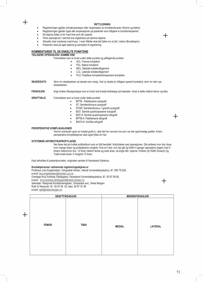
RETTLEDNINGRegistreringen gjelder primæroperasjon eller reoperasjon av korsbåndsruptur (fremre og bakre). Registreringen gjelder også alle reoperasjoner på pasienter som tidligere er korsbåndsoperert. Ett skjema fylles ut for hvert kne som blir operert. Flere operasjoner i samme kne registreres på samme skjema. Aktuelle ruter markeres med kryss. I noen tilfeller skal det fylles inn et tall i rutene (Brusklesjon). Pasienten skal på eget skjema gi samtykke til registrering.

KOMMENTARER TIL DE ENKELTE PUNKTENE TIDLIGERE OPERASJON I SAMME KNE

Forkortelser som er brukt under dette punktet og påfølgende punkter: ACL: Fremre korsbånd PCL: Bakre korsbånd MCL: Mediale kollateralligament LCL: Laterale kollateralligament PLC: Popliteus kompleks/bicepssene kompleks

SKADEDATO Skriv inn skadedatoen så eksakt som mulig. Ved ny skade av tidligere operert korsbånd, skriv inn den nye skadedatoen.

FIKSASJON Angi hvilken fiksasjonstype som er brukt ved å feste klistrelapp på baksiden. Husk å skille mellom femur og tibia.

GRAFTVALG Forkortelser som er brukt under dette punktet: BPTB; Patellarsene autograft ST: Semitendinosus autograft STGR: Semitendinosus + gracilis autograft BQT: Sentral quadricepssene autograft BQT-A: Sentral quadricepssene allograft BPTB-A: Patellarsene allograft BACH-A: Achilles allograft

PEROPERATIVE KOMPLIKASJONER Ved en eventuell ruptur av høstet graft e.l. skal det her nevnes hva som var det opprinnelige graftet. Andre peroperative komplikasjoner skal også fylles inn her.

SYSTEMISK ANTIBIOTIKAPROFYLAKSE Her føres det på hvilket antibiotikum som er blitt benyttet i forbindelse med operasjonen. Det anføres hvor stor dose,

hvor mange doser og profylaksens varighet. Hvis en f.eks. kun har gitt 2g Keflin 4 ganger operasjons dagen med 4 timers mellomrom dvs. 12 timer mellom første og siste dose, så angis det i skjema: Hvilken (A) Keflin Dose(A) 2g Totalt antall doser 4 Varighet 12 timer.

Kopi beholdes til pasientjournalen, originalen sendes til Haukeland Sykehus.

Kontaktpersoner vedrørende registreringsskjema erProfessor Lars Engebretsen, Ortopedisk Senter, Ullevål Universitetssykehus, tlf.: 950 79 529, e-post: [email protected] Knut Andreas Fjeldsgaard, Haukeland Universitetssykehus, tlf.: 55 97 56 80, e-post: [email protected]ær i Nasjonalt Korsbåndsregister, Ortopedisk avd., Helse Bergen: Ruth G Wasmuth, tlf.: 55 97 64 50, faks: 55 97 37 49 e-post: [email protected]

GRAFTFIKSASJON MENISKFIKSASJON

FEMUR TIBIAMEDIAL LATERAL

71

Paper I

72

308

National quality registries have been used in severalmedical specialties to improve health care inScandinavia,1,15,20,21,24,27,28,33 including Norway.3,17,21,23 Becauseof the inferior clinical results associated with some hip pros-thesis designs in the early 1980s,10 the nationwide NorwegianHip Arthroplasty Register (NAR) was established in 1987 withimplant revision as the main end point.14 Its aim was the early

detection of inferior results caused by implants, cements, orsurgical techniques.6,11 In 1994, the registry was expanded toinclude all joint replacements.11 In 1995, 2 papers12,13 werepublished that described the detection of inferior implants atan early stage, a finding only possible through registry studies.

The NAR is based on a simple reporting system (approx-imately 1 minute is required to complete a single-page reg-istration form) and the hospitals are provided withcontinuous feedback from the registry.11 These 2 factorsare believed to explain why the compliance rate of nearly100% has not declined during 20 years of operation.4,11

Immediately after each operation, the surgeon completesthe registration form, which is mailed to the NAR office.14

Patient identification and the different procedures, includ-ing the type of implant and cement used, are specified on

Development of a National CruciateLigament Surgery Registry

The Norwegian National Knee Ligament Registry

Lars-Petter Granan,*†‡ Roald Bahr,†‡ MD, PhD, Kjersti Steindal,‡§ Ove Furnes,‡§ll¶ MD, PhD,and Lars Engebretsen,†‡# MD, PhDFrom the †Oslo Sports Trauma Research Center, Norwegian School of Sport Sciences,Oslo, Norway, the ‡National Knee Ligament Registry, Bergen, Norway, the §NorwegianArthroplasty Register, Bergen, Norway, the llDepartment of Orthopaedics, Haukeland UniversityHospital, Bergen, Norway, the ¶Department of Surgical Sciences, University of Bergen, Norway,and the #Orthopaedic Center, Division of Neuroscience and Musculoskeletal Medicineand Faculty of Medicine, Ullevaal University Hospital, Oslo, Norway

Background: No prospective surveillance system exists for monitoring the outcome of cruciate ligament surgery.

Purpose: This article is intended to describe the development and procedures of the Norwegian National Knee LigamentRegistry (NKLR), including baseline results from the first 2 years of operation.

Study Design: Cohort study (prevalence); Level of evidence, 1.

Methods: The NKLR was established on June 7, 2004 to collect information prospectively on all cases of cruciate ligamentreconstruction surgery in Norway. Information on the details of surgery is gathered through a registration form completed by thesurgeon postoperatively, and a validated knee outcome score form is completed by the patients preoperatively and at follow-ups on all patients at 2, 5, and 10 years postoperatively. Hospital compliance was examined in 2005 and 2006.

Results: A total of 2793 primary cruciate ligament reconstruction surgeries were registered by 57 hospitals. This corresponds toan annual population incidence of primary anterior cruciate ligament reconstruction surgeries of 34 per 100 000 citizens (85 per100 000 citizens in the main at-risk age group of 16-39 years). After 21 months of operation, the NKLR had an overall compli-ance of 97% when compared with the hospital records.

Conclusions: A national population-based cruciate ligament registry has been developed, implemented, and maintained inNorway. The registry will each year enroll approximately 1500 primary cruciate ligament reconstruction cases. It is expected thatinadequate procedures and devices can be identified, as well as prognostic factors associated with good and poor outcomes,at least for the most frequent categories.

Keywords: orthopaedics; anterior cruciate ligament; registry; epidemiology; incidence; outcome

*Address correspondence to Lars-Petter Granan, Oslo Sports TraumaResearch Center, Norwegian School of Sport Sciences, PB 4014 UllevålStadion, 0806 Oslo, Norway (e-mail: [email protected])

No potential conflict of interest declared.

The American Journal of Sports Medicine, Vol. 36, No. 2DOI: 10.1177/0363546507308939© 2008 American Orthopaedic Society for Sports Medicine

© 2008 American Orthopaedic Society for Sports Medicine. All rights reserved. Not for commercial use or unauthorized distribution. at Universitet I Oslo on February 11, 2008 http://ajs.sagepub.comDownloaded from 73

Vol. 36, No. 2, 2008 National Cruciate Ligament Surgery Registry 309

the registration form. Feedback is given as annual nationalreports. In addition, each hospital receives a report on itsown activities and results, which can be compared with thenational average. A wide range of studies have been pub-lished based on the NAR database.11

In contrast to joint replacement surgery, for which nationalregistries have been established in Norway, Sweden (1979),Finland (1980), Denmark (1995), Australia (1999), NewZealand (1999), Canada (2000), Romania (2001), andEngland and Wales (2003), no national prospective surveil-lance system exists for monitoring the outcome of knee liga-ment surgery in a predefined population. Evidence from theScandinavian joint replacement registries indicates that anational knee ligament registry could be highly benefi-cial.12,13,16,26 First, treatment outcome can be improvedthrough feedback to the hospitals and surgeons from the reg-istries. Second, there are still several unresolved issuesrelated to cruciate ligament surgery and postoperative reha-bilitation methods. Some of these can and should beaddressed by conducting properly designed randomized con-trolled trials. However, because of practical, financial, orother restraints, such studies are often not possible. Also,some questions can only be answered by large cohort studies.This includes the detection of procedures and devices thatresult in premature failure.Third, large cohort studies can beused to identify prognostic factors associated with good andpoor outcomes.

This background served as the impetus for designing theNorwegian National Knee Ligament Registry (NKLR).This article describes the development and procedures ofthe first national knee ligament registry, including base-line results from the first 2 years of operation.

MATERIALS AND METHODS

Structure

A working group was established with members from NARand the Oslo Sports Trauma Research Center (OSTRC) in2002. The group designed the registry, constructed forms,planned the logistics, and contacted the hospitals. TheNKLR is owned by the Norwegian Orthopaedic Association(NOA), and a steering committee with 6 members isappointed jointly by NOA and OSTRC. Since the officialstart on June 7, 2004, the steering committee has beenresponsible for the budget, planning, and continuous eval-uation of the dataset.

Design

The NKLR is designed to collect information prospectivelyon all cases of cruciate ligament reconstruction surgery. Tobe included in the cohort, a patient should be a resident ofNorway undergoing primary or revision reconstruction sur-gery for an anterior cruciate ligament (ACL) and/or poste-rior cruciate ligament (PCL) injury at a Norwegian hospital.In addition, the NKLR also records all surgical proceduresto a knee joint that has previously undergone primary orrevision ACL and/or PCL reconstruction surgery.

Participation is voluntary, and all patients are asked tosign an informed consent form before surgery. The consentform contains information about the NKLR, the type ofinformation recorded, data protection, and the procedure forfollow-ups, and informs the patient that he or she may beinvited to participate in research projects at a later stage.The patients are also asked to complete a validated kneeoutcome score form, the Knee injury and OsteoarthritisOutcome Score (KOOS).22 The KOOS form is a knee-specificinstrument, developed to assess patients’ opinion abouttheir knees and associated problems, and was intended tobe used for knee injuries that could result in posttraumaticosteoarthritis.

The form includes 42 items in 5 separately scored sub-scales: pain (9 items), other symptoms (7 items), functionin activities of daily living (17 items), function in sportand recreation (5 items), and knee-related quality of life(4 items). Each item is responded to by marking 1 of 5response options on a Likert scale. The Western Ontarioand McMaster Universities (WOMAC) LK 3.02 items areincluded in the first 3 KOOS subscales. The KOOS is validand reliable for short-term and long-term follow-up studiesof knee injury and osteoarthritis.30-32 It is also valid forpatients in the age group 14 to 78 years of age. The KOOSwas considered reliable and responsive for assessment ofknee complaints in a recent comparative review of knee-specific outcome measures.7 Confidentiality is ensured forpatients and individual surgeons. The study has beenapproved by the Data Inspectorate as an expansion of theNAR concession.

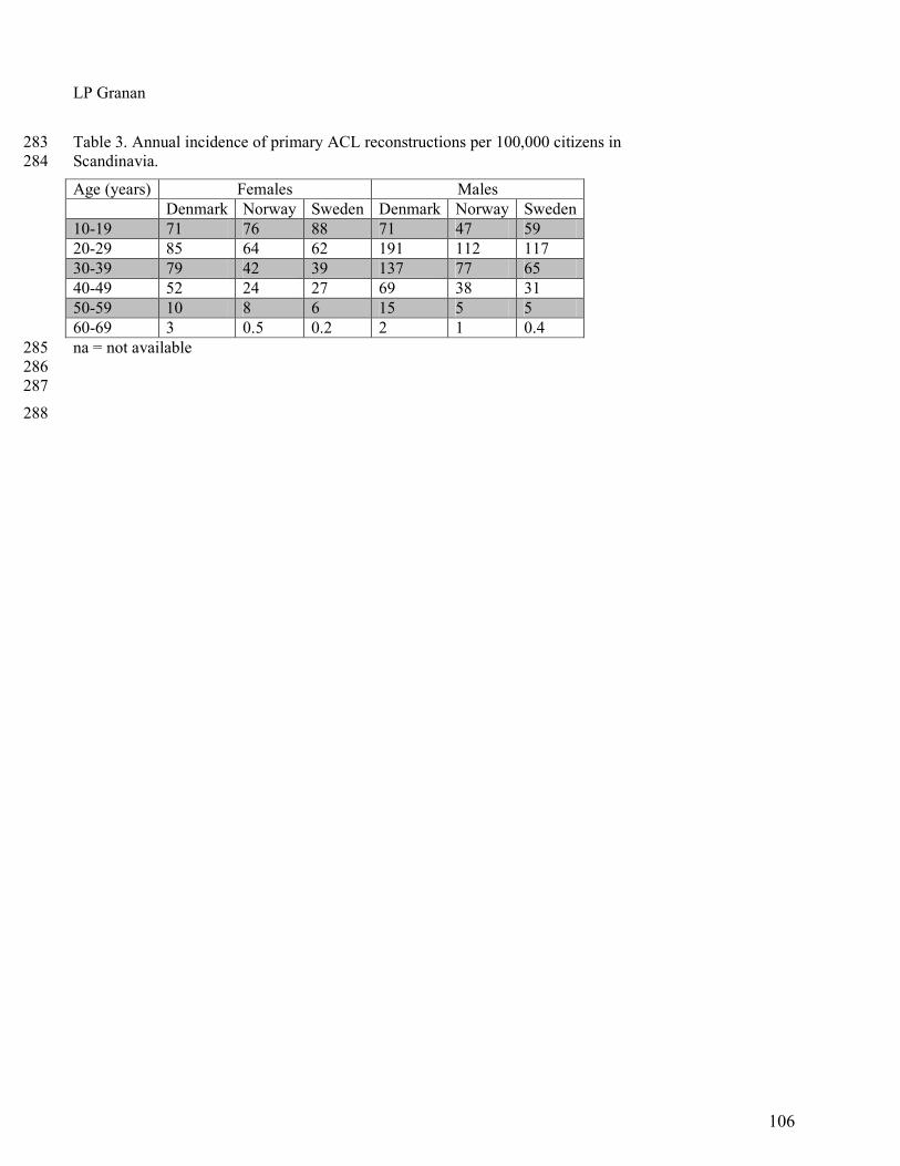
The registry makes use of both objective and subjectiveend points. Similar to NAR, the hard end points are revi-sion surgery after cruciate ligament surgery and total kneereplacement. Unlike NAR, the NKLR will include routinefollow-ups on all patients at 2, 5, and 10 years postopera-tively using KOOS score as a soft end point. The KOOSform will be dispatched from the NKLR secretariat at thetime for follow-ups. The NKLR will offer different ways ofreturning the completed KOOS forms, such as regular mailand Internet, as an attempt to ensure a high compliancerate. The KOOS form is not returned to the patient ifincomplete. Missing data are treated according to theguidelines for KOOS score calculation.31

Registration Process

After pilot testing at 3 hospitals, the registration form(Appendix 1) has been developed to collect information onthe details of surgery. One form is completed for each kneejoint undergoing surgical treatment. Similar to NAR, theform is completed by the surgeon immediately after sur-gery has been performed.

The data items recorded are a minimal set suited for apaper-based or web-based reporting system, not to exceed1 page. The items were chosen based on the following 3 cri-teria. Can the question addressed be clearly specified andjustified? Is the question clinically relevant? Can the itembe completed postoperatively while dictating the surgerynotes, not needing to seek information from other sources?

© 2008 American Orthopaedic Society for Sports Medicine. All rights reserved. Not for commercial use or unauthorized distribution. at Universitet I Oslo on February 11, 2008 http://ajs.sagepub.comDownloaded from 74

310 Granan et al The American Journal of Sports Medicine

Cartilage lesions are graded according to the InternationalCartilage Repair Society.34 To obtain accurate information onthe different fixation devices, it is recommended that the sur-geon report the catalog number of each device by using theunique bar-code stickers delivered by the manufacturers.The stickers contain all vital information about the device.The surgeon signs the form, but the surgeon’s identity is notrecorded, and thus cannot be traced in the registry.

One copy of the registration form is sent to NKLR andthe original is retained in the patient’s hospital chart. Onarrival at the NKLR, the KOOS and registration forms arechecked for completeness and entered into a computerizeddata management system. This is developed as an Oracledatabase (Oracle Corporation, Redwood Shores, Calif) withclerical and electronic data checks, as well as automatedcoding and reporting facilities. After registration, the dataare further checked to ensure the quality, eliminate possi-ble duplicates and illogical combinations in the form, andensure conformity between registration and KOOS forms.

A copy of the registration form is returned to the hospitalif the form is incomplete (eg, if essential data such as the dateof operation or the social security number are missing). If theform is not returned after 1 reminder or the data cannot befound, the form is marked as incomplete and labeled “miss-ing” for the missing data, thus retaining the possibility ofusing incomplete forms in the analysis.

The patients are identified by their unique social secu-rity number (including date of birth), which is assigned toall Norwegian residents. The social security number isused to link the KOOS and registration forms, and toupdate the registry annually with death and emigrationdata before extracting data files for analysis.

Compliance

A first baseline compliance study was carried out in March2005 covering the period October 1, 2004 through February28, 2005. The study covered primary ACL reconstructionsand ACL revision surgeries, not other procedures. Datafrom the NKLR were compared with the NorwegianPatient Register (NPR), which has been established by theMinistry of Health and Social Services to provide statisticsfrom the Norwegian hospital sector, as well as with patientdata from hospital records. The NPR has been used as agold standard by NAR.4 Ten hospitals participated, repre-senting all 5 health regions, hospitals with large and smallvolumes (cut-off was set at 30 annual ACL procedures),public and private hospitals, and hospitals with and with-out surgeons who were involved in developing NKLR.Based on preliminary data, we estimated that at least 250cases could be expected from these hospitals, which wouldgive the study sufficient power. All of the 10 invited hospi-tals agreed to participate.

A second study was performed in 2006 covering theperiod October 1, 2005 through February 28, 2006. Thisstudy used the same procedures as described for the base-line compliance study with 2 exceptions. Some of the hos-pitals dispatched the data electronically (electronic patientjournals), and the surgical log books were used as the goldstandard. This study covered 14 randomly chosen hospitalsparticipating in the NKLR.

Research and Information

Requests for data from the NKLR are encouraged, and datafiles are returned to the surgeon or hospital in question afterapproval of a written request addressed to the steeringcommittee. Only the official hospital contact can ask forpatient-identifiable information from his or her own hospi-tal. Some legal restrictions exist, primarily the combinationof NKLR with other population-based registries in Norway.Requests for more extensive data for research projects alsorequire a written application to the steering committee. Ifexternal researchers wish to combine data from the NKLRwith their own data files, specific approval is required fromthe Data Inspectorate and the appropriate RegionalCommittee for Medical Research Ethics.

Descriptive national data are provided in an annualreport, which is sent to all members of the NOA, all hospi-tals performing cruciate ligament surgery, and to thehealth authorities. This report is also published on thejoint website of NAR and NKLR (www.haukeland.no/nrl).In addition, each participating hospital will receivedescriptive statistics and outcome data for their own hos-pital, which they can compare with the national report.

Staff and Operating Costs

The NKLR employs a secretary (50% position), a computerengineer (50%), and an orthopaedic surgeon (20%) as theadministrative head of NKLR. In addition, each hospitalprovides secretarial assistance amounting to approximately10% of a full position. The total operating budget for 2006 forthe central NKLR office is 527 000 krones (approximately67 000 euros, or 91 000 US dollars). This cost does not includesalary for additional staff involved in various research proj-ects based on the NKLR. It is expected that the basic oper-ating costs will increase somewhat as the cohort andnumber of follow-ups increase year by year.

RESULTS

Descriptive Data

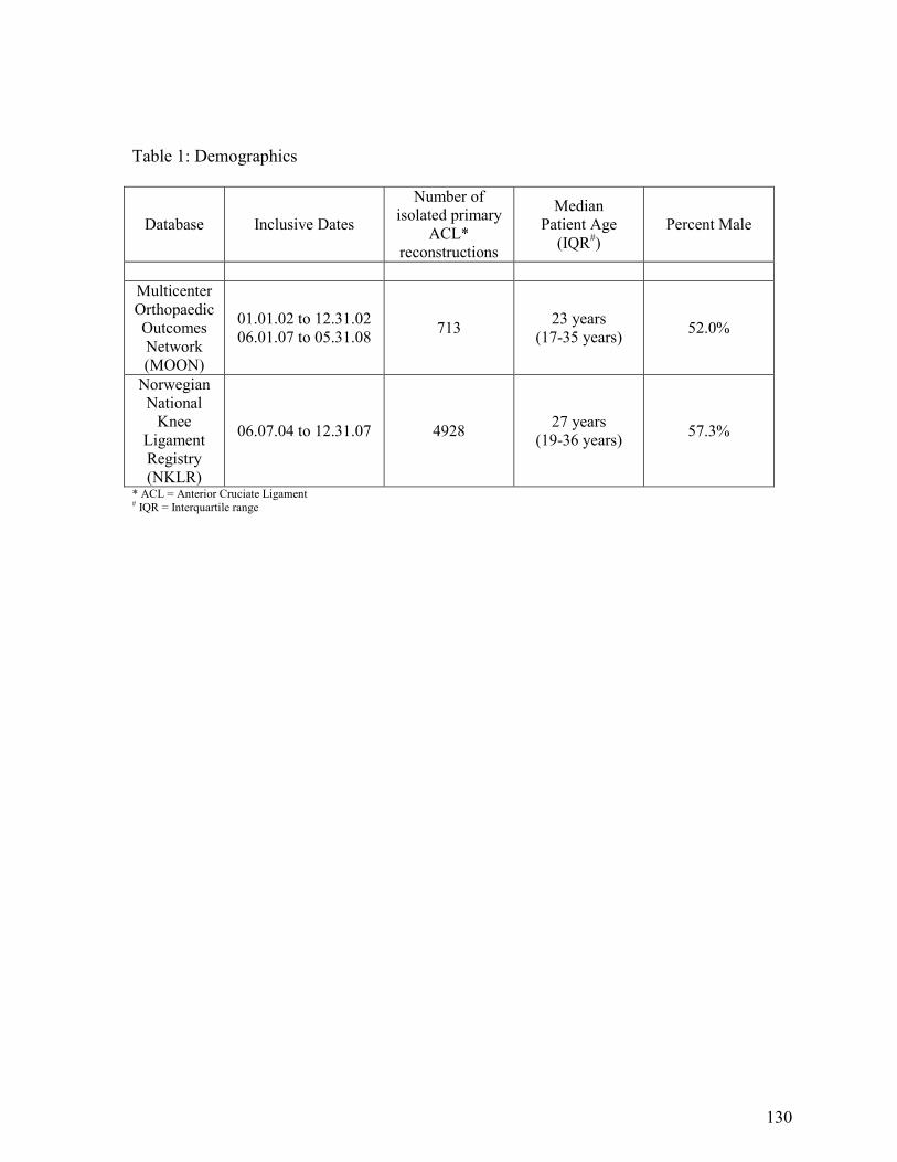
From June 7, 2004, until May 24, 2006 (687 days), 2793 pri-mary cruciate ligament reconstruction surgeries were regis-tered by 57 hospitals. This corresponds to an annual rate of1484 primary cruciate ligament reconstructions in Norway,1168 of them in the age group 16 through 39 years (the mainpopulation at risk). In 2005, there were 4 393 000 citizens inNorway, 1382000 of them aged 16 through 39 years.Thus, theannual population incidence of primary ACL reconstructionsurgeries was 34 per 100 000 citizens, while the incidence inthe 16 to 39 years age group was 85 per 100 000 citizens.

Of the 2793 cases recorded in the NKLR, 2714 were pri-mary ACL reconstructions, 25 were primary PCL recon-structions, and 54 were combined primary reconstructionsof both cruciate ligaments.

How Complete Are the Data?

The baseline compliance study identified 285 cases in theNKLR database, 332 in the hospital protocols, and 339 at

© 2008 American Orthopaedic Society for Sports Medicine. All rights reserved. Not for commercial use or unauthorized distribution. at Universitet I Oslo on February 11, 2008 http://ajs.sagepub.comDownloaded from 75

Vol. 36, No. 2, 2008 National Cruciate Ligament Surgery Registry 311

the NPR. Thus, after 4 to 9 months of operation, the NKLRhad a compliance of 84% in relation to the NPR among thehospitals participating. At this time, 51 out of a possibletotal of 56 hospitals and clinics (91%) took part.

The second compliance study identified 195 cases in theNKLR database, 202 in the protocols at the hospitals, and181 at the NPR (1 private hospital with 18 cases recorded inthe NKLR database did not report to the NPR). Thus, after16 to 21 months of operation, the NKLR had compliance of97% and 98% in relation to the hospital protocols (195/202)and NPR (177/181), respectively. By the end of the studyperiod, all hospitals and clinics (N = 57) participated in theNKLR, although the last hospital was not included until thefinal 2 months of the second compliance study period.

Primary ACL Reconstructions

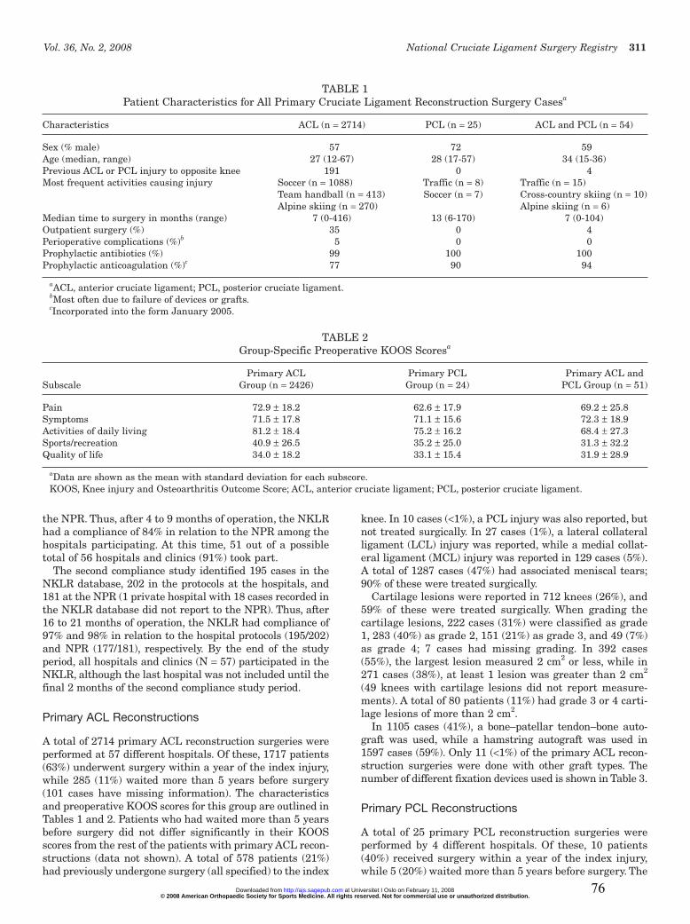
A total of 2714 primary ACL reconstruction surgeries wereperformed at 57 different hospitals. Of these, 1717 patients(63%) underwent surgery within a year of the index injury,while 285 (11%) waited more than 5 years before surgery(101 cases have missing information). The characteristicsand preoperative KOOS scores for this group are outlined inTables 1 and 2. Patients who had waited more than 5 yearsbefore surgery did not differ significantly in their KOOSscores from the rest of the patients with primary ACL recon-structions (data not shown). A total of 578 patients (21%)had previously undergone surgery (all specified) to the index

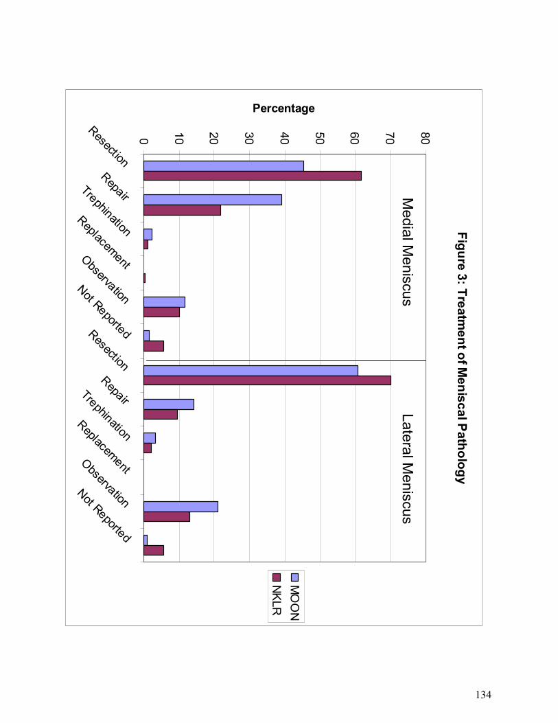
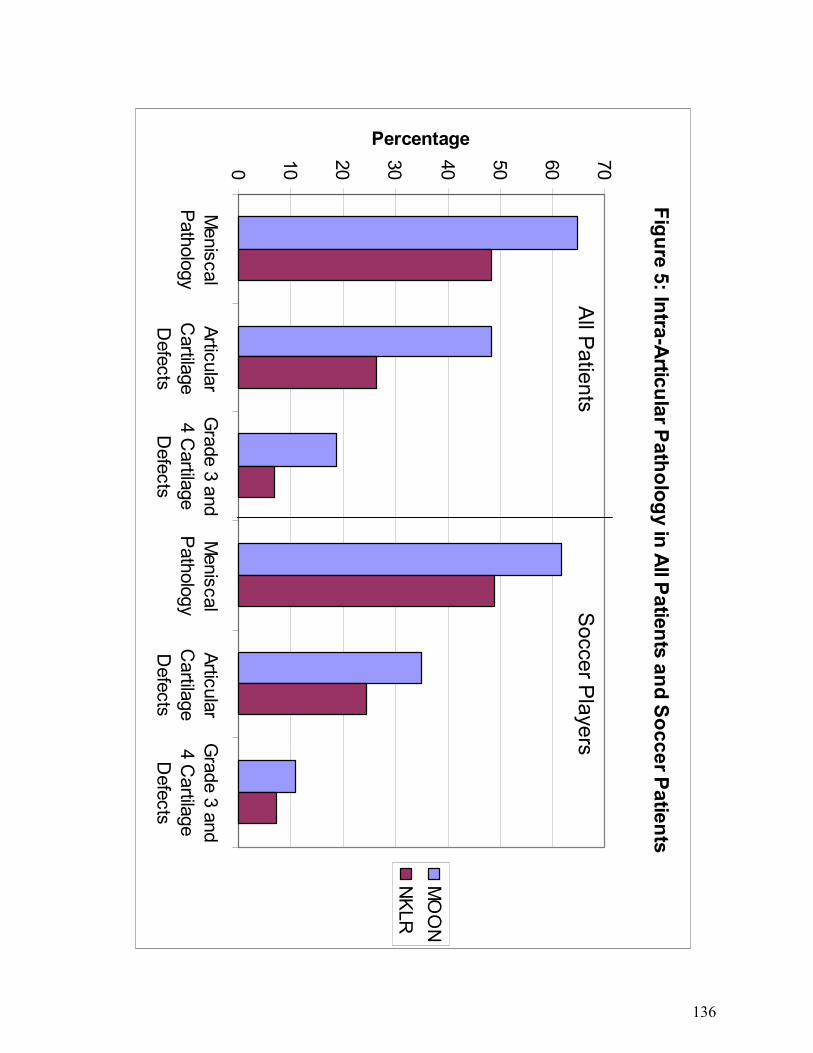
knee. In 10 cases (<1%), a PCL injury was also reported, butnot treated surgically. In 27 cases (1%), a lateral collateralligament (LCL) injury was reported, while a medial collat-eral ligament (MCL) injury was reported in 129 cases (5%).A total of 1287 cases (47%) had associated meniscal tears;90% of these were treated surgically.

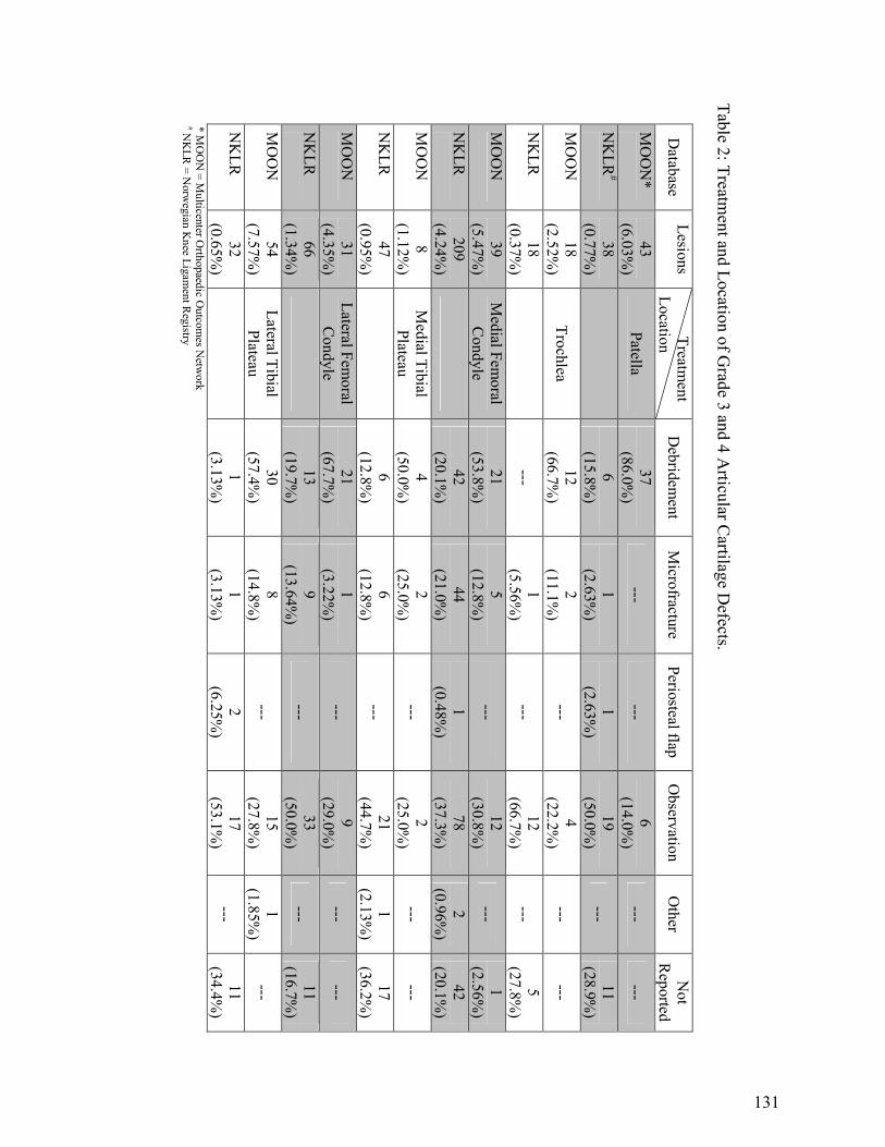
Cartilage lesions were reported in 712 knees (26%), and59% of these were treated surgically. When grading thecartilage lesions, 222 cases (31%) were classified as grade1, 283 (40%) as grade 2, 151 (21%) as grade 3, and 49 (7%)as grade 4; 7 cases had missing grading. In 392 cases(55%), the largest lesion measured 2 cm2 or less, while in271 cases (38%), at least 1 lesion was greater than 2 cm2

(49 knees with cartilage lesions did not report measure-ments). A total of 80 patients (11%) had grade 3 or 4 carti-lage lesions of more than 2 cm2.

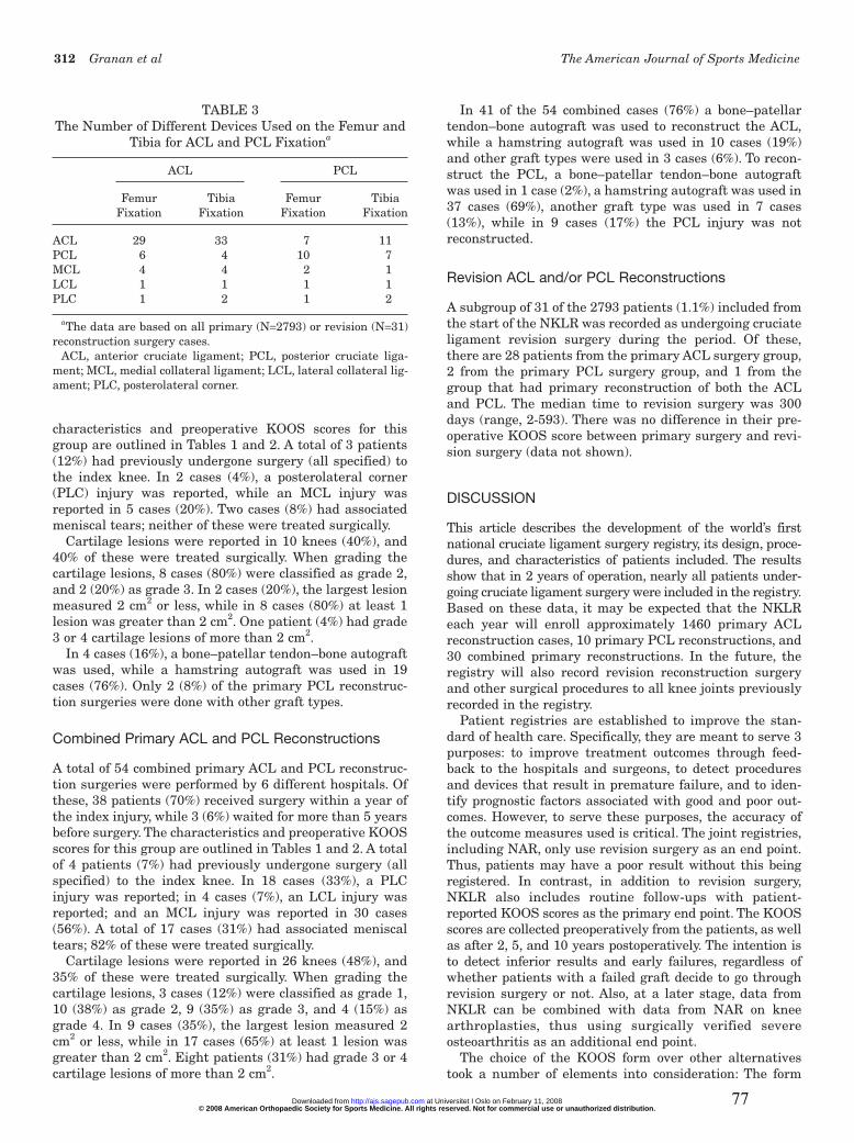
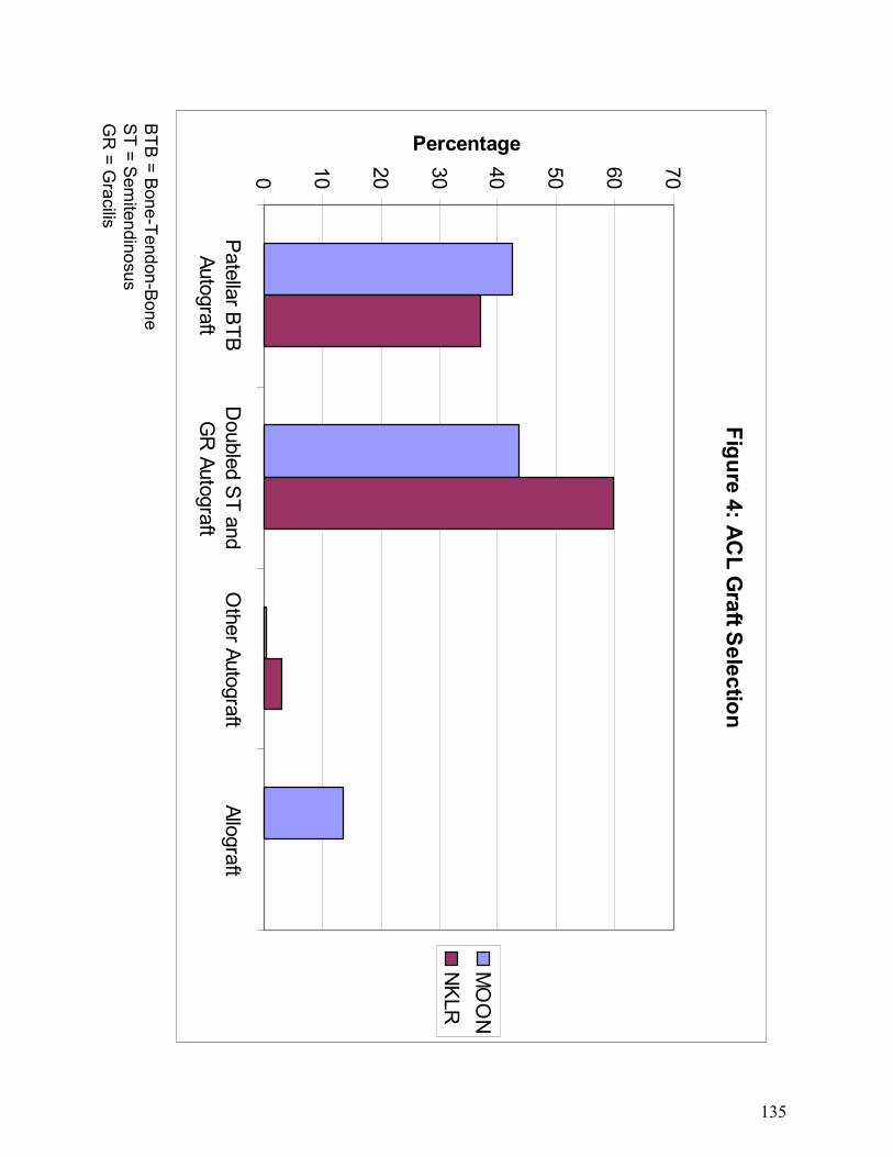
In 1105 cases (41%), a bone–patellar tendon–bone auto-graft was used, while a hamstring autograft was used in1597 cases (59%). Only 11 (<1%) of the primary ACL recon-struction surgeries were done with other graft types. Thenumber of different fixation devices used is shown in Table 3.

Primary PCL Reconstructions

A total of 25 primary PCL reconstruction surgeries wereperformed by 4 different hospitals. Of these, 10 patients(40%) received surgery within a year of the index injury,while 5 (20%) waited more than 5 years before surgery. The

TABLE 1Patient Characteristics for All Primary Cruciate Ligament Reconstruction Surgery Casesa

Characteristics ACL (n = 2714) PCL (n = 25) ACL and PCL (n = 54)

Sex (% male) 57 72 59Age (median, range) 27 (12-67) 28 (17-57) 34 (15-36)Previous ACL or PCL injury to opposite knee 191 0 4Most frequent activities causing injury Soccer (n = 1088) Traffic (n = 8) Traffic (n = 15)

Team handball (n = 413) Soccer (n = 7) Cross-country skiing (n = 10)Alpine skiing (n = 270) Alpine skiing (n = 6)

Median time to surgery in months (range) 7 (0-416) 13 (6-170) 7 (0-104)Outpatient surgery (%) 35 0 4Perioperative complications (%)b 5 0 0Prophylactic antibiotics (%) 99 100 100Prophylactic anticoagulation (%)c 77 90 94

aACL, anterior cruciate ligament; PCL, posterior cruciate ligament.bMost often due to failure of devices or grafts.cIncorporated into the form January 2005.

TABLE 2Group-Specific Preoperative KOOS Scoresa

Primary ACL Primary PCL Primary ACL and Subscale Group (n = 2426) Group (n = 24) PCL Group (n = 51)

Pain 72.9 ± 18.2 62.6 ± 17.9 69.2 ± 25.8Symptoms 71.5 ± 17.8 71.1 ± 15.6 72.3 ± 18.9Activities of daily living 81.2 ± 18.4 75.2 ± 16.2 68.4 ± 27.3Sports/recreation 40.9 ± 26.5 35.2 ± 25.0 31.3 ± 32.2Quality of life 34.0 ± 18.2 33.1 ± 15.4 31.9 ± 28.9

aData are shown as the mean with standard deviation for each subscore.KOOS, Knee injury and Osteoarthritis Outcome Score; ACL, anterior cruciate ligament; PCL, posterior cruciate ligament.

© 2008 American Orthopaedic Society for Sports Medicine. All rights reserved. Not for commercial use or unauthorized distribution. at Universitet I Oslo on February 11, 2008 http://ajs.sagepub.comDownloaded from 76

312 Granan et al The American Journal of Sports Medicine

characteristics and preoperative KOOS scores for thisgroup are outlined in Tables 1 and 2. A total of 3 patients(12%) had previously undergone surgery (all specified) tothe index knee. In 2 cases (4%), a posterolateral corner(PLC) injury was reported, while an MCL injury wasreported in 5 cases (20%). Two cases (8%) had associatedmeniscal tears; neither of these were treated surgically.

Cartilage lesions were reported in 10 knees (40%), and40% of these were treated surgically. When grading thecartilage lesions, 8 cases (80%) were classified as grade 2,and 2 (20%) as grade 3. In 2 cases (20%), the largest lesionmeasured 2 cm2 or less, while in 8 cases (80%) at least 1lesion was greater than 2 cm2. One patient (4%) had grade3 or 4 cartilage lesions of more than 2 cm2.

In 4 cases (16%), a bone–patellar tendon–bone autograftwas used, while a hamstring autograft was used in 19cases (76%). Only 2 (8%) of the primary PCL reconstruc-tion surgeries were done with other graft types.

Combined Primary ACL and PCL Reconstructions

A total of 54 combined primary ACL and PCL reconstruc-tion surgeries were performed by 6 different hospitals. Ofthese, 38 patients (70%) received surgery within a year ofthe index injury, while 3 (6%) waited for more than 5 yearsbefore surgery. The characteristics and preoperative KOOSscores for this group are outlined in Tables 1 and 2. A totalof 4 patients (7%) had previously undergone surgery (allspecified) to the index knee. In 18 cases (33%), a PLCinjury was reported; in 4 cases (7%), an LCL injury wasreported; and an MCL injury was reported in 30 cases(56%). A total of 17 cases (31%) had associated meniscaltears; 82% of these were treated surgically.

Cartilage lesions were reported in 26 knees (48%), and35% of these were treated surgically. When grading thecartilage lesions, 3 cases (12%) were classified as grade 1,10 (38%) as grade 2, 9 (35%) as grade 3, and 4 (15%) asgrade 4. In 9 cases (35%), the largest lesion measured 2cm2 or less, while in 17 cases (65%) at least 1 lesion wasgreater than 2 cm2. Eight patients (31%) had grade 3 or 4cartilage lesions of more than 2 cm2.

In 41 of the 54 combined cases (76%) a bone–patellartendon–bone autograft was used to reconstruct the ACL,while a hamstring autograft was used in 10 cases (19%)and other graft types were used in 3 cases (6%). To recon-struct the PCL, a bone–patellar tendon–bone autograftwas used in 1 case (2%), a hamstring autograft was used in37 cases (69%), another graft type was used in 7 cases(13%), while in 9 cases (17%) the PCL injury was notreconstructed.

Revision ACL and/or PCL Reconstructions

A subgroup of 31 of the 2793 patients (1.1%) included fromthe start of the NKLR was recorded as undergoing cruciateligament revision surgery during the period. Of these,there are 28 patients from the primary ACL surgery group,2 from the primary PCL surgery group, and 1 from thegroup that had primary reconstruction of both the ACLand PCL. The median time to revision surgery was 300days (range, 2-593). There was no difference in their pre-operative KOOS score between primary surgery and revi-sion surgery (data not shown).

DISCUSSION

This article describes the development of the world’s firstnational cruciate ligament surgery registry, its design, proce-dures, and characteristics of patients included. The resultsshow that in 2 years of operation, nearly all patients under-going cruciate ligament surgery were included in the registry.Based on these data, it may be expected that the NKLReach year will enroll approximately 1460 primary ACLreconstruction cases, 10 primary PCL reconstructions, and30 combined primary reconstructions. In the future, theregistry will also record revision reconstruction surgeryand other surgical procedures to all knee joints previouslyrecorded in the registry.

Patient registries are established to improve the stan-dard of health care. Specifically, they are meant to serve 3purposes: to improve treatment outcomes through feed-back to the hospitals and surgeons, to detect proceduresand devices that result in premature failure, and to iden-tify prognostic factors associated with good and poor out-comes. However, to serve these purposes, the accuracy ofthe outcome measures used is critical. The joint registries,including NAR, only use revision surgery as an end point.Thus, patients may have a poor result without this beingregistered. In contrast, in addition to revision surgery,NKLR also includes routine follow-ups with patient-reported KOOS scores as the primary end point. The KOOSscores are collected preoperatively from the patients, as wellas after 2, 5, and 10 years postoperatively. The intention isto detect inferior results and early failures, regardless ofwhether patients with a failed graft decide to go throughrevision surgery or not. Also, at a later stage, data fromNKLR can be combined with data from NAR on kneearthroplasties, thus using surgically verified severeosteoarthritis as an additional end point.

The choice of the KOOS form over other alternativestook a number of elements into consideration: The form

TABLE 3The Number of Different Devices Used on the Femur and

Tibia for ACL and PCL Fixationa

ACL PCL

Femur Tibia Femur Tibia Fixation Fixation Fixation Fixation

ACL 29 33 7 11PCL 6 4 10 7MCL 4 4 2 1LCL 1 1 1 1PLC 1 2 1 2

aThe data are based on all primary (N=2793) or revision (N=31)reconstruction surgery cases.

ACL, anterior cruciate ligament; PCL, posterior cruciate liga-ment; MCL, medial collateral ligament; LCL, lateral collateral lig-ament; PLC, posterolateral corner.

© 2008 American Orthopaedic Society for Sports Medicine. All rights reserved. Not for commercial use or unauthorized distribution. at Universitet I Oslo on February 11, 2008 http://ajs.sagepub.comDownloaded from 77

Vol. 36, No. 2, 2008 National Cruciate Ligament Surgery Registry 313

should be patient-based to allow for nonbiased outcomedata. The form should be self-explanatory, and timerequired to complete the form should be kept to a maxi-mum of 10 minutes to ensure good compliance at follow-ups. Finally, the form had to be validated for cruciateligament surgery. These requirements left us with twochoices: KOOS or International Knee DocumentationCommittee (IKDC) 2000.18,19 We chose the KOOS formbecause, in our opinion, it is far more user-friendly from apatient’s perspective than the IKDC 2000. However, itremains to be seen how well patients will comply with thefollow-up procedures.

To serve its first purpose, to improve treatment out-comes through continuous feedback to the participatinghospitals, each year hospitals are provided with results ontheir own patients and national data. This is based on theidea that hospitals able to compare their outcomes withnational averages will improve by following the betterexamples. An annual report is sent to all the members ofthe NOA, to all hospitals performing cruciate ligamentsurgery, and to the health authorities, and also publishedon the joint website of NAR and NKLR (www.hauke-land.no/nrl). The NKLR depends on participation from allorthopaedic surgeons performing cruciate ligament sur-gery, including those normally not involved in research.Feedback is therefore also important to maintain motiva-tion and interest in the registry, and we believe the report-ing procedure explains the high compliance with theregistry observed. Based on our previous experience withNAR, it may be expected that compliance will remain high.This is based on the premise that there will be no addi-tional demands on the surgeons except filling out theforms, and that NKLR will serve the hospitals with clini-cally relevant and important information.

The second purpose, to detect procedures and devicesthat result in premature failure, can be achieved based onrevision surgery or, if a revision has not been performed,deterioration of the KOOS score.29 The following exampleillustrates this point. A score of at least 60 points may beexpected with a successful outcome after surgery.31 Age-and sex-specific general population reference values arealso available for all 5 KOOS subscales.29 A change in theKOOS score of 10 points can be considered a clinically sig-nificant difference—as an improvement after surgery ordeterioration after graft failure.29 Thus, the number ofpatients needed to detect failure in a cohort study may becalculated. Assuming a more conservative estimate, that adifference of 20 points is sufficient to predict an inferiordevice or procedure, as few as 14 failures are needed, usingstandard statistical values. These estimates also apply ifthe purpose is to discover prognostic factors that are asso-ciated with good or poor outcomes. For example, there aremany patients with large cartilage lesions (>2 cm2) andlesions graded 3 or 4 that are of special interest as theirtreatment outcome may be less predictable. Thus, becauseit may be estimated that the registry will include 2-yearoutcome data on at least 6500 patients with isolated ACLreconstructions after 7 years of operation, it seems reason-able to assume that the registry will be able to provide rele-vant data on inadequate procedures and devices. However,

less common procedures and devices will be difficult toassess, and it should be noted that the frequency of devicesin use varies considerably (Table 3). Also, as shown in theresults, isolated PCL reconstructions and combinedACL/PCL reconstructions are much less frequent than iso-lated ACL reconstructions, and for these procedures it willbe difficult to study subgroups, even with a national registry.However, this may be achieved when the registries ofSweden, Denmark, and Norway are combined.

It may be argued that randomized controlled trials(RCTs) are better than cohort studies to assess the out-come of cruciate ligament surgery. Although RCTs arepreferable to address specific research questions, such ascomparing 1 surgical procedure to another, they are diffi-cult to organize, time-consuming, and costly. Therefore, it isoften not possible or even justified to conduct an RCT toaddress anything but major differences in procedures ordevices. One example may be minor changes in screwdesign or materials. A national registry can be used toassess results with minimal additional work or cost.However, it should be noted that in a nonrandomizedcohort study, confounding factors must be adjusted for,either by selection of homogeneous subgroups or by use ofa multiple regression model when analyzing the results.12

An important limitation of the registry is that only surgi-cally treated cruciate injuries are included. Some studieshave shown that most cruciate ligament-injured patientswill see medical care and thus could be entered into the reg-istry.9 However, because of logistic and diagnostic issues, wehave decided to not include this group at this stage.

The annual Norwegian population incidence of primaryACL reconstruction surgeries was 34 per 100 000 citizens,while the incidence in the 16- to 39-year-old age group was85 per 100 000 citizens, both higher than previously pub-lished. Based on a questionnaire to all Norwegian hospi-tals in 2001 and 2002-2003, we estimated the annualincidence to be 42 ACL surgeries per 100 000 citizens.8

However, because we do not know the ratio of surgicallytreated versus conservatively treated cases, the populationincidence of ACL injuries is not known. In Germany, thishas been estimated to be 32 per 100 000 citizens in the gen-eral population, and 70 per 100 000 citizens among themore physically active.25 A recent study from 1 emergencydepartment in Sweden reported that the physically activepopulation between 10 to 64 years of age had an annualincidence of ACL injuries of 81 per 100 000 citizens.5

However, the present study is the first extensive and com-plete population-based survey and from our data itappears that the true population incidence may be 50% to100% higher, as in our experience as many as 30% to 50%of all ACL-injured subjects do not undergo surgery.

In conclusion, this study shows that a national population-based cruciate ligament registry could be developed, imple-mented, and maintained in Norway, providing data on morethan 95% of all patients undergoing cruciate ligament sur-gery. The registry will each year enroll approximately 1460primary ACL reconstruction cases, 10 primary PCL recon-struction cases, and 30 cases of primary reconstruction ofboth cruciate ligaments. It may be expected that the registrycan enable us to identify inadequate procedures and devices,

© 2008 American Orthopaedic Society for Sports Medicine. All rights reserved. Not for commercial use or unauthorized distribution. at Universitet I Oslo on February 11, 2008 http://ajs.sagepub.comDownloaded from 78

314 Granan et al The American Journal of Sports Medicine

as well as prognostic factors associated with good and pooroutcomes, at least for the most frequent categories.

ACKNOWLEDGMENT

The NKLR is financed by the Oslo Sports Trauma ResearchCenter, which has been established through generous grantsfrom the Eastern Norway Regional Health Authority, PfizerAS, the Royal Norwegian Ministry of Culture, the NorwegianOlympic Committee & Confederation of Sport, and NorskTipping. In addition, the NKLR has been supported througha grant from the Norwegian Medical Association’s Fund forQuality Improvement. Lars-Petter Granan has been sup-ported by the Medical Research Curriculum at theUniversity of Oslo. The authors thank the NKLR secretariat,Ruth Gunvor Wasmuth and Marianne Wiese; medical stu-dents Kristoffer Solumshengslet and Karianne Ytterstad fortheir contribution with the compliance studies; and the staffand colleagues of the participating orthopaedic and surgicaldepartments for their cooperation.

REFERENCES

1. Akesson K. Bone and joint diseases around the world. Sweden: a briefupdate on burden and priority. J Rheumatol Suppl. 2003;67:38-40.

2. Bellamy N, Buchanan WW, Goldsmith CH, Campbell J, Stitt LW.Validation study of WOMAC: a health status instrument for measuringclinically important patient relevant outcomes to antirheumatic drugtherapy in patients with osteoarthritis of the hip or knee. J Rheumatol.1988;15:1833-1840.

3. Bergem AL. Norwegian Twin Registers and Norwegian twin studies:an overview. Twin Res. 2002;5:407-414.

4. Espehaug B, Furnes O, Havelin LI, Engesaeter LB, Vollset SE,Kindseth O. Registration completeness in the Norwegian ArthroplastyRegister. Acta Orthop. 2006;77:49-56.

5. Frobell RB, Lohmander LS, Roos HP. Acute rotational trauma to theknee: poor agreement between clinical assessment and magnetic reso-nance imaging findings. Scand J Med Sci Sports. 2007;17:109-114.

6. Furnes O, Espehaug B, Lie SA, Vollset SE, Engesaeter LB, Havelin LI.Early failures among 7,174 primary total knee replacements: a follow-up study from the Norwegian Arthroplasty Register 1994-2000. ActaOrthop Scand. 2002;73:117-129.

7. Garratt AM, Brealey S, Gillespie WJ. Patient-assessed health instru-ments for the knee: a structured review. Rheumatology (Oxford).2004;43:1414-1423.

8. Granan LP, Engebretsen L, Bahr R. Surgery after anterior cruciate lig-ament injuries in Norway [article in Norwegian]. Tidsskr NorLaegeforen. 2004;124:928-930.

9. Grontvedt T, Heir S, Rossvoll I, Engebretsen L. Five-year outcome of13 patients with an initially undiagnosed anterior cruciate ligamentrupture. Scand J Med Sci Sports. 1999;9:62-64.

10. Havelin LI. Hip Arthroplasty in Norway 1987-1994: The NorwegianArthroplasty Register. Bergen, Norway: University of Bergen; 1995.

11. Havelin LI, Engesaeter LB, Espehaug B, Furnes O, Lie SA, Vollset SE.The Norwegian Arthroplasty Register: 11 years and 73,000 arthro-plasties. Acta Orthop Scand. 2000;71:337-353.

12. Havelin LI, Espehaug B, Vollset SE, Engesaeter LB. Early asepticloosening of uncemented femoral components in primary total hip

replacement: a review based on the Norwegian Arthroplasty Register.J Bone Joint Surg Br. 1995;77:11-17.

13. Havelin LI, Espehaug B, Vollset SE, Engesaeter LB. The effect of thetype of cement on early revision of Charnley total hip prostheses: areview of eight thousand five hundred and seventy-nine primaryarthroplasties from the Norwegian Arthroplasty Register. J Bone JointSurg Am. 1995;77:1543-1550.

14. Havelin LI, Espehaug B, Vollset SE, Engesaeter LB, Langeland N. TheNorwegian arthroplasty register: a survey of 17,444 hip replacements1987-1990. Acta Orthop Scand. 1993;64:245-251.

15. Heaf J. The Danish Renal Biopsy Register. Kidney Int. 2004;66:895-897.16. Herberts P, Malchau H. How outcome studies have changed total hip

arthroplasty practices in Sweden. Clin Orthop Relat Res. 1997;344:44-60.17. Irgens LM. The Medical Birth Registry of Norway: epidemiological

research and surveillance throughout 30 years. Acta Obstet GynecolScand. 2000;79:435-439.

18. Irrgang JJ, Anderson AF, Boland AL, et al. Development and valida-tion of the international knee documentation committee subjectiveknee form. Am J Sports Med. 2001; 29:600-613.

19. Johnson DS, Smith RB. Outcome measurement in the ACL deficientknee: what’s the score? Knee. 2001;8:51-57.

20. Kallen B. The use of national health registers for studying environmen-tal causes of congenital defects. Rev Environ Health. 2005;20:57-64.

21. Kjaerheim K. Occupational cancer research in the Nordic countries.Environ Health Perspect. 1999;107 Suppl 2:233-238.

22. Knee injury and Osteoarthritis Outcome Score. http://www.koos.nu(Accessed September 2007).

23. Kvien TK, Uhlig T. The Oslo experience with arthritis registries. ClinExp Rheumatol. 2003;21(5 Suppl 31):S118-S122.

24. Lichtenstein P, De Faire U, Floderus B, Svartengren M, Svedberg P,Pedersen NL. The Swedish Twin Registry: a unique resource for clinical,epidemiological and genetic studies. J Intern Med. 2002;252:184-205.

25. Lobenhoffer P. Knee ligament injuries: anatomy, biomechanics, diag-nosis, indications [article in German]. Chirurg. 1999;70:219-230.

26. Malchau H, Herberts P, Eisler T, Garellick G, Soderman P. TheSwedish Total Hip Replacement Register. J Bone Joint Surg Am.2002; 84(Suppl 2):2-20.

27. Ohm KK, Derom C. Data collection on multiple births: establishing twinregisters and determining zygosity. Early Hum Dev. 2006;82:357-363.

28. Pahlman L, Gunnarsson U, Karlbom U. The influence on treatmentoutcome of structuring rectal cancer care. Eur J Surg Oncol. 2005;31:645-649.

29. Paradowski PT, Bergman S, Sunden-Lundius A, Lohmander LS, RoosEM. Knee complaints vary with age and gender in the adult population:population-based reference data for the Knee injury and OsteoarthritisOutcome Score (KOOS). BMC Musculoskelet Disord. 2006;7:38.

30. Roos EM, Roos HP, Ekdahl C, Lohmander LS. Knee injury andOsteoarthritis Outcome Score (KOOS): validation of a Swedish ver-sion. Scand J Med Sci Sports. 1998;8:439-448.

31. Roos EM, Roos HP, Lohmander LS, Ekdahl C, Beynnon BD. KneeInjury and Osteoarthritis Outcome Score (KOOS): development of aself-administered outcome measure. J Orthop Sports Phys Ther.1998;28:88-96.

32. Roos EM, Toksvig-Larsen S. Knee injury and Osteoarthritis OutcomeScore (KOOS): validation and comparison to the WOMAC in totalknee replacement. Health Qual Life Outcomes. 2003;1:17.

33. Sokka T. National databases and rheumatology research: I. longitudinaldatabases in Scandinavia. Rheum Dis Clin North Am. 2004;30:851-867.

34. The cartilage standard evaluation form/knee and cartilage repairassessment. Newsletter ICRS. 1998;Spring:5-8.

© 2008 American Orthopaedic Society for Sports Medicine. All rights reserved. Not for commercial use or unauthorized distribution. at Universitet I Oslo on February 11, 2008 http://ajs.sagepub.comDownloaded from 79

Vol. 36, No. 2, 2008 National Cruciate Ligament Surgery Registry 315

APPENDIX

© 2008 American Orthopaedic Society for Sports Medicine. All rights reserved. Not for commercial use or unauthorized distribution. at Universitet I Oslo on February 11, 2008 http://ajs.sagepub.comDownloaded from 80

Paper II

81

955

*Address correspondence to Lars-Petter Granan, MD, Oslo Sports Trauma Research Center, Norwegian School of Sport Sciences, PB 4014 Ullevål Stadion, 0806 Oslo, Norway (e-mail: [email protected]).

No potential conflict of interest declared.

The American Journal of Sports Medicine, Vol. 37, No. 5DOI: 10.1177/0363546508330136© 2009 American Orthopaedic Society for Sports Medicine

Timing of Anterior Cruciate Ligament Reconstructive Surgery and Risk of Cartilage Lesions and Meniscal Tears

A Cohort Study Based on the Norwegian National Knee Ligament Registry

Lars-Petter Granan,*†‡ MD, Roald Bahr,†‡ MD, PhD, Stein Atle Lie,§ll MSc, PhD, and Lars Engebretsen,†‡¶ MD, PhDFrom the †Oslo Sports Trauma Research Center, Norwegian School of Sport Sciences, Oslo, Norway, ‡National Knee Ligament Registry, Bergen, Norway, §Norwegian Arthroplasty Register, Department of Orthopaedic Surgery, Haukeland University Hospital, Bergen, Norway, llUniversity Research Bergen, Department of Health, Bergen, Norway, and ¶Orthopaedic Center, Ullevaal University Hospital and Faculty of Medicine, University of Oslo, Oslo, Norway

Background: There is inadequate evidence to determine when to perform surgery on anterior cruciate ligament–deficient knees.

Purpose: To study the association between timing of anterior cruciate ligament reconstruction and the risk of having meniscal tears and cartilage lesions.

Study Design: Cohort study (prognosis); Level of evidence, 2.

Methods: All patients registered in the Norwegian National Knee Ligament Registry who had undergone primary anterior cruciate ligament reconstruction from 2004 and throughout 2006 were reviewed. Logistic regression analyses were used to estimate the relationship between time from injury until anterior cruciate ligament surgery and the risk of meniscal tears or cartilage lesions.

Results: Of a total of 3475 patients, there were 909 patients (26%) with cartilage lesions, 1638 patients (47%) with meniscal tears, and 527 patients (15%) with both cartilage and meniscal lesions. The odds of a cartilage lesion in the adult knee (>16 years) increased by 1.006 (95% confidence interval, 1.003-1.010) for each month that elapsed from injury to surgery. The cartilage in young adults (17-40 years) deteriorated further with an increase in odds of 1.03 (95% confidence interval, 1.02-1.05) related to the aging in years of the patient. The odds for meniscal tears in young adults increased by 1.004 (95% confidence interval, 1.002-1.006) for each month that elapsed since injury. The presence of 1 degenerative lesion increased the odds of having the other degen-erative lesion by between 1.6 and 2.0 in all patient groups.

Conclusion: The odds of a cartilage lesion in the adult knee increased by nearly 1% for each month that elapsed from the injury date until the surgery date and that of cartilage lesions were nearly twice as frequent if there was a meniscal tear, and vice versa.

Keywords: registry; anterior cruciate ligament (ACL); timing; meniscal tears; cartilage lesions

in children with open physes until skeletal maturity is reached,10 timing of surgery in the adult population varies from the very first day after the injury to several years due to a long waiting list or the choice of the patient or surgeon. Surgery was frequently done acutely in the late 1970s and early 1980s, but a study by Shelbourne et al14 from 1991 on avoiding arthrofibrosis changed the field from a time- dependent to a function-dependent timing of surgery. Their data suggested that surgery should be performed after the swelling has subsided and range of motion is normal. A

The decision on when to perform surgery on an ACL-deficient knee varies among knee surgeons. Whereas there is some agreement on being conservative and delaying surgery

at UIO BIBLIOTEK FOR MEDISIN OG on May 5, 2009ajs.sagepub.comDownloaded from 82

956 Granan et al The American Journal of Sports Medicine

review of the literature on the treatment of ACL injuries by Beynnon et al1 concluded that “it appears that the time interval from ACL injury to reconstruction is not as impor-tant as the condition of the knee at the time of surgery.” Despite this, a recent study2 concluded that primary ACL reconstruction surgery should be carried out within 1 year after injury to minimize the risk of meniscal tears and degenerative changes.

The present study is based on data from the Norwegian Knee Ligament Registry (NKLR), established in 2004,4 with the aim to study the association between timing of ACL reconstruction and the risk of having meniscal tears and cartilage lesions in the ACL-injured knee.

MATERIALS AND METHODS

We reviewed all patients registered in the NKLR who had undergone primary ACL reconstruction surgery in Norway between June 7, 2004, and December 31, 2006.

The NKLR is a cohort designed to collect information prospectively on all cases of cruciate ligament reconstruction surgery performed in Norway. Because of logistic and diagnostic issues, patients not receiving surgical treatment for their ACL injuries are currently not included in the NKLR cohort.4 Thus, no control group is included in this study.

The NKLR makes use of both objective and subjective end points. The hard end points are revision surgery after cruciate ligament surgery and insertion of a total knee replacement. The NKLR includes routine follow-ups on all patients at 2, 5, and 10 years postoperatively using the Knee Injury and Osteoarthritis Outcome Score (KOOS)9 as a soft end point. The KOOS form is also completed preoperatively by the patients.

The NKLR has a compliance rate of 97% with respect to all reconstructive ACL surgeries in Norway. Further details about the registry are described in Granan et al (2008).4

From the NKLR, we obtained preoperative details about age at time of surgery, sex, date of injury and date of surgery, location of any associated meniscal tears, and location and grading (according to the International Cartilage Repair Society [ICRS])7 of any associated cartilage lesions.

The patients were divided into 3 different age groups according to age at time of surgery: children, 16 years and younger; young adults, 17 to 40 years; and older adults, 41 years and older. Children are expected to differ from adults due to skeletal immaturity, whereas older adults are expected to differ from younger adults due to the natural process of degenerative changes in the aging knee.

Logistic regression analyses were used to estimate the relationship between time from injury until primary reconstructive ACL surgery and the risk of meniscal tears or cartilage lesions. The risk for cartilage lesion (1) or not

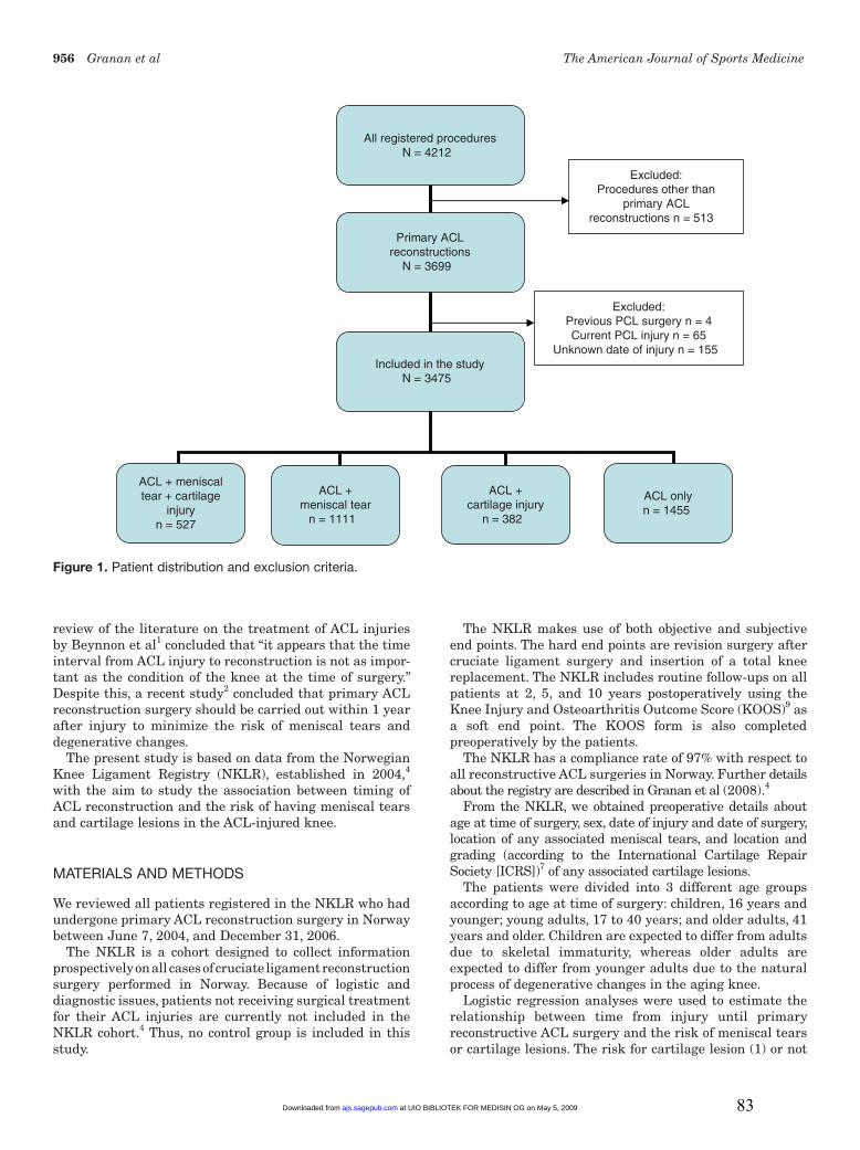
Figure 1. Patient distribution and exclusion criteria.

All registered proceduresN = 4212

ACL + meniscaltear + cartilage

injuryn = 527

ACL +meniscal tear

n = 1111

ACL onlyn = 1455

Excluded:Procedures other than

primary ACLreconstructions n = 513

Excluded:Previous PCL surgery n = 4Current PCL injury n = 65

Unknown date of injury n = 155 Included in the study

N = 3475

Primary ACLreconstructions

N = 3699

ACL +cartilage injury

n = 382

at UIO BIBLIOTEK FOR MEDISIN OG on May 5, 2009ajs.sagepub.comDownloaded from 83

Vol. 37, No. 5, 2009 Timing of ACL Reconstructive Surgery 957

(0), as well as for meniscal tears (1) or not (0), was studied using the logistic regression models. First, unadjusted analyses were performed to identify potential confounders. The relationships between time from injury until surgery and risk factors and between potential confounders and the risk of cartilage lesions or meniscal tears were calculated. Risk factors with a significant relationship (using P < .20) with time from injury until surgery and potential confounders with a significant relationship (using P < .20) to either cartilage lesion or meniscal tear prevalence were used as adjustment factors for potential confounding in the adjusted logistic regression models. The factors identified were age, sex, previous knee joint surgery (ie, surgery to medial collateral ligament [MCL], lateral collateral ligament [LCL], posterolateral corner [PLC], cartilage, medial meniscus, lateral meniscus, or other specified structure), current knee ligament injury (ie, LCL, MCL, and/or PLC), meniscal tears, and cartilage lesions. The analyses were stratified by age groups and adjusted for time to surgery, sex, age (as a continuous variable), previous knee joint surgery, current knee ligament injury, and the presence of cartilage lesions or meniscal tears at the time of surgery.

Unadjusted analysis was performed to estimate the mean difference in months from injury until surgery between risk factors and confounding factors. P values less than .05 were considered to be statistically significant. Odds ratios are presented with 95% confidence intervals (CIs).

All statistical analyses were performed using SPSS for Windows, version 15.0 (SPSS, Chicago, Illinois).

RESULTS

A total of 4212 procedures were registered in the NKLR, and 3699 of these were primary ACL reconstructions (Figure 1). After excluding patients with previous or cur-rent posterior cruciate ligament injury or surgery and cases in which the date of injury was unknown, we were left with 3475 knees. The median time from injury to surgery was 7 months (range, 9 days to 482 months). Of the 3475 cases identified, there were 1977 (57%) male and 1498 (43%) female patients, with a median age of 27 years (range, 12-67 years).

The number of patients, sex, age, distribution of current and previous surgeries, and distribution of meniscal and cartilage injuries across age groups are shown in Table 1. Of 246 cases with at least 1 cartilage lesion grade 3 or 4, 120 cases (49%) had 1 or more lesions larger than 2 cm2.

Among children, we were not able to detect a significant effect of time elapsed from injury until surgery on the prevalence of either cartilage lesions (Table 2) or meniscal tears (Table 3). The presence of cartilage lesions led to increased odds for the presence of meniscal tears (Table 3). Conversely, the odds for cartilage lesions were also increased in the presence of meniscal tears (Table 2). Within this age

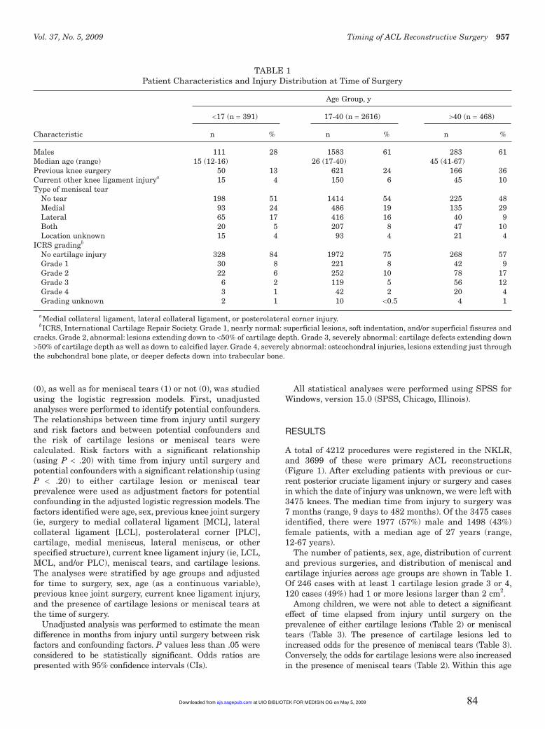
TABLE 1Patient Characteristics and Injury Distribution at Time of Surgery

Age Group, y

<17 (n = 391) 17-40 (n = 2616) >40 (n = 468)

Characteristic n % n % n %

Males 111 28 1583 61 283 61Median age (range) 15 (12-16) 26 (17-40) 45 (41-67) Previous knee surgery 50 13 621 24 166 36Current other knee ligament injurya 15 4 150 6 45 10Type of meniscal tear No tear 198 51 1414 54 225 48 Medial 93 24 486 19 135 29 Lateral 65 17 416 16 40 9 Both 20 5 207 8 47 10 Location unknown 15 4 93 4 21 4ICRS gradingb No cartilage injury 328 84 1972 75 268 57 Grade 1 30 8 221 8 42 9 Grade 2 22 6 252 10 78 17 Grade 3 6 2 119 5 56 12 Grade 4 3 1 42 2 20 4 Grading unknown 2 1 10 <0.5 4 1

aMedial collateral ligament, lateral collateral ligament, or posterolateral corner injury.bICRS, International Cartilage Repair Society. Grade 1, nearly normal: superficial lesions, soft indentation, and/or superficial fissures and

cracks. Grade 2, abnormal: lesions extending down to <50% of cartilage depth. Grade 3, severely abnormal: cartilage defects extending down >50% of cartilage depth as well as down to calcified layer. Grade 4, severely abnormal: osteochondral injuries, lesions extending just through the subchondral bone plate, or deeper defects down into trabecular bone.

at UIO BIBLIOTEK FOR MEDISIN OG on May 5, 2009ajs.sagepub.comDownloaded from 84

958 Granan et al The American Journal of Sports Medicine

group, we also found that the prevalence of meniscal tears decreased with age.

In the young adult group, there were several factors that influenced the prevalence of cartilage and meniscal lesions. An increase in odds with time to surgery was seen

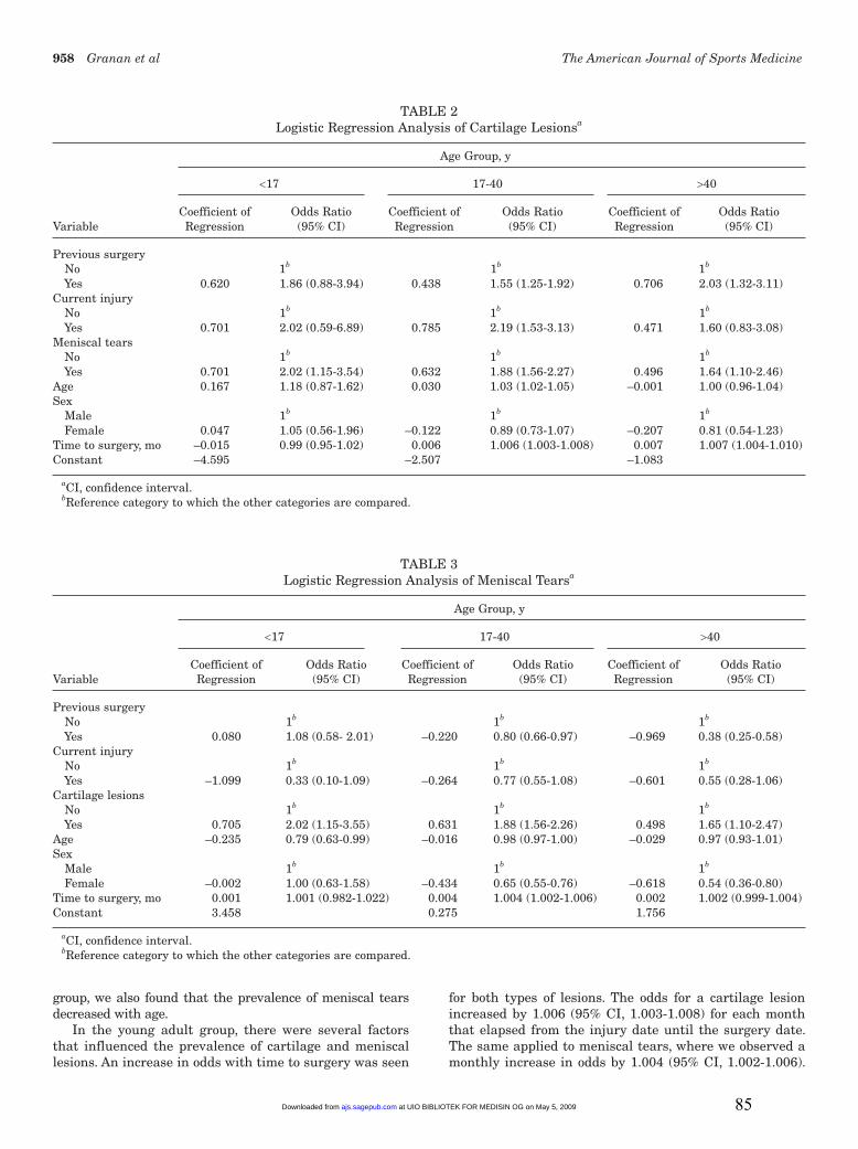
for both types of lesions. The odds for a cartilage lesion increased by 1.006 (95% CI, 1.003-1.008) for each month that elapsed from the injury date until the surgery date. The same applied to meniscal tears, where we observed a monthly increase in odds by 1.004 (95% CI, 1.002-1.006).

TABLE 2Logistic Regression Analysis of Cartilage Lesionsa

Age Group, y

<17 17-40 >40

Coefficient of Odds Ratio Coefficient of Odds Ratio Coefficient of Odds Ratio Variable Regression (95% CI) Regression (95% CI) Regression (95% CI)

Previous surgery No 1b 1b 1b

Yes 0.620 1.86 (0.88-3.94) 0.438 1.55 (1.25-1.92) 0.706 2.03 (1.32-3.11)Current injury No 1b 1b 1b

Yes 0.701 2.02 (0.59-6.89) 0.785 2.19 (1.53-3.13) 0.471 1.60 (0.83-3.08)Meniscal tears No 1b 1b 1b

Yes 0.701 2.02 (1.15-3.54) 0.632 1.88 (1.56-2.27) 0.496 1.64 (1.10-2.46)Age 0.167 1.18 (0.87-1.62) 0.030 1.03 (1.02-1.05) –0.001 1.00 (0.96-1.04)Sex Male 1b 1b 1b

Female 0.047 1.05 (0.56-1.96) –0.122 0.89 (0.73-1.07) –0.207 0.81 (0.54-1.23)Time to surgery, mo –0.015 0.99 (0.95-1.02) 0.006 1.006 (1.003-1.008) 0.007 1.007 (1.004-1.010)Constant –4.595 –2.507 –1.083

aCI, confidence interval.bReference category to which the other categories are compared.

TABLE 3Logistic Regression Analysis of Meniscal Tearsa

Age Group, y

<17 17-40 >40

Coefficient of Odds Ratio Coefficient of Odds Ratio Coefficient of Odds Ratio Variable Regression (95% CI) Regression (95% CI) Regression (95% CI)

Previous surgery No 1b 1b 1b

Yes 0.080 1.08 (0.58- 2.01) –0.220 0.80 (0.66-0.97) –0.969 0.38 (0.25-0.58)Current injury No 1b 1b 1b

Yes –1.099 0.33 (0.10-1.09) –0.264 0.77 (0.55-1.08) –0.601 0.55 (0.28-1.06)Cartilage lesions No 1b 1b 1b

Yes 0.705 2.02 (1.15-3.55) 0.631 1.88 (1.56-2.26) 0.498 1.65 (1.10-2.47)Age –0.235 0.79 (0.63-0.99) –0.016 0.98 (0.97-1.00) –0.029 0.97 (0.93-1.01)Sex Male 1b 1b 1b

Female –0.002 1.00 (0.63-1.58) –0.434 0.65 (0.55-0.76) –0.618 0.54 (0.36-0.80)Time to surgery, mo 0.001 1.001 (0.982-1.022) 0.004 1.004 (1.002-1.006) 0.002 1.002 (0.999-1.004)Constant 3.458 0.275 1.756

aCI, confidence interval.bReference category to which the other categories are compared.

at UIO BIBLIOTEK FOR MEDISIN OG on May 5, 2009ajs.sagepub.comDownloaded from 85

Vol. 37, No. 5, 2009 Timing of ACL Reconstructive Surgery 959

Previous surgery increased the odds for having a cartilage lesion (Table 2), whereas it decreased the odds for having a meniscal tear (Table 3). A current injury of the MCL, LCL, and/or PLC was associated with increased odds for cartilage lesions (Table 2). The presence of a meniscal tear increased the odds for cartilage lesions (Table 2) and vice versa (Table 3). The older the young adults were, the higher the odds were for a cartilage lesion (Table 2), whereas the odds for having a meniscal tear decreased with increasing age (Table 3). Being female reduced the odds of having a meniscal tear (Table 3), whereas there was no gender effect on the risk for cartilage lesions (Table 2).

In the older adult group, the odds for having a cartilage lesion increased by 1.007 (95% CI, 1.004-1.010) for each month that elapsed from the injury date until the surgery date, whereas there was no association between time until surgery and the odds for meniscal tears. The presence of previous surgery to knee ligaments, cartilage, and/or menisci increased the odds for having cartilage lesions (Table 2), whereas the odds for having meniscal tears were decreased (Table 3). An additional meniscal tear increased the odds for a cartilage lesion (Table 2) and vice versa (Table 3). Being female reduced the odds of having a meniscal tear (Table 3), but there was no effect on the odds for cartilage injuries.

Table 4 displays the mean differences in months from injury until surgery between sexes, previous knee joint surgery to the index knee, current knee ligament injury other than cruciate ligament injuries, patient age groups, and the presence of either meniscal tears or cartilage lesions.

DISCUSSION

The main findings of this study were that the odds for a cartilage lesion in the adult knee increased by nearly 1% for each month that elapsed from the injury date until the surgery date and that cartilage lesions were nearly twice as frequent if there were a meniscal tear and vice versa.

The main strength of our study is the large number of patients included. Another strong point is that the patients originated from a national and general population of ACL-injured patients. The main weakness of this study is that all details regarding the patients are solely based on the individual orthopaedic surgeons reporting to NKLR. The collected data regarding the condition of the cartilage and menisci are based on the arthroscopic findings of many different surgeons, and their estimations of cartilage injury location, size, and depth may vary. One level III study regarding ICRS scoring has been published suggesting that this system is valid for the assessment of cartilage repair and has been found to have good interpersonal value and be repeatable and, as such, is regarded as a precise tool in the evaluation of cartilage repair.9

In the present study, patients who had asymptomatic cartilage or meniscal injury before their ACL injuries represent a potential source of bias. One cannot be entirely sure that the cartilage and meniscal tears reported to the NKLR had been sustained at or after the index ligament injury. Another potential limitation is that patients who expect instability to be a problem or cannot afford instability problems (eg, manual laborers, professional athletes, those who perform pivoting leisure-time activities) are more likely to undergo surgery early in contrast to patients who receive surgery after having experienced at least 1 episode of instability or giving way of the knee. One might expect that older patients are more likely to try nonoperative treat-ment first and wait longer before undergoing surgery. This is an argument supported by the data presented in Table 4. In addition to this, previous data from Norway5 have estimated that at least 50% of patients with ACL injuries are treated nonoperatively. On the other hand, it is also likely that as time goes by, the chance of having surgery increases if you sustain further injuries to the knee. Then again, surgeons do have different practice profiles. Some are in favor of early surgery, some are leaning toward surgery after a thorough rehabilitation period, and some are somewhere between these 2 practice profiles. The consequence of one, some, or all these aspects is that we might overestimate the importance of time as a risk factor for developing degenerative lesions. The registration of preoperative KOOS data might to some degree counterbalance these limitations. One could argue that trying to formalize and register the patients’ reasons for delaying or undergoing reconstructive surgery would be a more desirable approach. The NKLR’s steering committee is currently reviewing this issue.

There are 2 other important variables that might bias the results; unfortunately, they are not yet part of the NKLR’s registration form. These are the patient’s weight and activity level. Either one of these factors is considered to increase the incidence of cartilage lesions and/or meniscal

TABLE 4Mean Difference in Months From Injury Until Surgery

Between Risk Factors and Confounding Factors

Mean 95% Confidence Variable Difference Interval

Previous surgery No –22 –26 to –19 Yes 0a Current injury No 13 6 to 19 Yes 0a Cartilage lesions No –16 –19 to –13 Yes 0a Meniscal tears No –4 –7 to –1 Yes 0a Age groups, y <17 –33 –39 to –27 17-40 –22 –27 to –18 >40 0a Sex Male –5 –8 to –2 Female 0a

aReference category to which the other categories are compared.

at UIO BIBLIOTEK FOR MEDISIN OG on May 5, 2009ajs.sagepub.comDownloaded from 86

960 Granan et al The American Journal of Sports Medicine

tears.6,12,15 Both factors are under consideration by the NKLR’s steering committee for inclusion in both the preoperative and postoperative patient assessments.

There are different opinions on whether reconstructive surgery will result in fewer degenerative changes in the ACL-deficient knee in the long run compared with nonoperative treatment. A recent article by Drogset et al3 suggested that early surgical intervention would be beneficial because the knees at an early stage had far less cartilage damage than did knees with late surgery. Our results confirm this. A recent study2 based on review of 183 cases concluded that primary ACL reconstruction surgery should be carried out within 12 months of injury to minimize the risk of meniscal tears and degenerative changes. In this study, presence and type of meniscal tear and type of degenerative change were recorded. The incidence of meniscal tears and degenerative change was assessed and related to the timing from injury to surgery. The patients were divided into an early group (surgery within 12 months of injury) and a late group (surgery more than 12 months from injury). Incidence of meniscal tears was significantly higher in patients undergoing reconstruction late compared with those in the early group (71% vs 42%).

Six percent of the patients with ACL injuries had additional ligament injuries. The presence of these additional injuries might be owing to more severe trauma or more instability and as such explain the reason for these patients receiving surgery 1 year earlier than did those without other ligament injuries. Whereas Beynnon et al1 found that ACL injuries are more prevalent among female athletes than male athletes, more ACL recon structions are performed on male athletes because more males participate in at-risk sports, such as team handball and soccer.

Regarding the children’s age group, the distribution in time from injury to surgery in relation to type of meniscal tear and ICRS grading does reflect relatively fewer findings (data not shown) in the children who received surgery in the latter end of the time scale. This probably reflects 2 tendencies among Norwegian orthopaedic surgeons. First, the most severely injured knees are operated on fairly soon. And second, in Norway, ACL reconstruction in children seldom occurs before the age of 14 years. This leads to a long time period between injury and stabilizing surgery for children with an early ACL tear. The protocol for these children consists of activity modification and use of a brace when performing knee-demanding activities. These data indicate that this approach does not lead to high incidence of meniscal tears and/or cartilage lesions.13

The change in odds for a single patient who chooses to have late surgery, the accumulated odds of 1 specific patient at a given time, and the difference in odds between 2 patients may be calculated using the coefficient of regression presented in Tables 2 and 3. This is illustrated in the following 2 examples (described in more detail in the appendix, available online at http://ajs.sagepub.com/supplemental/): a 34-year-old patient with previous surgery to the index knee has an additional ligament injury and a meniscal tear. The increase in odds for having a cartilage lesion for a 2-year difference in the timing of surgery is 1.2.

This illustrates that the statistical risk for cartilage lesions increases by 20% for a 2-year difference in the timing of surgery for patients belonging to the patient group of young adults with the same risk profile as presented in this example. This example only calculates the additional increased odds for having cartilage lesions if surgery is 2 years later and does not include the increased risk at time zero (ie, previous surgery to the index knee, additional ligament injury, and a meniscal tear giving a baseline odds of 1.3).

Another example illustrates the difference in odds between 2 patients. Patient A is a 17-year-old male with no previous surgery to the index knee, no additional ligament injuries, and no damaged menisci. Patient B is twice as old (34 years), is the same sex, has previous surgery to the index knee, and has an additional ligament injury and a meniscal tear. These risk profiles will give patient B a 10.6 times increased odds for having cartilage lesions in relation to patient A, which gives an increased odds of 960%, based on the logistic regression model.

On the basis of our results on adults, early surgery may be recommended. However, it is important to remember that many surgeons consider these patients to benefit from preoperative rehabilitation8,16 and that some patients may do well without surgery if they do not participate in high-risk activities.1,11 If sufficient improvement is not achieved within reasonable time, surgery should be considered. A reasonable cutoff can be calculated for each patient based on Tables 2 and 3.

Church and Keating2 specifically attempted to relate the development of degenerative changes in the knee to the timing of primary reconstruction surgery of the ACL. Our findings do concur with their main conclusions. To extend their findings, we have tried to provide both the physicians and the physical therapists with a new and more individualized tool to help in the decision making.

In conclusion, the odds for a cartilage lesion in the adult knee increased by nearly 1% for each month that elapsed from the injury date until the surgery date, and the presence of cartilage lesions was associated with a nearly 2-fold increase in the risk of having meniscal tears, and vice versa, independent of patient age. Our data suggest that early surgery is associated with fewer meniscal tears and cartilage injuries.

ACKNOWLEDGMENT

The Norwegian Knee Ligament Registry (NKLR) is financed by the Oslo Sports Trauma Research Center, which has been established through generous grants from the Eastern Norway Regional Health Authority, the Royal Norwegian Ministry of Culture, the Norwegian Olympic Committee & Confederation of Sport, and Norsk Tipping AS. In addition, the NKLR has been supported through a grant from the Norwegian Medical Association’s Fund for Quality Improvement. Lars-Petter Granan has been sup-ported by the Medical Research Curriculum at the University of Oslo. The authors thank Kjersti Steindal, computer engineer, for extracting the data from the NKLR; the NKLR secretaries, Ruth Gunvor Wasmuth and

at UIO BIBLIOTEK FOR MEDISIN OG on May 5, 2009ajs.sagepub.comDownloaded from 87

Vol. 37, No. 5, 2009 Timing of ACL Reconstructive Surgery 961

Marianne Wiese; and the staff and colleagues of the par-ticipating orthopaedic and surgical departments for their cooperation. In support of their research for or preparation of this article, one or more of the authors received grants or outside funding from Norwegian Eastern Health Corporate, Royal Norwegian Ministry of Culture, Norwegian Olympic Committee and Confederation of Sport, Norsk Tipping AS, and Pfizer AS. None of the authors received payments or other benefits or a commitment or agreement to provide such benefits from a commercial entity. No com-mercial entity paid or directed, or agreed to pay or direct, any benefits to any research fund, foundation, educational institution, or other charitable or nonprofit organization with which the authors are affiliated or associated.

REFERENCES

1. Beynnon BD, Johnson RJ, Abate JA, Fleming BC, Nichols CE. Treatment of anterior cruciate ligament injuries, part I. Am J Sports Med. 2005;33(10):1579-1602.

2. Church S, Keating JF. Reconstruction of the anterior cruciate liga-ment: timing of surgery and the incidence of meniscal tears and degenerative change. J Bone Joint Surg Br. 2005;87(12):1639-1642.

3. Drogset JO, Grontvedt T, Robak OR, Molster A, Viset AT, Engebretsen L. A sixteen-year follow-up of three operative techniques for the treat-ment of acute ruptures of the anterior cruciate ligament. J Bone Joint Surg Am. 2006;88(5):944-952.

4. Granan LP, Bahr R, Steindal K, Furnes O, Engebretsen L. Development of a national cruciate ligament surgery registry: the Norwegian National Knee Ligament Registry. Am J Sports Med. 2008;36(2):308-315.

5. Granan LP, Engebretsen L, Bahr R. Surgery for anterior cruciate liga-ment injuries in Norway [in Norwegian]. Tidsskr Nor Laegeforen. 2004;124(7):928-930.

6. Griffin TM, Guilak F. The role of mechanical loading in the onset and progression of osteoarthritis. Exerc Sport Sci Rev. 2005;33(4): 195-200.

7. International Cartilage Repair Society. The cartilage standard evalua-tion form/knee and cartilage repair assessment. Newsletter ICRS. Spring 1998:5-8.

8. Keays SL, Bullock-Saxton JE, Newcombe P, Bullock MI. The effec-tiveness of a pre-operative home-based physiotherapy programme for chronic anterior cruciate ligament deficiency. Physiother Res Int. 2006;11(4):204-218.

9. Knee Injury and Osteoarthritis Outcome Score. Available at: http://www.koos.nu. Accessed September 2008.

10. Kocher MS, Smith JT, Zoric BJ, Lee B, Micheli LJ. Transphyseal ante-rior cruciate ligament reconstruction in skeletally immature pubescent adolescents. J Bone Joint Surg Am. 2007;89(12):2632-2639.

11. Kostogiannis I, Ageberg E, Neuman P, Dahlberg L, Friden T, Roos H. Activity level and subjective knee function 15 years after anterior cruciate ligament injury: a prospective, longitudinal study of nonre-constructed patients. Am J Sports Med. 2007;35(7):1135-1143.

12. L’Hermette MF, Tourny-Chollet C, Polle G, Dujardin FH. Articular car-tilage, degenerative process, and repair: current progress. Int J Sports Med. 2006;27(9):738-744.

13. Moksnes H, Engebretsen L, Risberg MA. Performance-based func-tional outcome for children 12 years or younger following anterior cruciate ligament injury: a two to nine-year follow-up study. Knee Surg Sports Traumatol Arthrosc. 2008;16(3):214-223.

14. Shelbourne KD, Wilckens JH, Mollabashy A, DeCarlo M. Arthrofibrosis in acute anterior cruciate ligament reconstruction: the effect of timing of reconstruction and rehabilitation. Am J Sports Med. 1991;19(4): 332-336.

15. Wearing SC, Hennig EM, Byrne NM, Steele JR, Hills AP. Musculoskeletal disorders associated with obesity: a biomechanical perspective. Obes Rev. 2006;7(3):239-250.

16. Wilk KE, Reinold MM, Hooks TR. Recent advances in the rehabilita-tion of isolated and combined anterior cruciate ligament injuries. Orthop Clin North Am. 2003;34(1):107-137.

at UIO BIBLIOTEK FOR MEDISIN OG on May 5, 2009ajs.sagepub.comDownloaded from 88

Both examples make use of the coefficients of regression (young adults) drawn from table 2.

Example 1

A 34-year old (0.030 * 34) male with previous surgery to the index knee (0.438) has an additional ligament injury (0.785) and a meniscal tear (0.632). The increase in odds for having a cartilage lesion for a two-year (= 24 months) difference in the timing of surgery (0.006 * 24) may be calculated as follows. -2.507 is the constant for the young adults.

When inserting the values for these variables at a given time after injury

logit (pA0) = -2.507 + 0.030 * 34 + 0.438 + 0.785 + 0.632

and two years later (the patient is now 36 years: 0.030 * 36, instead of 0.030 * 34)

logit (pA1) = -2.507 + 0.030 * 36 + 0.438 + 0.785 + 0.632 + 0.006 * 24

The difference logit (pA1) – logit (pA0) is

logit (pA1) – logit (pA0) = 0.030 * (36 – 34) + 0.006 * 24 = 0.204

which gives an odds ratio of

exp(0.204) = 1.2.

To simplify the calculations the expression exp(0.204) can be exchanged with 2.7180.204. These calculations illustrate that that the risk for cartilage lesions increases by 20 % for a two-year difference in the timing of surgery. This example only calculates the additional increased odds for having cartilage lesions if surgery is two years later and do not include the increased risk at time 0 (i.e. previous surgery to the index knee, additional ligament injury and a meniscal tear giving a baseline odds of 1.3). This example does also illustrate how to calculate a reasonable cut off.

Example 2

Patient A is a 17 year old male (0.030 * 17) with no previous surgery to the index knee (0); no additional ligament injuries (0); and no damaged menisci (0). Patient B is twice as old (34 years; 0.030 * 34), same sex, with previous surgery to the index knee (0.438), and has an additional ligament injury (0.785) and a meniscal tear (0.632).

When inserting the values for these variables we will get for patient A

logit (pA) = -2.507 + 0.030 * 17 + 0 + 0 + 0

and for patient B

logit (pB) = -2.507 + 0.030 * 34 + 0.438 + 0.785 + 0.632

The difference logit (pB) – logit (pA) is

logit (pB) – logit (pA) = 0.030 * (34 – 17) + 0.438 + 0.785 + 0.632 = 2.365

which gives an odds ratio of

89

exp (2.365) = 10.6.

These calculations will give patient B a 10.6 times increased odds for having cartilage lesions in relation to patient A (i.e. an increased risk of 960 %).

90

Paper III

91

LP Granan

The Scandinavian ACL registries 2004-2007: baseline 1

epidemiology. 2

3

Lars-Petter Granan1,2, Martin Lind

3, Magnus Forssblad

4, and Lars Engebretsen

1,2,5 4

5

1Oslo Sports Trauma Research Center, Norwegian School of Sport Sciences, PB 4014 Ullevål 6

Stadion, 0806 Oslo, Norway 7

2National Knee Ligament Registry, Bergen, Norway 8

3Division of Sportstrauma, Orthopaedic Department, Aarhus University Hospital, Aarhus, 9

Denmark 10

4Capio Artro Clinic AB and Stockholm Sports Trauma Research Center, 11

Sophiahemmet, Stockholm, Sweden 12

5Orthopaedic Center, Ullevaal University Hospital and Faculty of Medicine, University of 13

Oslo, Norway 14

15

Correspondence LPG: [email protected] 16

17

92

LP Granan

Abstract 18

Background and purpose: No prospective surveillance systems have been available for 19

monitoring the outcome of cruciate ligament surgery in Scandinavia (Denmark, Norway and 20

Sweden). The present paper describes the Scandinavian ACL registries with their main 21

function, similarities and preliminary baseline results. 22

Methods: The Scandinavian registries were established in 2004 (Norway) and 2005 23

(Denmark and Sweden). The Danish and Swedish registries were developed based on the 24

Norwegian registry, and do not differ in any decisive way. In Denmark all hospitals and 25

clinics are legally bound to report to an approved national database. In Norway and Sweden 26

the registries are based on surgeons voluntarily reporting. 27

Results: The annual incidence of primary ACL reconstructions is higher in Denmark than in 28

Norway, except in females younger than 20 years. A similar approach to the patients exists 29

among the Scandinavian surgeons. Variations do however exist regarding choice of grafts, 30

implants, and treatment of simultaneous meniscal and cartilage injuries; the proportion of 31

ACL reconstructions performed as outpatient surgery; and the use of prophylactic 32

anticoagulation. The preoperative KOOS scores do not clinically significantly differ between 33

the Scandinavian registries, except for Denmark reporting more symptoms both pre- and 34

postoperatively. 35

Interpretation: The Scandinavian national ACL registries will generate new data about ACL 36

reconstructions. They will contribute important knowledge regarding ACL epidemiology. 37

They will be the only source for data on performance of a wide range of different implants 38

and techniques. They will hopefully have an impact on the selection of methods for ACL 39

reconstructions in Scandinavia and elsewhere. 40

41

93

LP Granan

Introduction 42

Scandinavian national arthroplasty registries have over the last two decades generated 43

important knowledge and served as an important quality control tool. Until Norway started 44

the world’s first national knee ligament registry in 2004 there were no prospective national 45

surveillance systems to monitor the outcome of knee ligament surgery (Granan et al. 2008). 46

We describe the 3 Scandinavian – Danish, Norwegian and Swedish – knee ligament registries 47

with their main function and similarities. Furthermore preliminary baseline results are 48

presented from the start of the registries until late 2007 for primary ACL reconstructions. 49

Patients and methods 50

The Scandinavian registries were established in June 2004 (Norway), January 2005 (Sweden) 51

and July 2005 (Denmark), the latter were developed based on the Norwegian registry. The 52

registries do not differ in any decisive way. Details on the Norwegian ACL registry have 53

previously been described by Granan et al. (2008). 54

The Norwegian and Swedish registries are based on surgeons voluntarily reporting to the 55

registries. In Denmark a law passed in June 2006 made it compulsory for all public and 56

private hospitals and clinics to report to the approved national, clinical databases. Reporting 57

to the databases in Denmark and Sweden is organized through a secured internet portal, thus 58

minimizing the costs of daily running. In Norway the registry relies on paper based reporting, 59

mainly due to the close cooperation with the Norwegian Arthroplasty Register (NAR) which 60

makes use of an identical system. 61

In Denmark 90 % of the orthopedic departments have been contributing to the registry with 62

an average compliance of 85 % of the performed primary ACL reconstructions. In Norway all 63

hospitals performing ACL surgeries have contributed with a total compliance of 97 %. In 64

94

LP Granan

Sweden some of the smaller hospitals with small volumes of ACL surgery have not been 65

included in the registry, yet still more than 71% of the hospitals have contributed to the 66

registry. 67

Follow up with KOOS forms are carried out by all 3 registries. In Denmark these follow ups 68

are done at 1, 5 and 10 years postoperatively; In Norway at 2, 5 and 10 years postoperatively; 69

and in Sweden at 1, 2, 5 and 10 years postoperatively. 70

All registries provide annual reports, national as well as for the individual hospitals. Sweden 71

also offers an on-line database where clinics can analyze their own statistics at any time. The 72

Danish database is managed by a special university center which manages all Danish national 73

orthopedic databases. In Norway the technical responsibility is with the Helse Vest IKT AS, 74

which manages all Norwegian national orthopedic databases. In Sweden the Capio Artro 75

Clinic in Stockholm is responsible for the registry on a daily basis. 76

For this present study on this cohort, data regarding common and comparable variables 77

(activities causing injury; age at injury and surgery; choice of graft; duration of surgery; 78

frequency of cartilage and meniscal injuries, meniscal resections, and cartilage treatments; 79

number of reconstructions and hospitals; graft fixation devices; outpatient surgery, pre 80

operative and post operative KOOS; prophylactic antibiotics and anticoagulation; sex; and 81

time to surgery) in relation to primary ACL reconstructions were extracted. 82

Ethics 83

In Norway the participation is voluntary, and all patients are asked to sign an informed 84

consent form before surgery. The consent form contains information about the Norwegian 85

ACL registry, the type of information that is recorded, data protection, the procedure for 86

follow ups and informs the patient that he or she may be invited to participate in research 87

projects at a later stage. The registration forms are signed by the surgeons, but they are not 88

95

LP Granan

possible to trace in the registry database since the surgeon’s identity is not recorded, due to a 89

mutual agreement among the Norwegian orthopaedic registries. For follow ups the patients 90

are identified by their unique social security number (including date of birth), which is 91

assigned to all Norwegian residents. The social security number is used to link the Knee 92

injury and Osteoarthritis Outcome Score (KOOS) and registration forms, and to update the 93

registry annually with data about knee arthroplasties from NAR, and death and emigration 94

data before obtaining data files for analysis. The Norwegian ACL registry has been approved 95

by the Norwegian Data Inspectorate as an expansion of the NAR concession. In Denmark and 96

Sweden, no consent is necessary for national clinical databases. In Denmark and Sweden the 97

social security number is used to access patients to the database and for identifying data. 98

Results 99

The total number of reported primary ACL reconstructions was 4972 in Denmark, 5329 in 100

Norway, and 7331 in Sweden. The distributions of male patients were 57 % in Norway, 58 % 101

in Sweden, and 60 % in Denmark. Of the Danish patients 1939 (39%) had simultaneous 102

meniscal injuries and 825 (17%) had cartilage injuries. In the Norwegian patients the 103

corresponding figures were 2914 (55%) and 1456 (27%), and in Sweden 2536 (35%) and 104

2001 (27%). The median age of the patients at the time of injury varied between 23 (Sweden) 105

and 27 years (Denmark), while the median age at the time of surgery varied between 25 106

(Sweden) and 30 years (Denmark). The median time, in months from injury to surgery varied 107

between 7 (Norway) and 10 (Sweden). At surgery the median duration varied between 68 min 108

(Denmark) and 71 min (Sweden). Outpatient surgery was performed in 38 % of the cases in 109

Norway, 56 % in Sweden and 79 % in Denmark. In all countries 99 % of the patients received 110

prophylactic antibiotics, while the use of prophylactic anticoagulation varied between 17 % in 111

Denmark and 78 % in Norway. These surgeries were conducted on 37 hospitals in Denmark, 112

96

LP Granan

52 hospitals in Sweden, and 60 hospitals in Norway. Hamstring autografts were the most 113

frequently used graft in all of Scandinavia (Norway 61 %; Denmark 71 %; Sweden 86 %). 114

Most often soccer was the cause of injury (Norway 40 %; Sweden 41 %; Denmark 50 %) 115

(Table 1). 116

The KOOS data, both preoperatively and postoperatively, displayed no national clinical 117

significant differences in any of the subscales, except for the poorer symptom scores in the 118

Danish patients (Table 2). The Danish KOOS data is based on 50 % of the patients in the 119

registry, while the Norwegian data constitutes 88 % of the registered patient population. 120

The annual incidence of primary ACL reconstructions in Norway was 34 per 100,000 citizens 121

(Granan et al. 2008), while in Denmark the incidence was 38/100,000 (Lind et al. 2008), and 122

in Sweden 32/100,000. On the other hand the real population at risk – that is 16-39 year age 123

group – had an incidence of 85 primary ACL reconstructions per 100,000 citizens in Norway 124

(Granan et al. 2008), while the Danish incidence was 91/100,000 for the 15-39 year age group 125

(Lind et al. 2008), and the Swedish incidence was 71/100,000 for the 20-39 year age group. 126

The annual incidence of primary ACL reconstructions for the different age groups in 127

Denmark, Norway and Sweden are displayed in table 3. 128

Discussion 129

In general the registries provided detailed epidemiological data. Based on conservative 130

estimates the Scandinavian ACL registries are expected to generate an annual average of 2500 131

patients in each of the Danish and Swedish registries, and 1600 patients in the Norwegian 132

registry. After 5 years more than 30,000 cases will be in the registries yielding data such as 133

the revision rates; KOOS; and the outcome related to various techniques and used implants. 134

It is also important to emphasize what the registries will not be able to demonstrate. There is 135

no radiographical follow-up of the ACL reconstructed patients. Consequently, data regarding 136

97

LP Granan

the development of radiographically verified osteoarthritis will not be obtainable. The choice 137

of not doing radiographic follow-ups is due to both financial restraints and the intention to not 138

put additional demands on the hospitals that are beyond their own follow-up routines. More 139

advanced investigations (e.g. gait analysis and muscle strength) are also omitted, due to the 140

same arguments. 141

It is the registries’ main intention to contribute to quality control and improvement of the 142

surgical cruciate ligament procedures. This may be done through establishing evidence based 143

national guidelines and protocols for surgical procedures and rehabilitation. To understand the 144

importance of reported failures, we need to know the actual number of reconstruction and 145

revision surgeries that are performed. Nordic arthroplasty registries have previously provided 146

accurate data of sufficient quality. The Norwegian ACL registry has calculated that if 14 147

patients with one specific fixation device fail, this may be considered a failure of that specific 148

device (Granan et al. 2008). This will enable the registries to give early warnings on poor 149

procedures and devices, and identify prognostic factors etc (Granan et al. 2008). 150

The registries must provide information for the orthopedic community at regular intervals on 151

the outcomes of surgical treatment of the cruciate ligaments with different methods. The hard 152

endpoints are clear and unequivocal, i.e. revision reconstruction and total knee replacement. 153

Causality of failure may not be sufficiently and accurately documented in the registries, but it 154

will provide information as to where there may be potential problems and direct future 155

analysis and studies toward these areas. Since the registries will provide real time information 156

and thus can be analyzed on an ongoing basis, they have the potential to reveal problems long 157

before they would be reported by traditional methods (e.g. RCTs). This will undoubtedly 158

benefit all interested parties, not at least the patients. 159

An important limitation in these registries is bias due to limitation in follow-ups. From the 160

Norwegian registry we know that baseline compliance is high both in respect to registration 161

98

LP Granan

forms (97 %) and KOOS forms (88 %). Mandatory reporting has been instituted in Denmark. 162

This might be the most important condition to obtain a high and sustainable compliance. Due 163

to the Scandinavian unique social security numbers it is easy to reach every patient, and thus 164

increase the response rate in the follow-ups. 165

There still are issues where the Scandinavian registries have no solutions. Due to logistic and 166

diagnostic issues, patients not receiving surgical treatment for their ACL injury are currently 167

not included in the Scandinavian registries. Thus, no data on the outcome of non-operatively 168

treated ACL injuries are obtained. Another limitation in these registries is the use of revision 169

as a primary end point. This is suboptimal since an unknown number of patients accept to live 170

with an inferior clinical outcome to avoid more surgery. However, if they undergo surgery for 171

debridement or arthroscopic surgery for other indications, they will be detected in the registry. 172

Knee arthroplasty has limitations as an endpoint because it can take several decades before a 173

patient with a poorly functioning knee is accepted as a knee arthroplasty candidate. Not all 174

patients with ACL insufficiencies develop osteoarthritis to a degree where knee arthroplasty is 175

indicated (Lohmander et al. 2007). 176

The registration of potential risk factors other than type of surgical procedure may be subject 177

to selection bias. The data items recorded are a minimal set suited for a paper-based or web-178

based reporting system, not to exceed one page. As such there has to be a careful, ongoing 179

selection of what is expected to be the most important risk factors. Thus, there is no way of 180

knowing the influence of the omitted variables. Finally there might be limitations due to 181

differences between Scandinavia and other countries in respect of indications for surgery and 182

patient success criteria. 183

Prospective national registries have several advantages. Inclusion of cases from an entire 184

nation generates a high volume of data. This in turn, will lead to the possibilities of drawing 185

early decisive conclusions. Another advantage is due to the nature of cohort studies, an 186

99

LP Granan

ongoing accumulation of short term and long term follow-up data. Finally there is the 187

advantage of monitoring development, implementation and evolution of new – and old – 188

techniques, implants, prophylactic medications and so forth. Although RCTs are the gold 189

standard in research methods and are immensely valuable for detailed testing, they are 190

insufficient when assessing techniques. A RCT aiming to demonstrate a 5 % difference in 191

revision rates after ACL surgery would need nearly 500 patients in each group, far more than 192

usually included in a typical RCT in knee ligament surgery. 193

Entirely web-based ACL registries are possible to develop – as demonstrated by the Danish 194

and Swedish registries – and are accessible and cost effective. Some restraints exist due to 195

various countries national legislation and infrastructure. Ultimately an emerging international 196

cooperation is expected to increase quality, open barriers and create an open minded 197

international discussion about methods and results in primary ACL reconstructions. 198

The different annual numbers in the Scandinavian registries are due to the differences in the 199

population sizes. Even though Norway is the smallest country it has the largest number of 200

hospitals. The explanation is likely to be due to a scattered population in a relatively long and 201

narrow country. 202

The data in Table 1 reflects the similar approach to the patients among the Scandinavian 203

surgeons. Some cultural variations do, however exist: The Swedish and Danish surgeons 204

prefer hamstring grafts to a much larger extent than the Norwegian surgeons. The reporting of 205

cartilage injuries in Norway was the first few years infested with flaws and inconsistency. 206

This might explain why Norwegian surgeons report more than 50 % as many cartilage injuries 207

as their Danish colleagues, and surgically treat less cartilage injuries than the Danes. 208

However, the Swedish data are identical to the Norwegian. On the other hand, Norway report 209

substantially more meniscal injuries than Denmark and Sweden, but treat relatively fewer 210

injuries. This probably reflects cultural and national attitudes. The variation in ACL 211

100

LP Granan

reconstructions performed as outpatient surgery probably reflects the variation in the 212

Scandinavian structure of the health care systems. The large variation in use of prophylactic 213

anticoagulation is of interest, however, postoperative trombo-embolic complications are not 214

recorded. 215

In respect of choice of autograft and fixation, the implants used (data not shown) in more than 216

2/3 of the cases varied between 1 and 3 different implants in the different registries. This 217

gives an overall total of 4 to 6 different implants when looking at various grafts and their 218

different fixation sites. This variation in the Scandinavian countries might be due to personal 219

preferences, skill of medical company sales team or local financial decisions, or more likely 220

combinations of these factors. There are no clinically significant differences in any KOOS 221

subscale either pre- or post-operatively among the Scandinavian countries. The only 222

exception is that Danish patients report clinically significant poorer symptoms score than their 223

Norwegian and Swedish counterparts, both pre-operatively and post-operatively. 224

Furthermore, the Danish and Swedish baseline KOOS data reveal an unsatisfactory 225

compliance rate, for unknown reasons. The baseline KOOS (Table 2) are the most 226

comprehensive data set published to date, and should be regarded as the reference values for 227

preoperative KOOS in ACL injured patients. 228

There are as yet no explanations to the large discrepancies among the Scandinavian incidence 229

data (Table 3). These differences must be investigated more thoroughly in separate studies. 230

The Scandinavian national ACL registries will generate new data about ACL reconstructions. 231

They will contribute to a better understanding of the ACL epidemiology. They will be the 232

only source for data on performance of a wide range of different implants and techniques. 233

They will influence the selection of methods for ACL reconstructions in both Scandinavia and 234

hopefully other countries in the future. 235

101

LP Granan

Contributions of authors 236

LPG designed the study, analyzed the Norwegian data, wrote the manuscript, and is 237

guarantor. LE participated in the design of the study and writing of the manuscript, and 238

supervised the work of LPG. ML participated in the design and writing of the manuscript, and 239

analyzed the Danish data. MF participated in the design and writing of the manuscript, and 240

analyzed the Swedish data. 241

Acknowledgements 242

The NKLR is financed by the Oslo Sports Trauma Research Center, which has been 243

established at the Norwegian School of Sport Sciences through generous grants from the 244

Royal Norwegian Ministry of Culture, the South-Eastern Norway Regional Health Authority, 245

the Norwegian Olympic Committee & Confederation of Sport, and Norsk Tipping AS. In 246

addition, the NKLR has been supported through a grant from the Norwegian Medical 247

Association's Fund for Quality Improvement. The Norwegian authors wish to thank Kjersti 248

Steindal, computer engineer, for extracting the data from the National Knee Ligament 249

Registry; NKLR secretaries, Merete Husøy, Ruth Gunvor Wasmuth and Marianne Wiese; and 250

the staff and colleagues of the participating orthopedic and surgical departments for their 251

cooperation. 252

253

102

LP Granan

References 254

Granan LP, Bahr R, Steindal K, Furnes O, Engebretsen L. Development of a national 255

cruciate ligament surgery registry: the Norwegian National Knee Ligament Registry. Am 256

J Sports Med 2008 Feb;36(2):308-15. 257

Havelin LI, Espehaug B, Vollset SE, Engesaeter LB. Early aseptic loosening of 258

uncemented femoral components in primary total hip replacement: a review based on the 259

Norwegian Arthroplasty Register. J Bone Joint Surg Br. 1995;77:11-17. 260

Havelin LI, Espehaug B, Vollset SE, Engesaeter LB. The effect of the type of cement on 261

early revision of Charnley total hip prostheses: a review of eight thousand five hundred 262

and seventy-nine primary arthroplasties from the Norwegian Arthroplasty Register. J 263

Bone Joint Surg Am. 1995;77:1543-1550. 264

Herberts P, Malchau H. How outcome studies have changed total hip arthroplasty 265

practices in Sweden. Clin Orthop Relat Res. 1997;344:44-60. 266

Lind M, Menhert F, Pedersen AB. The first results from the Danish ACL reconstruction 267

registry: epidemiologic and 2 year follow-up results from 5,818 knee ligament 268

reconstructions. Knee Surg Sports Traumatol Arthrosc. 2008 Oct 31. 269

Lohmander LS, Englund PM, Dahl LL, Roos EM. The long-term consequence of anterior 270

cruciate ligament and meniscus injuries: osteoarthritis. Am J Sports Med. 2007 271

Oct;35(10):1756-69. 272

Malchau H, Herberts P, Eisler T, Garellick G, Soderman P. The Swedish Total Hip 273

Replacement Register. J Bone Joint Surg Am. 2002; 84(Suppl 2):2-20. 274

103

LP Granan

Table 1. Variables in the registration forms reported to the Scandinavian ACL registries. 275

Characteristics Variable Denmark Norway Sweden

N 4972 5329 7331 Primary ACL

reconstructions Annual averageb 1886 1520 2444

Hospitals Total 37 60 52

Age at surgery Median (range) 30 (10-71) 27 (12-67) 25 (8-67)

Age at injury Median (range) 27 (7-70) 25 (6-65) 23 (5-66)

Gender Males 60% 57% 58%

Hamstring 71% 61% 86%

BPTB 22% 38% 14%

Grafts

Others 7% < 1% < 1%

Total 1939 (39%) 2914 (55%) 2536 (35%) Meniscal injuries

Resection 1591 (79%) 2002 (69%) 2007 (80%)

Total 825 (17%) 1456 (27%) 2001 (27%) Cartilage injuries

Treatment (%) 482 (55%) 293 (20%) 401 (20%)

Duration of surgerya Median in minutes

(range)

68 (30-210) 70 (10-240) 71 (14-330)

Time to surgery Median in months

(range)

9 (0-371) 7 (0-482) 10 (0-527)

Outpatient surgery 79% 38% 56%

Prophylactic

antibiotics

99% 99% 99%

Prophylactic

anticoagulation

17% 78% 41%

Most frequent

activities causing

injury

Soccer 50%

Team Handball

20%

Downhill

skiingc 14%

Soccer 40%

Team

Handball 15%

Downhill

skiingc 13%

Soccer 41%

Downhill

skiingc 13%

Floor ball 8%

na = not available 276

a = skin-to-skin time for isolated primary ACL reconstructions 277

b = figure is lower than expected average due to the inclusion of the very first months of the 278

individual registries’ running time 279

c = alpine skiing, telemark skiing and snowboarding 280

104

LP Granan

Table 2. KOOS scores, preoperative and follow ups, in the Scandinavian ACL registries. 281

When Subscale Denmark Norway Sweden

Pain 72 78 76

Symptoms 57 75 70

Function in ADL 79 88 85

Function in sport

and recreation 40 40 43

Preoperative

Knee related QOL 40 31 33

Pain 84 na 85

Symptoms 61 na 78

Function in ADL 90 na 92

Function in sport

and recreation 63 na 64

1 year post-

op.

Knee related QOL 60 na 60

Pain na 89 86

Symptoms na 86 80

Function in ADL na 97 92

Function in sport

and recreation na 70 66

2 years post-

op.

Knee related QOL na 69 62

na = not available 282

105

LP Granan

Table 3. Annual incidence of primary ACL reconstructions per 100,000 citizens in 283

Scandinavia. 284

Age (years) Females Males

Denmark Norway Sweden Denmark Norway Sweden

10-19 71 76 88 71 47 59

20-29 85 64 62 191 112 117

30-39 79 42 39 137 77 65

40-49 52 24 27 69 38 31

50-59 10 8 6 15 5 5

60-69 3 0.5 0.2 2 1 0.4

na = not available 285

286

287

288

106

Paper IV

107

Cross-cultural Comparison of Patients Undergoing ACL Reconstruction in the United States and Norway

Corresponding Author's Institution: Vanderbilt Orthopaedic Institute

Magnussen RA1, Granan LP

2,3, Dunn WR

1, Amendola A

1, Andrish JT

1, Brophy R

1,

Carey JL, Flanigan D1, Huston LJ

1, Jones M

1, Kaeding CC

1, McCarty EC

1, Marx RG

1,

Matava MJ1, Parker RD

1, Vidal A

1, Wolcott M

1, Wolf BR

1, Wright RW

1, Spindler K

1,

Engebretsen L2,3,4

.

1 Vanderbilt University School of Medicine, Nashville, TN, USA

2 Oslo Sports Trauma Research Center, Norwegian School of Sport Sciences, Oslo,

Norway

3 National Knee Ligament Registry, Bergen, Norway

4 Orthopaedic Center, Ullevaal University Hospital and Faculty of Medicine, University

of Oslo, Norway

108

Abstract

Background: Data from large prospectively collected ACL cohorts are being utilized to

address clinical questions regarding ACL injury demographics and outcomes of ACL

reconstruction. These data are affected by patient and injury factors as well as surgical

factors associated with the site of data collection. The aim of this paper is to compare

primary ACL reconstruction data from patient cohorts in the United States and Norway,

demonstrating the similarities and differences between two large cohorts.

Methods: Primary ACL reconstruction data from the Multicenter Orthopaedic Outcomes

Network (MOON) in the United States and the Norwegian Knee Ligament Registry

(NKLR) were compared to identify similarities and differences in patient demographics,

activity at injury, preoperative Knee injury and Osteoarthritis Outcome Score (KOOS),

time to reconstruction, intraarticular pathology, and graft choice.

Results: 713 patients from the MOON cohort were compared with 4928 patients from

the NKLR. A higher percentage of males (NKLR 57%, MOON 52%; p < 0.01) and

increased patient age (NKLR 27 years, MOON 23 years; p < 0.001) were noted in the

NKLR population. The most common sports associated with injury in the MOON cohort

were basketball (20%), soccer (17%), and American football (14%); while soccer (42%),

handball (26%), and downhill skiing (10%) were most common in the NKLR. Median

time to reconstruction was 2.4 (Interquartile range [IQR] 1.2 - 7.2) months in the MOON

cohort and 7.9 (IQR 4.2 - 17.8) months in the NKLR cohort (p < 0.001). Both meniscal

tears (MOON 65%, NKLR 48%; p < 0.001) and articular cartilage defects (MOON 46%,

NKLR 26%; p < 0.001) were more common in the MOON cohort. Hamstring autografts

(MOON 44%, NKLR 63%) and patellar tendon autografts (MOON 42%, NKLR 37%)

109

were commonly utilized in both cohorts. Allografts were much more frequently utilized

in the MOON cohort (MOON 13%, NKLR 0.04%; p < 0.001).

Conclusions: Significant diversity in patient, injury, and surgical factors exist among

large prospective cohorts collected in different locations. Surgeons should investigate and

consider the characteristics of these cohorts when applying knowledge gleaned from

these groups to their own patient populations.

110

Introduction

The anterior cruciate ligament (ACL) is the most frequently injured ligament in the knee

and its subsequent reconstruction is a commonly performed orthopaedic procedure. The

evolution from primary repair through an open arthrotomy to arthroscopically assisted

ACL reconstruction has allowed clinically stable ligament reconstruction in most

patients.[1, 2] The arthroscopic approach decreases trauma to the knee joint and

minimizes scarring while modern accelerated rehabilitation techniques reliably restore

the knee range of motion and quadriceps strength necessary for resumption of sporting

activities.[3-6]

However, a multitude of issues surrounding ACL surgery and postoperative rehabilitation

remain unresolved. Some issues can and should be addressed by conducting properly

designed randomized controlled trials. However, large prospective longitudinal cohorts

are increasingly utilized as the most practical study design for collecting clinically

relevant outcome data and prognostic factors. This study design can track the incidence

of ACL graft failure, provide information on postoperative activity level and patient

oriented outcome scores, and identify prognostic factors associated with outcome data.

Prospective ACL reconstruction cohorts are ongoing in Norway and the United States to

determine both prognosis and predictors of outcomes following ACL reconstruction. The

Multicenter Orthopaedic Outcomes Network (MOON) was established in 2002 to

determine the prognosis and predictors of ACL reconstruction outcomes.[7] Similarly,

the Norwegian National Knee Ligament Registry (NKLR) was established in 2004 after

review of evidence from the Scandinavian joint replacement registries indicated that a

national knee ligament registry could be highly beneficial.[8-11] These cohorts have been

111

utilized in the publication of papers describing the incidence of reconstruction in the

general population[12] and the prevalence of ACL graft failure following ACL

reconstruction,[7] and are expected to provide data to address many other clinical

questions in the future. A key question in the analysis and interpretation of outcomes

from these two prospective databases is their applicability to geographically and

culturally diverse populations. Different patient demographics, injury mechanisms,

preoperative treatment algorithms, surgical techniques, and patient expectations make for

markedly different patient populations throughout the world. Attempts to generalize

results from one specific population to another could lead to inaccurate conclusions

unless the similarities and clinically relevant differences are known.

The aim of this paper is to compare primary isolated ACL reconstruction data from the

MOON cohort and NKLR, demonstrating the similarities and differences between these

two populations on opposite sides of the Atlantic Ocean. We describe patient

demographics, activity leading to injury, time from injury to reconstruction, preoperative

Knee injury and Osteoarthritis Outcome Score (KOOS), meniscal and articular cartilage

findings and treatments at reconstruction, and graft selection for reconstruction. We

hypothesize that there are statistically and clinically relevant differences between the

cohorts as well as important similarities that should be noted by surgeons attempting to

extrapolate results from such databases to their own patients.

112

Materials and Methods

Prospective Data Collection in MOON and NKLR

The MOON group began enrolling all ACL reconstruction patients at seven academic

medical centers in the United States in 2002. A prospective longitudinal cohort design

was established to determine the prognosis and identify predictors of outcome.

Preoperatively, subjects complete a 13-page form that included the mechanism of injury;

time from injury to reconstruction; additional injuries before reconstruction; a series of

validated patient-oriented outcome questionnaires including KOOS[13] - which includes

the Western Ontario and McMaster Osteoarthritis Index (WOMAC),[14] Marx activity

score,[15] SF-36,[16] and International Knee Documentation Committee (IKDC)

score;[17] sports participation history; co-morbidities; demographics; and any ongoing

therapies.[7, 18, 19] The surgeon completes a detailed examination under anesthesia

including the contralateral knee and detailed operative assessment and treatment of

meniscus and articular cartilage injuries using the standard modified Outerbridge

score.[20] The details of ACL reconstruction technique and rehabilitation milestones are

also recorded. MOON enrolls approximately 500 patients undergoing primary ACL

reconstruction annually.

The NKLR is designed to collect information prospectively on all cases of cruciate

ligament reconstruction in Norway. Data collected includes mechanism of injury, time

since injury, intraarticular findings (meniscal and chondral pathology), method of

ligament reconstruction, and treatment of any other pathology. Cartilage lesions are

graded according to the International Cartilage Repair Society (ICRS).[21] The patients

113

are also asked to complete the KOOS form in advance of surgery. Approximately 1600

patients undergoing primary ACL reconstruction are enrolled annually.

Retrospective Data Collection for this Analysis

After approval was obtained from appropriate institutional review boards, data from both

the MOON cohort and NKLR were accessed. Each prospectively collected database

included information about patient demographics (age and sex), activity associated with

injury, time from injury to reconstruction, preoperative KOOS scores, meniscal and

articular cartilage findings and treatments at reconstruction, and graft choice for

reconstruction. These data were compiled from two years of MOON data on all primary

ACL reconstructions performed between January 1 and December 31, 2002, and between

June 1, 2007 and May 31, 2008; and from three and one half years of NKLR data on all

primary ACL reconstructions performed between June 7, 2004, and December 31, 2007.

Data Analysis

Pearson’s chi-square test was utilized to compare the proportion of men and women and

the incidence of meniscal pathology in each cohort. Nonparametric methods (Mann-

Whitney U test) were utilized to compare patient age and time from injury to

reconstruction between the two groups as the data did not fit a normal distribution. A

score in each of the five KOOS subscales was calculated for each patient utilizing the

KOOS scoring sheet as published online.[13] Mean and standard deviations for each

subscale were calculated for all patients for whom data was available in the respective

databases and compared using a t-test as the data fit a normal distribution.

114

Source of Funding

There was no outside funding for this study comparing primary isolated ACL

reconstructions in the MOON and NKLR cohorts. The MOON group and NKLR have

external funding sources as outlined in the acknowledgement section. These funding

sources play no role in data collection or analysis.

Results

Demographics (Table 1)

During the data collection period, 950 ACL reconstructions were enrolled in the MOON

cohort. Revision ACL reconstruction was performed in 132 patients (13.9%), leaving 818

primary ACL reconstructions. Concurrent PCL, MCL, LCL, or posterolateral corner

injury was noted in 105 patients (12.8%) who were excluded, leaving 713 patients with

an undergoing primary ACL reconstruction for this analysis. During the data collection

period, 5720 ACL reconstructions were logged in the NKLR. Revision ACL

reconstruction was performed in 391 patients (6.8%), leaving 5329 ACL reconstructions

for analysis. Concurrent PCL, MCL, LCL, or posterolateral corner injury was noted in

401 patients (7.5%) who were excluded, leaving 4928 patients with an isolated ACL

reconstruction for this analysis.

The median age at reconstruction in the MOON cohort was 23 years (Interquartile range

[IQR], 17-35), while the median for patients in the NKLR population was 27 years (IQR,

19-36 years) (Mann-Whitney U test, p < 0.001). The MOON cohort included 371 male

patients (52.0%) and the NKLR population included 2825 male patients (57.3%) (Chi

squared, p < 0.01).

115

Activity Associated with Injury (Figure 1)

In the MOON cohort, ACL injuries were associated with a sport in 88.5% of those for

whom an injury mechanism was known. The most frequent activities associated with

ACL injury in the MOON cohort were basketball (19.8%), soccer (16.8%), American

football (13.5%), skiing (6.73%), other sports injuries (19.4%), work injuries (2.67%),

motor vehicle accidents (1.12%), and other non-sport activities (6.03%). Injury

mechanism was unknown in 3.22% of patients and not reported in 10.8% of patients.

In the NKLR, ACL injuries were associated with a sport in 86.7% of those for whom an

injury mechanism was known. The most common activities associated with injury in the

NKLR population were soccer (41.6%), handball (15.5%), downhill skiing (10.2%), other

sports injuries (17.3%), work injuries (2.54%), motor vehicle accidents (1.85%), and

other non-sport activities (8.28%). Injury mechanism was unknown in 1.12% of patients

and not reported in 1.40% of patients.

Time from Injury to Reconstruction

A specific date of injury was known in 457 patients in the MOON cohort, allowing

calculation of the median time from injury to reconstruction in 64.1% of patients. The

median time from injury to reconstruction was 2.4 months (IQR, 1.2 – 7.2 months). A

specific date of injury was known in 4672 patients in the NKLR population, allowing

calculation of the median time to injury in 94.8% of patients. The median time from

injury to reconstruction in the NKLR population was 7.9 months (IQR, 4.2-17.8 months)

(Mann-Whitney U test, p < 0.001).

116

Pre-operative KOOS (Figure 2)

A preoperative KOOS was available for 643 patients (90.2%) in the MOON cohort and

for 4182 patients (84.9%) in the NKLR population. Patients in both databases exhibited

higher scores in the pain, other symptoms, and function in activity of daily living (ADL)

subscales than in the function in sport and recreation (sport/rec) and knee related quality

of life (QOL) subscales. Statistically significant differences between the two databases

were noted in each KOOS subscale except knee related quality of life; however, only the

difference in the “other symptoms” subscale exceeded the 8 points previously described

as the minimum clinically significant difference.[22, 23] Differences in the other KOOS

subscales are too small to be clinically significant.

Meniscal Pathology and Treatment (Figure 3)

In the MOON cohort, 461 patients (64.7%) had meniscal pathology. There were 273

medial tears and 319 lateral tears. In the NKLR population, 2386 patients (48.4%) had

meniscal pathology. There were 1642 medial tears and 1235 lateral tears. The prevalence

of meniscal pathology was significantly higher in the MOON cohort (Chi squared, p <

0.001).

In the MOON cohort, medial meniscal lesions were treated with resection (45.4%), repair

(39.2%), trephination (2.2%), or observation (11.7%). Lateral meniscal lesions were

treated with resection (60.8%), repair (14.1%), trephination (3.1%), or observation

(21.0%). In the NKLR population, medial meniscal lesions were treated with resection

(61.6%), repair (21.7%), trephination (1.0%), replacement (0.1%), or observation

(10.0%). Lateral meniscal lesions were treated with resection (70.1%), repair (9.4%),

117

trephination (2.0%), or observation (13.0%). Treatment was not reported in 1.2% of

patients in the MOON database and 5.5% of patients in the NKLR.

Resection was more frequently utilized in the NKLR population for all meniscal lesions,

while repair and observation were more common in the MOON cohort (Chi square, p <

0.05). Trephination alone and replacement were rare in both databases.

Articular Cartilage Pathology and Treatment (Table 2)

In the MOON cohort, 326 patients (45.7%) had an articular cartilage injury of any type

noted at reconstruction. Modified Outerbridge grade 3 or 4 lesions were noted in 133

patients (18.6%). Grade 3 and 4 lesions were most commonly located on the lateral tibial

plateau, patella, and medial femoral condyle. In the NKLR population, 1302 patients

(26.4%) were noted to have an articular cartilage injury of any type at reconstruction.

ICRS grade 3 or 4 lesions were noted in 343 patients (6.9%). Grade 3 and 4 lesions were

most commonly located on the medial and lateral femoral condyles. The incidence of

articular cartilage pathology was significantly higher in the MOON cohort (Chi squared,

p < 0.001).

In the MOON cohort, cartilage debridement (chondroplasty) was the most common

treatment for grade 3 and 4 articular cartilage defects in all locations (64.8%).

Observation alone was also common (24.9%). Microfracture was also utilized (14.4%),

most commonly on the medial and lateral tibial plateaus and the medial femoral condyle.

In the NKLR population, observation alone was most commonly utilized for grade 3 and

4 articular cartilage lesions in all locations (43.9%). Cartilage debridement (16.6%) and

microfracture (15.1%) were frequently utilized, with microfracture utilized most

118

commonly on the medial and lateral femoral condyles and medial tibial plateau. In no

cases in either the MOON or NKLR populations were mosaicplasty or autogenous

chondrocyte implantation (ACI) utilized. Observation alone was generally utilized for

grade 1 and 2 articular cartilage lesions in both cohorts.

Graft selection (Figure 4)

In the MOON cohort, the most common grafts were doubled semitendinosus ad gracilis

autograft (309 patients, 43.6%) and patellar tendon autograft (300 patients, 42.4%). Other

autografts accounted for four patients (0.56%) while allograft was utilized in 95 patients

(13.4%). In the NKLR population, the most common grafts were doubled semitendinosus

and gracilis autograft (2932 patients, 59.5%), patellar tendon autograft (1830 patients,

37.1%). Other autograft accounted for 148 patients (2.96%) while allograft was utilized

in two patients (0.04%). The use of allograft was significantly higher in the MOON

cohort than in the NKLR (Chi square, p < 0.001)

Soccer Subgroup Analysis (Figure 5)

Soccer was the only sport contributing a large number of patients in both populations.

The MOON cohort contained 120 patients (16.8%) who injured their ACL playing

soccer. They were 45.8% male and had a median age of 18 (IQR, 16-28). Meniscal

pathology was noted in 74 soccer players (61.7%); articular cartilage pathology was

identified in 42 soccer players (35.0%), and 13 patients (10.8%) were noted to have grade

3 or 4 articular cartilage defects. The NKLR population contained 2050 patients (41.6%)

who injured their ACL playing soccer. They were 72.3% male and had a median age of

25 (IQR, 19-33). Meniscal pathology was noted in 1004 soccer players (49.0%); articular

119

cartilage pathology was identified in 503 soccer players (24.5%); and 144 patients (7.0%)

were noted to have grade 3 or 4 articular cartilage defects.

Discussion

Prospective cohorts are the most practical clinical research design to define prognosis and

identify modifiable predictors of outcomes. These two prospective cohorts are among the

largest in the world to assess outcomes of ACL reconstruction, with multiple

investigators collecting data on two continents.[12, 19, 24, 25] This wealth of data is

available to physicians everywhere via a multitude of electronic sources, and knowledge

gleaned from these data influences care of countless patients. A key question for

physicians is how applicable these data are to their individual patient populations. Our

comparison of demographic and treatment data from the MOON cohort with those from

the NKLR draws attention to differences and similarities that can arise in data collected

in different environments.

Demographics of patients undergoing ACL reconstruction can vary considerably. Our

data demonstrate that patients injuring their ACL and undergoing reconstruction in

Norway are on average older than patients in the MOON cohort in the United States.

Similarly, a higher percentage of males were present in the NKLR population. These

differences are likely due to differences in the geographic regions of data collection as

well as differences in the manner of database collection. For example, in the United

States a large percentage of athletes compete for high school and college sports teams,

which do not exist in Norway where most athletes compete for club teams. Whereas

many Americans cease playing team sports at the completion of school, many

Norwegians continue to play for club teams long after finishing school, possibly

120

explaining the older demographics noted in the NKLR population. Gender differences

may be explained by differences in sport participation rates among men and women in

the two countries or differences in the frequency of utilization of injury prevention

training protocols in female athletes. These protocols have been heavily researched and

instituted in Norway, possibly decreasing the incidence of ACL tears in females

athletes.[26] Additionally, as a national registry, the NKLR gathers a much broader cross

section of surgeons than the MOON cohort, which is comprised exclusively of surgeons

at seven academic institutions in the United States. The MOON patient group may not be

a complete cross section of patients with ACL tears in the United States, as the majority

of ACL reconstructions in the country are performed by surgeons in private practice.

Similarly, healthcare system differences may introduce bias into which patients present to

surgeons for reconstruction given that not all Americans have insurance and easy access

to providers. Finally, treatment algorithms for ACL injuries differ between the two

countries, with nonoperative management of ACL injuries attempted much more

frequently in Norway. It has been estimated that 50% of ACL injuries in Norway are

treated nonoperatively,[12, 27] while surveys of the centers participating in MOON data

collection place the nonoperative treatment rate at 5 - 10%. This difference may in part be

related to the increased patient age noted in the Norwegian data.

The activity associated with the injury to the ACL reflects the national popularity of

various sports and activities and varies greatly between the two databases, as soccer was

the only sport representing greater than 10% of injuries in both databases. However, the

overall percentage of patients who injured themselves during a sporting activity was

121

similar. Greater than 85% of those who recalled a specific injury related it to sports in

both groups.

Differences in the number of associated intraarticular injuries at the time of ACL

reconstruction were also large. These differences are likely in part explained by different

injury mechanisms associated with different sports. Different bone bruise patterns are

known to occur in contact versus noncontact ACL injuries and the activity at the time of

injury highly influences whether contact is involved in the injury.[28] Differences in the

number of intraarticular injuries in the two databases may also be due to interrater

differences in identifying and describing pathology. While the ICRS and modified

Outerbridge cartilage grading systems have shown good interrater reliability, the

determination of whether small meniscal fibrillation and degeneration amounts to a tear is

quite subjective and subject to bias.[20, 29] These differences may also reflect intrinsic

differences in data collection methods. MOON data are recorded by a small number of

surgeons who all play a role in data collection and publication while NKLR data are

recorded by numerous surgeons throughout the country, the majority of whom are only

involved in data collection.

The poorer preoperative score on the “other symptoms” KOOS subscale noted in patients

in the MOON database are not related to the increased additional intraarticular pathology

in this group. Clinically significant difference remains even when comparing patients

without intraarticular pathology other than ACL injury (data not shown). The difference

may be related to differences in time from injury to KOOS in the two patient groups. The

KOOS was obtained immediately preoperatively in both databases leading to a larger

time between injury and KOOS in the NKLR group.

122

In order to eliminate differences in activity at injury as a confounding variable, we

compared patients from both databases who were injured playing soccer. Differences

were again noted between the two groups in amount of intraarticular pathology.

However, further analysis reveals that differences still exist between the two groups. The

overall differences in both age and gender between the two populations are even larger in

the soccer subset. These demographic differences may explain differences in the rates of

intraarticular injury between the two groups, or they may be related to other, unknown

factors.

Differences in treatment philosophy greatly influence the timing of ACL reconstruction

as well as the choice of ACL graft and treatment of associated intraarticular pathology.

The median time from injury to reconstruction in the NKLR population was three times

that in the MOON cohort. While some have hypothesized that increased time to

reconstruction may increase the incidence of intraarticular pathology,[30-32] our data do

not support this concept, as a greater incidence of intraarticular pathology was noted in

the MOON cohort in spite of much earlier reconstruction. The fact that an increased

percentage of meniscal tears in the NKLR group involved the medial meniscus may

support the hypothesis, as the medial meniscus is known to be a restraint to anterior tibial

translation in the case of ACL deficiency. However, as above, one must be wary of

differences in patient demographics and injury mechanism when making this comparison.

Similarly, the fact that 35% of patients in the MOON database were unable to identify a

specific injury date may lead to an underestimation of median time to reconstruction in

this group.

123

ACL graft choice was relatively similar between groups, with hamstring and patellar

tendon autograft being the most common grafts in both. Allograft was much more

frequently utilized in the MOON cohort at the time of data collection although the

increased failure rates previously reported among younger patients will likely decrease

that number in the future.[33, 34]

While treatment of meniscal tears was similar between the two databases the approaches

to grade 3 and 4 articular cartilage defects were quite different. Surgeons in the MOON

cohort were much more likely to report debriding cartilage while surgeons in the NKLR

were more likely to treat lesions with observation. This difference may be real or due to

differences in classification resulting from semantics – does one refer to a small amount

of cartilage shaving as debridement or simply as observation? Surgeons utilized

microfracture and abrasion techniques at similar rates in both databases.

This paper addresses differences in these databases related only to patient and injury

characteristics and findings and techniques utilized at reconstruction. We have not

reviewed any outcome data. However, multiple studies have documented the influence

that intraarticular pathology at the time of reconstruction can have on outcome.[5, 35, 36]

These data must therefore be carefully considered when reviewing outcome data from

these and other such databases and applying it to other populations.

Conclusions

Important differences exist between the MOON and NKLR populations related to patient

demographics, activity leading to injury, time to reconstruction, presence and treatment

of intraarticular pathology, and graft selection. However, multiple similarities also exist,

124

including the almost identical percentage of injuries due to sport as well as similarities in

preoperative KOOS scores, particularly the sport and activity function and knee related

quality of life subscales. Similar differences potentially exist between other databases

from various locations around the world. Surgeons should investigate the patient and

surgical characteristics of such databases when applying knowledge gleaned from these

groups to their own patient populations.

125

References

1. Fu FH, Bennett CH, Ma CB, Menetrey J, Latterman C. Current trends in anterior

cruciate ligament reconstruction, part II: operative procedures and clinical

correlations. Am J Sports Med. 2000;28:124-129.

2. Salmon LJ, Russell VJ, Refshauge K, Kader D, Connolly C, Linklater J,

Pinczewski LA. Long-term outcome of endoscopic anterior cruciate ligament

reconstruction with patellar tendon autograft: minimum 13 year review. Am J

Sports Med. 2006;34(5):721-732.

3. Lephart SM, Kocher MS, Harner CD. Quadriceps strength and functional capacity

after anterior cruciate ligament reconstruction: patellar tendon autograft versus

allograft Am J Sports Med. 1993;21:738-743.

4. Shelbourne KD, Gray T. Anterior cruciate ligament reconstruction with

autogenous patellar tendon graft followed by accelerated rehabilitation: a two- to

nine-year followup. Am J Sports Med. 1997;25:786-795.

5. Shelbourne KD, Gray T. Results of anterior cruciate ligament reconstruction

based on meniscus and articular cartilage status at the time of surgery Am J Sports

Med. 2000;28(4):446-452.

6. Myer GD, Paterno MV, Ford KR, Hewett TE. Neuromuscular training techniques

to target deficits before return to sport after anterior cruciate ligament

reconstruction. J Strength Cond Res. 2008;22(3):987-1014.

7. Wright RW, Dunn WR, Amendola A, Andrish JT, Bergfeld J, Kaeding CC, Marx

RG, McCarty EC, Parker RD, Wolcott M, Wolf BR, Spindler KP. Risk of tearing

the intact anterior cruciate ligament in the contralateral knee and rupturing the

anterior cruciate ligament graft during the first 2 years after anterior cruciate

ligament reconstruction: a prospective MOON cohort study. Am J Sports Med.

2007;35(7):1131-1134.

8. Havelin LI, Espehaug B, Vollset SE, Engesaeter LB. The effect of the type of

cement on early revision of Charnley total hip prostheses. A review of eight

thousand five hundred and seventy-nine primary arthroplasties from the

Norwegian Arthroplasty Register. J Bone Joint Surg Am. 1995;77(10):1543-1550.

9. Havelin LI, Espehaug B, Vollset SE, Engesaeter LB. Early aseptic loosening of

uncemented femoral components in primary total hip replacement. A review

based on the Norwegian Arthroplasty Register. J Bone Joint Surg Br.

1995;77(1):11-17.

10. Herberts P, Malchau H. How outcome studies have changed total hip arthroplasty

practices in Sweden. Clin Orthop Relat Res. 1997(344):44-60.

126

11. Malchau H, Herberts P, Eisler T, Garellick G, Soderman P. The Swedish Total

Hip Replacement Register. J Bone Joint Surg Am. 2002;84-A Suppl 2:2-20.

12. Granan LP, Bahr R, Steindal K, Furnes O, Engebretsen L. Development of a

national cruciate ligament surgery registry: the Norwegian National Knee

Ligament Registry. Am J Sports Med. 2008;36(2):308-315.

13. Knee injury and Osteoarthritis Outcome Score. Available at: http://www.koos.nu/.

Accessed March, 2008.

14. Bellamy N, Buchanan WW, Goldsmith CH, Campbell J, Stitt LW. Validation

study of WOMAC: a health status instrument for measuring clinically important

patient relevant outcomes to antirheumatic drug therapy in patients with

osteoarthritis of the hip or knee. J Rheumatol. 1988;15(12):1833-1840.

15. Marx RG, Stump TJ, Jones EC, Wickiewicz TL, Warren RF. Development and

evaluation of an activity rating scale for disorders of the knee. Am J Sports Med.

2001;29(2):213-218.

16. Ware JE, Jr., Sherbourne CD. The MOS 36-item short-form health survey (SF-

36). I. Conceptual framework and item selection. Med Care. 1992;30(6):473-483.

17. Hefti F, Muller W, Jakob RP, Staubli HU. Evaluation of knee ligament injuries

with IKDC form. Knee Surg Sports Traumatol Arthrosc. 1993;1:226-234.

18. Spindler KP, McCarty EC, Warren TA, Devin C, Connor JT. Prospective

comparison of arthroscopic medial meniscal repair technique: inside-out suture

versus entirely arthroscopic arrows. Am J Sports Med. 2003;31(6):929-934.

19. Spindler KP, Warren TA, Callison JC, Jr., Secic M, Fleisch SB, Wright RW.

Clinical outcome at a minimum of five years after reconstruction of the anterior

cruciate ligament. J Bone Joint Surg Am. 2005;87(8):1673-1679.

20. Marx RG, Connor J, Lyman S, Amendola A, Andrish JT, Kaeding C, McCarty

EC, Parker RD, Wright RW, Spindler KP. Multirater agreement of arthroscopic

grading of knee articular cartilage. Am J Sports Med. 2005;33(11):1654-1657.

21. The cartilage standard evaluation form/knee and cartilage repair assessment.

Newsletter ICRS. 1998;Spring:5-8.

22. Ehrich EW, Davies GM, Watson DJ, Bolognese JA, Seidenberg BC, Bellamy N.

Minimal perceptible clinical improvement with the Western Ontario and

McMaster Universities osteoarthritis index questionnaire and global assessments

in patients with osteoarthritis. J Rheumatol. 2000;27(11):2635-2641.

23. Roos EM, Lohmander LS. The Knee injury and Osteoarthritis Outcome Score

(KOOS): from joint injury to osteoarthritis. Health Qual Life Outcomes.

2003;1:64.

127

24. Eriksson E. A European ACL register. Knee Surg Sports Traumatol Arthrosc.

2006;14(4):309.

25. Lind M, Menhert F, Pedersen AB. The first results from the Danish ACL

reconstruction registry: epidemiologic and 2 year follow-up results from 5,818

knee ligament reconstructions. Knee Surg Sports Traumatol Arthrosc.

2009;17(2):117-124.

26. Tegnander A, Olsen OE, Moholdt TT, Engebretsen L, Bahr R. Injuries in

Norwegian female elite soccer: a prospective one-season cohort study. Knee Surg

Sports Traumatol Arthrosc. 2008;16(2):194-198.

27. Granan LP, Engebretsen L, Bahr R. [Surgery for anterior cruciate ligament

injuries in Norway]. Tidsskr Nor Laegeforen. 2004;124(7):928-930.

28. Viskontas DG, Giuffre BM, Duggal N, Graham D, Parker D, Coolican M. Bone

bruises associated with ACL rupture: correlation with injury mechanism. Am J

Sports Med. 2008;36(5):927-933.

29. Smith GD, Taylor J, Almqvist KF, Erggelet C, Knutsen G, Garcia Portabella M,

Smith T, Richardson JB. Arthroscopic assessment of cartilage repair: a validation

study of 2 scoring systems. Arthroscopy. 2005;21(12):1462-1467.

30. Bellabarba C, Bush-Joseph CA, Bach BR, Jr. Patterns of meniscal injury in the

anterior cruciate-deficient knee: a review of the literature. Am J Orthop.

1997;26(1):18-23.

31. Tandogan RN, Taser O, Kayaalp A, Taskiran E, Pinar H, Alparslan B, Alturfan A.

Analysis of meniscal and chondral lesions accompanying anterior cruciate

ligament tears: relationship with age, time from injury, and level of sport. Knee

Surg Sports Traumatol Arthrosc. 2004;12(4):262-270.

32. Granan LP, Bahr R, Lie SA, Engebretsen L. Timing of anterior cruciate ligament

reconstruction surgery and risk of cartilage lesions and meniscal tears: a cohort

study based on the Norwegian Knee Ligament Registry. Am J Sports

Med.Accepted.

33. Kaeding CC, Pedroza A, Aros BC, Dunn WR, Pifel E, Spindler KP. Independent

predictors of ACL reconstruction failure from the MOON prospective

longitudinal cohort. Paper presented at: American Orthopaedic Society for Sports

Medicine, 2008; Orlando, FL.

34. Luber KT, Greene PY, Barrett GR. Allograft anterior cruciate ligament

reconstruction in the young, active patient (Tegner activity level and failure rate).

Paper presented at: American Orthopaedic Society for Sports Medicine, 2008;

Orlando, FL.

128

35. Aglietti P, Buzzi R, Giron F, Simeone AJV, Zaccherotti G. Arthroscopic-assisted

anterior cruciate ligament reconstruction with the central third patellar tendon: a

5-8-year follow-up. Knee Surg, Sports, Traumatol, Arthrosc. 1997;5:138-144.

36. Wu WH, Hackett T, Richmond JC. Effect of meniscal and articular surface status

on knee stability, function, and symptoms after anterior cruciate ligament

reconstruction: a long-term prospective study. Am J Sports Med. 2002;30(6):845-

850.

129

Table 1: Demographics

Database Inclusive Dates

Number of

isolated primary

ACL*

reconstructions

Median

Patient Age

(IQR#)

Percent Male

Multicenter

Orthopaedic

Outcomes

Network

(MOON)

01.01.02 to 12.31.02

06.01.07 to 05.31.08 713

23 years

(17-35 years) 52.0%

Norwegian

National

Knee

Ligament

Registry

(NKLR)

06.07.04 to 12.31.07 4928 27 years

(19-36 years) 57.3%

* ACL = Anterior Cruciate Ligament # IQR = Interquartile range

130

Table 2

: Treatm

ent an

d Locatio

n of Grade 3 and 4 Articu

lar Cartilag

e Defects.

* MOON = Multicen

ter Orthopaedic O

utcomes N

etwork

# NKLR = Norwegian Knee L

igament Registry

Datab

ase Lesio

ns

Treatm

ent

Locatio

n

Debridement

Micro

fracture

Perio

steal flap

Observ

ation

Other

Not

Reported

MOON*

43

(6.03%)

Patella

37

(86.0%)

--- ---

6

(14.0%)

--- ---

NKLR#

38

(0.77%)

6

(15.8%)

1

(2.63%)

1

(2.63%)

19

(50.0%)

--- 11

(28.9%)

MOON

18

(2.52%)

Trochlea

12

(66.7%)

2

(11.1%)

--- 4

(22.2%)

--- ---

NKLR

18

(0.37%)

---

1

(5.56%)

--- 12

(66.7%)

--- 5

(27.8%)

MOON

39

(5.47%)

Medial F

emoral

Condyle

21

(53.8%)

5

(12.8%)

--- 12

(30.8%)

--- 1

(2.56%)

NKLR

209

(4.24%)

42

(20.1%)

44

(21.0%)

1

(0.48%)

78

(37.3%)

2

(0.96%)

42

(20.1%)

MOON

8

(1.12%)

Medial T

ibial

Plateau

4

(50.0%)

2

(25.0%)

--- 2

(25.0%)

--- ---

NKLR

47

(0.95%)

6

(12.8%)

6

(12.8%)

--- 21

(44.7%)

1

(2.13%)

17

(36.2%)

MOON

31

(4.35%)

Lateral F

emoral

Condyle

21

(67.7%)

1

(3.22%)

--- 9

(29.0%)

--- ---

NKLR

66

(1.34%)

13

(19.7%)

9

(13.64%)

--- 33

(50.0%)

--- 11

(16.7%)

MOON

54

(7.57%)

Lateral T

ibial

Plateau

30

(57.4%)

8

(14.8%)

--- 15

(27.8%)

1

(1.85%)

---

NKLR

32

(0.65%)

1

(3.13%)

1

(3.13%)

2

(6.25%)

17

(53.1%)

--- 11

(34.4%)

131

Figure 1: Activty Associated with ACL Injury

0 5

10

15

20

25

30

35

40

45Soccer

American Football*

BasketballHandball*

Dow

nhill skiing

Snowboarding*

Gym

nastics/AerobicsBaseball*Volleyball

Other sport

Work related

Motor vehicle crash

Other N

on-sportUnknow

nNot reported

Percentage

MOON

NKLR

* These sports had specific categories in only one database. In the other database they are represented under “other sport”

132

Figure 2: Pre-operative KOOS

0

10

20

30

40

50

60

70

80

90

100

Pain

Symptoms

ADL

Sport/R

ec

QOL

KOOS

MOON

NKLR

* p < 0.002

#p < 0.001

*

*

*#

133

Figure 3: Treatment of M

eniscal Pathology

0

10

20

30

40

50

60

70

80

Resection

Repair

TrephinationReplacem

entObservationNot ReportedR

esection

Repair

TrephinationReplacem

entObservationNot R

eported

Percentage

MOON

NKLR

Medial Meniscus

Lateral Meniscus

134

Figure 4: ACL Graft S

election

0

10

20

30

40

50

60

70

Patellar BTB

Autograft

Doubled ST and

GR Autograft

Other Autograft

Allograft

Percentage

MOON

NKLR

BTB = Bone-Tendon-Bone

ST = Semitendinosus

GR = Gracilis

135

Figure 5: In

tra-Artic

ular Pathology in All P

atients and Soccer Patients

0

10

20

30

40

50

60

70

Meniscal

Pathology

Articular

Cartilage

Defects

Grade 3 and

4 Cartilage

Defects

Meniscal

Pathology

Articular

Cartilage

Defects

Grade 3 and

4 Cartilage

Defects

Percentage

MOON

NKLR

All Patients

Soccer Players

136

Figure Legends

Figure 1: The activity associated with ACL injury is shown. Greater than 85 % of

patients with a known mechanism of injury were injured playing a sport.

Figure 2: Pre-operative KOOS scores and statistically significant differences are shown.

A clinically significant difference (greater than 8 points) is noted only in the “other

symptoms” subscale.

Figure 3: Treatment of medial and lateral meniscal pathology in both the MOON and

NKLR databases is shown. Resection is more commonly utilized in the NKLR database

while repair (medial meniscus) and observation (lateral meniscus) are more common in

the MOON database.

Figure 4: Graft selection for ACL reconstruction is shown. Hamstring autograft is more

commonly utilized in the NKLR database while patellar tendon autograft is more

common in the MOON database. Allograft is utilized much more frequently in the

MOON database.

Figure 5: The incidence of meniscal and articular cartilage pathology in the MOON and

NKLR databases are shown in all patients in the in the soccer subgroup. Higher rates are

noted in the MOON database in both groups but the differences are smaller in the soccer

subgroup.

137


Recommended